Embed Size (px)
Citation preview
Distinct Dictation of Japanese Encephalitis Virus-InducedNeuroinflammation and Lethality via Triggering TLR3and TLR4 Signal PathwaysYoung Woo Han1., Jin Young Choi1., Erdenebelig Uyangaa1, Seong Bum Kim1, Jin Hyoung Kim2,
Bum Seok Kim1, Koanhoi Kim3, Seong Kug Eo1*
1 College of Veterinary Medicine and Bio-Safety Research Institute, College of Natural Science, Chonbuk National University, Jeonju, Republic of Korea, 2 Department of
Biology, College of Natural Science, Chonbuk National University, Jeonju, Republic of Korea, 3 Department of Pharmacology, School of Medicine, Pusan National
University, Yangsan, Republic of Korea
Abstract
Japanese encephalitis (JE) is major emerging neurologic disease caused by JE virus. To date, the impact of TLR molecules onJE progression has not been addressed. Here, we determined whether each TLR modulates JE, using several TLR-deficientmouse strains (TLR2, TLR3, TLR4, TLR7, TLR9). Surprisingly, among the tested TLR-deficient mice there were contrastingresults in TLR32/2 and TLR42/2 mice, i.e. TLR32/2 mice were highly susceptible to JE, whereas TLR42/2 mice showedenhanced resistance to JE. TLR3 ablation induced severe CNS inflammation characterized by early infiltration ofinflammatory CD11b+Ly-6Chigh monocytes along with profoundly increased viral burden, proinflammatory cytokine/chemokine expression as well as BBB permeability. In contrast, TLR42/2 mice showed mild CNS inflammation manifested byreduced viral burden, leukocyte infiltration and proinflammatory cytokine expression. Interestingly, TLR4 ablation providedpotent in vivo systemic type I IFN innate response, as well as ex vivo type I IFN production associated with strong inductionof antiviral PRRs (RIG-I, MDA5), transcription factors (IRF-3, IRF-7), and IFN-dependent (PKR, Oas1, Mx) and independent ISGs(ISG49, ISG54, ISG56) by alternative activation of IRF3 and NF-kB in myeloid-derived DCs and macrophages, as compared toTLR32/2 myeloid-derived cells which were more permissive to viral replication through impaired type I IFN innate response.TLR4 ablation also appeared to mount an enhanced type I IFN innate and humoral, CD4+ and CD8+ T cell responses, whichwere mediated by altered immune cell populations (increased number of plasmacytoid DCs and NK cells, reducedCD11b+Ly-6Chigh monocytes) and CD4+Foxp3+ Treg number in lymphoid tissue. Thus, potent type I IFN innate and adaptiveimmune responses in the absence of TLR4 were closely coupled with reduced JE lethality. Collectively, these results suggestthat a balanced triggering of TLR signal array by viral components during JE progression could be responsible fordetermining disease outcome through regulating negative and positive factors.
Citation: Han YW, Choi JY, Uyangaa E, Kim SB, Kim JH, et al. (2014) Distinct Dictation of Japanese Encephalitis Virus-Induced Neuroinflammation and Lethality viaTriggering TLR3 and TLR4 Signal Pathways. PLoS Pathog 10(9): e1004319. doi:10.1371/journal.ppat.1004319
Editor: Jay A. Nelson, Oregon Health and Science University, United States of America
Received October 11, 2013; Accepted July 9, 2014; Published September 4, 2014
Copyright: � 2014 Han et al. This is an open-access article distributed under the terms of the Creative Commons Attribution License, which permits unrestricteduse, distribution, and reproduction in any medium, provided the original author and source are credited.
Funding: This study was supported by the National Research Foundation of Korea (NRF) grant funded by the Korea Government (MISP, 2013R1A4A1069486 and2012R1A2A1A03670284, http://www.nrf.re.kr). The funders had no role in study design, data collection and analysis, decision to publish, or preparation of themanuscript.
Competing Interests: The authors have declared that no competing interests exist.
* Email: [email protected]
. These authors contributed equally to this work.
Introduction
Due to rapid changes in climate and demography, vector-
transmitted arboviral diseases pose an increasing threat to global
health and welfare [1–3]. Among the most severe arboviral
infections known to affect the human race are those caused by
members of the Flavivirus genus of the Flaviviridae. As such,
flaviviruses, including Japanese encephalitis (JE), West Nile (WN),
dengue, and tick-borne encephalitis virus (TBEV), are major
emerging human pathogens, affecting millions of individuals
worldwide. In addition, neurological disease frequently occurs
upon infection with emerging flaviviruses, such as JEV, WNV, and
TBEV [1–3]. Among neurotrophic flaviviruses, JEV is the most
prevalent cause of viral encephalitis in the world, with approxi-
mately 67,900 cases reported annually [4]. Of these cases, about
25–30% are fatal and 50% result in permanent neuropsychiatric
sequelae [4], for which JE is considered to be more fatal than West
Nile encephalitis resulting in a fatality of 3–5% (1,100 death/
29,000 symptomatic infection) [5]. Indeed, more than 60% of the
world’s population inhabit JE endemic areas which include eastern
and southern Asia, and the virus is currently spreading to
previously unaffected regions, such as Indonesia, Pakistan, and
the northern area of Australia [6,7].
Considerable progress in understanding the kinetics and
mechanisms of JEV dissemination and pathogenesis has been
made in murine models [1–3]. However, the molecular patho-
genesis of JE still remains elusive. After peripheral amplification of
the virus in dendritic cells (DC) and macrophages as primary
target cells, the virus gains entry into the CNS through blood-
brain barrier (BBB). While JEV infects and kills neurons directly
PLOS Pathogens | www.plospathogens.org 1 September 2014 | Volume 10 | Issue 9 | e1004319

[8], viral replication within microglia/glia and infiltrated
monocytes leads to indirect neuronal killing via the secretion of
pro-inflammatory cytokines (such as IL-6 and TNF-a) and soluble
mediators which cause neuronal death [9]. Thus, it is believed that
uncontrolled over-activation of microglia/glia and infiltrated
monocytes during JE progression is one of the key factors in
indirect neuronal cell death [9]. JEV-specific T cells and virus-
neutralizing IgM and IgG are considered in part to play a role in
the clearance of virus from peripheral lymphoid tissues, as well as
from the CNS [7]. However, innate immune responses are
considered to play a more crucial role in the early control of JEV
infection due to delayed establishment of adaptive immunity, and
may also be responsible for generating pathological levels of
inflammation. Type I IFN gene expression and signaling are
essential components of innate immune programs and control
various viral infections, and thus may be potentially required for
host control of JEV infection [10–13]. Studies in genetically
deficient models suggest that type I IFN production after WNV
infection is triggered by recognition of viral pathogenic-associated
molecular patterns (PAMPs) through cytoplasmic helicases RIG-I
and MDA5 as host PRRs [14–16], and thus the ablation of these
molecules, their downstream signaling molecules (IPS-1), or
transcriptional activators (IRF-3 and IRF-7) results in a greatly
enhanced susceptibility to WNV infection [14–16]. However, type
I IFN innate responses have also evolved through the recognition
of membrane-bound cell-surface or intracellular Toll-like recep-
tors (TLRs) [17,18]. While RIG-I and MDA5 helicases recognize
single- and double-stranded RNA in the cytosol and signal
through IPS-1, TLRs on the cell surface or within endosomes
recognize single- and double-stranded RNA and viral compo-
nents, and subsequently transmit intracellular signals through
adaptor molecules MyD88 and/or TRIF. The role of TLR signal
pathway through MyD88 and/or TRIF in restricting flaviviral
infection, as well as in modulating immune responses, remains less
clear because of conflicting and intricate data [19–21]. Moreover,
the impact of each TLR signal pathway on JE progression has not
been addressed to date. We therefore became interested in
identifying the key TLR molecule(s) which regulate JEV-induced
neurological disease.
TLRs function as intermediates by interacting with products of
viral replication, and transmitting signals to a cascade of adaptors
and kinases that ultimately lead to the activation of transcription of
cytokines and type I IFN genes. TLR3 recruits the adaptor
molecule TRIF to induce type I IFN gene via interactions with
TRAF3, TBK1, and IKKe, which, in turn, activate the latent
transcription factors IRF-3 and IRF-7, whereas other TLRs
associating with the adaptor protein MyD88 form a complex with
TRAF6, IRAK1, and IRAK4 to activate kinases that regulate
IRF-5 and IRF-7. Notably, TLR4 signal pathway uses both
adaptor molecules MyD88 and TRIF to initiate the production of
cytokine and type I IFN proteins. In viral infection, four TLRs,
including TLR3, TLR7, TLR8 and TLR9, seem to play critical
roles in the recognition of viral nucleic acid components, and
TLR2 and TLR4 were shown to detect viral components such as
envelope glycoproteins [22–26]. We have previously shown that
JEV can modulate innate immune responses and subsequent
adaptive responses in MyD88-dependent and independent path-
ways [27], which indicate that JEV may be recognized by certain
TLR signal pathways, thereby affecting the outcome of JEV-
induced neurological diseases. Therefore, we aimed to determine
whether each TLR signal pathway modulated neurological disease
caused by JEV infection, using several TLR-deficient mice (TLR2,
TLR3, TLR4, TLR7, TLR9). Surprisingly, among the tested
TLR-deficient mouse strains we found a contrasting result in
TLR32/2 and TLR42/2 mice, i.e. TLR32/2 mice were highly
susceptible to JE, whereas TLR42/2 mice showed markedly
enhanced resistance to JE. Subsequently, we investigated the
pathologic feature, type I IFN innate and adaptive immunity of
TLR32/2 and TLR42/2 mice during JE progression. TLR32/2
mice displayed severe neuroinflammatory reactions as well as
enhanced BBB permeability by failure of the early control of viral
replication, whereas TLR42/2 mice elicited the effective regula-
tion of viral replication and subsequent inflammatory reaction by
inducing potent type I IFN innate immune responses against JEV.
Notably, our data revealed that TLR4 ablation provided potent
type I IFN innate responses through enhanced induction of
antiviral ISG genes by alternative activation of IRF-3 and NF-kB
in myeloid-derived DCs and macrophages. Also, TLR42/2 mice
showed an alteration of plasmacytoid DC subpopulation and
CD4+Foxp3+ regulatory T cells, which were closely associated
with enhanced type I IFN innate immune and JEV-specific CD4+
and CD8+ T cell responses. These results suggest that the balanced
triggering of TLR array during JE progression plays a pivotal role
in predicting the outcome of neurological disease.
Results
Contrasting regulation of JE by triggering TLR3 and TLR4signal pathway
It is believed that intracellular signaling through TLRs regulates
host responses against various bacterial and viral infections.
However, to date, the impact of TLR signaling on JEV-induced
neuroinflammatory diseases and how this response is propagated
and regulates in vivo innate and adaptive immunity have not been
defined. To this end, we assessed the impact of each TLR
molecule on JE, by evaluating the susceptibility of TLR22/2,
TLR32/2, TLR42/2, TLR72/2, and TLR92/2 mice to JEV
infection (1.46107 pfu) (Figure S1). The ablation of TLR2,
TLR7, and TLR9 molecules did not significantly affect the
progression of encephalitis caused by JEV. However, somewhat
Author Summary
Japanese encephalitis (JE) is major emerging encephalitis,and more than 60% of global population inhabits JEendemic areas. The etiological virus is currently spreadingto previously unaffected regions due to rapid changes inclimate and demography. However, the impact of TLRmolecules on JE progression has not been addressed todate. We found that the distinct outcomes of JEprogression occurred in TLR3 and TLR4-dependent man-ner, i.e. TLR32/2 mice were highly susceptible, whereasTLR42/2 mice showed enhanced resistance to JE. TLR3ablation induced severe CNS inflammation manifested byearly CD11b+Ly-6Chigh monocyte infiltration, high expres-sion of proinflammatory cytokines, as well as increasedBBB permeability. In contrast, TLR4 ablation providedpotent type I IFN innate response in infected mice, as wellas in myeloid-derived cells closely associated with stronginduction of antiviral ISG genes, and also resulted inenhanced humoral, CD4+, and CD8+ T cell responses alongwith altered plasmacytoid DC and CD4+Foxp3+ Tregnumber. Thus, potent type I IFN innate and adaptiveimmune responses in the absence of TLR4 were coupledwith reduced JE lethality. Our studies provide an insightinto the role of each TLR molecule on the modulation ofJE, as well as its mechanism of neuroinflammation controlduring JE progression.
Distinct Dictation of JE by TLR3 and TLR4 Pathways
PLOS Pathogens | www.plospathogens.org 2 September 2014 | Volume 10 | Issue 9 | e1004319

surprisingly, TLR3- and TLR4- triggered molecular signaling
pathways were observed to induce a completely contrasting
regulation of JE. While all TLR32/2 mice succumbed to
neuroinflammatory diseases caused by JEV infection
(p = 0.0153), TLR42/2 mice showed enhanced resistance to JE,
compared to wild-type mice (p = 0.0819). This contrasting
regulation of JE by TLR3 and TLR4 molecules was more
apparent (p = 0.0314 for TLR3 and p = 0.0342 for TLR4), when
we evaluated the susceptibility of TLR32/2 and TLR42/2 mice
to neuroinflammatory diseases after infection with a higher dose of
JEV (2.86107 pfu) (Figure 1A). Also, the ablation of both TLR3
and TLR4 molecules induced a highly increased susceptibility to
encephalitis caused by JEV infection (p = 0.0122). Likewise,
TLR32/2 mice infected with JEV showed more rapid signs of
neurological disorder starting from 3 days pi, whereas TLR42/2
mice showed delayed signs of neurological disorder with a lower
frequency of occurrence, compared to wild-type mice (Fig-ure 1B). To further examine the contrasting roles of TLR3 and
TLR4 molecules, we assessed viral burden within lymphoid and
the CNS tissues (Figure 1C). TLR32/2 mice were found to
exhibit 100–1,000-fold elevated viral load in spleen, brain, and
spinal cord, but TLR42/2 mice retained significantly lower viral
loads with 10–100-fold decreased levels in the spleen, brain, and
spinal cord, compared to those of wild-type mice. In addition,
since two genetic backgrounds of mouse strains used for TLR32/2
and TLR42/2 mice could complicate the comparison of
susceptibility to JE, we directly compared the susceptibility to JE
between TLR32/2 mice and wild-type mice, using TLR32/2
mice derived from the same genetic background (H-2b) as
TLR42/2 mice. As expected, all TLR32/2 (H-2b) mice
succumbed to JE after infection with two different doses of JEV
(1.46107 and 2.86107 pfu), while wild-type (H-2b) mice showed
similar 50% and 70% mortality to wild-type mice of mouse strain
(H-2d) used for TLR32/2 mice, respectively (Figure S2A and B).
This indicates that the genetic background of mouse strains used in
this study did not affect the progression of neuroinflammation
caused by JEV infection. Also, TLR32/2 (H-2b) mice showed
faster neurological disorder and severely reduced body weight by
JEV infection. Supportively, TLR32/2 (H-2b) mice retained
higher viral burden within lymphoid and the CNS tissues (FigureS2C). Collectively, these results clearly indicate that triggering
signal pathways through TLR3 and TL4 molecules differentially
affect the outcome of neuroinflammatory disease caused by JEV
infection and in vivo viral replication.
TLR3, but not TLR4, is essential for the control of CNSinflammation following JEV infection
To further characterize the CNS inflammation caused by JEV
infection, we assessed the infiltration of CD11b+Ly-6Chigh cells
into the CNS, as it has been demonstrated that CD11b+Ly-6Chigh
cells have properties of inflammatory monocytes [28]. Our results
revealed that nearly identical percentage of CD11b+Gr-1high
neutrophil was retained in the brain of TLR32/2 and wild-type
mice 3 days following JEV infection, whereas a markedly higher
frequency of infiltrated CD11b+Ly-6Chigh monocytes in TLR32/2
mice was observed with 10–20-fold increased levels 3 days after
JEV infection, as compared to wild-type mice (Figure 2A).
However, there were no significant changes in the proportion of
CD11b+Gr-1high neurophils and CD11b+Ly-6Chigh inflammatory
monocytes infiltrated in the brain of TLR42/2 mice, following
JEV infection. Also, the absolute number of inflammatory
CD11b+Ly-6Chigh monocytes infiltrated in the brain of TLR32/2
mice increased 100–200-fold, whereas TLR42/2 mice showed no
significant changes in the absolute number of infiltrated monocytes
or neutrophils following JEV infection (Figure 2B). To further
determine whether the activation of infiltrated CD11b+Ly-6Chigh
monocytes could be affected by the ablation of TLR3 and TLR4
molecules, we characterized the phenotypes of infiltrated
CD11b+Ly-6Chigh monocytes. However, we found that there were
no significant changes in phenotypic levels (CD40, CD80, CD86,
MHC I, MHC II, F4/80) of brain infiltrated CD11b+Ly-6Chigh
monocytes between TLR32/2 and TLR42/2 mice (data not
shown). It has been shown that microglia cells contribute to the
pathogenesis of encephalitis caused by some neurotrophic viruses
such as WNV [28,29]. Thus, triple-color staining (CD11c/CD11b/
CD45) was used to distinguish the resting and activated microglia.
Based on the CNS myeloid cell classification of Ford et al. [30],
equivalent percentages and absolute numbers of resting microglia
(CD11bintCD45intCD11c2) were observed in brains of TLR32/2
and TLR42/2 mice following JEV infection. However, the
frequency and absolute number of activated microglia
(CD11bhighCD45highCD11c2) were increased 4–5-fold in
TLR32/2 mice (Figure 2C and D). To confirm the effect of
TLR3 and TLR4 molecules on patterns of leukocyte accumulation
within the CNS, histological and confocal examinations were
performed. Histological examination revealed that increased BBB
permeability in JEV-infected TLR32/2 mice was associated with
perivascular cuffing, while JEV infection of TLR42/2 mice elicited
reduced numbers of infiltrating foci (Figure 2E). Similarly,
significantly higher numbers of infiltrated CD11b+ cells
were detected in TLR32/2 mice by confocal microscopy, and
a small subset of CD11b+ myeloid cells co-stained positive with
JEV antigen (Figure 2F). Taken together, these results demon-
strate that TLR3-induced signal pathway is essential for the control
of neuroinflammation caused by JEV infection, while TLR4
molecules may be dispensable to provide resistance to fatal
encephalitis.
In terms of severe neuroinflammation in TLR32/2 mice, the
expression levels of cytokines and chemokines within the CNS
can be required for further explain encephalitis, because
encephalitis caused by neurotrophic viruses is indirectly derived
from CNS degeneration caused by robust immunological
responses, such as the uncontrolled secretion of cytokines and
chemokines, and resultant activation of microglia and astrocytes
[7–9]. Therefore, we examined the expression of cytokines and
chemokines in inflammatory tissues. We found that JEV infection
of TLR32/2 mice induced a highly enhanced expression of IL-6
and TNF-a in the CNS, including brain and spinal cord, whereas
moderate changes in the expression of pro-inflammatory cyto-
kines were observed in TLR42/2 mice (Figure 3A and B). Also,
the expression levels of chemokines including CCL2, CCL3,
CCL4, CCL5, and CXCL10, which are involved in the
migration of leukocytes into the CNS, was increased 10–1,000-
fold in the brain and spinal cord of TLR32/2 mice (Figure 3Cand D). To further characterize how TLR3 and TLR4
molecules modulate the inflammatory reaction to JEV infection,
we measured the levels of systemic IL-6 in serum of JEV-infected
mice at 4 and 6 days pi. A trend towards more rapid induction
and increased levels of IL-6 were observed in serum of TLR32/2
mice compared to those of the wild-type mice (Figure 3E).
However, no detectable differences in serum TNF-a levels were
observed in TLR32/2 or wild-type mice, since all samples
clustered near the limit of detection by ELISA. Also, it was note
worthy that TLR4 ablation induced no significant induction of
systemic IL-6 and TNF-a. These results demonstrate that in the
absence of TLR3, but not TLR4 molecules, greater pro-
inflammatory cytokine and chemokine responses are induced
during JE progression.
Distinct Dictation of JE by TLR3 and TLR4 Pathways
PLOS Pathogens | www.plospathogens.org 3 September 2014 | Volume 10 | Issue 9 | e1004319

TLR3, but not TLR4, regulates JEV-induced BBBdisintegrity
Since BBB integrity is known to be damaged by neurotrophic
virus-induced inflammation, such as WNV infection [20,21], we
assessed whether the ablation of TLR3 and TLR4 molecules could
modulate BBB permeability and, possibly, allow for the earlier
entry of virus and leukocytes within the CNS. Changes in BBB
integrity over time following JEV infection, as revealed by
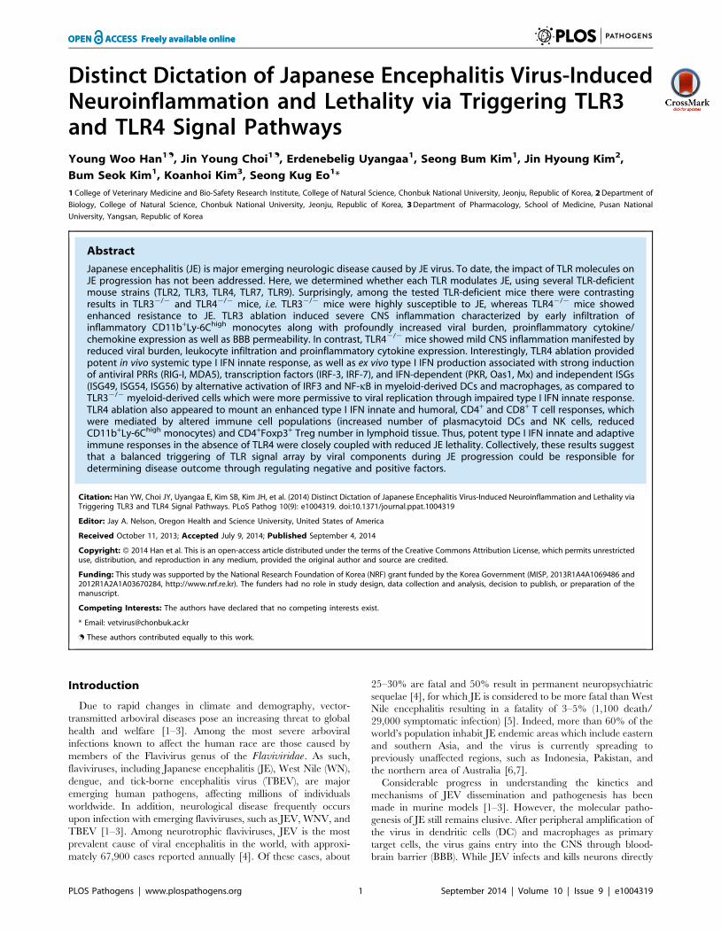
extravasated Evans blue dye, showed that JEV infection of
TLR32/2 mice gave rise to increased BBB permeability 3 days pi
(Figure 4A). In contrast, TLR42/2 mice showed no significant
change in BBB permeability following JEV infection. Supportively,
the ablation of TLR3 molecules was found to induce increased
BBB permeability by JEV infection, when the amount of
extravasated Evans blue dye within the brain was measured by
photometric analysis (Figure 4B). Notably, TLR32/2 mice
apparently retained increased amounts of extravasated Evans blue
dye in the brain 3 days pi, compared to those of wild-type mice.
This demonstrates that the ablation of TLR3, but not TLR4
molecule, is able to regulate BBB integrity following JEV infection.
The spread of JEV in the brain of TLR32/2 and TLR42/2
mice after intracranial inoculationTLR32/2 and TLR42/2 mice showed distinct viral burdens in
the CNS, which were closely associated with lethality to JE. This
phenotype could be due to differential dissemination from the
periphery and/or an independent antiviral effect in the CNS. To
test this, wild-type, TLR32/2, and TLR42/2 mice were
inoculated with 103 pfu of JEV directly into the cerebral cortex
via the intracranial (IC) route, and viral burdens in sub-tissues of
brain (cortex, olfactory bulb, hippocampus, brain stem, cerebel-
lum, and spinal cord) were monitored (Figure 5A–F). Wild-type
as well as TLR32/2 and TLR42/2 mice showed rapid and
complete mortality following IC infection of JEV, and there was
no significant difference in the average survival time between wild-
type and KO mice following IC infection of JEV (data not shown).
Interestingly, TLR32/2 and TLR42/2 mice showed slightly
lower levels of median viral burden in several sub-tissues of the
brain. These data suggest that TLR3 and TLR4 molecules had no
regulatory function on viral dissemination within the CNS
following introduction, but appeared to have a subtle role in
Figure 1. Contrasting regulation of JE by triggering TLR3 and TLR4 signal pathway. A. Susceptibility of TLR32/2, TLR42/2, and TLR3/42/2
mice to JE. Four- to five-week-old mice (n = 10–18) were inoculated with JEV (2.86107 pfu), and the survival rate was examined over 15 days. B. Ratioof mice showing neurologic disorder during JE progression. Mice infected with JEV were examined every 6 h from 4 to 7 days pi. C. Viral burden inlymphoid and inflammatory tissues during JE progression. Viral burden in spleen, brain, and spinal cord of mice infected with JEV was assessed byreal-time qRT-PCR at the indicated days pi. The viral RNA load was expressed by viral RNA copy number per microgram of total RNA (n = 5). Eachsymbol represents the level of an individual mouse; horizontal line indicates the median of each group.doi:10.1371/journal.ppat.1004319.g001
Distinct Dictation of JE by TLR3 and TLR4 Pathways
PLOS Pathogens | www.plospathogens.org 4 September 2014 | Volume 10 | Issue 9 | e1004319

Figure 2. Enhanced inflammation of the CNS in TLR32/2 mice following JEV infection. A and B. Early infiltration of inflammatoryCD11b+Ly-6Chigh monocytes. After heart perfusion at the 3rd day pi, the frequency (A) and absolute number (B) of CD11b+Ly-6Chigh monocytes andCD11b+Gr-1high granulocytes infiltrated into brain were analyzed by flow cytometric analysis. C and D. Percentage and number of resting microgliaand activated microglia/macrophages. The frequency (C) and total number (D) of CD11bintCD45int (resting microglia) and CD11bhighCD45high
(activated microglia/macrophages) were determined at the 3rd day pi. The values in the representative dot-plot denote the average of the indicatedcell population obtained from three individual experiment (n = 3–5). The bar in graph represents the average 6 SD of the indicated cell number. E.H&E-stained brain tissue sections. Histological examinations were performed at the 4th day pi. The arrows denote the area of interest. F.Representative confocal microscopic images. Brain sections from TLR32/2 and TLR42/2 mice which were infected with JEV were co-stained for JEVantigen (red), the nuclear stain DAPI (blue), and the microglia/macrophage cell-specific marker CD11b (green) at 4 days pi. The data arerepresentative of sections from at least five mice per group. ***, p,0.001 compared with the levels of the indicated group.doi:10.1371/journal.ppat.1004319.g002
Distinct Dictation of JE by TLR3 and TLR4 Pathways
PLOS Pathogens | www.plospathogens.org 5 September 2014 | Volume 10 | Issue 9 | e1004319

regulating viral replication in the CNS. In addition, we examined
the expression of pro-inflammatory cytokine (IL-6 and TNF-a),
chemokine (CCL2), and type I IFN (IFN-a and IFN-b). The
expression of such cytokines in sub-tissues of brain following IC
infection of JEV was consistently the same between wild-type and
KO mice (Figure 5G). The accumulation of CD11b+Ly-6Chigh
leukocytes in the brain was slightly, but not significantly, higher in
TLR32/2 mice following IC infection of JEV, as compared to
wild-type mice, and TLR42/2 mice showed no significant
change in leukocyte accumulation by IC infection of JEV
(Figure 5H). Collectively, these results imply that TLR3 and
TLR4 molecules have different roles in controlling the dissem-
ination of JEV from the periphery into the CNS, rather than a
regulatory role(s) on viral dissemination within the CNS after
CNS invasion.
Type I IFN responses are not blunted in TLR32/2 micefollowing JEV infection
It has been demonstrated that TLR3 molecules, in concert with
RIG-I, MDA5, and TLR7, recognize viral RNA and induce type I
IFNs through activation of adaptor molecule TRIF and subse-
quent transcription regulators IRF-3 and IRF-7. Also, triggering
signal pathway by TLR4 molecule can activate IRF-3, IRF-5, and
IRF-7 through adaptor molecules TRIF and MyD88, thereby
inducing the production of type I IFNs (IFN-a and b) [31–33].
Therefore, since TLR3 and TLR4 molecules contribute to the
generation of a normal IFN response through activation of IRF-3,
IRF-5, and IRF-7 after infection with neurotrophic virus [14,15],
we tested whether the ablation of TLR3 and TLR4 molecules
affected type I innate responses in JEV infection. Our data
revealed that the expressions of IFN-a and b mRNA were
Figure 3. TLR3 ablation induces huge production of pro-inflammatory cytokines in inflammatory tissues. A–D. The expression of pro-inflammatory cytokine and chemokine in inflammatory tissues. The expression of pro-inflammatory cytokines and chemokines in brain (A and C) andspinal cord (B and D) was determined by real-time qRT-PCR 4 days pi. Each symbol represents the level of an individual mouse; horizontal lineindicates median of each group. E. Systemic production of pro-inflammatory IL-6 cytokine. The levels of IL-6 in sera were determined by cytokineELISA at the indicated day pi. Data represent the average 6 SD derived from at least five mice per group. ***, p,0.001 compared with the levels ofthe wild-type mice.doi:10.1371/journal.ppat.1004319.g003
Distinct Dictation of JE by TLR3 and TLR4 Pathways
PLOS Pathogens | www.plospathogens.org 6 September 2014 | Volume 10 | Issue 9 | e1004319
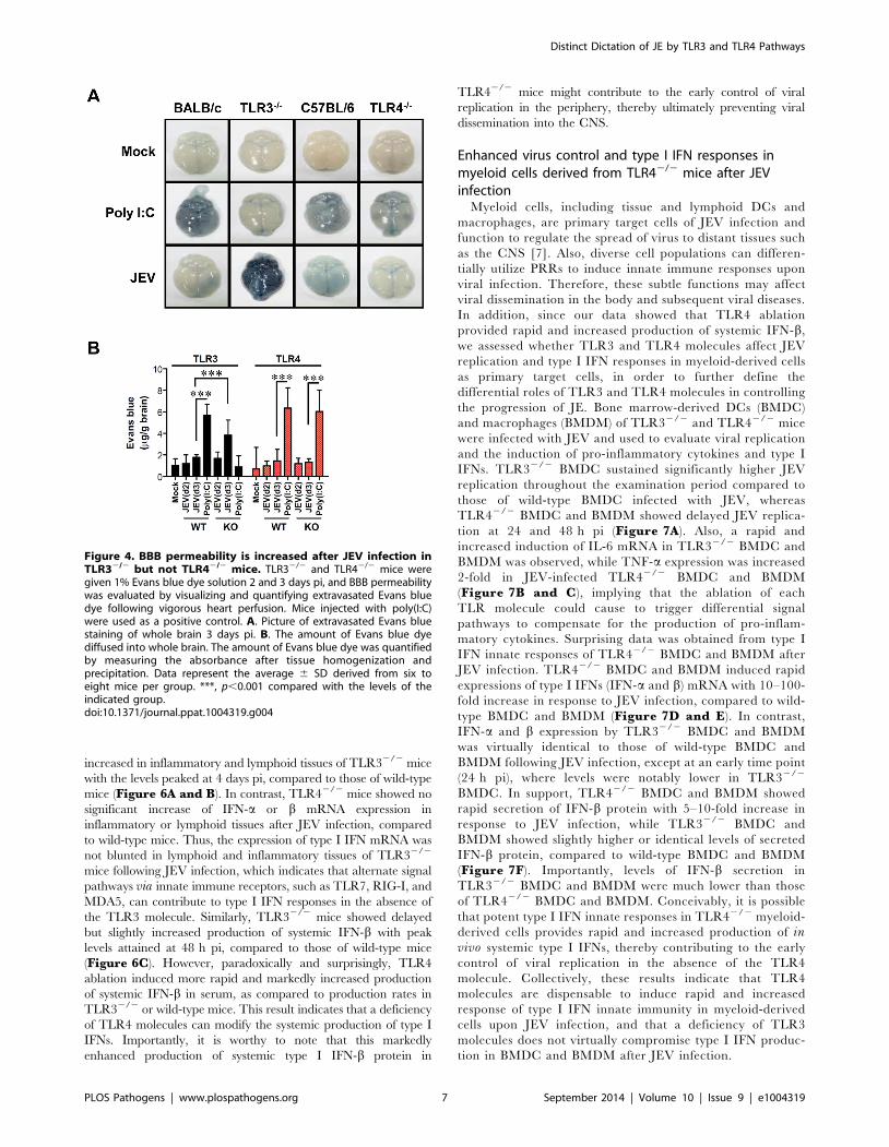
increased in inflammatory and lymphoid tissues of TLR32/2 mice
with the levels peaked at 4 days pi, compared to those of wild-type
mice (Figure 6A and B). In contrast, TLR42/2 mice showed no
significant increase of IFN-a or b mRNA expression in
inflammatory or lymphoid tissues after JEV infection, compared
to wild-type mice. Thus, the expression of type I IFN mRNA was
not blunted in lymphoid and inflammatory tissues of TLR32/2
mice following JEV infection, which indicates that alternate signal
pathways via innate immune receptors, such as TLR7, RIG-I, and
MDA5, can contribute to type I IFN responses in the absence of
the TLR3 molecule. Similarly, TLR32/2 mice showed delayed
but slightly increased production of systemic IFN-b with peak
levels attained at 48 h pi, compared to those of wild-type mice
(Figure 6C). However, paradoxically and surprisingly, TLR4
ablation induced more rapid and markedly increased production
of systemic IFN-b in serum, as compared to production rates in
TLR32/2 or wild-type mice. This result indicates that a deficiency
of TLR4 molecules can modify the systemic production of type I
IFNs. Importantly, it is worthy to note that this markedly
enhanced production of systemic type I IFN-b protein in
TLR42/2 mice might contribute to the early control of viral
replication in the periphery, thereby ultimately preventing viral
dissemination into the CNS.
Enhanced virus control and type I IFN responses inmyeloid cells derived from TLR42/2 mice after JEVinfection
Myeloid cells, including tissue and lymphoid DCs and
macrophages, are primary target cells of JEV infection and
function to regulate the spread of virus to distant tissues such
as the CNS [7]. Also, diverse cell populations can differen-
tially utilize PRRs to induce innate immune responses upon
viral infection. Therefore, these subtle functions may affect
viral dissemination in the body and subsequent viral diseases.
In addition, since our data showed that TLR4 ablation
provided rapid and increased production of systemic IFN-b,
we assessed whether TLR3 and TLR4 molecules affect JEV
replication and type I IFN responses in myeloid-derived cells
as primary target cells, in order to further define the
differential roles of TLR3 and TLR4 molecules in controlling
the progression of JE. Bone marrow-derived DCs (BMDC)
and macrophages (BMDM) of TLR32/2 and TLR42/2 mice
were infected with JEV and used to evaluate viral replication
and the induction of pro-inflammatory cytokines and type I
IFNs. TLR32/2 BMDC sustained significantly higher JEV
replication throughout the examination period compared to
those of wild-type BMDC infected with JEV, whereas
TLR42/2 BMDC and BMDM showed delayed JEV replica-
tion at 24 and 48 h pi (Figure 7A). Also, a rapid and
increased induction of IL-6 mRNA in TLR32/2 BMDC and
BMDM was observed, while TNF-a expression was increased
2-fold in JEV-infected TLR42/2 BMDC and BMDM
(Figure 7B and C), implying that the ablation of each
TLR molecule could cause to trigger differential signal
pathways to compensate for the production of pro-inflam-
matory cytokines. Surprising data was obtained from type I
IFN innate responses of TLR42/2 BMDC and BMDM after
JEV infection. TLR42/2 BMDC and BMDM induced rapid
expressions of type I IFNs (IFN-a and b) mRNA with 10–100-
fold increase in response to JEV infection, compared to wild-
type BMDC and BMDM (Figure 7D and E). In contrast,
IFN-a and b expression by TLR32/2 BMDC and BMDM
was virtually identical to those of wild-type BMDC and
BMDM following JEV infection, except at an early time point
(24 h pi), where levels were notably lower in TLR32/2
BMDC. In support, TLR42/2 BMDC and BMDM showed
rapid secretion of IFN-b protein with 5–10-fold increase in
response to JEV infection, while TLR32/2 BMDC and
BMDM showed slightly higher or identical levels of secreted
IFN-b protein, compared to wild-type BMDC and BMDM
(Figure 7F). Importantly, levels of IFN-b secretion in
TLR32/2 BMDC and BMDM were much lower than those
of TLR42/2 BMDC and BMDM. Conceivably, it is possible
that potent type I IFN innate responses in TLR42/2 myeloid-
derived cells provides rapid and increased production of invivo systemic type I IFNs, thereby contributing to the early
control of viral replication in the absence of the TLR4
molecule. Collectively, these results indicate that TLR4
molecules are dispensable to induce rapid and increased
response of type I IFN innate immunity in myeloid-derived
cells upon JEV infection, and that a deficiency of TLR3
molecules does not virtually compromise type I IFN produc-
tion in BMDC and BMDM after JEV infection.
Figure 4. BBB permeability is increased after JEV infection inTLR32/2 but not TLR42/2 mice. TLR32/2 and TLR42/2 mice weregiven 1% Evans blue dye solution 2 and 3 days pi, and BBB permeabilitywas evaluated by visualizing and quantifying extravasated Evans bluedye following vigorous heart perfusion. Mice injected with poly(I:C)were used as a positive control. A. Picture of extravasated Evans bluestaining of whole brain 3 days pi. B. The amount of Evans blue dyediffused into whole brain. The amount of Evans blue dye was quantifiedby measuring the absorbance after tissue homogenization andprecipitation. Data represent the average 6 SD derived from six toeight mice per group. ***, p,0.001 compared with the levels of theindicated group.doi:10.1371/journal.ppat.1004319.g004
Distinct Dictation of JE by TLR3 and TLR4 Pathways
PLOS Pathogens | www.plospathogens.org 7 September 2014 | Volume 10 | Issue 9 | e1004319

Figure 5. The spread of JEV in the brain of TLR32/2 and TLR42/2 mice after intracranial inoculation. TLR32/2 and TLR42/2 mice wereinoculated with JEV (103 pfu) by intracranial injection. Brains were harvested on days 2 and 4 pi, and then used for the determination of viral spreadand cytokine expression. A–F. Viral burden in each sub-tissue of brain. The CNS tissues were separated into cortex (A), olfactory bulb (B),hippocampus (C), brain stem (D), cerebellum (E), and spinal cord (F). Viral burden was determined by real-time qRT-PCR. The viral RNA load wasexpressed by viral RNA copy number per microgram of total RNA. Each symbol represents the level of an individual mouse; the horizontal lineindicates the median of each group. G. The expression levels of pro-inflammatory cytokine and type I IFN genes in each sub-tissue of brain. Theexpression levels were expressed by the indicated target gene levels relative to those in the mock-infected group. The bar represents the average 6SD of the indicated target gene levels obtained from each group (n = 5). Cor, cortex; Olf, olfactory bulb; Hip, hippocampus; BS, brain stem; Cer,cerebellum; SC, spinal cord. H. Leukocyte accumulation in the CNS of TLR32/2 and TLR42/2 mice after intracranial infection of JEV. TLR32/2 andTLR42/2 mice were inoculated with JEV (103 pfu) by intracranial injection, and brains were harvested on day 2. Leukocyte populations were isolatedby vigorous heart perfusion and then determined by flow cytometric analysis. The values in the representative dot-plots denote the average of theindicated cell population obtained from three individual experiments.doi:10.1371/journal.ppat.1004319.g005
Distinct Dictation of JE by TLR3 and TLR4 Pathways
PLOS Pathogens | www.plospathogens.org 8 September 2014 | Volume 10 | Issue 9 | e1004319
Distinct Dictation of JE by TLR3 and TLR4 Pathways
PLOS Pathogens | www.plospathogens.org 9 September 2014 | Volume 10 | Issue 9 | e1004319
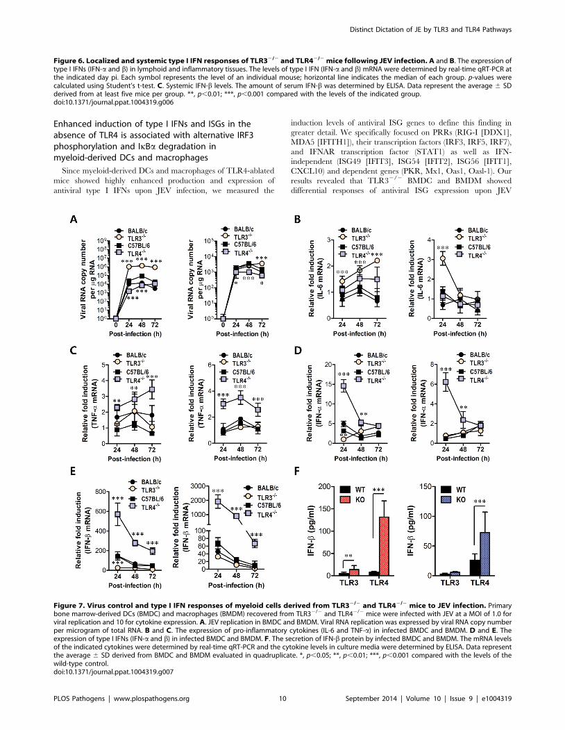
Enhanced induction of type I IFNs and ISGs in theabsence of TLR4 is associated with alternative IRF3phosphorylation and IkBa degradation inmyeloid-derived DCs and macrophages
Since myeloid-derived DCs and macrophages of TLR4-ablated
mice showed highly enhanced production and expression of
antiviral type I IFNs upon JEV infection, we measured the
induction levels of antiviral ISG genes to define this finding in
greater detail. We specifically focused on PRRs (RIG-I [DDX1],
MDA5 [IFITH1]), their transcription factors (IRF3, IRF5, IRF7),
and IFNAR transcription factor (STAT1) as well as IFN-
independent (ISG49 [IFIT3], ISG54 [IFIT2], ISG56 [IFIT1],
CXCL10) and dependent genes (PKR, Mx1, Oas1, Oasl-1). Our
results revealed that TLR32/2 BMDC and BMDM showed
differential responses of antiviral ISG expression upon JEV
Figure 6. Localized and systemic type I IFN responses of TLR32/2 and TLR42/2 mice following JEV infection. A and B. The expression oftype I IFNs (IFN-a and b) in lymphoid and inflammatory tissues. The levels of type I IFN (IFN-a and b) mRNA were determined by real-time qRT-PCR atthe indicated day pi. Each symbol represents the level of an individual mouse; horizontal line indicates the median of each group. p-values werecalculated using Student’s t-test. C. Systemic IFN-b levels. The amount of serum IFN-b was determined by ELISA. Data represent the average 6 SDderived from at least five mice per group. **, p,0.01; ***, p,0.001 compared with the levels of the indicated group.doi:10.1371/journal.ppat.1004319.g006
Figure 7. Virus control and type I IFN responses of myeloid cells derived from TLR32/2 and TLR42/2 mice to JEV infection. Primarybone marrow-derived DCs (BMDC) and macrophages (BMDM) recovered from TLR32/2 and TLR42/2 mice were infected with JEV at a MOI of 1.0 forviral replication and 10 for cytokine expression. A. JEV replication in BMDC and BMDM. Viral RNA replication was expressed by viral RNA copy numberper microgram of total RNA. B and C. The expression of pro-inflammatory cytokines (IL-6 and TNF-a) in infected BMDC and BMDM. D and E. Theexpression of type I IFNs (IFN-a and b) in infected BMDC and BMDM. F. The secretion of IFN-b protein by infected BMDC and BMDM. The mRNA levelsof the indicated cytokines were determined by real-time qRT-PCR and the cytokine levels in culture media were determined by ELISA. Data representthe average 6 SD derived from BMDC and BMDM evaluated in quadruplicate. *, p,0.05; **, p,0.01; ***, p,0.001 compared with the levels of thewild-type control.doi:10.1371/journal.ppat.1004319.g007
Distinct Dictation of JE by TLR3 and TLR4 Pathways
PLOS Pathogens | www.plospathogens.org 10 September 2014 | Volume 10 | Issue 9 | e1004319

infection (Figure 8A and B). TLR32/2 BMDC showed less
induction of PRR genes (RIG-I and MDA5) and their transcrip-
tion factors (IRF-3 and IRF-7), but member of genes (ISG49,
ISG54, ISG56, CXCL10) that are induced in IFNAR2/2 cells
(i.e., are IFN-independent) [34,35] were expressed in TLR32/2
BMDC with slightly higher levels, compared to those of wild-type
BMDC. This result was consistent with the fact that TLR32/2
BMDC showed slightly higher or identical secretion of
IFN-b compared to wild-type BMDC (Figure 7F), because
IFN-independent ISG genes can also be induced through ISRE
binding of ISGF3 complex initiated by type I IFN receptor [15].
In contrast, TLR32/2 BMDM showed less induction of IFN-
independent ISG genes (ISG49, ISG54, ISG56, CXCL10) as well
as IFN-dependent ISG genes (PKR, Mx1, Mx2) and members of
the 29-59-oligoadenylate synthetase family (Oas1, Oasl-1), com-
pared to wild-type BMDM. This result implies that macrophages
could be more compromised in the inductiveness of type I IFN
innate responses than DCs, if the TLR3 molecule was ablated.
Figure 8. ISG induction, phosphorylation of IRFs, and IkBa degradation in primary myeloid cells derived from TLR32/2 and TLR42/2
mice after JEV infection. A and B. Clustered heatmap showing the expression of IRF, ISG, and RLR genes in infected BMDC and BMDM. Primarybone marrow-derived DCs (BMDC) and macrophages (BMDM) recovered from TLR32/2 and TLR42/2 mice were infected with JEV at a MOI of 10 ormock-infected (M), and employed to analyze the induction of IRF, ISG, and RLR genes at 24 and 48 h pi. The expression of each IRF, ISG, and RLR genewas normalized to b-actin after determining mRNA levels by real-time qRT-PCR, and displayed as the average of at least four independent samples,according to the indicated color on a log2 scale. C and D. Expression and phosphorylation of IRF3, IRF7, and STAT1 and IkBa degradation. BMDC andBMDM derived from TLR32/2 and TLR42/2 mice were infected with JEV at 10 MOI or mock-infected (M). At 6, 12, 24, and 48 h after infection, cellswere lysed, separated by SDS-PAGE and analyzed by western blot to detect unphosphorylated and phosphorylated form of target proteins usingspecific Abs. One representative picture of at least three experiments is shown.doi:10.1371/journal.ppat.1004319.g008
Distinct Dictation of JE by TLR3 and TLR4 Pathways
PLOS Pathogens | www.plospathogens.org 11 September 2014 | Volume 10 | Issue 9 | e1004319

The prominent induction of antiviral ISG genes was observed in
TLR42/2 BMDC and BMDM after JEV infection (Figure 8Aand B). TLR42/2 BMDC showed enhanced expression of PRR
genes (MDA-5) and its transcription factors (IRF-3, IRF-5, IRF-7),
and IFN-dependent genes (PKR, Oasl-1), as well as IFN-
independent genes (ISG49, ISG 54, ISG 56, CXCL10). Also,
TLR42/2 BMDM showed much more apparently and highly
induced expression of antiviral ISG genes after JEV infection
compared to those of wild-type BMDM and other cells, because
TLR42/2 BMDM induced the expression of all tested ISG genes
(PRRs, transcription factors, IFN-dependent and independent
genes) with higher levels than other cells. Notably, TLR42/2
BMDM showed markedly enhanced induction of both IFN-
dependent (PKR, Oas1, Oasl-1, Mx1, Mx2) and independent
genes (ISG49, ISG54, ISG56, CXCL10), compared to TLR32/2
BMDM that showed less induction of such genes. Therefore, these
results support that a deficiency of TLR4 molecule provides more
efficient type I IFN innate immune responses in DCs and
macrophages following JEV infection.
To further define the induction of antiviral IFN-independent
and dependent ISG genes in JEV-infected TLR32/2 and
TLR42/2 DCs and macrophages, the activation state of
associated transcription factors was examined by western blot. In
line with antiviral ISG induction data, TLR32/2 BMDC
displayed decreased expression of IRF-3 and IRF-7 at 6–48 h
and 48 h pi, respectively, and phosphorylated form of IRF-3 was
not detected in both TLR32/2 and TLR42/2 BMDC (Fig-ure 8C), which supports that enhanced induction of antiviral
IFN-independent ISG genes (ISG49, ISG54, ISG 56, CXCL10) in
TLR32/2 and TLR42/2 BMDC may be caused by stimulation of
IFNAR signal through increased IFN-b secretion [15]. Since
slightly delayed phosphorylation of STAT1, an IFNAR transcrip-
tion factor, was observed in TLR32/2 and TLR42/2 BMDC,
other pathways to activate NF-kB were also considered to
contribute to enhanced induction of IFN-independent ISG genes.
Interestingly, this notion can be explained by the result that faster
degradation of IkBa was detected in TLR42/2 BMDC. IkBaproteins are phosphorylated via IkB kinase (IKK) activated by
signal transducers, and are subsequently degraded after release of
NF-kB [36]. Therefore, these results suggest that TLR42/2
BMDC could have evolved as yet unknown pathway(s) to activate
NF-kB upon JEV infection, thereby inducing enhanced expression
of type I IFNs and ISG genes. In addition, somewhat interestingly,
transiently phosphorylated form of IRF-3 was strongly detected in
TLR42/2 BMDM, but not TLR32/2 BMDM, as early as 6 and
12 h pi (Figure 8D). Also, TLR42/2 BMDM showed prolonged
and strong phosphorylation of STAT1 after JEV infection,
compared to wild-type BMDM. Therefore, it was considered that
activation of IRF3 and STAT1 in TLR42/2 BMDM derived
potent type I IFN production as well as the induction of broad
antiviral IFN-independent and IFN-dependent ISG genes.
Induction of type I IFN and ISGs in primary corticalneurons derived from TLR3 and 4-deficient mice after JEVinfection
Neurons may be the main target cell of JEV infection in the
CNS, and their death is a key factor in pathogenesis and
neurological sequelae [8]. To examine whether TLR3 and TLR4
molecules can regulate JEV replication in neurons, primary
cortical neurons generated from wild-type as well as TLR32/2
and TLR42/2 mice were infected with JEV, and virus yield, type I
IFN responses and ISG expression were evaluated. It was likely
that wild-type neurons were more permissive to JEV infection than
DCs or macrophages, because infection of neurons with 10-fold
less virus (MOI 0.1 versus 1.0) produced over ,105 viral RNA
within 24 h (Figure 7A and Figure 9A). In the absence of TLR3
molecule, JEV replicated faster, resulting in a 1.5–2.0-fold increase
in infectious virus production between 24 h and 48 h pi, as
compared to infected wild-type neurons. The ablation of TLR4
molecule showed earlier replication of JEV at 24 h pi, but the
levels of virus were similar in both wild-type and TLR42/2
neurons at 48 h pi (Figure 9A). Biphasic type I IFN mRNA
induction was observed, with slightly higher levels at 24 h pi but
much lower at 48 h pi in TLR32/2 neurons, compared to wild-
type neurons (Figure 9B). In contrast, TLR42/2 neurons showed
transient induction of IFN-b at 24 h pi, after which IFN-b mRNA
levels were comparable in both wild-type and TLR42/2 neurons.
The secretion of IFN-b protein in culture media was markedly
lower in TLR32/2 neurons at 48 h pi, while TLR42/2 neurons
showed increased expression and production of IFN-b at both
24 h and 48 h pi, as compared to those of wild-type neurons
(Figure 9B). Also, it seemed that the expression of antiviral ISGs
in TLR32/2 neurons followed type I IFN responses; hence,
ISG49 and ISG56 showed transient increases at 24 h pi but much
lower expression at 48 h pi (Figure 9C). Also, a higher expression
of RIG-I and MDA-5, a cytosolic PRRs of viral RNA, was
observed in TLR32/2 neurons, but their transcription factor IRF-
3 was shown with decreased expression levels, as compared to
wild-type neurons. It was thought that this caused the reduction in
IFN-b production in TLR32/2 neurons at 48 h pi. TLR42/2
neurons showed transiently higher expression of ISG54 and
MDA5 at 24 h pi, but the decreased levels of RIG-I and IRF-7
expression was observed at 48 h pi. Collectively, these results
suggest that TLR3 may have an independent and subordinate role
in triggering type I IFN innate responses in cortical neurons,
because type I IFN responses and ISGs expression were much
decreased at a later time point (48 h pi). Also, TLR42/2 cortical
neurons appeared to induce less potent type I IFN innate immune
responses than TLR42/2 DCs and macrophages, which indicates
that specific types of cells differentially trigger innate immune
responses following JEV infection.
Enhanced type I IFN innate and JEV-specific CD4+/CD8+ Tcell responses in TLR42/2 mice are associated withaltered number of plasmacytoid DCs and CD4+Foxp3+
Treg cells in lymphoid tissuesTLR signal pathway through MyD88 and/or TRIF adaptor
molecules is required in some cases for antigen-specific antibody
responses [37,38], which may contribute to the control of JEV
dissemination and replication in the brain. Our data revealed that
TLR3 ablation showed slightly, but not significantly, increased
level of IgM and IgG, while TLR42/2 mice showed significantly
increased levels of JEV-specific IgM and IgG, compared to wild-
type mice (Figure S3A). Also, JEV infection showed marginally
increased numbers of CD4+, CD8+ T, and CD19+ B cells with
activated phenotypes, as corroborated by the expression of surface
markers, such as CD69, CD44, and CD80; however TLR3 and
TLR4 molecules did not show apparently regulatory functions in
T and B lymphocytes (Table S1). Since effector antigen-specific
CD4+ and CD8+ T cell responses are also required for the control
and clearance of JEV in the CNS as well as in peripheral tissues
[7], we evaluated whether the ablation of TLR3 and TLR4
molecules altered JEV antigen-specific CD4+ and CD8+ T cell
responses. A deficiency of TLR3 molecules resulted in a similar
percentage and absolute number of CD4+ and CD8+ T cells
expressing IFN-c and TNF-a, whereas TLR42/2 mice showed
an increased percentage and absolute number of IFN-c and
Distinct Dictation of JE by TLR3 and TLR4 Pathways
PLOS Pathogens | www.plospathogens.org 12 September 2014 | Volume 10 | Issue 9 | e1004319

TNF-a-producing CD4+ and CD8+ T cells (Figure S3B–E).
Along with potent type I IFN innate responses, these data indicate
that TLR4 ablation could provide enhanced antigen-specific
responses, thereby contributing in part to the control of virus
replication and dissemination. Therefore, to further characterize
the immunological parameters associated with potent type I IFN
Figure 9. Induction of type I IFNs and ISGs in primary cortical neurons derived from TLR32/2 and TLR42/2 mice after JEV infection.Primary cortical neurons generated from TLR32/2 and TLR42/2 mice were infected at an MOI of 0.1, and viral replication and type I IFNs responses at24 and 48 h pi were analyzed. A. JEV replication. JEV replication was determined by both real-time qRT-PCR and focus-forming assay. B. Theexpression of type I IFNs (IFN-a and IFN-b) mRNA and IFN-b secretion in primary cortical neurons infected by JEV. C. Induction of IRFs, ISGs, and RLRsgene in infected primary cortical neurons. The mRNA levels of the indicated gene were determined by real-time qRT-PCR, and IFN-b levels in culturemedia were determined by ELISA. Data represent the average 6 SD derived from primary cortical neurons quadruplicate. *, p,0.05; **, p,0.01; ***,p,0.001 compared with the levels of the indicated group.doi:10.1371/journal.ppat.1004319.g009
Distinct Dictation of JE by TLR3 and TLR4 Pathways
PLOS Pathogens | www.plospathogens.org 13 September 2014 | Volume 10 | Issue 9 | e1004319

innate and adaptive immune responses in JEV-infected TLR42/2
mice, we analyzed the immune cellular components related to type
I IFN innate and adaptive immune responses. TLR32/2 and
TLR42/2 mice were challenged with JEV, and spleens were
harvested at 3 and 5 days pi. At the early phase of infection,
analysis of the spleen provides an insight into how TLR3 and
TLR4 molecules modulate innate immune and inflammatory
responses immediately after infection, because JEV was adminis-
tered intraperitoneally. Analysis of lymphoid CD8a+ and myeloid
CD11b+ DC subsets revealed that JEV-infected TLR32/2 and
TLR42/2 mice exhibited similar increases in both DC subsets,
compared to those of infected wild-type mice (Figure 10A).
However, somewhat surprisingly, the ablation of TLR4 molecule
resulted in a highly increased number of CD11cintPDCA-1high
plasmacytoid DC (pDC) subset, which is known as a potent
cellular component to produce type I IFNs in response to viral
infection [39]. Thus, it was considered that highly increased pDC
number might contribute in part to enhanced production of
systemic IFN-b in TLR42/2 mice. TLR42/2 mice also showed a
decreased frequency of inflammatory CD11c2CD11b+Ly-6Chigh
monocytes and no significant changes in the absolute number,
whereas a significant increased number, but not frequency, of
inflammatory monocytes was observed in TLR32/2 mice,
compared to that in wild-type mice (Figure 10B and C). This
implies that TLR42/2 mice exhibit a mild inflammatory reaction
in the spleen. In addition, a deficiency of TLR4 molecule provided
an increased number of NK cells at 5 days pi, but TLR3 molecule
had no modulatory function on NK cell number (Figure 10D).
Moreover, since CD4+CD25+Foxp3+ Treg cells contribute to the
dampening of innate and adaptive immune responses during acute
viral infection [40], we addressed the frequency and number of
CD4+CD25+Foxp3+ Treg cells in the spleen. We found that the
frequency and absolute number of CD4+CD25+Foxp3+ Treg cells
were increased 1.5–2-fold in response to JEV infection in wild-type
mice (Figure 10E and F). TLR32/2 mice showed identical
increase of CD4+CD25+Foxp3+ Treg cells to wild-type mice, while
in TLR42/2 mice a reduced frequency and absolute number of
CD4+CD25+Foxp3+ Treg cells was observed, which indicates that
TLR4 molecule could be involved in the increase of
CD4+CD25+Foxp3+ Treg cell numbers following JEV infection.
Collectively, these results suggest that increased number of
CD11cintPDCA-1high pDC subpopulation and reduced
CD4+CD25+Foxp3+ Treg cells are closely associated with
enhanced type I IFN innate immunity and JEV-specific CD4+
and CD8+ T cell responses in TLR42/2 mice.
Discussion
Although recognition of ssRNA virus, such as flavivirus, viacytosolic helicase RIG-I and MDA5 may be dominant to induce
type I IFN innate responses [14–16], the role of TLRs as first-front
line of innate immune receptors in the extracellular space,
including the cell membrane and endosome, remains still
undefined in flaviviral infections, due to conflicting and intricate
data. Furthermore, despite the pathological importance of JE as a
major cause of acute encephalitis, the role of TLR signal pathways
in JE progression has not been fully explored to date. Here, we
observed strikingly contrasting regulation of JE via TLR3 and
TLR4 signal pathways; TLR3 ablation elicited highly enhanced
susceptibility to JE, whereas TLR4 ablation provided significantly
enhanced resistance to JE rather than inducing increased
susceptibility. In the present study, interesting clues to such
contrasting regulation of JE by TLR3 and TLR4 molecules were
derived from the differential induction of type I IFN innate
responses in TLR32/2 and TLR42/2 mice. Notably, TLR4
ablation induced potent type I IFN innate responses through
enhanced induction of antiviral ISG genes by alternative
activation of IRF-3 and NF-kB in DCs and macrophages.
Additionally, altered CD11cintPDCA-1high pDC and
CD4+CD25+Foxp3+ Treg number in TLR42/2 mice appeared
to contribute in part to enhanced type I IFN innate as well as JEV-
specific T cell responses. Collectively, potent type I IFN innate and
adaptive immune responses generated in peripheral lymphoid
tissues after JEV infection were closely coupled with a reduced JE
lethality in TLR42/2 mice. These findings imply that the
balanced triggering of TLR signal array by viral components
during JE progression could be responsible for determining the
outcome of disease through negative and positive regulatory
factors.
There are several conflicting reports on the role of TLR3
signaling pathway in neurological diseases caused by viral infection
[41,42]. A deficiency of TLR3 in humans predisposes to a genetic
risk factor for herpes simplex virus encephalitis [43] and influenza
A virus-induced encephalopathy [44], but TLR32/2 mice infected
with influenza [45], punta toro [46], and vaccinia viruses [47]
showed improved survival and decreased production of inflam-
matory cytokines. Strikingly conflicting role of TLR3 signal
pathway was derived from an infection model with WNV
[20,21]. While TLR3 ablation protected mice from WNV lethal
infection by decreased systemic TNF-a and IL-6 production and
BBB permeability [20], there is a report demonstrating that TLR3
molecules are essential in protecting from WNV infection [21].
Our results favor the latter report. TLR32/2 BMDC, but not to
BMDM, showed defective type I IFN innate responses at an early
time (24 h pi), which may allow early viral replication. This result
is in contrast to that of WNV infection, where TLR3 molecule did
not modulate WNV replication and IFN induction in primary
myeloid cells [21]. Although TLR32/2 BMDC is more permissive
to JEV replication, JEV-infected TLR32/2 BMDC elicited similar
levels of type I IFN responses to wild-type BMDC with delayed
kinetics, and TLR32/2 mice also showed no blunted type I IFN
responses in lymphoid and local tissues. This suggests that
enhanced tissue tropism and rapid viral entry into the CNS is
not affected by locally induced type I IFN responses. Type I IFN
responses of TLR32/2 mice were considered not to be attenuated
since cytosolic RIG-I and MDA5 molecules are intact. However,
TLR3 molecule appeared to play more important role in inducing
type I IFN responses of neuron cells than BMDC and BMDM,
because TLR32/2 neuron cells showed a markedly reduced
expression and production of type I IFN and ISGs at a late time
(48 h pi), thereby promoting viral replication. This implies that
TLR3 molecule had differential modulatory functions on type I
IFN innate responses and JEV replication in a cell-type restricted
manner. However, considering that TLR32/2 and TLR42/2
mice showed no difference in CNS replication of JEV following IC
infection, subtle changes of CNS system, such as innate responses
of microglia and astrocyte, appear to modulate the in vivo spread
of directly inoculated JEV in the CNS.
Increased BBB permeability by systemic TNF-a and IL-6
appears to promote an earlier entry of virus into the CNS. In
contrast to WNV infection, where TLR32/2 mice showed no
change in BBB permeability [21], TLR32/2 mice, but not
TLR42/2 mice, elicited increased BBB permeability associated
with a huge production of systemic IL-6. Also, this result was in
contrast with a previous study in which TLR32/2 mice showed
reduced cytokine (e.g., TNF-a and IL-6) responses, BBB
permeability, neuroinvasion, and mortality following infection
with mammalian cell-passaged WNV [20]. Nonetheless, our
Distinct Dictation of JE by TLR3 and TLR4 Pathways
PLOS Pathogens | www.plospathogens.org 14 September 2014 | Volume 10 | Issue 9 | e1004319
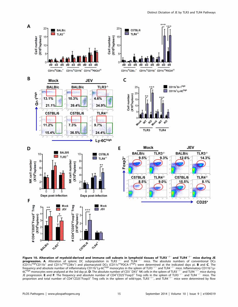
Figure 10. Alteration of myeloid-derived and immune cell subsets in lymphoid tissues of TLR32/2 and TLR42/2 mice during JEprogression. A. Alteration of splenic DC subpopulation in TLR32/2 and TLR42/2 mice. The absolute numbers of conventional DCs(CD11chighCD11b+ and CD11chighCD8a+) and plasmacytoid DCs (CD11cintPDCA-1high) were determined at the indicated days pi. B and C. Thefrequency and absolute number of inflammatory CD11b+Ly-6Chigh monocytes in the spleen of TLR32/2 and TLR42/2 mice. Inflammatory CD11b+Ly-6Chigh monocytes were analyzed at the 3rd day pi. D. The absolute number of CD32DX5+ NK cells in the spleen of TLR32/2 and TLR42/2 mice duringJE progression. E and F. The frequency and absolute number of CD4+CD25+Foxp3+ Treg cells in the spleen of TLR32/2 and TLR42/2 mice. Theproportion and total number of CD4+CD25+Foxp3+ Treg cells in the spleen of wild-type, TLR32/2, and TLR42/2 mice were determined by flow
Distinct Dictation of JE by TLR3 and TLR4 Pathways
PLOS Pathogens | www.plospathogens.org 15 September 2014 | Volume 10 | Issue 9 | e1004319

results showed some similarities with previous reports using WNV,
such as increased viral burden in peripheral tissues. Although the
impact of TLR3 molecule on BBB permeability is likely to differ,
depending on the context and details of the model, virus-culture
conditions, and the viral strain being tested, the failure of early
viral clearance in the periphery of TLR32/2 mice may ultimately
cause enhanced inflammatory reactions, thereby increasing BBB
permeability and viral load in the CNS. One intriguing result in
this study was that TLR72/2 mice showed no change in
susceptibility to JE, since TLR7 molecule can recognize ssRNA
of JEV. This result was in contrast with the report that the TLR7
molecule is involved in modulating the progression of WNV
encephalitis via an IL-23-dependent accumulation of leukocytes in
the CNS [48]. Although systemic levels of proinflammatory
cytokines and type I IFNs were higher in TLR72/2 than in wild-
type mice [48], it is expected that splenic pDC or circulating pDC
from TLR32/2 mice may also have contributed to the type I IFN
responses, because TLR7 signal pathway was intact in TLR32/2
mice. In addition, we previously found that TLR2 molecule had
modulatory function in cross-presentation of OVA protein using
JEV-infected TLR22/2 mice, suggesting that JEV infection may
be also be recognized by TLR2 molecule [49]. However, in this
study, TLR2 signal pathway had no impact on the progression of
JE. One trivial explanation of this result is that TLR2 signal
pathway was not involved in inducing pathologic disease by JEV
infection, no matter what OVA cross-presentation is regulated by
JEV infection in a TLR2-dependent manner.
The most intriguing result in this study was that TLR42/2 mice
showed markedly enhanced resistance to JE. To date, the role of
TLR4 signal pathway in inducing innate and adaptive immune
response against JEV and other flaviviruses has not been defined.
Our results demonstrate that TLR4 ablation strongly induces invivo systemic type I IFN innate responses, as well as type I IFN
expression and production from myeloid-derived cells upon JEV
infection. This presumably promotes early clearance of virus. In
spite of the existence of TLR4 prototype ligand, LPS, a growing
number of reports suggest that TLR4 molecule is biologically
relevant, and is responsive to viral proteins, including those of
Ebola virus [23], hepatitis C virus [24], and respiratory syncytial
virus [25], leading to the induction of proinflammatory cytokines.
We are not sure whether the induction of potent type I IFN innate
responses in the absence of TLR4 signal pathway was mediated
directly by enhanced signal transduction of other PRRs, such as
TLR3, RIG-I, and MDA5, and/or indirectly by soluble factors
produced from host cells by viral infection, i.e. DAMPs. However,
our results provide one explanation as to how TLR42/2 myeloid-
derived cells induce potent type I IFN innate responses, i.e.enhanced activation of NF-kB through unknown pathway(s) in
DCs, and transient activation of IRF3 at 6–12 h pi and prolonged
activation of STAT1 in macrophages. The expression of antiviral
ISG genes in myeloid-derived cells after JEV infection was induced
by both direct (by IRF-3) and indirect (by IFN-b production and
IFNAR signaling) pathways. Considering that only small faction
(10–20%) of myeloid-derived cells is infected by JEV [49],
uninfected myeloid-derived cells are thought to substantially
contribute to antiviral ISG induction through stimulation of
IFNAR signal after binding with secreted IFN-b proteins. This
notion was supported by two results, i.e. 1) induction of IFN-
dependent genes (PKR, Mx1, Oas1) in TLR32/2 BMDC,
TLR42/2 BMDC and BMDM with increased secretion of
IFN-b after JEV infection, and 2) no detection of phosphorylated
IRF-3 except in TLR42/2 BMDM. Also, transient activation of
IRF3 and prolonged activation of STAT1 explains strong
induction of both IFN-independent ISG (ISG49, ISG54, ISG56,
CXCL10) and dependent genes (PKR, Oas1, Mx1, Mx2) in
TLR42/2 macrophages. Although NF-kB activation in DCs and
IRF-3 and STAT1 activation in macrophages after JEV infection
support potent type I IFN innate responses in the absence of
TLR4 molecule, how these signal molecules are activated remains
still undefined. Therefore, future studies will be required to
delineate the mechanistic and functional intermediates that link
and regulate NF-kB, IRF-3 and STAT1 signal pathway in the
absence of TLR4 molecule.
In addition, our results is strengthened by a recent report that
TLR42/2 or TLR4 antagonist-treated mice are highly refractory
to influenza-induced lethality, due to blocking inflammation by
host-derived, oxidized phospholipid that potently stimulates TLR4
[50,51]. One similarity with our data is that mice treated with
TLR4 antagonist, Eritoran, or TLR42/2 mice had reduced lung
pathology to infection with influenza virus, which is characterized
by the reduction of viral burden and proinflammatory cytokine
expression. However, it is not certain whether Eritoran-treated or
TLR42/2 mice displayed rapid and enhanced type I IFN innate
responses after infection with influenza virus. Thus, it is
worthwhile identifying whether blocking TLR4 signal pathway
by antagonists such as Eritoran, affects JE progression through the
induction of potent type I IFN innate responses. This study will
provide valuable insights into developing therapeutic strategies to
viral encephalitis caused by neurotrophic virus such as JEV and
WNV. Analogously, in the absence of TLR4 molecule, the
enhanced expansion of CD11b+Ly-6Chigh ‘‘inflammatory mono-
cytes’’ was not observed in comparison with TLR32/2 mice,
which was suggestive that in TLR42/2 mice mild inflammatory
responses were elicited in the spleen. This monocyte subset
migrates to the site of infection, secretes pro-inflammatory
cytokines, and thereby exacerbates immunopathologic diseases
[28]. Thus, the aberrant recruitment and expansion of these
CD11b+Ly-6Chigh inflammatory monocytes may also contribute
to JE immunopathogenesis in TLR32/2 mice.
The production and response of type I IFN is considered to be a
major linkage point between innate and adaptive immunity,
because IFN-a/b sustains B cell activation and differentiation
[52,53], expands antigen-specific CD8+ T cells [54], CD4+ T cells
[55], and activation of NK cells [56]. Therefore, another
intriguing finding of this study was the global alteration of
immune responses in TLR42/2 mice. This suggests that TLR4
molecule is largely dispensable for the efficient link between innate
and adaptive immunity in JEV infection. Infection of TLR42/2
mice with JEV exhibited the expansion of pDC and NK cells, and
enhanced JEV-specific CD4+ and CD8+ T cell responses, which
are involved in viral clearance at early and late phases of infection,
respectively. Also, it is likely that increased number of pDCs
contributed in part to the potent induction of type I IFN innate
responses in TLR42/2 mice. In addition, TLR42/2 mice showed
limited expansion of CD4+CD25+Foxp3+ Tregs, which have been
known to suppress innate and effector T cells, thus preventing or
controlling reactivity to self-antigen and pathogens, and thereby
blunting severe inflammation and maintaining antigen-specific T
cytometric analysis at 7 days pi. The values in the representative dot-plots denote the average of the indicated cell populations obtained from threeindividual experiments (n = 3–4). The bar in graph represents the average 6 SD of the total number of the indicated cell population. *, p,0.001; **,p,0.01; ***, p,0.05 compared with the levels of the indicated group.doi:10.1371/journal.ppat.1004319.g010
Distinct Dictation of JE by TLR3 and TLR4 Pathways
PLOS Pathogens | www.plospathogens.org 16 September 2014 | Volume 10 | Issue 9 | e1004319

cell homeostasis [40]. The role of CD4+CD25+Foxp3+ Tregs in
acute viral diseases is still debatable [57,58]. Recent work
implicates CD4+Foxp3+ Tregs in the control of WNV pathogen-
esis, wherein peripheral expansion of Treg was associated with
mild inflammation, but reduced Treg levels were associated with
WNV encephalitis [57]. However, while CD4+Foxp3+ Tregs that
were adoptively transferred 2 days prior to JEV infection made the
recipients vulnerable to JE, CD4+Foxp3+ Tregs that were
adoptively transferred 2 days after infection provided resistance
to JE (unpublished personal data). This suggests that CD4+Foxp3+
Tregs elicit dual-phased roles during the progression of JEV-
induced neurological disorders. More importantly, Treg induction
during a viral infection is considered to be a detrimental response
that promotes virus persistence without benefits to the host
[59,60]. One trivial explanation of CD4+Foxp3+ Treg role is that
initially low number of CD4+Foxp3+ Tregs in TLR42/2 mice
may promote the expansion of effector CD4+ and CD8+ T cells
specific for JEV antigen as well as innate immune responses,
thereby inducing enhanced anti-viral response and virus-specific
CTL to promote early viral clearance.
JE pathogenesis in the murine model may be altered by the
route of peripheral administration, virus-propagation condition,
and viral strains [7,20,21]. It is also possible that the genetic
background of mice affects the immunopathogenesis of JE.
However, we found that two backgrounds of mouse strains used
for TLR32/2 and TLR42/2 mice showed comparable mortality
and similar clinical signs after JEV infection, which indicates that
JE pathogenesis is unaffected by genetic background of mouse
strains used in this study. Although JEV infected via i.p. route does
not directly reflect natural infection mediated by intradermal or
intramuscular route after biting of mosquitoes, JEV infected viai.p. route displays entirely similar pathogenesis to natural infection,
due to peripheral amplification in the spleen. Also, since mice
infected i.p. with JEV usually exhibited neurological disorder at
4–5 days pi, rapid innate immune responses are more critical to
control JE progression than adaptive T cell responses, which take
time to develop. Indeed, the role of T cells in flavivirus encephalitis
is less clear. This is, in part, due to variation of virus strain, the
infection dose, the route of administration, mouse strain and age of
the mice. Therefore, considering that the character of CD4+ and
CD8+ T cells specific for JEV is also governed by innate immune
responses initiated by recognition of PRRs, triggering of each PRR
by direct viral components and/or host factors derived from
infection could affect innate immune responses to shape adaptive
immune responses, thereby influencing JE pathogenesis. A better
understanding of the mechanisms that govern the induction of
protective immunity plays a critical role in developing novel
therapeutic strategies against JE.
Materials and Methods
Animals and ethics statementC57BL/6 (H-2b) and BALB/c (H-2d) mice (4–6 weeks old) were
purchased from Samtako (O-San, Korea). TLR2 (H-2b), TLR3
(H-2d and H-2b), TLR4 (H-2b), TLR7 (H-2d), and TLR9 (H-2b)-
deficient mice have been described elsewhere [33]. TLR3/42/2
mice that are deficient in both TLR3 and TLR4 molecules were
generated by backcrossing with TLR3 and TLR4-deficient mice.
All mice were genotyped and bred in the animal facilities of
Chonbuk National University. All experimental procedures were
pre-approved and adhered to the guidelines set by the Institutional
Animal Care and Use Committees (IACUC), Chonbuk National
University (Permission code 2013-0028), which is fully accredited
by the Korea Association for Laboratory Animal Sciences
(KALAS).
Cells and virusesJEV Beijing-1 strain was obtained from Green Cross Research
Institute (Suwon, Korea) and propagated in the mosquito cell line
(C6/36) using DMEM supplemented with 2% FBS, penicillin
(100 U/ml), and streptomycin (100 U/ml). The C6/36 cultures
were infected with JEV Beijing-1 at a multiplicity of infection
(MOI) of 0.1, and were incubated in a humidified CO2 incubator
for 1 h at 28uC. After absorption, the inoculum was removed, and
7 ml of a maintenance medium containing 2% FBS was added.
Approximately 6–7 days pi, cultures of the host cells showing an
80–90% cytopathic effect were harvested. The virus stocks were
titrated by conventional plaque assay or focus-forming assay, and
were stored in aliquots at 280uC until use.
Antibodies and reagentsThe mAbs used for the flow cytometric analysis and other
experiments were obtained from eBioscience (San Diego, CA) or
BD Biosciences (San Diego, CA) which include: fluorescein
isothiocyanate (FITC) conjugate-anti-CD3e (145-2C11), CD4
(RM4-5), CD8 (53-6.7), CD44 (IM7), CD62L (MEL-14), CD69
(H1.2F3), Ly-6G (1A8), anti-rabbit IgG, phycoerythrin (PE)
conjugate-anti-mouse-CD11b (M1/70), Foxp3 (FJK-16s), IFN-c(XMG1.2), goat anti-mouse IgG, peridinin chlorophyll protein
complex (PerCP) conjugate-anti-Ly-6C (HK1.4), PE-Cyanine dye
(Cy7)-anti-mouse NK1.1 (PK136), allophycocyanin (APC) conju-
gate-anti-mouse-CD25 (PC62.5), Ly-6G (Gr-1), TNF-a (MP6-
XT22). The peptides of the defined I-Ab-restricted epitopes JEV
NS1132–145 (TFVVDGPETKECPD), H-2Db-restricted epitope
JEV NS4B215–223 (SAVWNSTTA) [61], and H-2d-restricted
epitope JEV E60–68 (CYHASVTDI) [62] were chemically synthe-
sized at Peptron Inc. (Daejeon, Korea). Poly(I:C) was purchased
from Sigma-Aldrich (St. Louis, MO). JEV-specific primers for the
detection of viral RNA (JEV10,564–10,583 forward, 59-CCC
TCA GAA CCG TCT CGG AA-39 and JEV10,862–10,886
reverse, 59-CTA TTC CCA GGT GTC AAT ATG CTG T-39)
[27] and primers specific for cytokines, type I IFNs (IFN-a/b), and
ISGs (Table S2) were synthesized at Bioneer Corp. (Daejeon,
Korea) and used for PCR amplification of target genes.
Quantitative real-time RT-PCR for viral burden andcytokine expression
Viral burden, cytokine (IL-1b, IL-6, TNF-a, IFN-a, and IFN-b)
and chemokine (CCL2, CCL3, CCL4, CCL5, and CXCL10)
expression in inflammatory and lymphoid tissues were determined
by conducting quantitative SYBR Green-based real-time RT-
PCR (real-time qRT-PCR). Mice were infected intraperitoneally
(i.p.) with JEV (1.4 6107 PFU) and tissues including brain, spinal
cord, and spleen were harvested at 2, 3, 4, and 6 days pi following
extensive cardiac perfusion with Hanks balanced salt solution
(HBSS). Total RNAs extracted from tissues using easyBLUE
(iNtRON, INC., Daejeon, Korea) were employed for real-time
qRT-PCR using a CFX96 Real-Time PCR Detection system (Bio-
Rad Laboratories, Hercules, CA). Following reverse transcription
of total RNAs with High-Capacity cDNA Reverse Transcription
Kits (Applied Biosystems, Foster, CA), the reaction mixture
contained 2 ml of template cDNA, 10 ml of 26 SYBR Primix Ex
Taq, and 200 nM primers at a final volume of 20 ml. The
reactions were denatured at 95uC for 30 s, and then subjected to
45 cycles of 95uC for 5 s, and 60uC for 20 s. After the reaction
cycle was completed the temperature was increased from 65uC to
Distinct Dictation of JE by TLR3 and TLR4 Pathways
PLOS Pathogens | www.plospathogens.org 17 September 2014 | Volume 10 | Issue 9 | e1004319

95uC at a rate of 0.2uC/15 s, and the fluorescence was measured
every 5 s to construct a melting curve. A control sample that
contained no template DNA was run with each assay, and all
determinations were performed at least in duplicate to ensure
reproducibility. The authenticity of the amplified product was
determined by melting curve analysis. The relative ratio of viral
RNA in the infected samples to uninfected samples was
determined. All data were analyzed using the Bio-Rad CFX
Manager, version 2.1 analysis software (Bio-Rad Laboratories).
Cytokine and type I IFN ELISA(i) IL-6 cytokine ELISA. Sandwich ELISA was used to
determine the levels of IL-6 cytokines in sera and culture
supernatants. ELISA plates were coated with IL-6 (MP5-20F3)
anti-mouse Ab purchased from eBioscience, and then incubated
overnight at 4uC. The plates were washed three times with PBS
containing 0.05% Tween (PBST), after which they were blocked
with 3% nonfat-dried milk for 2 h at 37uC. The sera and culture
supernatant and standards for recombinant cytokine proteins
(PeproTech, Rocky Hill, NJ) were added to the plates, which were
then incubated for 2 h at 37uC. The plates were washed with
PBST again and biotinylated IL-6 (MP5-32C11) Ab was added.
Next, the plates were incubated overnight at 4uC, followed by
washing with PBST and subsequent incubation with peroxidase-
conjugated streptavidine (eBioscience) for 1 h. Color development
was then performed by the addition of a substrate (ABTS) solution.
Cytokine concentrations were determined with an automated
ELISA reader and SoftMax Pro3.4 according to comparisons with
two concentrations of standard cytokine proteins.
(ii) Type I IFN (IFN-b) ELISA. A commercial ELISA kit
(PBL Biomedical Laboratories, Piscataway, NJ) was used to
measure levels of secreted IFN-b protein in sera and cell culture
supernatants, according to the manufacturer’s protocol.
Analysis of leukocytes infiltrated into the CNSMice infected with JEV were perfused with 30 ml of HBSS on
day 3 pi via cardiac puncture of the left ventricle. Brains were then
harvested, and homogenized by gently pressing them through 100-
mesh tissue sieve, after which they were digested with 25 mg/ml of
collagenase type IV (Worthington Biochem, Freehold, NJ),
0.1 mg/ml trypsin inhibitor Na-p-tosyl-L-lysine chloromethyl
ketone, 10 mg/ml DNase I (Amresco, Solon, OH), and 10 mM
HEPE in HBSS for 1 h at 37uC with shaking. Cells were separated
by using Optiprep density gradient (18/10/5%) centrifugation at
400 6g for 30 min (Axis-Shield, Oslo, Norway), after which cells
were collected from 18% to 10% interface and washed twice with
PBS. Cells were then counted and stained for CD11b, Gr-1,
Ly6G, Ly6C, CD3, CD4, CD8, and NK1.1 with directly
conjugated antibodies (eBioscience) for 30 min at 4uC. Finally,
the cells were fixed with 10% formaldehyde. Data collection and
analysis were performed with a FACSCalibur flow cytometer
(Becton Dickson Medical Systems, Sharon, MA) and FlowJo (Tree
Star, San Carlos, CA) software.
Histological examinations and confocal microscopyBrain derived from mock and JEV-infected mice were embedded in
paraffin and 10-mm sections were prepared and stained with
hematoxylin and eosin (H&E). Sections were analyzed using a Nikon
Eclipse E600 microscope (Nikon, Tokyo, Japan). For confocal
microscopy of brain tissue, brains were collected and frozen in
optimum cutting temperature (OCT) compound (Sakura Finetechnical
Co., Tokyo, Japan) following vigorous perfusion with HBSS. 6–7 mm
thick sections were then cut, air-dried, and fixed in cold solution (1:1
mixture of acetone and methanol) for 15 min at 220uC. Non-specific
binding was blocked with 10% normal goat serum, and samples were
then permeabilized with 0.1% Triton X-100. Staining was performed
by incubating sections overnight in moist chamber at 4uC with anti-JEV
and biotin conjugated anti-mouse CD11b (BD Biosciences, San Diego,
CA) antibody. Primary antibodies were detected with secondary FITC-
conjugated streptavidin and PE-conjugated goat anti-mouse Ab. Nuclei
were counterstained with DAPI (496-diamidino-2-phenylindole) (Sig-
ma-Aldrich). Finally, the fluorescence was observed by confocal laser
scanning microscope (CalZeiss, Zena, Germany).
Evaluation of BBB permeabilityBlood–brain barrier (BBB) permeability was determined by
visualizing and quantifying extravasated Evans blue dye into the
brain, as described earlier with some modification [20,21]. Briefly,
JEV-infected mice were injected i.p. with 800 ml of 1% (w/v)
Evans Blue dye (Sigma-Aldrich) 2 and 3 days pi, and perfused viaintracardiac puncture with HBSS 1 h later. Brains were subse-
quently removed, weighed, and stored at 280uC following
visualization with a high resolution digital camera. For Evans
blue quantification, brain tissues were homogenized in 1 ml of
PBS, and 1 ml of 100% trichloroacetic acid (TCA) (Sigma-
Aldrich) was then added to the homogenate to precipitate proteins.
The mixture was then vigorously shaken to precipitate the proteins
for 2 min and cooled for 30 min at 4uC. After centrifugation
(30 min at 4,000 6g), the absorbance of the supernatant was
measured at 620 nm using a spectrophotometer. The content of
Evans blue was valued as micrograms of dye per gram of brain
tissue by using a standard curve.
Primary cell culture and infection(i) Myeloid-derived DCs and macrophages. Myeloid-
derived DCs (BMDC) and macrophages (BMDM) were prepared
from bone marrow cells of TLR32/2, TLR42/2, and WT mice,
as described earlier with some modification [27]. Briefly, for
BMDC, bone marrow cells (36105 cells/ml) from femurs and
tibiae were cultured in RPMI 1640 supplemented with 2 ng/ml
GM-CSF and 10 ng/ml IL-4. On day 3, another 6 ml of fresh
complete medium containing 2 ng/ml GM-CSF and 10 ng/ml
IL-4 was added, and half of the medium was changed on day 6.
On day 8, non-adherent and loosely adherent DCs were harvested
by vigorous pipetting. Cells were then characterized by flow
cytometric analysis, which revealed that the culture generally
consisted of .85% CD11c+ cells (25% CD11c+CD11b+ and 65%
CD11c+CD8a+). BMDM was prepared by culturing bone marrow
cells in DMEM supplemented containing 30% L929 cell-
conditioned medium (LCCM) as a source of macrophage-colony
stimulating factor (M-CSF). On day 3, another 6 ml of fresh
complete medium containing 30% LCCM was added, and half of
the medium was changed on day 6. The cultured cells were
harvested following 8-day incubation and were analyzed by flow
cytometry. The prepared BMDMs were composed of .85% F4/
80+ cells that consisted of 99.2% F4/80+CD11b+ and ,1% F4/
80+CD11c+ cells. Prepared BMDC and BMDM were infected
with JEV at a MOI of 1.0 for viral replication and 10 MOI for
cytokine expression.
(ii) Primary cortical neurons. Primary cortical neurons
were prepared from 15-day-old embryo as described previously
[29]. Cortical neurons were seeded in 12-well poly-D-lysine/
laminin-coated plates in DMEM containing 5% FBS and 5%
horse serum for 24 h, and then cultured for 4 days with
Neurobasal medium containing B27 supplement and L-glutamine
(Invitrogen, Carlsbad, CA). Primary cortical neurons were infected
with JEV at a 0.1 MOI for viral replication and type I IFN
responses.
Distinct Dictation of JE by TLR3 and TLR4 Pathways
PLOS Pathogens | www.plospathogens.org 18 September 2014 | Volume 10 | Issue 9 | e1004319

Western blottingBMDC and BMDM infected with JEV were lysed in RIPA
buffer (10 mM Tris, 150 mM NaCl, 0.02% sodium azide, 1%
sodium deoxycholate, 1% Triton X-100, 0.1% SDS, pH 7.4)
supplemented with protease inhibitors (iNtRON Biotech, Dae-
jeon, Korea). Samples (15 mg) were resolved by electrophoresis on
10 to 12.5% SDS-polyacrylamide gels. After proteins were
transferred to PVDF Immobilon-P Transfer Membrane (Milli-
pore, Billerica, MA), blots were blocked with 5% non-fat dried
milk or 3% BSA overnight at 4uC, and probed with the following
panel of primary antibodies: rabbit anti-IRF-3, phospho-IRF-3
(Ser396), STAT1, phospho-STAT1 (Tyr701), mouse anti-IkBa(Amino-terminal Antigen) antibodies (Cell Signaling, Danvers,
MA), and rabbit anti-IRF-7, phospho-IRF-7 (Ser471+Ser472)
antibodies (Bioss Inc, Woburn, MA). Western blots were incubated
with peroxidase-conjugated secondary antibodies (SouthernBiotech,
Birmingham, AL) and visualized with WEST-ZOL Plus
Immunoblotting detection reagents (iNtRON Biotech) using chemi-
documentation system (Fusion Fx7, Vilber Lourmat, Cedex1,
France).
JEV-specific antibody and CD4+/CD8+ T-cell responsesJEV-specific IgM and IgG levels in sera of infected mice
were determined by conventional ELISA using JEV E
glycoprotein antigen (Abcam, Cambridge, UK). JEV-specific
CD4+ and CD8+ T cell responses were determined by
intracellular IFN-c and TNF-a staining in response to antigen
stimulation. Mice were infected i.p. with 2.8 6106 PFU of JEV
and were sacrificed at day 7 pi and splenocytes were prepared.
The erythrocytes were depleted by treating single-cell suspen-
sions with ammonium chloride-containing Tris buffer (NH4Cl-
Tris) for 5 min at 37uC. The splenocytes were cultured in
96-well culture plates (56105 cells/well) in the presence of
synthetic peptide epitopes (NS1132–145 and NS4B215–225 or
E60–68) for 12 h and 6 h in order to observe CD4+ and CD8+ T
cell responses, respectively. CD4+ responses of BALB/c genetic
background (H-2d) strain groups were evaluated by stimulation
with UV-irradiated virus for 12 h at 37uC. Monensin at the
concentration of 2 mM was added to antigen-stimulated cells
6 h before harvest. The cells were washed twice with PBS, and
surface stained for FITC-anti-CD4 or CD8 antibodies for
30 min at 4uC, and then washed twice with PBS containing
monensin. After fixation, the cells were washed twice with
permeabilization buffer (eBioscience) and then stained with
PE-anti-IFN-c, or APC-anti-TNF-a in permeabilization buffer
for 30 min at room temperature. Finally, the cells were washed
twice with PBS and fixed using fixation buffer. Sample analysis
was performed with FACS Calibur flow cytometer (Becton
Dickson Medical Systems, Sharon, MA) and FlowJo (Tree
Star, San Carlos, CA) software.
Analysis of myeloid-derived and immune cell subsets inlymphoid tissues
Splenocytes from infected mice were isolated and digested with
collagenase (Roche) and type I DNase in serum-free RPMI media
at 37uC for 40 min with mechanical disruption. Cells were
washed twice with RPMI media containing 10% FBS before
FACS staining. Myeloid-derived and immune cells were stained
antibodies specific for CD11c, CD11b, B220, CD3, CD4, CD8a,
NK1.1, DX5, Gr-1, Ly-6C, PDCA-1, CD25, and intracellular
Foxp3. Finally, the cells were fixed with 10% formaldehyde, and
analyzed with FACS Calibur flow cytometer and FlowJo
software.
Statistical analysisAll data were expressed as the average 6 standard deviation,
and statistically significant differences between groups were
analyzed by unpaired two-tailed Student’s t-test for in vitroexperiments and immune cell analysis or ANOVA and post-hoc
test for multiple comparisons of the mean. The statistical
significance of viral burden and in vivo cytokine gene expression
were evaluated by Mann-Whitney test or unpaired two-tailed
Student’s t-test. Kaplan-Meier survival curves were analyzed by
the log-rank test. A p-value #0.05 was considered significant. All
data were analyzed using Prism software (GraphPadPrism4, San
Diego, CA).
Supporting Information
Figure S1 Susceptibility of TLR22/2, TLR32/2, TLR42/2,
TLR72/2, TLR92/2 mice to lethal encephalitis caused by JEV
infection. TLR22/2 (A), TLR32/2 (B), TLR42/2 (C), TLR72/2
(D), and TLR92/2 (E) mice (n = 7–13) were infected with JEV
(1.46107 pfu) and were then monitored for mortality over 15 days.
Data represent the proportion of surviving mice relative to
challenged mice. Survival differences were statistically significant
(B, p = 0.0153; C, p = 0.0819).
(PDF)
Figure S2 Susceptibility and viral burden of TLR32/2 mice
(C57BL/6 genetic background) in lethal encephalitis caused by
JEV infection. TLR32/2 mice derived from C57BL/6 genetic
background (H-2b) were infected with two doses, 1.46107 pfu (A)
and 2.86107 pfu (B), of JEV, and then monitored for mortality,
paralysis rate and body weight. The survival rate was examined
over 15 days, and ratio of mice showing neurological disease
during JE progression was examined every 6 h from 4 to 7 days pi.
The change of body weight was expressed as the average
percentage 6 SD of weight relative to the time of challenge
(n = 9–13). (C) Viral burden in lymphoid and inflammatory tissues
during JE progression. Viral burden in spleen, brain, and spinal
cord of TLR32/2 (C57BL/6 genetic background) mice infected
with JEV was assessed by real-time qRT-PCR at the indicated
days pi. The viral RNA load was expressed by viral RNA copy
number per microgram of total RNA (n = 5). Each symbol
represents the level of an individual mouse; horizontal line
indicates the median of each group.
(PDF)
Figure S3 TLR4 is dispensable to induce adequate adaptive
immune responses specific for JEV antigen. (A) JEV-specific IgM
and IgG levels. The levels of JEV-specific IgM and IgG in sera of
TLR32/2 and TLR42/2 mice infected with sub-lethal dose of
JEV (2.86106 pfu) were determined by ELISA at the indicated
days pi. (B and C) JEV antigen-specific CD4+ T cell responses.
Splenocytes were stimulated with UV-inactivated JEV (5 moi) for
TLR32/2 mice and NS1132–145 peptide (2 mg/ml) for TLR42/2
mice at 7 days pi. The frequency (B) and total number (C) of JEV-
specific CD4+ T cells were enumerated by the combined staining
of surface CD4 and intracellular cytokines (IFN-c and TNF-a). (D
and E) JEV antigen-specific CD8+ T cell responses. Splenocytes
were stimulated with E60–68 and NS4B215–225 peptides for
TLR32/2 and TLR42/2 mice at 7 days pi, respectively. The
frequency (D) and total number (E) of JEV-specific CD8+ T cells
were enumerated by the combined staining of surface CD8 and
intracellular cytokines (IFN-c and TNF-a). The values in
representative dot-plots denote the average of the indicated cell
populations obtained from three individual experiment (n = 3–4).
The bar in the graph represents the average 6 SD of total number
Distinct Dictation of JE by TLR3 and TLR4 Pathways
PLOS Pathogens | www.plospathogens.org 19 September 2014 | Volume 10 | Issue 9 | e1004319
of the indicated cell population. *, p,0.001; **, p,0.01; ***, p,
0.05 compared with the levels of the indicated group.
(PDF)
Table S1 Summary of the percentage and activation of splenic
lymphocyte subsets in TLR3 and TLR4-deficient mice following
JEV infection. TLR3 and TLR4-deficient (KO) mice (n = 3) were
infected i.p. with JEV (1.46107 pfu/mouse). Splenocytes were
prepared 3 days pi, stained with the indicated Abs, and were then
analyzed by flow cytometry. Results are expressed as the average
6 SD of positive cells (CD69+, CD62LLow, CD44high, or CD80+)
in a given cell subset (CD4+, CD8+, or CD19+).
(PDF)
Table S2 Specific primers for cytokines, chemokines, and type I
IFNs, and ISGs used in real-time qRT-PCR.
(PDF)
Acknowledgments
We would like to thank Dr. Md Masudur Rahman for discussion and his
help to write manuscript.
Author Contributions
Conceived and designed the experiments: YWH JYC SKE. Performed the
experiments: YWH JYC EU SBK JHK. Analyzed the data: YWH JYC
SKE. Contributed reagents/materials/analysis tools: KK. Wrote the
paper: YWH JYC SKE. Contributed histological examination: BSK.
References
1. Mackenzie JS, Gubler DJ, Petersen LR (2004) Emerging flaviviruses: the spread
and resurgence of Japanese encephalitis, West Nile and dengue viruses. Nat Med
10: S98–109.
2. Sips GJ, Wilschut J, Smit JM (2012) Neuroinvasive flavivirus infections. Rev
Med Virol 22: 69–87.
3. Wilson MR (2013) Emerging viral infections. Curr Opin Neurol 26: 301–306.
4. Center for Disease(2013) Japanese encephalitis surveillance and immunization -
Asia and the Western pacific, 2012. MMWR Morb Mortal Wkly Rep 62: 658–
662.
5. Center for Disease (2009) West Nile Virus activity - Human disease cases
reported. 2005–2009. Http://www.cdc.gov/ncidod/dvbid/westnile/
sur&control.htm.
6. Solomon T (2006) Control of Japanese encephalitis–within our grasp?
N Engl J Med 355: 869–871.
7. Ghosh D, Basu A (2009) Japanese encephalitis-a pathological and clinical
perspective. PLoS Negl Trop Dis 3: e437.
8. Chen CJ, Ou YC, Lin SY, Raung SL, Liao SL, et al. (2010) Glial activation
involvement in neuronal death by Japanese encephalitis virus infection. J Gen
Virol 91: 1028–1037.
9. Ghoshal A, Das S, Ghosh S, Mishra MK, Sharma V, et al. (2007)
Proinflammatory mediators released by activated microglia induces neuronal
death in Japanese encephalitis. Glia 55: 483–496.
10. Le Bon A, Tough DF (2002) Links between innate and adaptive immunity via
type I interferon. Curr Opin Immunol 14: 432–436.
11. Wang BX, Fish EN (2012) The yin and yang of viruses and interferons. Trends
Immunol 33: 190–197.
12. Welsh RM, Bahl K, Marshall HD, Urban SL (2012) Type 1 interferons and
antiviral CD8 T-cell responses. PLoS Pathog 8: e1002352.
13. Paun A, Pitha PM (2007) The innate antiviral response: new insights into a
continuing story. Adv Virus Res 69: 1–66.
14. Suthar MS, Ma DY, Thomas S, Lund JM, Zhang N, et al. (2010) IPS-1 is
essential for the control of West Nile virus infection and immunity. PLoS Pathog
6: e1000757.
15. Lazear HM, Lancaster A, Wilkins C, Suthar MS, Huang A, et al. (2013)
IRF-3, IRF-5, and IRF-7 coordinately regulate the type I IFN response in
myeloid dendritic cells downstream of MAVS signaling. PLoS Pathog 9:
e1003118.
16. Loo YM, Fornek J, Crochet N, Bajwa G, Perwitasari O, et al. (2008) Distinct
RIG-I and MDA5 signaling by RNA viruses in innate immunity. J Virol 82:
335–345.
17. Brennan K, Bowie AG (2010) Activation of host pattern recognition receptors by
viruses. Curr Opin Microbiol 13: 503–507.
18. Khoo JJ, Forster S, Mansell A (2011) Toll-like receptors as interferon-regulated
genes and their role in disease. J Interferon Cytokine Res 31: 13–25.
19. Scholle F, Mason PW (2005) West Nile virus replication interferes with both
poly(I:C)-induced interferon gene transcription and response to interferon
treatment. Virology 342: 77–87.
20. Wang T, Town T, Alexopoulou L, Anderson JF, Fikrig E, et al. (2004) Toll-like
receptor 3 mediates West Nile virus entry into the brain causing lethal
encephalitis. Nat Med 10: 1366–1373.
21. Daffis S, Samuel MA, Suthar MS, Gale M, Jr., Diamond MS (2008) Toll-like
receptor 3 has a protective role against West Nile virus infection. J Virol 82:
10349–10358.
22. Arpaia N, Barton GM (2011) Toll-like receptors: key players in antiviral
immunity. Curr Opin Virol 1: 447–454.
23. Okumura A, Pitha PM, Yoshimura A, Harty RN (2010) Interaction between
Ebola virus glycoprotein and host toll-like receptor 4 leads to induction of
proinflammatory cytokines and SOCS1. J Virol 84: 27–33.
24. Duesberg U, von dem Bussche A, Kirschning C, Miyake K, Sauerbruch T, et al.
(2002) Cell activation by synthetic lipopeptides of the hepatitis C virus (HCV)–
core protein is mediated by toll like receptors (TLRs) 2 and 4. Immunol Lett 84:
89–95.
25. Kurt-Jones EA, Popova L, Kwinn L, Haynes LM, Jones LP, et al. (2000) Pattern
recognition receptors TLR4 and CD14 mediate response to respiratory syncytial
virus. Nat Immunol 1: 398–401.
26. Yew KH, Carpenter C, Duncan RS, Harrison CJ (2012) Human cytomega-
lovirus induces TLR4 signaling components in monocytes altering TIRAP,
TRAM and downstream interferon-beta and TNF-alpha expression. Plos One 7:
e44500.
27. Aleyas AG, George JA, Han YW, Rahman MM, Kim SJ, et al. (2009)
Functional modulation of dendritic cells and macrophages by Japanese
encephalitis virus through MyD88 adaptor molecule-dependent and -indepen-
dent pathways. J Immunol 183: 2462–2474.
28. Getts DR, Terry RL, Getts MT, Muller M, Rana S, et al. (2008) Ly6c+‘‘inflammatory monocytes’’ are microglial precursors recruited in a pathogenic
manner in West Nile virus encephalitis. J Exp Med 205: 2319–2337.
29. Szretter KJ, Daffis S, Patel J, Suthar MS, Klein RS, et al. (2010) The innate
immune adaptor molecule MyD88 restricts West Nile virus replication and
spread in neurons of the central nervous system. J Virol 84: 12125–12138.
30. Ford AL, Foulcher E, Lemckert FA, Sedgwick JD (1996) Microglia induce CD4
T lymphocyte final effector function and death. J Exp Med 184: 1737–1745.
31. Kondo T, Kawai T, Akira S (2012) Dissecting negative regulation of Toll-like
receptor signaling. Trends Immunol 33: 449–458.
32. Sasai M, Yamamoto M (2013) Pathogen recognition receptors: ligands and
signaling pathways by Toll-like receptors. Int Rev Immunol 32: 116–133.
33. Kawai T, Akira S (2007) Antiviral signaling through pattern recognition
receptors. J Biochem 141: 137–145.
34. Grandvaux N, Servant MJ, tenOever B, Sen GC, Balachandran S, et al. (2002)
Transcriptional profiling of interferon regulatory factor 3 target genes: direct
involvement in the regulation of interferon-stimulated genes. J Virol 76: 5532–
5539.
35. Scherbik SV, Stockman BM, Brinton MA (2007) Differential expression of
interferon (IFN) regulatory factors and IFN-stimulated genes at early times after
West Nile virus infection of mouse embryo fibroblast. J Virol 81: 12005–12018.
36. Diaz-Meco MT, Moscat J (2012) The atypical PKCs in inflammation: NF-kB
and beyond. Immunol Rev 246:154–167.
37. Desmet CJ, Ishii KJ (2012) Nucleic acid sensing at the interface between innate
and adaptive immunity in vaccination. Nat Rev Immunol 12: 479–491.
38. Michallet MC, Rota G, Maslowski K, Guarda G (2013) Innate receptors for
adaptive immunity. Curr Opin Microbiol 16: 296–302.
39. Wang Y, Swiecki M, McCartney SA, Colonna M (2011) dsRNA sensors and
plasmacytoid dendritic cells in host defense and autoimmunity. Immunological
Reviews 243: 74–90.
40. Rowe JH, Ertelt JM, Way SS (2012) Foxp3(+) regulatory T cells, immune
stimulation and host defence against infection. Immunology 136: 1–10.
41. Perales-Linares R, Navas-Martin S (2013) Toll-like receptor 3 in viral
pathogenesis: friend or foe? Immunology 140(2):153–67.
42. Zhang SY, Herman M, Ciancanelli MJ, Perez de Diego R, Sancho-Shimizu V,
et al. (2013) TLR3 immunity to infection in mice and humans. Curr Opin
Immunol 25: 19–33.
43. Zhang SY, Jouanguy E, Ugolini S, Smahi A, Elain G, et al. (2007) TLR3
deficiency in patients with herpes simplex encephalitis. Science 317: 1522–1527.
44. Hidaka F, Matsuo S, Muta T, Takeshige K, Mizukami T, et al. (2006) A
missense mutation of the Toll-like receptor 3 gene in a patient with influenza-
associated encephalopathy. Clin Immunol 119: 188–194.
45. Le Goffic R, Balloy V, Lagranderie M, Alexopoulou L, Escriou N, et al. (2006)
Detrimental contribution of the Toll-like receptor (TLR)3 to influenza A virus-
induced acute pneumonia. PLoS Pathog 2: e53.
46. Gowen BB, Hoopes JD, Wong MH, Jung KH, Isakson KC, et al. (2006) TLR3
deletion limits mortality and disease severity due to Phlebovirus infection.
J Immunol 177: 6301–6307.
47. Hutchens M, Luker KE, Sottile P, Sonstein J, Lukacs NW, et al. (2008) TLR3
increases disease morbidity and mortality from vaccinia infection. J Immunol
180: 483–491.
Distinct Dictation of JE by TLR3 and TLR4 Pathways
PLOS Pathogens | www.plospathogens.org 20 September 2014 | Volume 10 | Issue 9 | e1004319
48. Town T, Bai F, Wang T, Kaplan AT, Qian F, et al. (2009) Toll-like receptor 7
mitigates lethal West Nile encephalitis via interleukin 23-dependent immune cell
infiltration and homing. Immunity 30: 242–253.
49. Aleyas AG, Han YW, Patil AM, Kim SB, Kim K, et al. (2012) Impaired cross-
presentation of CD8alpha+ CD11c+ dendritic cells by Japanese encephalitis
virus in a TLR2/MyD88 signal pathway-dependent manner. Eur J Immunol
42: 2655–2666.
50. Nhu QM, Shirey K, Teijaro JR, Farber DL, Netzel-Arnett S, et al. (2010) Novel
signaling interactions between proteinase-activated receptor 2 and Toll-like
receptors in vitro and in vivo. Mucosal Immunol 3: 29–39.
51. Shirey KA, Lai W, Scott AJ, Lipsky M, Mistry P, et al. (2013) The TLR4
antagonist Eritoran protects mice from lethal influenza infection. Nature 497:
498–502.
52. Coro ES, Chang WL, Baumgarth N (2006) Type I IFN receptor signals directly
stimulate local B cells early following influenza virus infection. J Immunol 176:
4343–4351.
53. Fink K, Lang KS, Manjarrez-Orduno N, Junt T, Senn BM, et al. (2006) Early
type I interferon-mediated signals on B cells specifically enhance antiviral
humoral responses. Eur J Immunol 36: 2094–2105.
54. Kolumam GA, Thomas S, Thompson LJ, Sprent J, Murali-Krishna K (2005)
Type I interferons act directly on CD8 T cells to allow clonal expansion and
memory formation in response to viral infection. J Exp Med 202: 637–650.
55. Havenar-Daughton C, Kolumam GA, Murali-Krishna K (2006) Cutting Edge:
The direct action of type I IFN on CD4 T cells is critical for sustaining clonal
expansion in response to a viral but not a bacterial infection. J Immunol 176:
3315–3319.56. Gerosa F, Gobbi A, Zorzi P, Burg S, Briere F, et al. (2005) The reciprocal
interaction of NK cells with plasmacytoid or myeloid dendritic cells profoundly
affects innate resistance functions. J Immunol 174: 727–734.57. Lanteri MC, O’Brien KM, Purtha WE, Cameron MJ, Lund JM, et al. (2009)
Tregs control the development of symptomatic West Nile virus infection inhumans and mice. J Clin Invest 119: 3266–3277.
58. Stross L, Gunther J, Gasteiger G, Asen T, Graf S, et al. (2012) Foxp3+regulatory T cells protect the liver from immune damage and compromise viruscontrol during acute experimental hepatitis B virus infection in mice.
Hepatology 56: 873–883.59. Molling JW, de Gruijl TD, Glim J, Moreno M, Rozendaal L, et al. (2007) CD4(+
)CD25hi regulatory T-cell frequency correlates with persistence of humanpapillomavirus type 16 and T helper cell responses in patients with cervical
intraepithelial neoplasia. Int J Cancer 121: 1749–1755.
60. Perrella A, Arenga G, Pisaniello D, Rampone B, Di Costanzo GG, et al. (2009)Elevated CD4+/CD25+ T-cell frequency and function during hepatitis C virus
recurrence after liver transplantation. Transplant Proc 41: 1761–1766.61. Trobaugh DW, Yang L, Ennis FA, Green S (2010) Altered effector functions of
virus-specific and virus cross-reactive CD8+ T cells in mice immunized with
related flaviviruses. Eur J Immunol 40: 1315–1327.62. Takada K, Masaki H, Konishi E, Takahashi M, Kurane I (2000) Definition of
an epitope on Japanese encephalitis virus (JEV) envelope protein recognized byJEV-specific murine CD8+ cytotoxic T lymphocytes. Arch Virol 145: 523–534.
Distinct Dictation of JE by TLR3 and TLR4 Pathways
PLOS Pathogens | www.plospathogens.org 21 September 2014 | Volume 10 | Issue 9 | e1004319