Embed Size (px)
Citation preview

Dissecting desaturation: plants prove
advantageous
Chris Somerville is at the Carnegie
Institution of Washington, 290
Panama Street, Stanford, CA
94305, USA; and John Browse is at
the Institute for Biological
Chemistry, Washington State
University, Pullman, WA
99164-6340, USA.
148
Fatty acid desaturases play important roles in controlling the
physical properties of membranes and in the synthesis of signal
molecules such as prostaglandins and pheromones. Most
desaturases are membrane proteins that have been recalcitrant to
characterization by conventional biochemical methods. Only one
enzyme of this class has been characterized fi-om animals or fimgi.
In this context, plants have proved to be useful sources of
experimental materials. Substantial progress has been made in
characterizing and manipulating nine classes of desaturases that
control the fatty acid composition of both plant membranes and
plant storage lipids, which account for -30% of the
calories in the human diet.
The introduction of a single double bond between carbons 9 and 10 of stearic acid (C,,:,) decreases the melting temperature by 56°C. The ‘fluid mosaic’ nature of membranes that define cells and organelles is made possible by the presence in all eukaryotic cells of stearoyl-CoA or stearoyl-ACP desaturases that carry out this reaction. Plants and animals also catalyse a number of other desaturation reactions (Fig. 1). Because of the ubiquitous availability of certain polyunsaturated fatty acids from dietary sources, vertebrate cells do not have the ability to synthesize the essential fatty acid C18:2(9r rZcj but contain desatur- ases that introduce unsaturations at positions not commonly found in plants. These desaturases, which are required for the synthesis of arachidonic acid and derivatives, have not been characterized because the enzymes are integral membrane proteins that have proved difficult to isolate.
FIGURE 1
Pathways for biosynthesis of some of the unsaturated fatty acids formed from C,,:, in plants and vertebrates. Some
invertebrates and lower orders of animals and fungi
synthesize C, 8:2(9c1 2c) and other fatty acids. Depending on the species, plants synthesize many additional fatty acids
not shown here, including C,8:3~6c9c,2c~ and
%I3 (&,I 151 ‘lc) (Ref. 38). Vertebrates and other animals also
synthesize CZo:S(5C 8c 11 c 14c 17C)’ I, I I
0 1996 Elsevier Science Ltd PII: 50962.8924(96)10002-7
Although most of the plant desaturases are also bio- chemically intractable, the use of both genetic and biochemical methods has facilitated the characteriz- ation of nine functionally different plant desatur- aseslJ. In addition, investigation of mutants of Arabidopsis that are deficient in each of the seven classes of desaturases in this organism has provided novel insights into the biological functions of glycerolipid polyunsaturation3. These advances have raised new questions, concerning the mechanisms controlling membrane and storage lipid composi- tion, that have emphasized the importance of un- derstanding the cellular organization of the pathways involved in glycerolipid biosynthesis.
Two pathways of glycerolipid synthesis Membrane biogenesis in higher plants involves a
relatively massive flux of fatty acids or lipids from one organelle to another (Fig. 2). The quantitatively most significant flux is between the endoplasmic reticulum (ER) and the chloroplast, which contains as much as 85% of leaf cell glycerolipids. In epider- ma1 cells, there is also substantial flux of wax and cutin monomers from the ER to the extracellular space. The mechanisms by which lipids move be- tween the ER and the chloroplasts, or from the ER to the outside of the cell, are unknown.
Fatty acid biosynthesis takes place exclusively in the plastids (see Box 1). As in bacteria, the reactions of the fatty acid synthesis cycle are catalysed by monofunctional enzymes that comprise a type II fatty acid synthase complexl. During synthesis, the fatty acids are esterified to a small acyl-carrier protein (AU) that provides aqueous solubility. Following synthesis in the plastid, fatty acyl-ACP esters are either used directly for lipid synthesis by the prokary- otic pathway within the plastid (Fig. 2) or hydrolysed to free fatty acids by specific thioesterases4. The fatty acids traverse the two envelope membranes of the plastid by an unknown mechanism, and are con- verted to acyl-CoA esters by an acyl-CoA synthetase associated with the chloroplast envelope. Inter- estingly, acyl-CoA synthetase and a fatty acid
Plant Animal 18:0 18:0
18:l (9c) 18:l (9c)
+ diet 18:2 (SC, 12~) h18:2 (SC, 12~)
I I
+ + 18:3 (SC, 12c, 15~) 18:3 (6c, 9c, 12~)
+ 20:3 (8c, Ilc, 14~)
+ 20:4 (5c, 8c, llc, 14~)
trends in CELL BIOLOGY (Vol. 6) April 1996

mmm 1 Endoplasmic reticulum
PtdGro, Ptdlns PtdEtn
e/a fad3
By*= *,8Em&E dy8z (16:O) (l&:0) (l&O) (16:O)
Cytoplasm
(16:O)
..D/ D..’ -a-
ia:2 Ia:2 l&2 l&2
18~3 $a:3 *a:3 la:3 (16:O)
? SL,
16:0 18:2
Chloroplast
FIGURE 2
An abbreviated scheme for lipid synthesis in leaves of Arabidopsis. The set of reactions that are confined to the shaded area within the chloroplast is termed the prokaryotic pathway; those that involve glycerolipid synthesis in the endoplasmic reticulum (ER) and subsequent transfer to the chloroplast constitute the eukaryotic pathway. The width of the arrows represents the relative flux through the various steps of the pathways. The breaks in the pathway represent some of the sites at which mutations have been characterized in Arabidopsis (adapted, with permission, from Ref. 3).
The pathways begin with the synthesis of C,,:, in the chloroplast as an acyl-carrier protein (AU) thioester. Most of the C,,:, is elongated to C,,:, but some is used directly for the synthesis of lipid within the chloroplast by esterification to the sn-2 position of lysophosphatidic acid (LPA) to produce phosphatidic acid (PA). The PA is used for the synthesis of phosphatidylglycerol (PtdCro) or diacylglycerol (DAG). The DAG is subsequently converted to monogalactosyl diacylglycerol (MGD), digalactosyl diacylglycerol (DCD) or sulpholipid (SL). The fatty acids are thought to be desaturated following attachment of the various headgroups to the diacylglycerol moiety. The location of the enzymes within the chloroplasts is not yet known.
In Arabidopsis, -50% of the fatty acids are exported from the chloroplast as free fatty acids, converted to acyl-CoA esters on the outer envelope, then utilized for lipid synthesis by acyltransferases located in the ER. The major glycerolipids of the extrachloroplast membranes are phosphatidylcholine (PtdCho), phosphatidylethanolamine (PtdEtn) and phosphatidylinositol (Ptdlns). Approximately 50% of the lipid synthesized in the ER is transported ultimately back to the chloroplast by an unknown mechanism. Lipids synthesized within the ER always have an 18-carbon fatty acid at the sn-2 position of the glycerol moiety, whereas lipids synthesized in the chloroplast always have a C,6:0 at the WI-2 position.
transport protein have been implicated recently in transmembrane transport of fatty acids in adipo- cytess. Thus, the association of acyl-CoA synthetase with the chloroplast envelope suggests that similar mechanisms may be involved in export of fatty acids in animals and plants.
The synthesis of glycerolipids in plants takes place almost exclusively in the plastids and the ER. However, the relative amount of glycerolipid syn- thesis in these two cellular compartments may vary in different tissues or in different plant species. In species such as pea and barley, more than 95% of leaf glycerolipids are synthesized in the ER. By contrast, in so called ‘16:3 plants’, such as spinach and Arabidopsis, as much as 50% of cellular glycerolipid
in leaf cells is synthesized within the chloroplasts. Since c16:3(7c lOc,13c) is a major product of the pathway of glycerolip’id synthesis in the chloroplast, the rela- tive flux through the chloroplast pathway can be readily determined by the presence of this fatty acid (hence the name)3. Most other plants fall somewhere between these extremes, and some appear to be able to vary the relative flux between the two pathways depending upon growth temperature.
An abbreviated schematic diagram of the two path- ways of glycerolipid synthesis in Arubidopsis leaves is shown in Figure 2. The ‘eukaryotic’ pathway refers to the sequence of reactions involved in the synthesis of lipids in the ER, the transfer of some lipid between the ER and the plastid, and the further modification
trends in CELL BIOLOGY (Vol. 6) April 1996 149
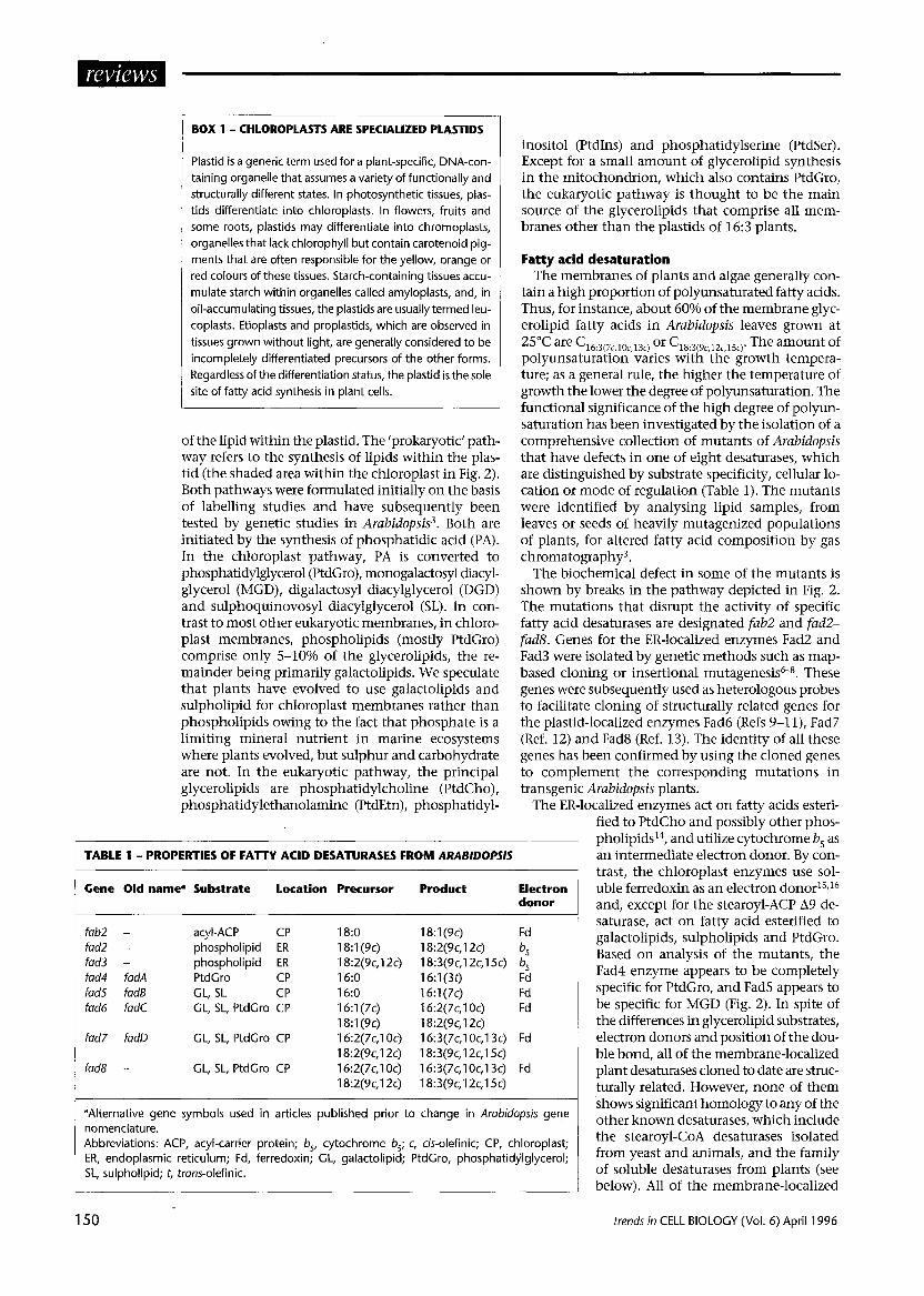
BOX 1 - CHLOROPLASTS ARE SPECIALIZED PLASTIDS
Plastid is a generic term used for a plant-specific, DNA-con- taining organelle that assumes a variety of functionally and structurally different states. In photosynthetic tissues, plas- tids differentiate into chloroplasts. In flowers, fruits and some roots, plastids may differentiate into chromoplasts, organelles that lack chlorophyll but contain carotenoid pig- ments that are often responsible for the yellow, orange or red colours of these tissues. Starch-containing tissues accu- mulate starch within organelles called amyloplasts, and, in oil-accumulating tissues, the plastids are usually termed leu- coplasts. Etioplasts and proplastids, which are observed in tissues grown without light, are generally considered to be incompletely differentiated precursors of the other forms. Regardless of the differentiation status, the plastid is the sole site of fatty acid synthesis in plant cells.
of the lipid within the plastid. The ‘prokaryotic’ path- way refers to the synthesis of lipids within the plas- tid (the shaded area within the chloroplast in Fig. 2). Both pathways were formulated initially on the basis of labelling studies and have subsequently been tested by genetic studies in Arabidopsis3. Both are initiated by the synthesis of phosphatidic acid (PA). In the chloroplast pathway, PA is converted to phosphatidylglycerol (PtdGro), monogalactosyl diacyl- glycerol (MGD), digalactosyl diacylglycerol (DGD) and sulphoquinovosyl diacylglycerol (SL). In con- trast to most other eukaryotic membranes, in chloro- plast membranes, phospholipids (mostly PtdGro) comprise only 5-10% of the glycerolipids, the re- mainder being primarily galactolipids. We speculate that plants have evolved to use galactolipids and sulpholipid for chloroplast membranes rather than phospholipids owing to the fact that phosphate is a limiting mineral nutrient in marine ecosystems where plants evolved, but sulphur and carbohydrate are not. In the eukaryotic pathway, the principal glycerolipids are phosphatidylcholine (PtdCho), phosphatidylethanolamine (PtdEtn), phosphatidyl-
inositol (PtdIns) and phosphatidylserine (PtdSer). Except for a small amount of glycerolipid synthesis in the mitochondrion, which also contains PtdGro, the eukaryotic pathway is thought to be the main source of the glycerolipids that comprise all mem- branes other than the plastids of 16:3 plants.
Fatty acid desaturation The membranes of plants and algae generally con-
tain a high proportion of polyunsaturated fatty acids. Thus, for instance, about 60% of the membrane glyc- erolipid fatty acids in Arabidopsis leaves grown at 25”c are %:3(7c lOc,13c) or G3:3(9c,l2c,l5c)~ The amount of polyunsaturation varies with the growth tempera- ture; as a general rule, the higher the temperature of growth the lower the degree of polyunsaturation. The functional significance of the high degree of polyun- saturation has been investigated by the isolation of a comprehensive collection of mutants of Arabidopsis that have defects in one of eight desaturases, which are distinguished by substrate specificity, cellular lo- cation or mode of regulation (Table 1). The mutants were identified by analysing lipid samples, from leaves or seeds of heavily mutagenized populations of plants, for altered fatty acid composition by gas chromatography”.
TABLE 1 - PROPERTIES OF FAlTY ACID DESATURASES FROM ARAB/DO/T/S I
Gene Old name0 Substrate Location Precursor Product
acyl-ACP CP phospholipid ER phospholipid ER PtdCro CP CL, SL CP CL, SL, PtdGro CP
CL, SL, PtdCro CP
GL, SL, PtdGro CP
The biochemical defect in some of the mutants is shown by breaks in the pathway depicted in Fig. 2. The mutations that disrupt the activity of specific fatty acid desaturases are designated fab2 and fad2- fads. Genes for the ER-localized enzymes Fad2 and Fad3 were isolated by genetic methods such as map- based cloning or insertional mutagenesis6-s. These genes were subsequently used as heterologous probes to facilitate cloning of structurally related genes for the plastid-localized enzymes Fad6 (Refs 9-ll), Fad7 (Ref. 12) and Fad8 (Ref. 13). The identity of all these genes has been confirmed by using the cloned genes to complement the corresponding mutations in transgenic Arabidopsis plants.
The ER-localized enzymes act on fatty acids esteri- fied to PtdCho and possibly other phos- pholipids14, and utilize cytochrome b, as an intermediate electron donor. By con- trast, the chloroplast enzymes use sol- uble ferredoxin as an electron donor*5J6 and, except for the stearoyl-ACP A9 de- saturase, act on fatty acid esterified to galactolipids, sulpholipids and PtdGro. Based on analysis of the mutants, the Fad4 enzyme appears to be completely specific for PtdGro, and Fad5 appears to be specific for MGD (Fig. 2). In spite of the differences in glycerolipid substrates, electron donors and position of the dou- ble bond, all of the membrane-localized plant desaturases cloned to date are struc- turally related. However, none of them shows significant homology to any of the other known desaturases, which include the stearoyl-CoA desaturases isolated from yeast and animals, and the family of soluble desaturases from plants (see below). All of the membrane-localized
Electron donor
fab2 - fad2 - fad3 - fad4 fadA fad5 fad6 fad6 fadC
fad7 fadll
fad8 -
1 B:O 18:1(9c) Fd 181 (SC) 18:2(9c,lZc) b, 18:2(9c,12c) 18:3(9c,12c,l5c) b, 16:0 16:1(3t) Fd 16:O 16:1(7c) Fd 16:1(7c) 16:2(7c,l Oc) Fd 18:1(9c) 18:2(9c,12c) 16:2(7c,l Oc) 16:3(7c,l Oc,l3c) Fd 18:2(9c,12c) 18:3(9c,12c,l5c) 16:2(7c,l Oc) 16:3(7c,l Oc,l3c) Fd 18:2(9c,12c) 18:3(9c,12c,l5c)
aAlternative gene symbols used in articles published prior to change in Arabidopsis geve nomenclature. Abbreviations: ACP, acyl-carrier protein; b,, cytochrome b,; c, cis-olefinic; CP, chloroplast; ER, endoplasmic reticulum; Fd, ferredoxin; CL, galactolipid; PtdGro, phosphatidylglycerol; SL, sulpholipid; t, trans-olefinic.
150 trends in CELL BIOLOGY (Vol. 6) April 1996

desaturases from plants, as well as the desaturases from animals and yeast, contain a pair of histidine- rich sequences (HXXHH) that have been proposed to bind two iron molecules that are required for catalysis17,18.
From analysis of the effects of the mutations on glycerolipid fatty acid composition, it appears that a mutant is available for every desaturase in Arabidopsis except stearoyl-ACP A9 desaturase, which is encoded by multiple copies of the gene. Thus, the fab2 mu- tation, which is thought to inactivate one of the stearoyl-ACP desaturase genes, causes a significant in- crease in stearate but does not eliminate the produc- tion of polyunsaturated fatty acids19.
Until recently, the stearoyl-ACP A9 desaturase from plants was the only known soluble desaturase. The enzyme was purified from several plants, and genes encoding it have been isolated from a large number of species. In the first example of the genetic engi- neering of plant oils, an antisense version of a gene for this enzyme from Brassica nap was used to increase the stearate content of the seed oil in transgenic B. napus plants 20. Recently, it has become apparent that the stearoyl-ACP A9 desaturase is the prototype of a distinct family of structurally similar enzymes that introduce the double bond at various locations along the acyl chain. Species-specific genes have been isolated recently from coriander (Coriandn~m sativum) and black-eyed Susan (Thunbergia alata) for two additional isoforms, which catalyse desaturations at the A4 and A6 positions of stearic acidZ1J2.
The solubility of these enzymes has greatly facili- tated structural and mechanistic studies. By express- ing an active form of the castor bean stearoyl-ACP A9 desaturase in Escherichia coli, diffracting crystals have been produced Z3~that are being used to obtain the tertiary structure of the enzyme. The sequences of the three known soluble desaturases are sufficiently homologous that it may be possible to deduce the structural basis for the position of double-bond in- sertion by mapping the sequences of the A4 and A6 desaturases onto the structure of the A9 desaturase. Mossbauer spectrometry of the recombinant castor enzyme from cultures of E. coli grown in 57Fe revealed the presence of a binuclear iron centre of the type found in methane monooxygenasez4. Assuming that this site is also found in other fatty acid desaturases, it provides an attractive explanation for many prop- erties of the desaturases, such as the observation that the mammalian stearoyl-CoA desaturase reaction re- quires a two-electron transfe?.
A central question concerns the mechanisms that control desaturation; what factors determine the level of desaturation of a specific glycerolipid in a particular membrane? In cyanobacteria, the steady- state level of a Al2 desaturase mRNA is inversely cor- related with temperature. Cells grown at 30°C have approximately 10% of the amount of this mRNA compared with cells grown at 10°C (Ref. 26). The stimulatory effect of low temperature on desaturase gene expression can be mimicked by using non- lethal catalytic hydrogenation to reduce the number of double bonds in membranes of living cells27. Thus, it appears that cyanobacteria have a mechanism that
senses a physical property of the membrane and regulates the level of desaturase mRNA accordingly.
Most higher plants also increase the level of mem- brane glycerolipid desaturation when grown at low temperature. However, with one exception, the amounts of mRNAs for the genes encoding desatu- rases in Arabidopsis do not appear to change signifi- cantly in response to changes in growth temperature. The one exception concerns the fad8 gene, which ex- hibits a strong increase in mRNA abundance at low temperature13. However, the amount of fad8 mRNA is not altered in fad7 ftzd8 double mutants, which are highlY deficient in c16:3(7c,10c,13c) and C18:3(9c,12c,15c)~ If there were a mechanism that regulated desaturase gene expression in response to changes in the physi- cal properties of membranes, we should expect that, in the fad7 fad8 mutant, this mechanism would de- tect the large change in membrane composition caused by the mutations and would signal an increase in desaturase gene expression. The fact that none of the fad mutations alters fad gene expression indicates that, if there is such a sensing mechanism in higher plants, it must act at the post-transcriptional level. Indeed, a post-translational mechanism has recently been shown to regulate the PtdCho:PtdEtn ratio in the ER of yeast 28. Presumably, the complexities asso- ciated with regulating the composition of a dozen in- tracellular membranes in higher plants requires post- transcriptional mechanisms that can adjust the composition of each membrane independently. Two incompletely characterized mutations, designated ela and rod1 (Fig. 2), cause increased accumulation of C18:3c9r 12c lscj and C18:.(9cj, respectively. Since these mutations do not appear to affect the structural genes for the corresponding desaturases, it is possible that they affect factors that regulate the desaturases of the eukaryotic pathway.
lnterorganelle lipid transfer Several lines of evidence indicate the existence of
regulatory mechanisms that coordinate the activity of the two pathways for glycerolipid synthesis in plants. The act1 mutants of Arubidopsis are deficient in ac- tivity of the chloroplast acyl-ACP:sn-glycerol-3-phos- phate acyltransferase (EC 2.3.1.15) - the first enzyme of the prokaryotic pathway (Fig. 2). The deficiency of this activity is compensated for by increased synthesis of chloroplast glycerolipids via the eukaryotic path- wayz9. These and related results indicate that, even in the face of a major disruption of one of the pathways of glycerolipid synthesis, mechanisms exist that ensure the synthesis and transfer of enough glycerolipids to support normal rates of membrane biogenesis. This striking example raises many unanswered questions. In particular, how is the demand for increased glyc- erolipid synthesis communicated to the lipid bio- synthetic pathways during membrane expansion?
A partial answer to the foregoing question lies in the recognition that, at least for the ER and the plas- tid, lipid traffic between the membranes is bidirec- tional. Detailed analysis of the effects of the various fad mutations on the fatty acid composition of mem- branes indicates that, even though the enzymes are located exclusively in one compartment or the other,
trends in CELL BIOLOGY (Vol. 6) April 1996 151

FIGURE 3
Decreased fatty acid unsaturation in the fab2 mutant of Arabidopis (a) leads to formation of leaf mesophyll cells that
are small compared with those of the wild-type (b). Trichome cells that arise from the epidermis are normal in
size (reproduced, with permission, from Ref. 33).
most of the mutations affect the composition of both chloroplast and extrachloroplast membranes30. Thus, lipids are transferred from the ER to the chloroplast (and other membranes), but lipids are also transferred from the chloroplast to extrachloroplast membranes. As most of the chloroplast glycerolipids are not found in other membranes, these lipids must be converted to some other form (e.g. PtdCho or PtdEtn) before or immediately after transfer. Similarly, PtdCho, which is the principal glycerolipid in the extrachloroplast membranes, is found only in the outer leaflet of the chloroplast envelope, and chloroplasts completely lack PtdEtn, the other major component of the ER. Thus, it seems likely that PtdCho, or another lipid, is transferred and is converted to one of the common chloroplast lipids in the chloroplast envelope before being transferred to the inner membranes of the chloroplast.
A confounding problem for this scenario is that there is no evidence for vesicular traffic between the ER and the plastids. Thus, there has been sustained interest in the possible role of lipid transfer proteins (LTPs) as carriers of lipids between the various mem- branes. However, it appears that the plant LTPs, which were originally identified by in vitro assays, are actually located outside of the plasma membrane in the cell-wall space of epidermal cells. The function of
152
these LTPs is unknown, but, based on their location, it has been proposed that LTPs participate in trans- ferring wax and cutin monomers from the plasma membrane to the cell surface31,32. This is clearly an area of study that would benefit from more detailed investigation. It seems unlikely that it will be poss- ible to understand the factors that control the amount and composition of membranes without de- tailed knowledge of how lipid moves between mem- branes and of which species move.
The biological role of polyunsaturation A dramatic example of an effect of membrane glyc-
erolipid fatty acid composition on plant growth and development is afforded by the fub2 mutant, which is characterized by elevated C,,:, (Ref. 33). The fab2 mutant plants are dwarfs that grow to less than about 10% of the size of the wild type. Many cell types in the mutant, including leaf mesophyll and epidermal cells, fail to expand, whereas others, such as stomata1 cells and trichomes, develop to normal size (Fig. 3). Growth at high temperature ameliorates the effects of the mutation on growth and development with- out changing the fatty acid composition. These effects can be explained in a general sense by the known physical properties of lipids. At normal growth temperatures, an increased proportion of C1aZo in membranes would be expected to decrease fluidity or lead to the formation of domains containing gel- phase lipids. It appears that some aspect of the pro- cesses leading to cellular expansion is dependent on membrane fluidity or a related physical property of one or more membranes. These effects would be ame- liorated by growth at high temperatures because ther- mal energy weakens the van der Waals interactions between adjacent fatty acids in the membrane.
With the exception of the fub2 mutant, none of the single-gene desaturase mutants shows any visible phenotype at normal growth temperatures (ZS’C) or following several days of exposure to low tempera- tures. Thus, it appears that polyunsaturation is not required for the normal function of the various mem- branes. However, wild-type alleles of the fud2, fad5 and fad6 genes are required for the normal growth of plants during extended exposure to low temperature. The fad2 mutant, which is deficient in C18:2(9c lZrj and C18:3(9c,12c,15c) lipids in extrachloroplast membranes, ceases growth after transfer to 4°C but does not show visible signs of tissue damage for several weeks34. Similarly, plants with the fad.5 or fad6 mutations, which affect primarily the composition of chloroplast glycerolipids, fail to accumulate normal levels of chlorophyll when grown at low temperature. By con- trast, tissue that is allowed to develop at normal tem- peratures does not show any deleterious effects (e.g. loss of chlorophyll) following transfer to low tem- perature. Morphometric analyses of chloroplast mem- branes from fad.5 and fad6 plants grown at low tem- perature indicated that the reduced accumulation of chlorophyll was associated with an up to fourfold de- crease in the amount of chloroplast membranes in the mutants at low temperature35. This was interpreted as suggesting an important role for polyunsaturation in membrane biogenesis at low temperature.
trends in CELL BIOLOGY (Vol. 6) April 1996

Direct evidence for an effect of fatty acid unsatu- ration on membrane biogenesis was recently obtained from an analysis of the effects of low temperature on the turnover of the Dl protein of the photosystem II reaction centre36. Under conditions of high irradi- ante, the membrane-localized Dl protein is normally damaged and replaced at a high rate. The rate of damage and repair of the Dl protein was measured in transgenic tobacco plants in which the proportion of chloroplast PtdGro-containing saturated fatty acids was altered by expression of an isoform of glycerol-3-phosphate acyltransferase with a prefer- ence for saturated fatty acyl substrates36. At low temperature, the rate of turnover of the Dl protein was much slower in the transgenic plants with high levels of saturated fatty acids than in the wild type. It is unknown whether the change in turnover rate is due to a change in the rate of removal of damaged protein or in the rate of insertion of new Dl protein. Similar results were also observed in a mutant of Synechocystis that was deficient in polyunsatu- ration37. These studies have provided a promising new focus and novel experimental resources for the investigation of long-standing questions concerning the role of polyunsaturation.
We anticipate that some of the insights gained from studies of lipid synthesis in plants and cyanobacteria will permit new advances in under- standing comparable questions concerning non- plant organisms. Indeed, we have noticed in the pub- lic data bases the existence of uncharacterized partial cDNA clones from animals that show sequence hom- ology to some of the desaturases from Arabidopsis.
References 1 OHLROCCE, J. and BROWSE, 1. (1995) Plant Cell 7,957-970 2 GIBSON, S., FALCONE, D., BROWSE, J. and SOMERVILLE, C. R.
(1994) Plant Cell Environ. 17, 627-637 3 SOMERVILLE, C. R. and BROWSE, J. (1991) Science 252, 80-87 4 SCHAFFER, J. C. and LODISH, H. F. (1994) Cell 79,427-436 5 JONES, A., MAELOR, H. and VOELKER, T. A. (1995) Plant Cell 7,
359-371 6 ARONDEL, V., LEMIEUX, B., HWANG, I., GIBSON, S.,
GOODMAN, H. and SOMERVILLE, C. R. (1992) Science 258, 1353-l 355
7 YADAV, N. et al. (1994) P/ant Cell 6, 147-l 58 8 OKULEY, I., LIGHTNER, j., FELDMANN, K., YADAV, N., LARK, E.
and BROWSE, 1. (1994) P/ant Cell 6, 147-l 58 9 HITZ, W. D., CARLSON, T. I., BOOTH, J. R., KINNEY, A. J.,
STECCA, K. L. and YADAV, N. 5. (1994) P/ant Physiol. 105, 635-641
IO FALCONE, D., GIBSON, S., LEMIEUX, B. and SOMERVILLE, C. R. (1994) Plant Physiol. 106, 1453-l 459
11 SCHMIDT, H., DRESSLHAUS, T., BUCK, F. and HEINZ, E. (1994) P/ant MO/. Biol. 26, 631-642
12 IBA, K. et a/. (1993) 1. Biol. Chem. 268, 24099-24105 13 GIBSON, S., ARONDEL, V., I&A, K. and SOMERVILLE, C. R.
(1994) Plant fhysiol. 106, 1615-I 621 14 SPERLING, P., LINSCHEID, M., STOECKER, S., MUEHLBACH, H. P.
and HEINZ, E. (1993) I. Biol. Chem. 268, 26935-26940 15 SCHMID, T. H. and HEINZ, E. (1990) Proc. Nat/ Acad. Sci. USA
87,9477-9480 16 SCHMIDT, H. and HEINZ, E. (1993) Biochem. I. 289, 777-782 17 SHANKLIN, J., WHITTLE, E. and FOX, B. G. (1994)
trends in CELL BIOLOGY (Vol. 6) April 1996
Biochemistry 33,12787-l 2794 18 FOX, B. G., SHANKLIN, J., Al, J., LOEHR, T. M. and
SANDERS-LOEHR, J. (1994) Biochemistry 33,12776-l 2786 19 LIGHTNER, J., WU, 1. and BROWSE, J. (1994) Plant Physiol.
106,1443-1451 20 KNUTZON, D. S., THOMPSON, G. A., RADKE, S. E.,
JOHNSON, W. B., KNAUF, V. C. and KRIDL, J. C. (1992) hoc. Nat/ Acad. Sci. USA 89, 2624-2628
21 CAHOON, E. B., SHANKLIN, J. and OHLROGGE, J. (1992) hoc. Not/ Acad. ki. USA 89, 11184-l 1188
22 CAHOON, E.G., CRAMNER, A. M., SHANKLIN, J. and OHLROGGE, J. B. (1994) I. Biol. Chem. 269,27519-27526
23 SCHNEIDER, G., LINDQVIST, Y., SHANKLIN, J. and SOMERVILLE, C. R. (1992) 1. Mol. Biol. 225, 561-564
24 FOX, B. G., SHANKLIN, J., SOMERVILLE, C. R. and MeNCK, E. (1993) Proc. Nat/ Acad. Sci. USA 90, 2486-2490
25 ENOCH, H. G., CATALA, A. and STRIllMAllER, P. (1976) 1. Biol. Cbem. 251,5095-5103
26 LOS, D., HORVATH, I., VIGH, L. and MURATA, N. (1993) FEBS lett. 318, 57-60
27 VIGH, L., LOS, D., HORVATH, I. and MURATA, N. (1993) Proc. Nat/ Acad. Sci. USA 90, 9090-9094
28 SKINNER, H. B., MCGEE, T., McMASTER, C. R., FRY, M. R., BELL, R. M. and BANKAITIS, V. (1995) Proc. Nat/ Acad. Sci. USA 92,112-116
29 KUNST, L., BROWSE, J. and SOMERVILLE, C. R. (1988) Proc. Nat/ Acad. Sci. USA 85,4143-4147
30 BROWSE, J., McCONN, M., JAMES, D. and MIQUEL, M. (1993) 1. Biol. Chem. 268,16345-l 6351
31 STERK, P., BOOIF, H., SCHELLEKENS, G. A., VAN KAMMEN, A. and DE VRIES, 5. C. (1991) Plant Cell 3, 907-921
32 THOMA, S. L., KANEKO, Y. and SOMERVILLE, C. R. (1993) Plant]. 3, 427-437
33 LIGHTNER, J., JAMES, D. W., Jr, DOONER, H. K. and BROWSE, J. (1994) P/ant/. 6,401412
34 MIQUEL, M., JAMES, D., DOONER, H. and BROWSE, J. (1993) hoc. Nat/ Acad. Sci. USA 90, 6208-6212
3.5 HUGLY, 5. and SOMERVILLE, C. R. (1992) Plant Physiol. 99, 197-202
36 MOON, B. Y., HIGASHI, S. I., COMBOS, Z. and MURATA, N. (1995) Proc. Nat/ Acad. Sci. USA 92, 6219-6223
37 GOMBOS, Z., WADA, H. and MURATA, N. (1994) Proc. Nat/ Acad. Sci. USA 91, 8787-8791
38 VAN DE LOO, F., FOX, B. and SOMERVILLE, C. R. (1993) in Plant lipids (Moore, T., ed.), pp. 91-126, CRC Press
LipidSi
153