Embed Size (px)
Citation preview

138 WMJ • JUNE 2012
• • •
Author Affiliations: Department of Hematology/Oncology, Marshfield Clinic Weston Center, Weston, Wis (Onitilo, Depke); A.T. Still University School of Osteopathic Medicine, Mesa, Ariz (Demos-Bertrand); Department of Laboratory Pathology, Marshfield Clinic, Marshfield, Wis (Resnick); Department of Hematology/Oncology, Marshfield Clinic at Ministry St. Michael’s Hospital, Stevens Point, Wis (Engel).
Corresponding Author: Adedayo A. Onitilo, MD; Department of Hematology/Oncology, Marshfield Clinic Weston Center, 3501 Cranberry Blvd, Weston, WI 54476; ph 715.393.1400; fax 715.393.1399; e-mail [email protected].
CASE REPORT
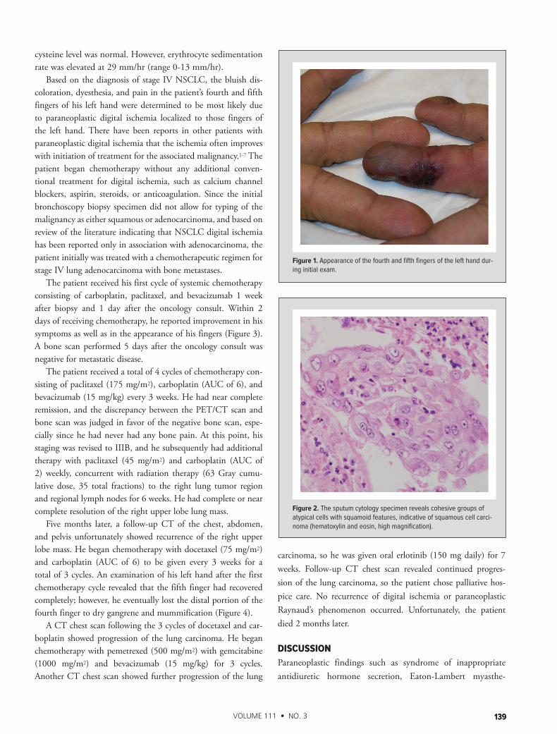
CASE REPORTA 40-year-old white man with a 20-pack-per-year history of smoking ini-tially presented with cough, fatigue, and decreased appetite. He was diagnosed with pneumonia by chest radiograph and treated with antibiotics without improvement. Two weeks later, the distal phalanxes of the fourth and fifth fingers
of his left hand became bluish-colored with dyesthesia and pain (Figure 1). The temperature of these fingers was normal and peripheral pulses were palpable. There was no sign or history of arterial trauma and no known family or patient history of Raynaud’s phenomenon. A venous Doppler ultrasound of the left arm was negative for deep vein thrombosis. No other sites of superficial ischemia were observed. His symptoms persisted, even with pain management with acetaminophen and hydroco-done (Vicodin) prescribed as needed. Three weeks after initial presentation, a computed tomography (CT) chest scan showed a 4.5-centimeter right-sided bronchial mass, complete obstruc-tion of the bronchus, and collapse of the right upper lobe with bilateral hilar and mediastinal lymphadenopathy. Two days later, bronchoscopy with biopsy and brochioaveolar lavage was performed. The specimen, while demonstrating non-small cell carcinoma, did not allow for more precise classification of the tumor. Subsequently, sputum cytology was collected, reviewed by a cytopathologist, and ultimately revealed a squamous cell carcinoma (Figure 2). Positron emission tomography/CT (PET/CT) scan 1 week later showed a right lung mass (stan-dardized uptake value [SUV] >10) extending into the medias-tinum and mildly increased activity in dorsal and lumbar verte-bral bodies and the pelvis (SUV 3.5).
The patient presented for an oncology consult the next day. Based on the PET/CT, which indicated bone metastases, he was diagnosed with stage IV non-small cell lung carcinoma (NSCLC). A thrombophilia workup to determine hypercoagu-lability state was performed. Tests for cryoglobulin, antiphos-pholipid antibody, antinuclear antibody, anticardiolipin antibody, and lupus anticoagulant all were negative, and homo-
INTRODUCTIONDigital ischemia as a paraneoplastic syndrome of lung carci-noma is an unusual finding. It may be a complication of its own or may be associated with paraneoplastic Raynaud’s phe-nomenon. Only 13 cases of digital ischemia, or paraneoplastic Raynaud’s phenomenon accompanied by digital ischemia, in association with lung carcinoma were found during a review of the literature utilizing PubMed and OVID and the search terms “lung carcinoma,” “Raynaud’s phenomenon,” “digi-tal ischemia,” and “paraneoplastic.” We report a patient who, based on the literature review, is the youngest patient to date with digital ischemia associated with lung carcinoma and the only one with a squamous cell differentiation tumor type.
ABSTRACTWe report the case of a 40-year-old man who presented with digital ischemia and squamous cell lung carcinoma. Based on review of the literature, to our knowledge this case represents the youngest patient with lung carcinoma associated with digital ischemia and the only one with this type of tumor. The patient’s digital ischemia symptoms improved rapidly with systemic chemotherapy; however, he did eventually lose the distal portion of 1 finger to dry gangrene and mummification.
Adedayo A. Onitilo, MD, MSCR, FACP; Jennifer Demos-Bertrand, BS; Jill Depke, MSN, AOCNP; Jeffrey M. Resnick, MD; Jessica Engel, MSN, AOCNP
Digital Ischemia as a Paraneoplastic Consequence of Squamous Cell Lung Carcinoma
CME available. See page 142 for more information.
139VOLUME 111 • NO. 3 139
carcinoma, so he was given oral erlotinib (150 mg daily) for 7 weeks. Follow-up CT chest scan revealed continued progres-sion of the lung carcinoma, so the patient chose palliative hos-pice care. No recurrence of digital ischemia or paraneoplastic Raynaud’s phenomenon occurred. Unfortunately, the patient died 2 months later.
DISCUSSIONParaneoplastic findings such as syndrome of inappropriate antidiuretic hormone secretion, Eaton-Lambert myasthe-
cysteine level was normal. However, erythrocyte sedimentation rate was elevated at 29 mm/hr (range 0-13 mm/hr).
Based on the diagnosis of stage IV NSCLC, the bluish dis-coloration, dyesthesia, and pain in the patient’s fourth and fifth fingers of his left hand were determined to be most likely due to paraneoplastic digital ischemia localized to those fingers of the left hand. There have been reports in other patients with paraneoplastic digital ischemia that the ischemia often improves with initiation of treatment for the associated malignancy.1-7 The patient began chemotherapy without any additional conven-tional treatment for digital ischemia, such as calcium channel blockers, aspirin, steroids, or anticoagulation. Since the initial bronchoscopy biopsy specimen did not allow for typing of the malignancy as either squamous or adenocarcinoma, and based on review of the literature indicating that NSCLC digital ischemia has been reported only in association with adenocarcinoma, the patient initially was treated with a chemotherapeutic regimen for stage IV lung adenocarcinoma with bone metastases.
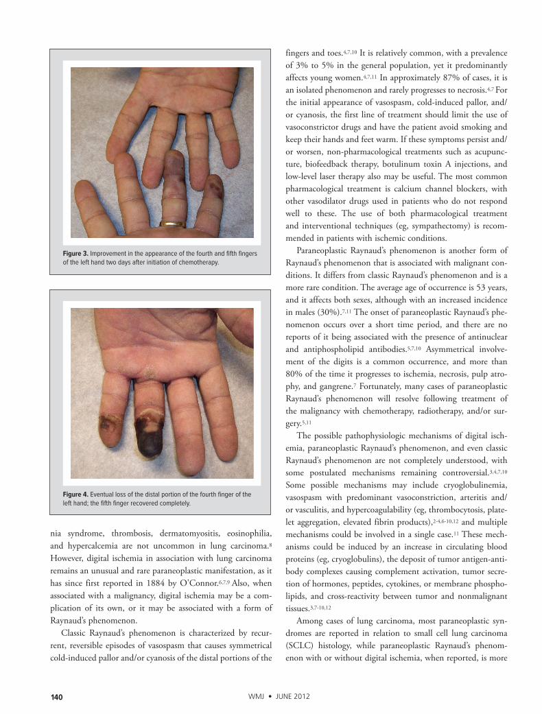
The patient received his first cycle of systemic chemotherapy consisting of carboplatin, paclitaxel, and bevacizumab 1 week after biopsy and 1 day after the oncology consult. Within 2 days of receiving chemotherapy, he reported improvement in his symptoms as well as in the appearance of his fingers (Figure 3). A bone scan performed 5 days after the oncology consult was negative for metastatic disease.
The patient received a total of 4 cycles of chemotherapy con-sisting of paclitaxel (175 mg/m2), carboplatin (AUC of 6), and bevacizumab (15 mg/kg) every 3 weeks. He had near complete remission, and the discrepancy between the PET/CT scan and bone scan was judged in favor of the negative bone scan, espe-cially since he had never had any bone pain. At this point, his staging was revised to IIIB, and he subsequently had additional therapy with paclitaxel (45 mg/m2) and carboplatin (AUC of 2) weekly, concurrent with radiation therapy (63 Gray cumu-lative dose, 35 total fractions) to the right lung tumor region and regional lymph nodes for 6 weeks. He had complete or near complete resolution of the right upper lobe lung mass.
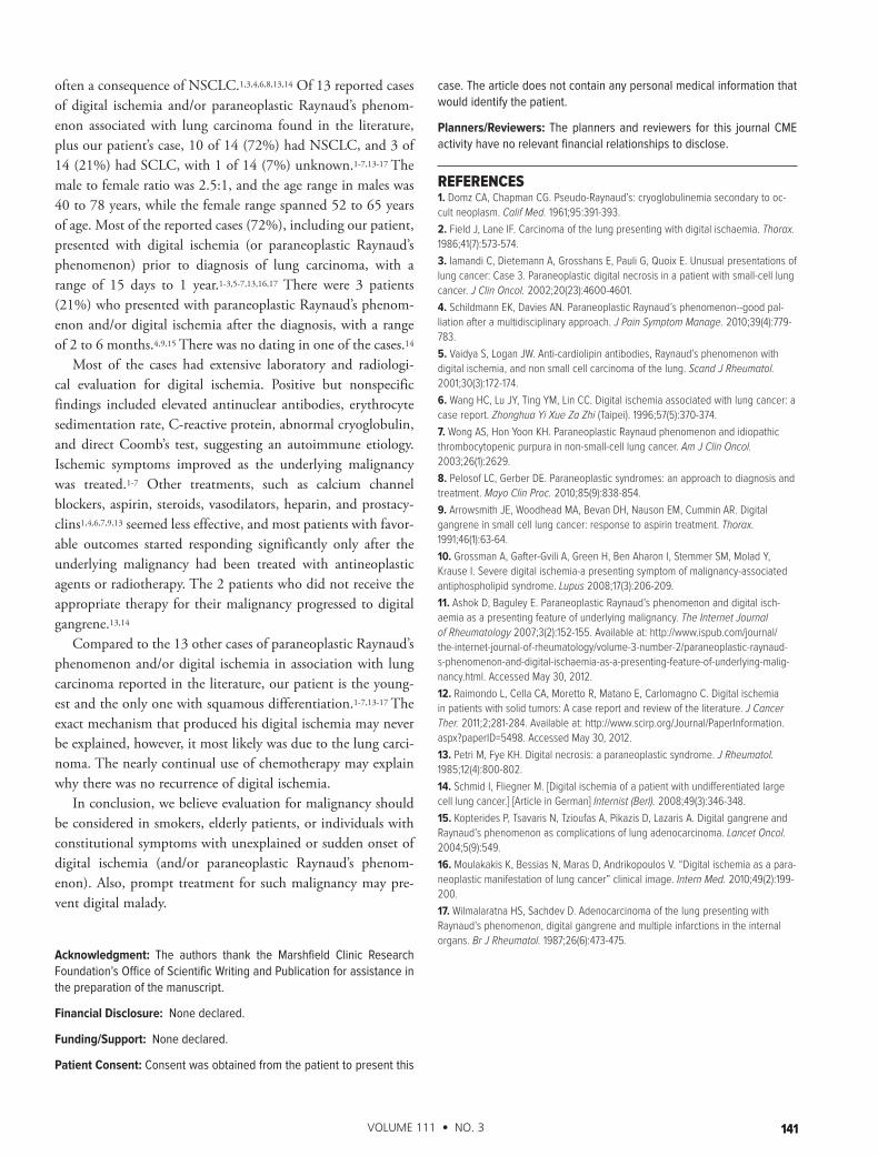
Five months later, a follow-up CT of the chest, abdomen, and pelvis unfortunately showed recurrence of the right upper lobe mass. He began chemotherapy with docetaxel (75 mg/m2) and carboplatin (AUC of 6) to be given every 3 weeks for a total of 3 cycles. An examination of his left hand after the first chemotherapy cycle revealed that the fifth finger had recovered completely; however, he eventually lost the distal portion of the fourth finger to dry gangrene and mummification (Figure 4).
A CT chest scan following the 3 cycles of docetaxel and car-boplatin showed progression of the lung carcinoma. He began chemotherapy with pemetrexed (500 mg/m2) with gemcitabine (1000 mg/m2) and bevacizumab (15 mg/kg) for 3 cycles. Another CT chest scan showed further progression of the lung
Figure 1. Appearance of the fourth and fifth fingers of the left hand dur-ing initial exam.
Figure 2. The sputum cytology specimen reveals cohesive groups of atypical cells with squamoid features, indicative of squamous cell carci-noma (hematoxylin and eosin, high magnification).
140 WMJ • JUNE 2012
fingers and toes.4,7,10 It is relatively common, with a prevalence of 3% to 5% in the general population, yet it predominantly affects young women.4,7,11 In approximately 87% of cases, it is an isolated phenomenon and rarely progresses to necrosis.4,7 For the initial appearance of vasospasm, cold-induced pallor, and/or cyanosis, the first line of treatment should limit the use of vasoconstrictor drugs and have the patient avoid smoking and keep their hands and feet warm. If these symptoms persist and/or worsen, non-pharmacological treatments such as acupunc-ture, biofeedback therapy, botulinum toxin A injections, and low-level laser therapy also may be useful. The most common pharmacological treatment is calcium channel blockers, with other vasodilator drugs used in patients who do not respond well to these. The use of both pharmacological treatment and interventional techniques (eg, sympathectomy) is recom-mended in patients with ischemic conditions.
Paraneoplastic Raynaud’s phenomenon is another form of Raynaud’s phenomenon that is associated with malignant con-ditions. It differs from classic Raynaud’s phenomenon and is a more rare condition. The average age of occurrence is 53 years, and it affects both sexes, although with an increased incidence in males (30%).7,11 The onset of paraneoplastic Raynaud’s phe-nomenon occurs over a short time period, and there are no reports of it being associated with the presence of antinuclear and antiphospholipid antibodies.5,7,10 Asymmetrical involve-ment of the digits is a common occurrence, and more than 80% of the time it progresses to ischemia, necrosis, pulp atro-phy, and gangrene.7 Fortunately, many cases of paraneoplastic Raynaud’s phenomenon will resolve following treatment of the malignancy with chemotherapy, radiotherapy, and/or sur-gery.5,11
The possible pathophysiologic mechanisms of digital isch-emia, paraneoplastic Raynaud’s phenomenon, and even classic Raynaud’s phenomenon are not completely understood, with some postulated mechanisms remaining controversial.3,4,7,10
Some possible mechanisms may include cryoglobulinemia, vasospasm with predominant vasoconstriction, arteritis and/or vasculitis, and hypercoagulability (eg, thrombocytosis, plate-let aggregation, elevated fibrin products),2-4,6-10,12 and multiple mechanisms could be involved in a single case.11 These mech-anisms could be induced by an increase in circulating blood proteins (eg, cryoglobulins), the deposit of tumor antigen-anti-body complexes causing complement activation, tumor secre-tion of hormones, peptides, cytokines, or membrane phospho-lipids, and cross-reactivity between tumor and nonmalignant tissues.3,7-10,12
Among cases of lung carcinoma, most paraneoplastic syn-dromes are reported in relation to small cell lung carcinoma (SCLC) histology, while paraneoplastic Raynaud’s phenom-enon with or without digital ischemia, when reported, is more
nia syndrome, thrombosis, dermatomyositis, eosinophilia, and hypercalcemia are not uncommon in lung carcinoma.8
However, digital ischemia in association with lung carcinoma remains an unusual and rare paraneoplastic manifestation, as it has since first reported in 1884 by O’Connor.6,7,9 Also, when associated with a malignancy, digital ischemia may be a com-plication of its own, or it may be associated with a form of Raynaud’s phenomenon.
Classic Raynaud’s phenomenon is characterized by recur-rent, reversible episodes of vasospasm that causes symmetrical cold-induced pallor and/or cyanosis of the distal portions of the
Figure 3. Improvement in the appearance of the fourth and fifth fingers of the left hand two days after initiation of chemotherapy.
Figure 4. Eventual loss of the distal portion of the fourth finger of the left hand; the fifth finger recovered completely.
141VOLUME 111 • NO. 3 141
case. The article does not contain any personal medical information that would identify the patient.
Planners/Reviewers: The planners and reviewers for this journal CME activity have no relevant financial relationships to disclose.
REFERENCES1. Domz CA, Chapman CG. Pseudo-Raynaud’s: cryoglobulinemia secondary to oc-cult neoplasm. Calif Med. 1961;95:391-393.2. Field J, Lane IF. Carcinoma of the lung presenting with digital ischaemia. Thorax. 1986;41(7):573-574.3. Iamandi C, Dietemann A, Grosshans E, Pauli G, Quoix E. Unusual presentations of lung cancer: Case 3. Paraneoplastic digital necrosis in a patient with small-cell lung cancer. J Clin Oncol. 2002;20(23):4600-4601.4. Schildmann EK, Davies AN. Paraneoplastic Raynaud’s phenomenon--good pal-liation after a multidisciplinary approach. J Pain Symptom Manage. 2010;39(4):779-783.5. Vaidya S, Logan JW. Anti-cardiolipin antibodies, Raynaud’s phenomenon with digital ischemia, and non small cell carcinoma of the lung. Scand J Rheumatol. 2001;30(3):172-174.6. Wang HC, Lu JY, Ting YM, Lin CC. Digital ischemia associated with lung cancer: a case report. Zhonghua Yi Xue Za Zhi (Taipei). 1996;57(5):370-374.7. Wong AS, Hon Yoon KH. Paraneoplastic Raynaud phenomenon and idiopathic thrombocytopenic purpura in non-small-cell lung cancer. Am J Clin Oncol. 2003;26(1):2629.8. Pelosof LC, Gerber DE. Paraneoplastic syndromes: an approach to diagnosis and treatment. Mayo Clin Proc. 2010;85(9):838-854.9. Arrowsmith JE, Woodhead MA, Bevan DH, Nauson EM, Cummin AR. Digital gangrene in small cell lung cancer: response to aspirin treatment. Thorax. 1991;46(1):63-64.10. Grossman A, Gafter-Gvili A, Green H, Ben Aharon I, Stemmer SM, Molad Y, Krause I. Severe digital ischemia-a presenting symptom of malignancy-associated antiphospholipid syndrome. Lupus 2008;17(3):206-209.11. Ashok D, Baguley E. Paraneoplastic Raynaud’s phenomenon and digital isch-aemia as a presenting feature of underlying malignancy. The Internet Journal of Rheumatology 2007;3(2):152-155. Available at: http://www.ispub.com/journal/the-internet-journal-of-rheumatology/volume-3-number-2/paraneoplastic-raynaud-s-phenomenon-and-digital-ischaemia-as-a-presenting-feature-of-underlying-malig-nancy.html. Accessed May 30, 2012.12. Raimondo L, Cella CA, Moretto R, Matano E, Carlomagno C. Digital ischemia in patients with solid tumors: A case report and review of the literature. J Cancer Ther. 2011;2;281-284. Available at: http://www.scirp.org/Journal/PaperInformation.aspx?paperID=5498. Accessed May 30, 2012.13. Petri M, Fye KH. Digital necrosis: a paraneoplastic syndrome. J Rheumatol. 1985;12(4):800-802.14. Schmid I, Fliegner M. [Digital ischemia of a patient with undifferentiated large cell lung cancer.] [Article in German] Internist (Berl). 2008;49(3):346-348.15. Kopterides P, Tsavaris N, Tzioufas A, Pikazis D, Lazaris A. Digital gangrene and Raynaud’s phenomenon as complications of lung adenocarcinoma. Lancet Oncol. 2004;5(9):549.16. Moulakakis K, Bessias N, Maras D, Andrikopoulos V. “Digital ischemia as a para-neoplastic manifestation of lung cancer” clinical image. Intern Med. 2010;49(2):199-200.17. Wilmalaratna HS, Sachdev D. Adenocarcinoma of the lung presenting with Raynaud’s phenomenon, digital gangrene and multiple infarctions in the internal organs. Br J Rheumatol. 1987;26(6):473-475.
often a consequence of NSCLC.1,3,4,6,8,13,14 Of 13 reported cases of digital ischemia and/or paraneoplastic Raynaud’s phenom-enon associated with lung carcinoma found in the literature, plus our patient’s case, 10 of 14 (72%) had NSCLC, and 3 of 14 (21%) had SCLC, with 1 of 14 (7%) unknown.1-7,13-17 The male to female ratio was 2.5:1, and the age range in males was 40 to 78 years, while the female range spanned 52 to 65 years of age. Most of the reported cases (72%), including our patient, presented with digital ischemia (or paraneoplastic Raynaud’s phenomenon) prior to diagnosis of lung carcinoma, with a range of 15 days to 1 year.1-3,5-7,13,16,17 There were 3 patients (21%) who presented with paraneoplastic Raynaud’s phenom-enon and/or digital ischemia after the diagnosis, with a range of 2 to 6 months.4,9,15 There was no dating in one of the cases.14
Most of the cases had extensive laboratory and radiologi-cal evaluation for digital ischemia. Positive but nonspecific findings included elevated antinuclear antibodies, erythrocyte sedimentation rate, C-reactive protein, abnormal cryoglobulin, and direct Coomb’s test, suggesting an autoimmune etiology. Ischemic symptoms improved as the underlying malignancy was treated.1-7 Other treatments, such as calcium channel blockers, aspirin, steroids, vasodilators, heparin, and prostacy-clins1,4,6,7,9,13 seemed less effective, and most patients with favor-able outcomes started responding significantly only after the underlying malignancy had been treated with antineoplastic agents or radiotherapy. The 2 patients who did not receive the appropriate therapy for their malignancy progressed to digital gangrene.13,14
Compared to the 13 other cases of paraneoplastic Raynaud’s phenomenon and/or digital ischemia in association with lung carcinoma reported in the literature, our patient is the young-est and the only one with squamous differentiation.1-7,13-17 The exact mechanism that produced his digital ischemia may never be explained, however, it most likely was due to the lung carci-noma. The nearly continual use of chemotherapy may explain why there was no recurrence of digital ischemia.
In conclusion, we believe evaluation for malignancy should be considered in smokers, elderly patients, or individuals with constitutional symptoms with unexplained or sudden onset of digital ischemia (and/or paraneoplastic Raynaud’s phenom-enon). Also, prompt treatment for such malignancy may pre-vent digital malady.
Acknowledgment: The authors thank the Marshfield Clinic Research Foundation’s Office of Scientific Writing and Publication for assistance in the preparation of the manuscript.
Financial Disclosure: None declared.
Funding/Support: None declared.
Patient Consent: Consent was obtained from the patient to present this

The mission of WMJ is to provide a vehicle for professional communication and continuing education for Midwest physicians and other health professionals.
WMJ (ISSN 1098-1861) is published by the Wisconsin Medical Society and is devoted to the interests of
the medical profession and health care in the Midwest. The managing editor is responsible for oversee-
ing the production, business operation and contents of the WMJ. The editorial board, chaired by the
medical editor, solicits and peer reviews all scientific articles; it does not screen public health, socioeco-
nomic, or organizational articles. Although letters to the editor are reviewed by the medical editor, all
signed expressions of opinion belong to the author(s) for which neither WMJ nor the Wisconsin Medical
Society take responsibility. WMJ is indexed in Index Medicus, Hospital Literature Index, and Cambridge
Scientific Abstracts.
For reprints of this article, contact the WMJ at 866.442.3800 or e-mail [email protected].
© 2012 Wisconsin Medical Society