Embed Size (px)
Citation preview
Part 2
Digestive System
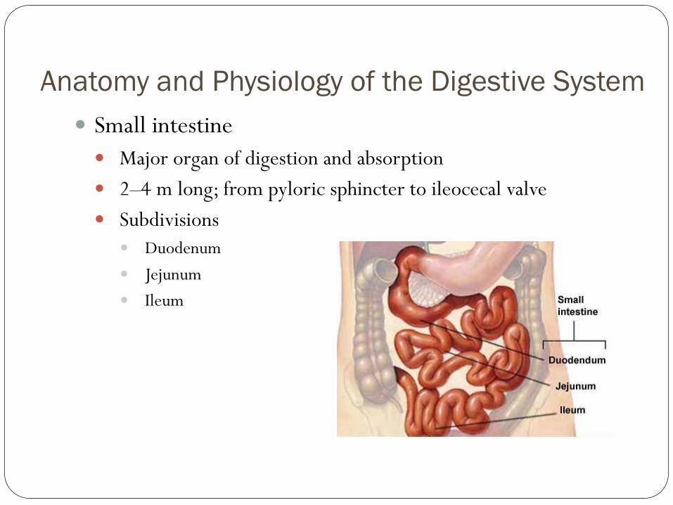
Anatomy and Physiology of the Digestive System
Small intestine
Major organ of digestion and absorption
2–4 m long; from pyloric sphincter to ileocecal valve
Subdivisions
Duodenum
Jejunum
Ileum

Anatomy and Physiology of the Digestive System
Small intestine
Structural modifications
Villi
Intestinal glands
Mucosa
Submucosa
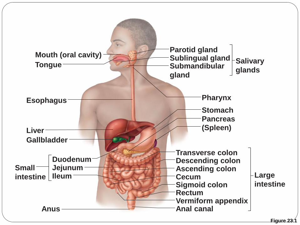
Figure 23.1
Mouth (oral cavity)
Tongue
Esophagus
Liver
Gallbladder
Anus
Duodenum Jejunum Ileum
Small
intestine
Parotid gland Sublingual gland Submandibular
gland
Salivary
glands
Pharynx
Stomach Pancreas (Spleen)
Transverse colon Descending colon Ascending colon Cecum Sigmoid colon Rectum Vermiform appendix Anal canal
Large
intestine
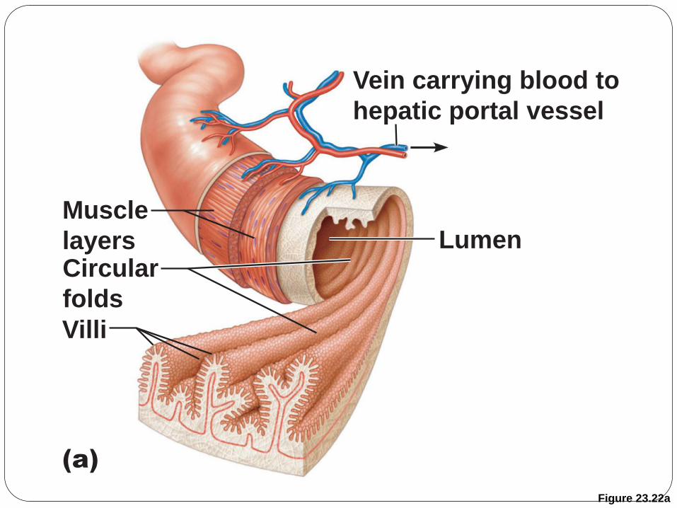
Figure 23.22a
Vein carrying blood to
hepatic portal vessel
Muscle
layers Circular
folds
Villi
(a)
Lumen
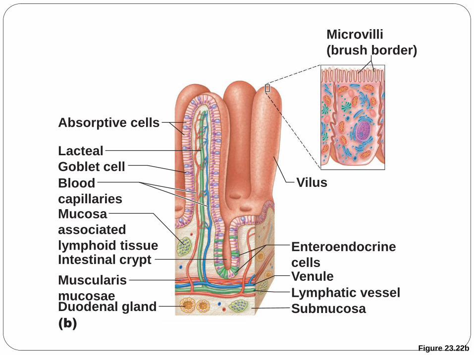
Figure 23.22b
(b)
Absorptive cells
Lacteal
Intestinal crypt
Mucosa
associated
lymphoid tissue
Muscularis
mucosae Duodenal gland Submucosa
Enteroendocrine
cells Venule
Lymphatic vessel
Goblet cell
Blood
capillaries
Vilus
Microvilli
(brush border)
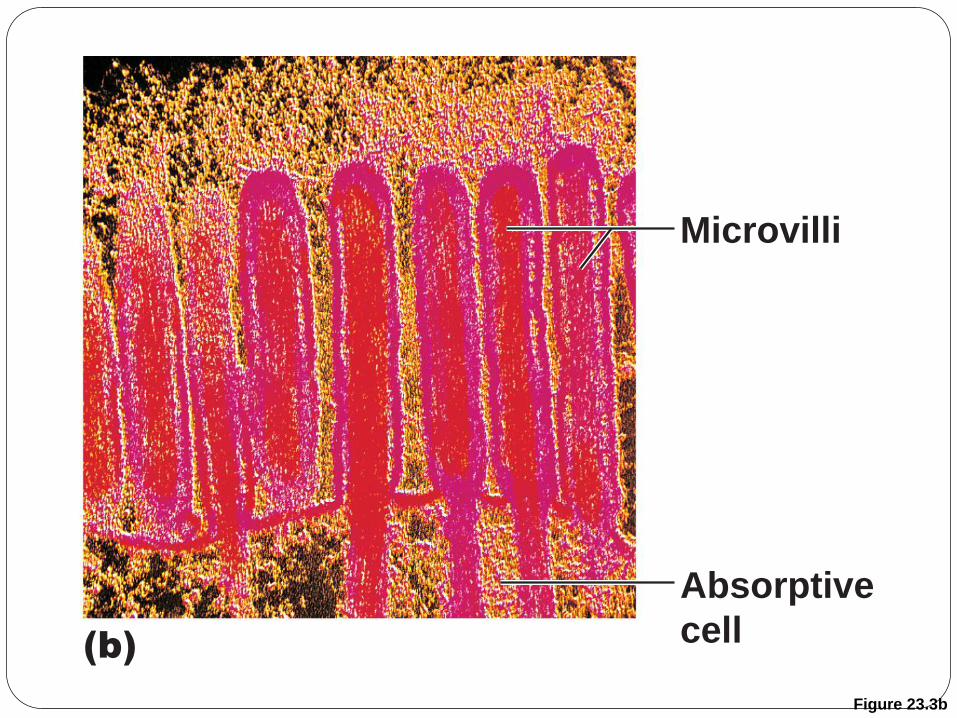
Figure 23.3b
(b)
Microvilli
Absorptive
cell
Anatomy and Physiology of the Digestive System
Chemical digestion in the small intestine
Food entering SI = partially digested
Intestinal juice
Water, mucous
Crypt cells produce lysozyme

Figure 23.22b
(b)
Absorptive cells
Lacteal
Intestinal crypt
Mucosa
associated
lymphoid tissue
Muscularis
mucosae Duodenal gland Submucosa
Enteroendocrine
cells Venule
Lymphatic vessel
Goblet cell
Blood
capillaries
Villus
Microvilli
(brush border)

Anatomy and Physiology of the Digestive System
Chemical digestion in the small intestine
Pancreatic juice
Enzymes
Amylase
o Carbohydates
Lipase
o Fats
Trypsinogen, chymotrypsinogen, carboxypeptidase
o Activated to digest protein
Sodium bicarbonate
Neutralize stomach acid
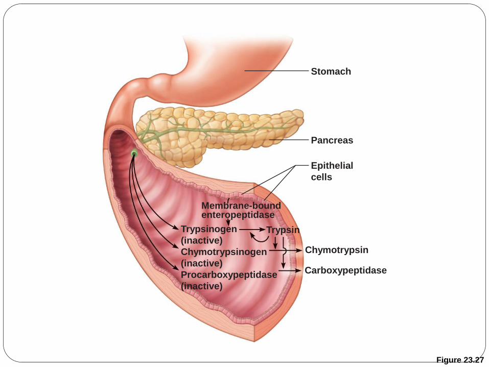
Figure 23.27
Stomach
Pancreas
Epithelial
cells
Trypsinogen
(inactive)
Chymotrypsinogen
(inactive)
Procarboxypeptidase
(inactive)
Trypsin
Chymotrypsin
Carboxypeptidase
Membrane-bound enteropeptidase

Anatomy and Physiology of the Digestive System
Chemical digestion in the small intestine
Bile
Emilsify lipids
Disaccharides and peptidases
Protective mucous secreted as well

Figure 23.21
Jejunum
Mucosa
with folds
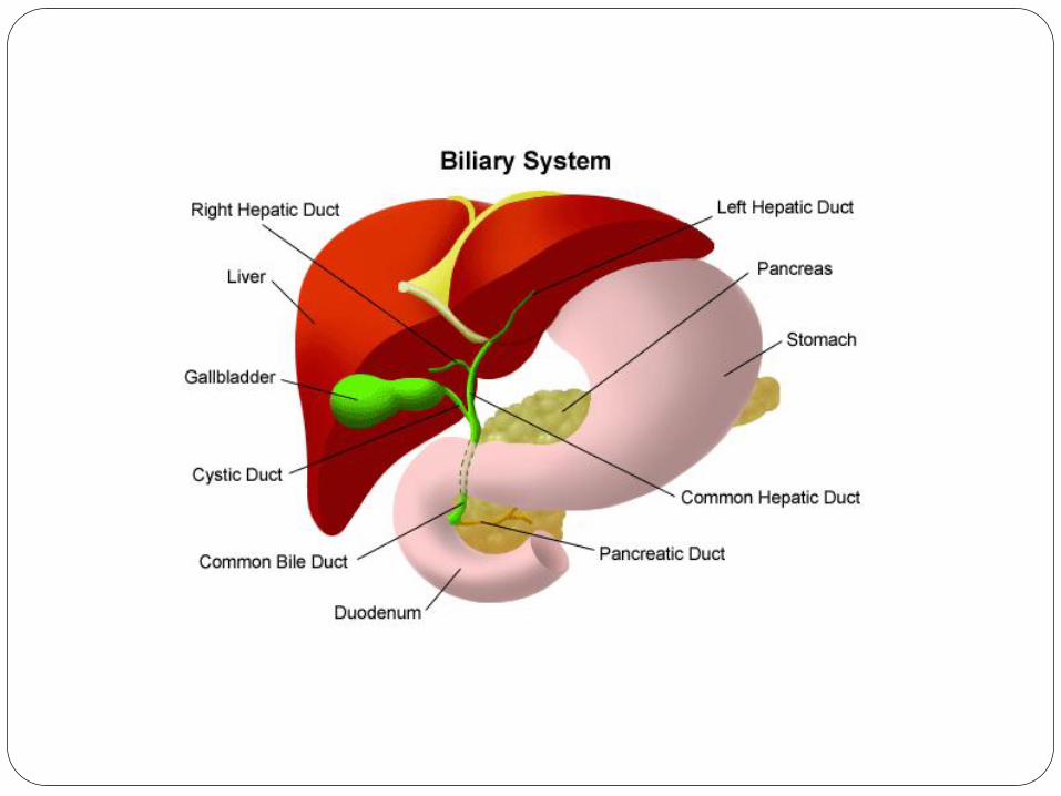
Cystic duct
Duodenum Hepatopancreatic
ampulla and sphincter
Gallbladder
Right and left
hepatic ducts
of liver
Bile duct and sphincter
Main pancreatic duct
and sphincter
Pancreas
Tail of pancreas
Head of pancreas
Common hepatic duct
Major duodenal
papilla
Accessory pancreatic duct

Anatomy and Physiology of the Digestive System
Accessory digestive organs
Liver
Pancreas
Gallbladder
Anatomy and Physiology of the Digestive System
Accessory digestive organs
Liver
Largest internal surface area of any body organ
Blood supply
Hepatic artery
Hepatic-portal vein
Hepatic vein
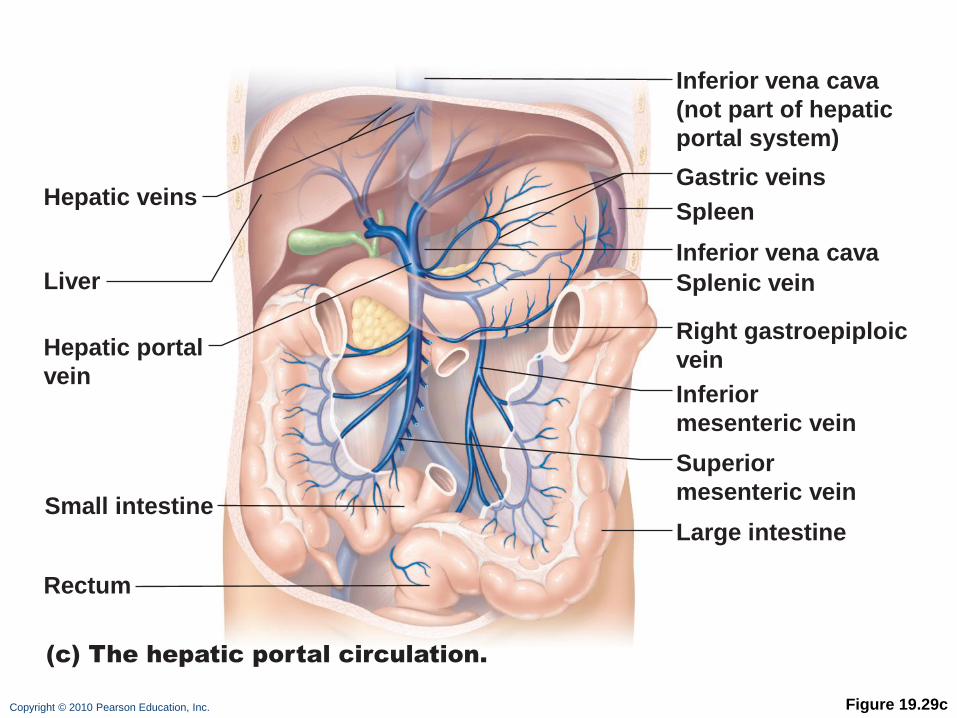
Copyright © 2010 Pearson Education, Inc. Figure 19.29c
(c) The hepatic portal circulation.
Hepatic veins
Liver
Spleen
Gastric veins
Inferior vena cava
Inferior vena cava
(not part of hepatic
portal system)
Splenic vein
Right gastroepiploic
vein
Inferior
mesenteric vein
Superior
mesenteric vein
Large intestine
Hepatic portal
vein
Small intestine
Rectum
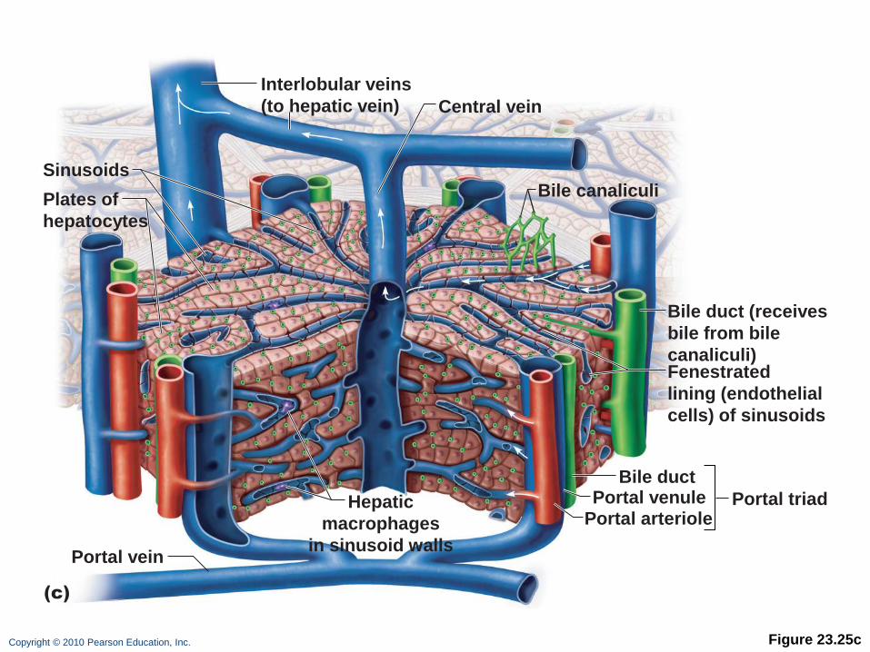
Copyright © 2010 Pearson Education, Inc. Figure 23.25c
(c)
Interlobular veins
(to hepatic vein) Central vein
Sinusoids
Portal triad
Plates of
hepatocytes
Portal vein
Fenestrated
lining (endothelial
cells) of sinusoids
Bile duct (receives
bile from bile
canaliculi)
Bile duct
Portal arteriole Portal venule Hepatic
macrophages
in sinusoid walls
Bile canaliculi

Anatomy and Physiology of the Digestive System
Accessory digestive organs
Liver
Microscopic compartments = lobules
Lined by hepatocytes = screen blood
o Store nutrients
o Manage toxins
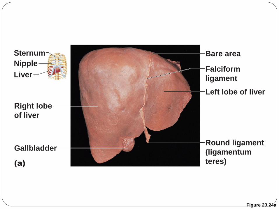
Figure 23.24a
Sternum
Nipple
Liver
Right lobe
of liver
Gallbladder
(a)
Bare area
Falciform
ligament
Left lobe of liver
Round ligament
(ligamentum
teres)
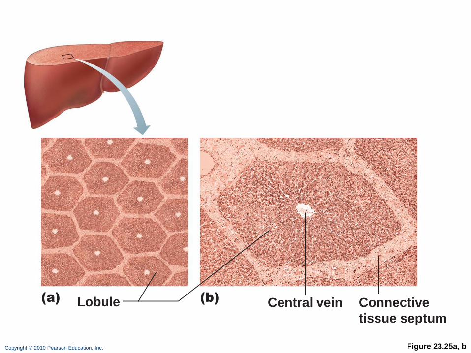
Copyright © 2010 Pearson Education, Inc. Figure 23.25a, b
(a) (b) Lobule Central vein Connective
tissue septum
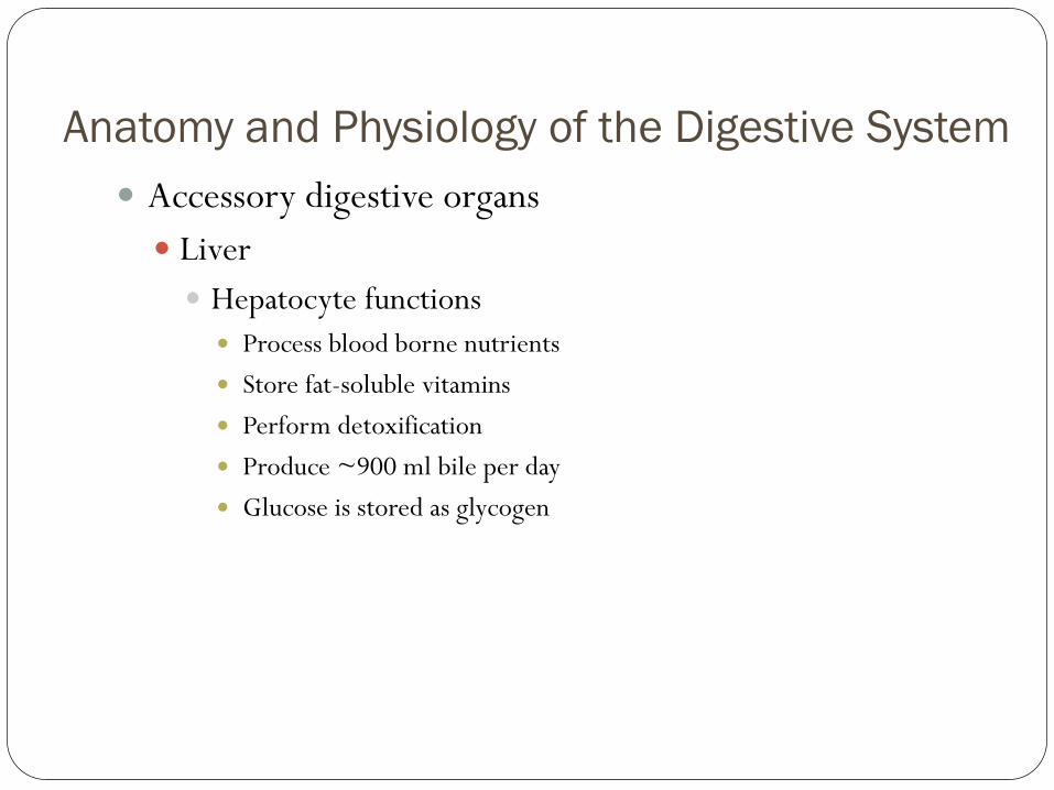
Anatomy and Physiology of the Digestive System
Accessory digestive organs
Liver
Hepatocyte functions
Process blood borne nutrients
Store fat-soluble vitamins
Perform detoxification
Produce ~900 ml bile per day
Glucose is stored as glycogen
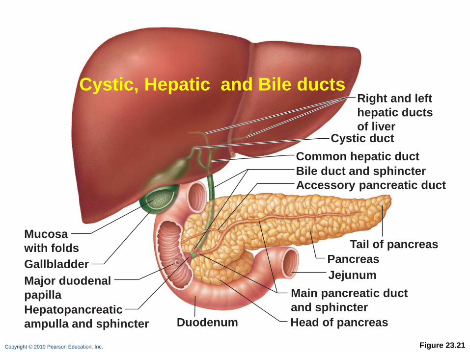
Copyright © 2010 Pearson Education, Inc. Figure 23.21
Jejunum
Mucosa
with folds
Cystic duct
Duodenum Hepatopancreatic
ampulla and sphincter
Gallbladder
Right and left
hepatic ducts
of liver
Bile duct and sphincter
Main pancreatic duct
and sphincter
Pancreas
Tail of pancreas
Head of pancreas
Common hepatic duct
Major duodenal
papilla
Accessory pancreatic duct
Cystic, Hepatic and Bile ducts
Anatomy and Physiology of the Digestive System
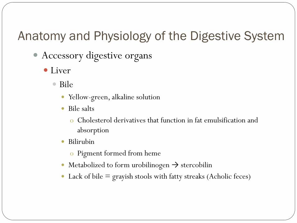
Accessory digestive organs
Liver
Bile
Yellow-green, alkaline solution
Bile salts
o Cholesterol derivatives that function in fat emulsification and
absorption
Bilirubin
o Pigment formed from heme
Metabolized to form urobilinogen → stercobilin
Lack of bile = grayish stools with fatty streaks (Acholic feces)
Anatomy and Physiology of the Digestive System
Accessory digestive organs
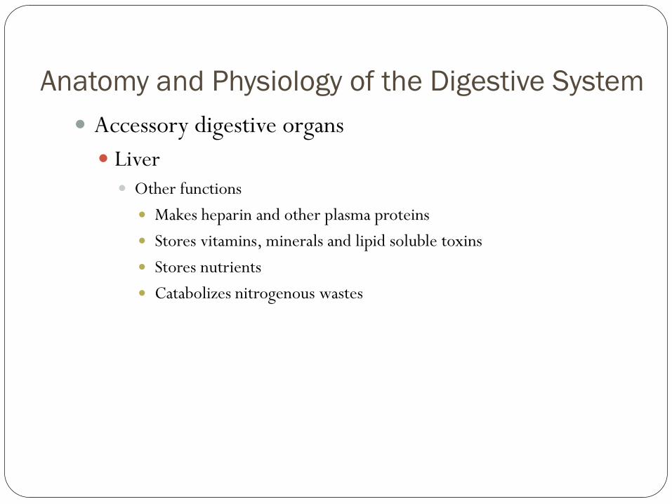
Liver Other functions
Makes heparin and other plasma proteins
Stores vitamins, minerals and lipid soluble toxins
Stores nutrients
Catabolizes nitrogenous wastes

Anatomy and Physiology of the Digestive System
Accessory digestive organs
Liver
Gallbladder
Thin-walled muscular sac on the ventral surface of the liver
Stores and concentrates bile by absorbing its water and ions
Releases bile via the cystic duct
o Flows into the bile duct

Anatomy and Physiology of the Digestive System
Accessory digestive organs
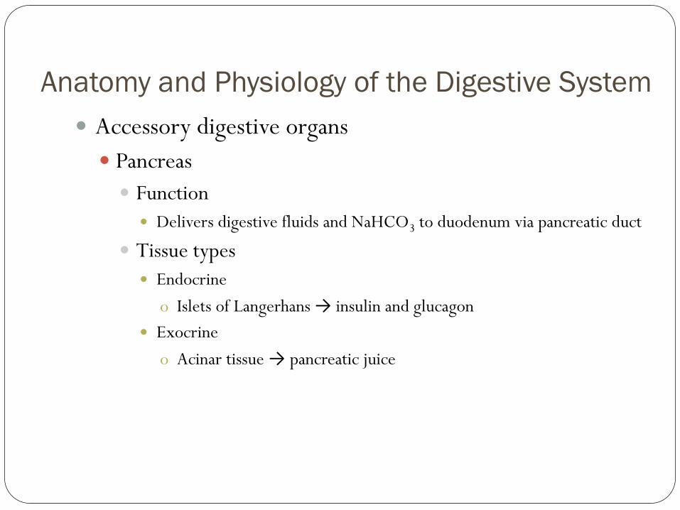
Pancreas
Function
Delivers digestive fluids and NaHCO3 to duodenum via pancreatic duct
Tissue types
Endocrine
o Islets of Langerhans → insulin and glucagon
Exocrine
o Acinar tissue → pancreatic juice

Figure 23.1
Mouth (oral cavity)
Tongue
Esophagus
Liver
Gallbladder
Anus
Duodenum Jejunum Ileum
Small
intestine
Parotid gland Sublingual gland Submandibular
gland
Salivary
glands
Pharynx
Stomach Pancreas (Spleen)
Transverse colon Descending colon Ascending colon Cecum Sigmoid colon Rectum Vermiform appendix Anal canal
Large
intestine

Anatomy and Physiology of the Digestive System
Accessory digestive organs
Pancreas
Secretion mediated by hormones
Secretin
o Released in response to acid
o Stimulates release of base
Cholecystokinin
o Released when protein and fat enter intestine
o Stimulates the release of bile and pancreatic juice

Figure 23.21
Jejunum
Mucosa
with folds
Cystic duct
Duodenum Hepatopancreatic
ampulla and sphincter
Gallbladder
Right and left
hepatic ducts
of liver
Bile duct and sphincter
Main pancreatic duct
and sphincter
Pancreas
Tail of pancreas
Head of pancreas
Common hepatic duct
Major duodenal
papilla
Accessory pancreatic duct
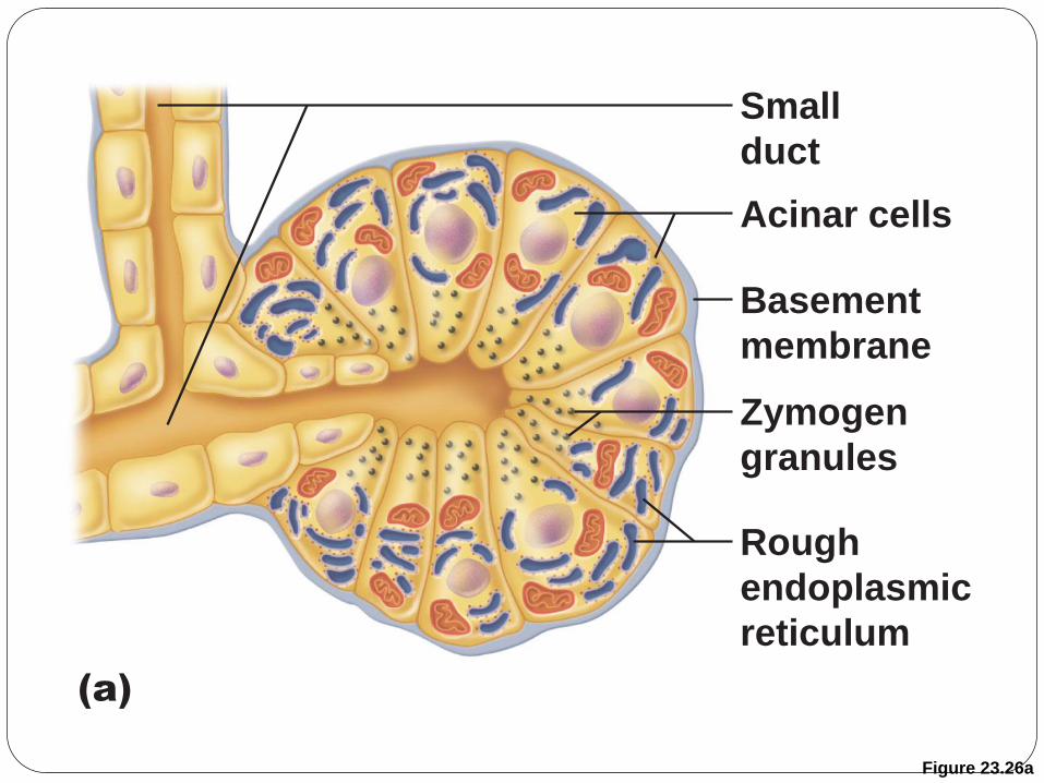
Figure 23.26a
Small
duct
Acinar cells
Basement
membrane
Zymogen
granules
Rough
endoplasmic
reticulum
(a)
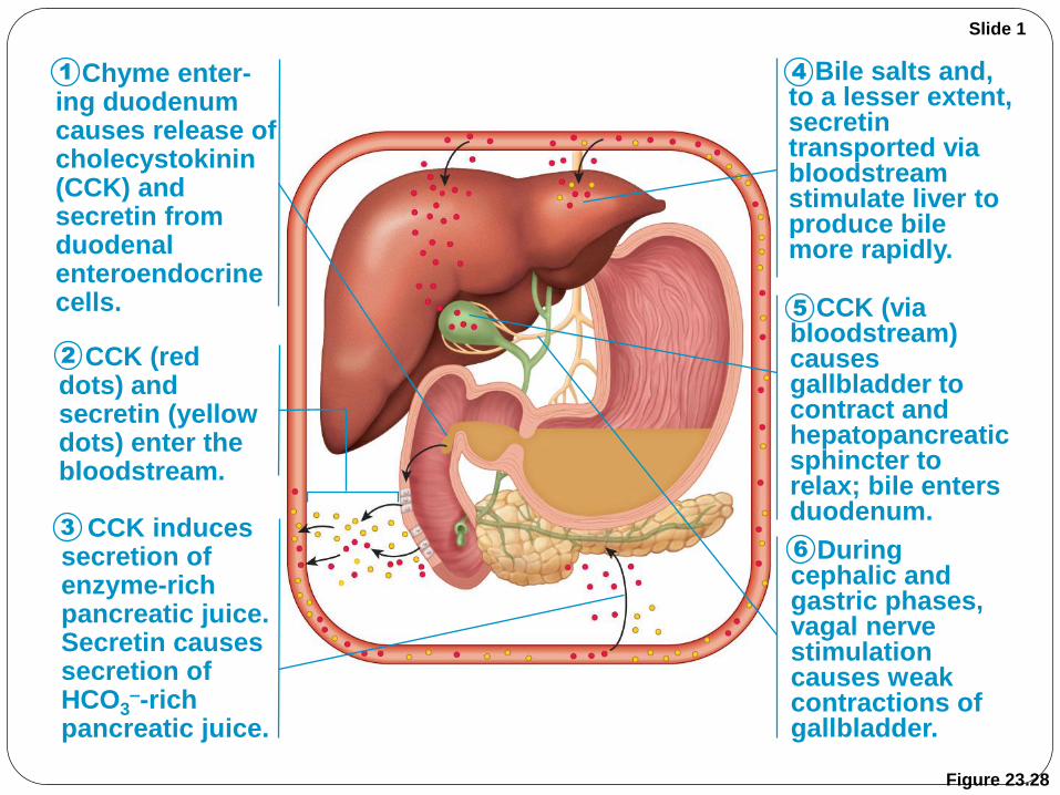
Figure 23.28
Chyme enter- ing duodenum causes release of cholecystokinin (CCK) and secretin from duodenal enteroendocrine cells.
CCK (red dots) and secretin (yellow dots) enter the bloodstream.
CCK induces secretion of enzyme-rich pancreatic juice. Secretin causes secretion of HCO3
–-rich pancreatic juice.
Bile salts and, to a lesser extent, secretin transported via bloodstream stimulate liver to produce bile more rapidly.
CCK (via bloodstream) causes gallbladder to contract and hepatopancreatic sphincter to relax; bile enters duodenum.
During cephalic and gastric phases, vagal nerve stimulation causes weak contractions of gallbladder.
Slide 1
1
2
3
4
5
6
Anatomy and Physiology of the Digestive System
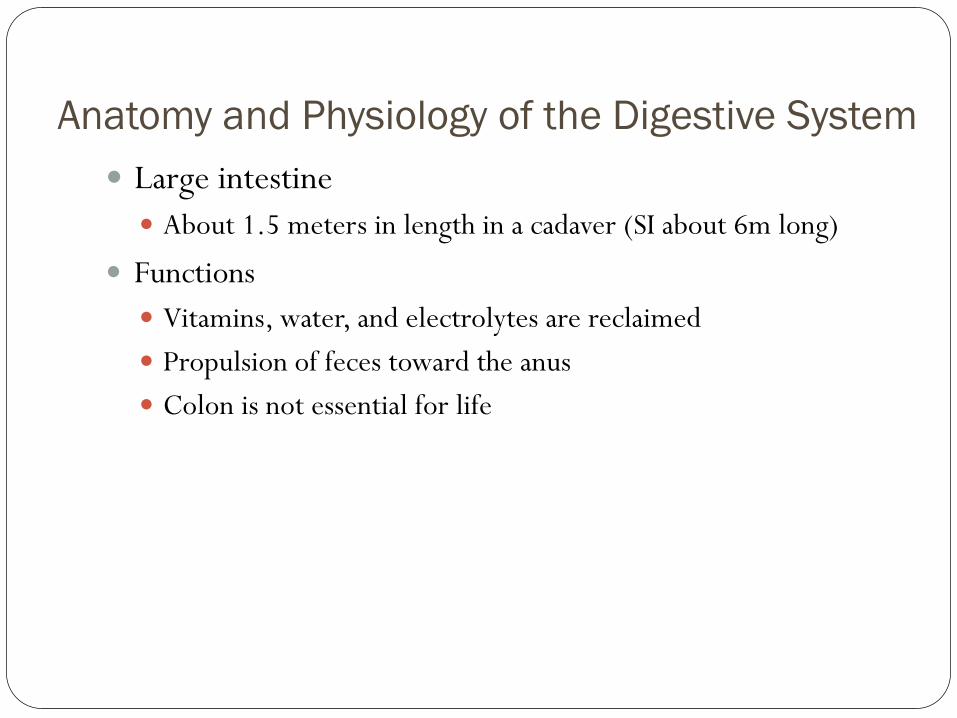
Large intestine
About 1.5 meters in length in a cadaver (SI about 6m long)
Functions
Vitamins, water, and electrolytes are reclaimed
Propulsion of feces toward the anus
Colon is not essential for life
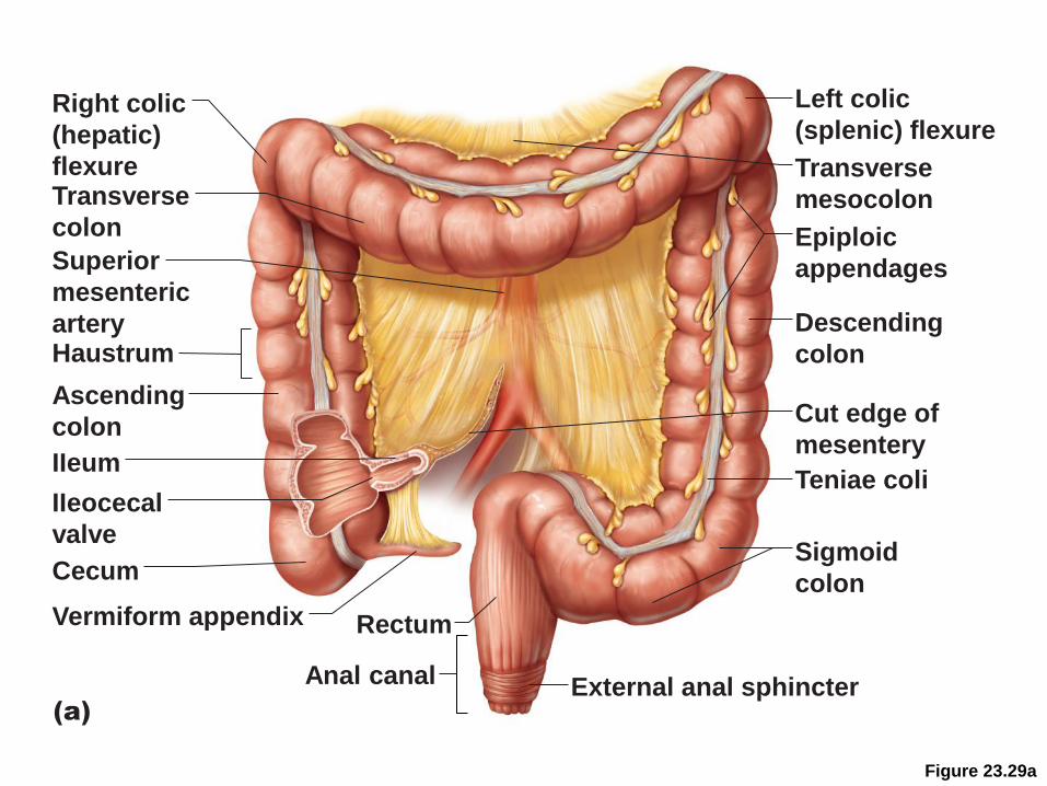
Figure 23.29a
Left colic
(splenic) flexure
Transverse
mesocolon
Epiploic
appendages
Descending
colon
Teniae coli
Sigmoid
colon
Cut edge of
mesentery
External anal sphincter
Rectum
Anal canal
(a)
Right colic
(hepatic)
flexure Transverse
colon
Superior
mesenteric
artery Haustrum
Ascending
colon
IIeum
IIeocecal
valve
Vermiform appendix
Cecum

Anatomy and Physiology of the Digestive System
Regions
Cecum
Colon
Rectum
Anal canal
Anatomy and Physiology of the Digestive System
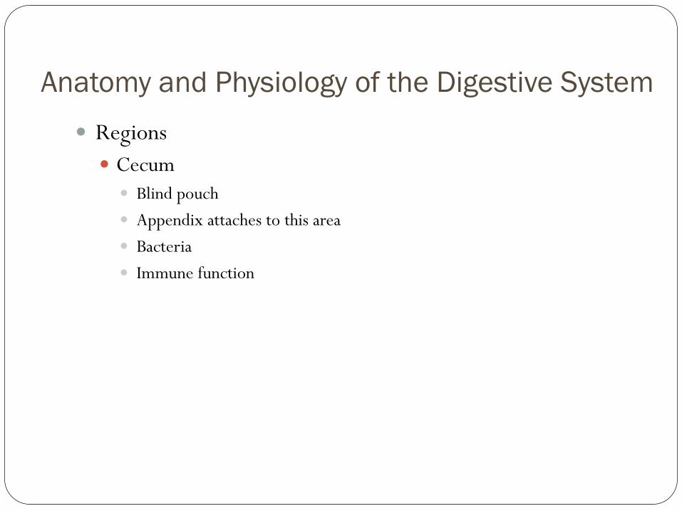
Regions
Cecum
Blind pouch
Appendix attaches to this area
Bacteria
Immune function
Anatomy and Physiology of the Digestive System
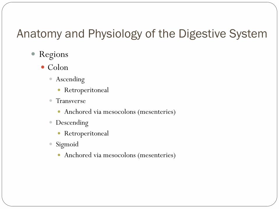
Regions
Colon
Ascending
Retroperitoneal
Transverse
Anchored via mesocolons (mesenteries)
Descending
Retroperitoneal
Sigmoid
Anchored via mesocolons (mesenteries)
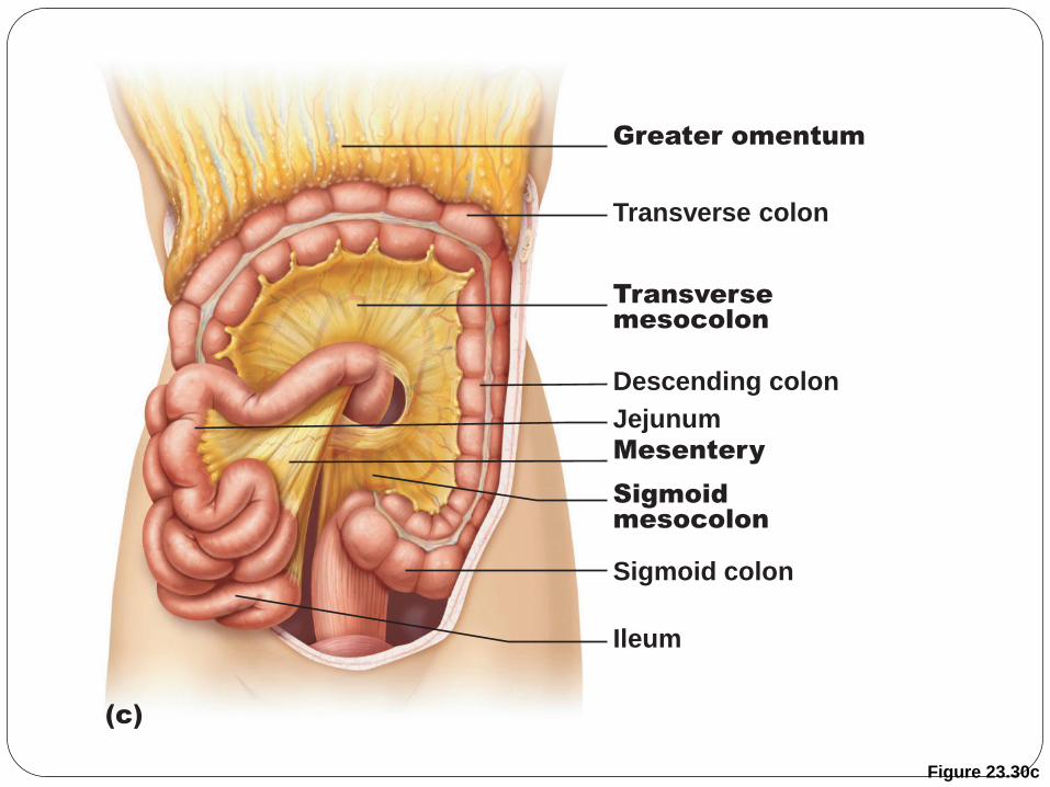
Figure 23.30c
Transverse colon
Greater omentum
Descending colon
Jejunum
Mesentery
Transverse
mesocolon
Sigmoid
mesocolon
Sigmoid colon
Ileum
(c)

Figure 23.30d
(d)
Pancreas
Liver
Lesser omentum
Stomach
Duodenum
Transverse
mesocolon
Greater omentum
Mesentery
Jejunum
Visceral peritoneum
Urinary bladder
Transverse colon
Ileum
Parietal peritoneum
Rectum
Anatomy and Physiology of the Digestive System
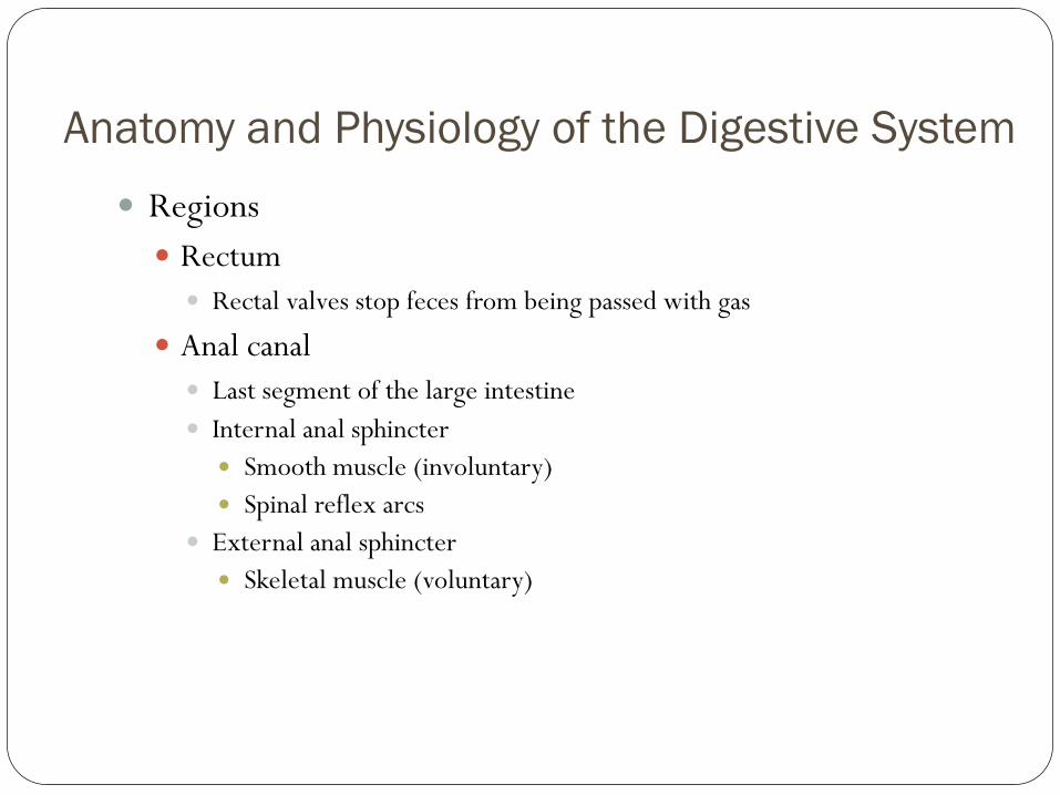
Regions
Rectum
Rectal valves stop feces from being passed with gas
Anal canal
Last segment of the large intestine
Internal anal sphincter
Smooth muscle (involuntary)
Spinal reflex arcs
External anal sphincter
Skeletal muscle (voluntary)
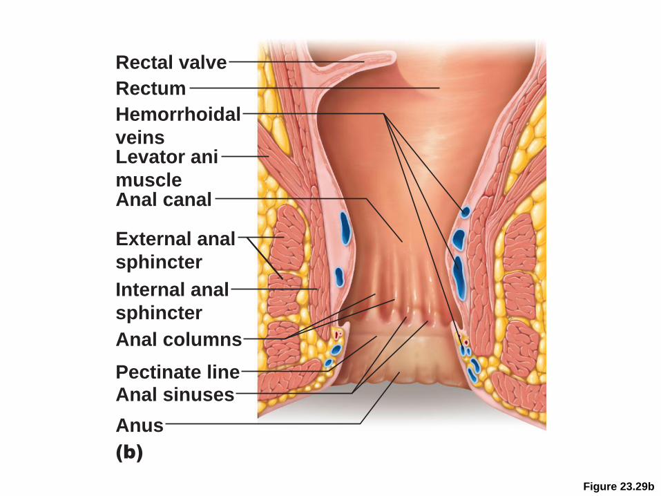
Figure 23.29b
(b)
Rectal valve
Rectum
Anal canal
Levator ani
muscle
Anus
Anal sinuses
Anal columns
Internal anal
sphincter
External anal
sphincter
Hemorrhoidal
veins
Pectinate line
Anatomy and Physiology of the Digestive System
Defectation
Mass movements force feces into rectum
Distension initiates spinal defecation reflex
Parasympathetic signals Stimulate contraction of the sigmoid colon and rectum
Relax the internal anal sphincter
Conscious control allows relaxation of external anal sphincter
Valsalva’s maneuver
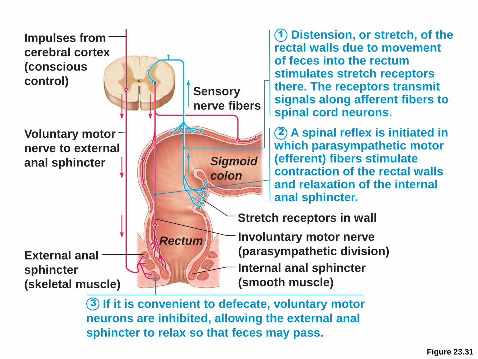
Figure 23.31
Impulses from
cerebral cortex
(conscious
control)
Voluntary motor
nerve to external
anal sphincter
External anal
sphincter
(skeletal muscle)
Internal anal sphincter
(smooth muscle)
Sensory
nerve fibers
Involuntary motor nerve
(parasympathetic division)
Stretch receptors in wall
Rectum
Sigmoid
colon
3
1
2
Distension, or stretch, of the rectal walls due to movement of feces into the rectum stimulates stretch receptors there. The receptors transmit signals along afferent fibers to spinal cord neurons.
A spinal reflex is initiated in which parasympathetic motor (efferent) fibers stimulate contraction of the rectal walls and relaxation of the internal anal sphincter.
If it is convenient to defecate, voluntary motor
neurons are inhibited, allowing the external anal
sphincter to relax so that feces may pass.
Questions?