Embed Size (px)
Citation preview
1/15/2009
1
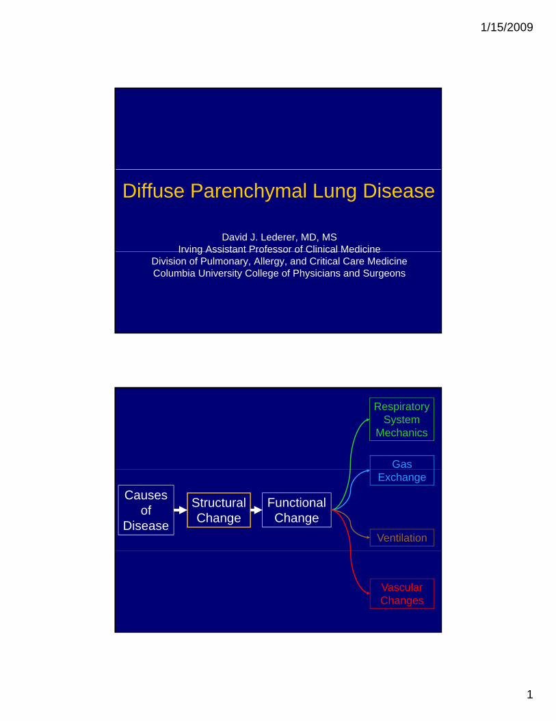
Diffuse Parenchymal Lung Disease
David J. Lederer, MD, MSIrving Assistant Professor of Clinical MedicineIrving Assistant Professor of Clinical Medicine
Division of Pulmonary, Allergy, and Critical Care MedicineColumbia University College of Physicians and Surgeons
Respiratory System
Mechanics
Gas
Causes of
Disease
Structural Change
FunctionalChange
Exchange
Ventilation
VascularChanges
1/15/2009
2
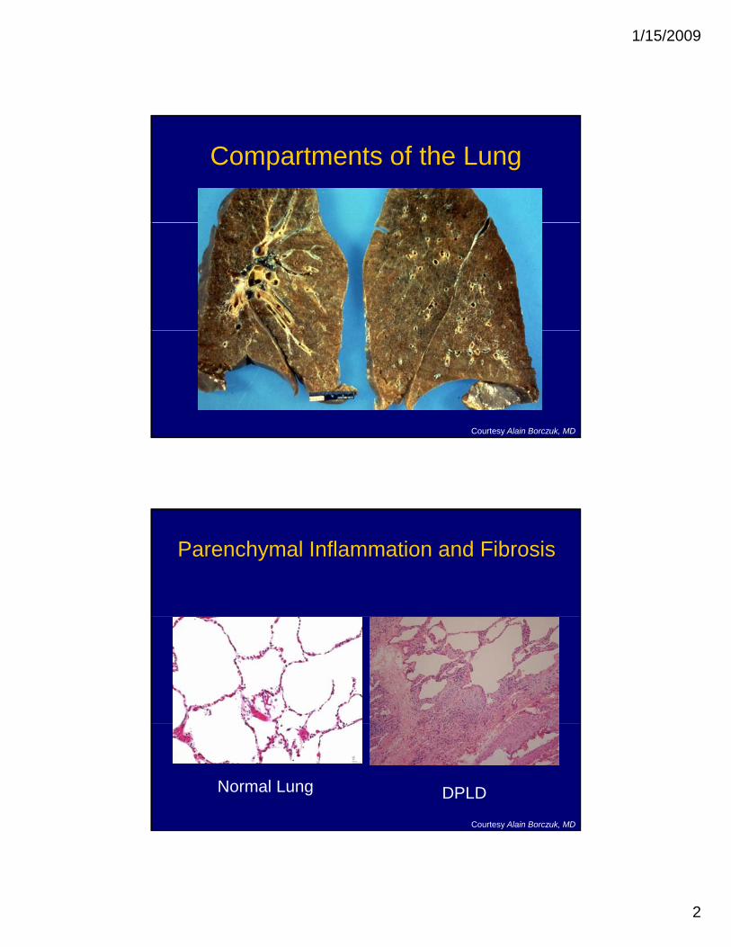
Compartments of the Lung
Courtesy Alain Borczuk, MD
Parenchymal Inflammation and Fibrosis
Normal Lung DPLD
Courtesy Alain Borczuk, MD

1/15/2009
3
Overview
• Terminology and classification scheme• Pathophysiology• Clinical manifestations• Pathogenesis• Management
Alphabet Soup
1/15/2009
4
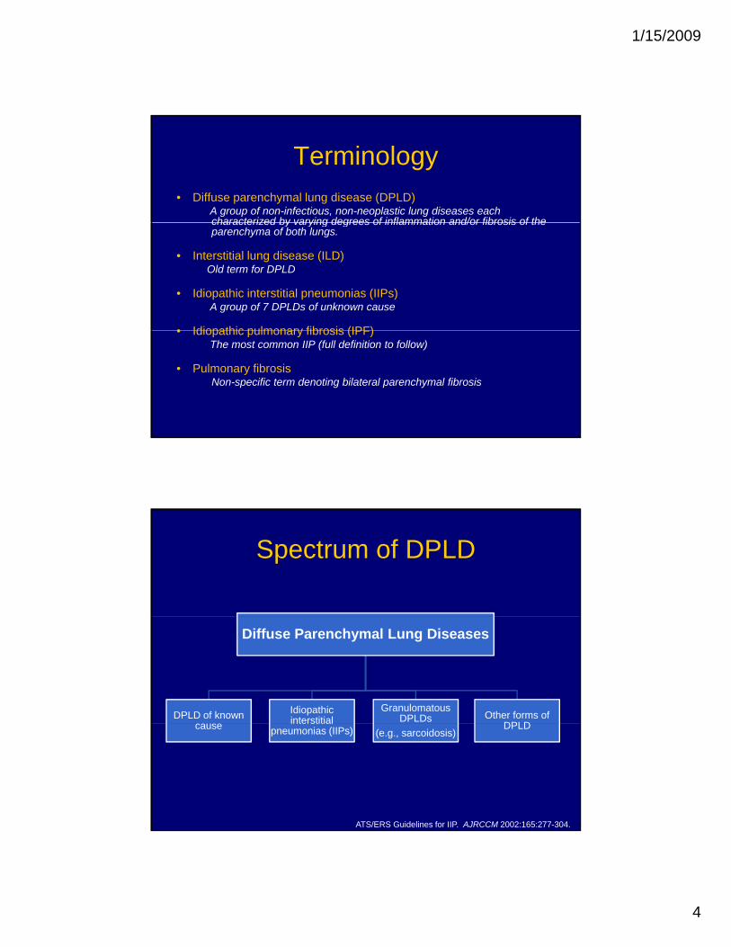
Terminology• Diffuse parenchymal lung disease (DPLD)
A group of non-infectious, non-neoplastic lung diseases each characterized by varying degrees of inflammation and/or fibrosis of thecharacterized by varying degrees of inflammation and/or fibrosis of the parenchyma of both lungs.
• Interstitial lung disease (ILD)Old term for DPLD
• Idiopathic interstitial pneumonias (IIPs)A group of 7 DPLDs of unknown cause
• Idiopathic pulmonary fibrosis (IPF)• Idiopathic pulmonary fibrosis (IPF)The most common IIP (full definition to follow)
• Pulmonary fibrosisNon-specific term denoting bilateral parenchymal fibrosis
Spectrum of DPLD
Diffuse Parenchymal Lung Diseases
DPLD of known cause
Idiopathic interstitial
Granulomatous DPLDs Other forms of
DPLD
ATS/ERS Guidelines for IIP. AJRCCM 2002:165:277-304.
cause e s apneumonias (IIPs) (e.g., sarcoidosis)
DPLD
1/15/2009
5
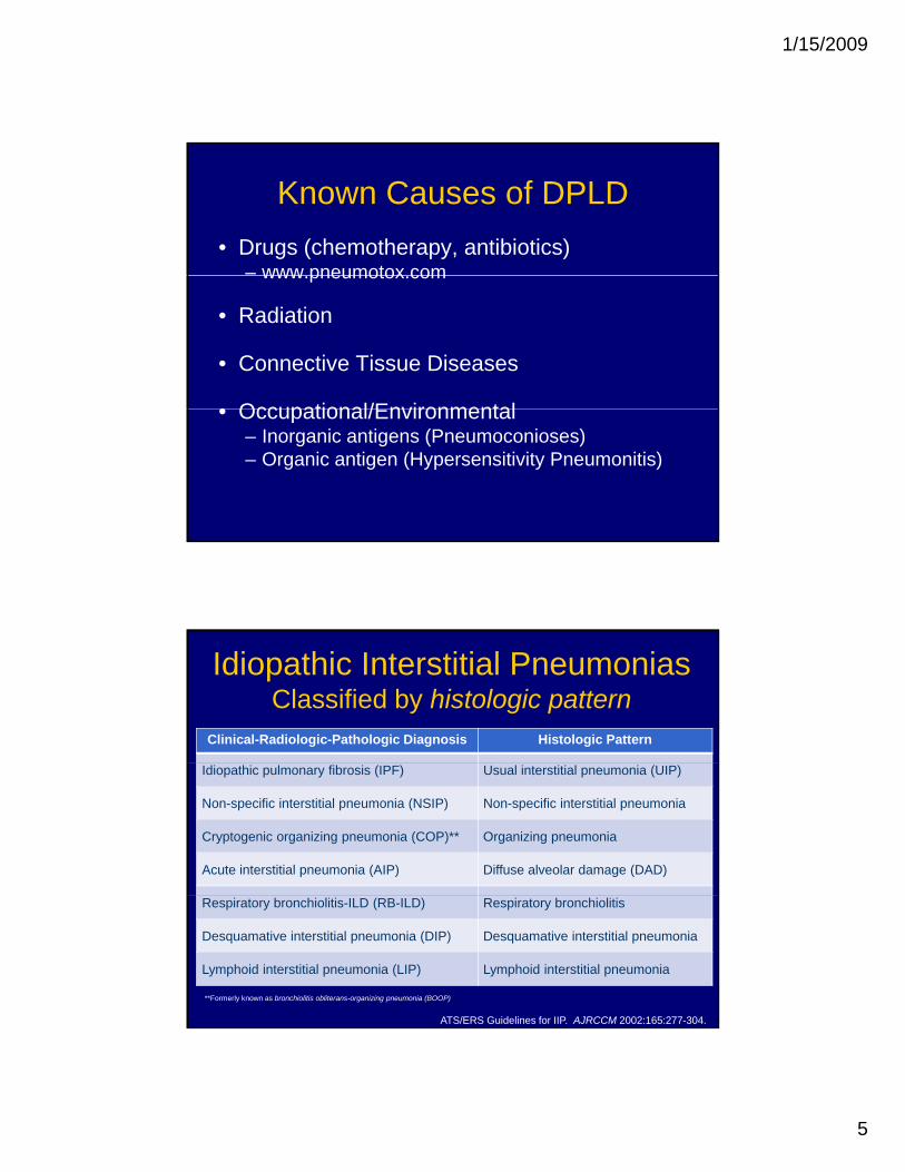
Known Causes of DPLD• Drugs (chemotherapy, antibiotics)
– www pneumotox comwww.pneumotox.com
• Radiation
• Connective Tissue Diseases
• Occupational/Environmental• Occupational/Environmental– Inorganic antigens (Pneumoconioses)– Organic antigen (Hypersensitivity Pneumonitis)
Idiopathic Interstitial PneumoniasClassified by histologic pattern
Clinical-Radiologic-Pathologic Diagnosis Histologic Pattern
Idiopathic pulmonary fibrosis (IPF) Usual interstitial pneumonia (UIP)
Non-specific interstitial pneumonia (NSIP) Non-specific interstitial pneumonia
Cryptogenic organizing pneumonia (COP)** Organizing pneumonia
Acute interstitial pneumonia (AIP) Diffuse alveolar damage (DAD)
Respiratory bronchiolitis-ILD (RB-ILD) Respiratory bronchiolitis
Desquamative interstitial pneumonia (DIP) Desquamative interstitial pneumonia
Lymphoid interstitial pneumonia (LIP) Lymphoid interstitial pneumonia
**Formerly known as bronchiolitis obliterans-organizing pneumonia (BOOP)
ATS/ERS Guidelines for IIP. AJRCCM 2002:165:277-304.

1/15/2009
6
Usual interstitial pneumonia is the histologic pattern of IPF
Fibroblastic foci are a key histological finding in UIP
Visscher & Myers. Proc Am Thorac Soc 2006;3:322-9.
1/15/2009
7
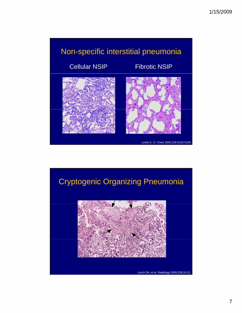
Non-specific interstitial pneumonia
Cellular NSIP Fibrotic NSIP
Leslie K. O. Chest 2005;128:513S-519S
Cryptogenic Organizing Pneumonia
Lynch DA, et al. Radiology 2005;236:10-21.
1/15/2009
8
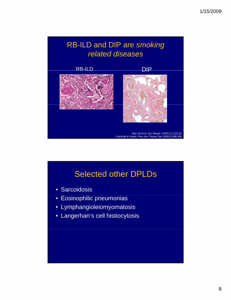
RB-ILD and DIP are smoking related diseases
RB-ILD DIPRB ILD DIP
Ryu JH,et al. Eur Respir J 2001;17:122-32.Caminati & Harari. Proc Am Thorac Soc 2006:3;299-306.
Selected other DPLDs
• Sarcoidosis• Eosinophilic pneumonias• Lymphangioleiomyomatosis• Langerhan’s cell histiocytosis
1/15/2009
9
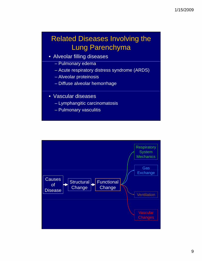
Related Diseases Involving the Lung Parenchyma
• Alveolar filling diseases– Pulmonary edema– Acute respiratory distress syndrome (ARDS)– Alveolar proteinosis– Diffuse alveolar hemorrhage
• Vascular diseases– Lymphangitic carcinomatosis– Pulmonary vasculitis
Respiratory System
Mechanics
Gas
Causes of
Disease
Structural Change
FunctionalChange
Exchange
Ventilation
VascularChanges
1/15/2009
10
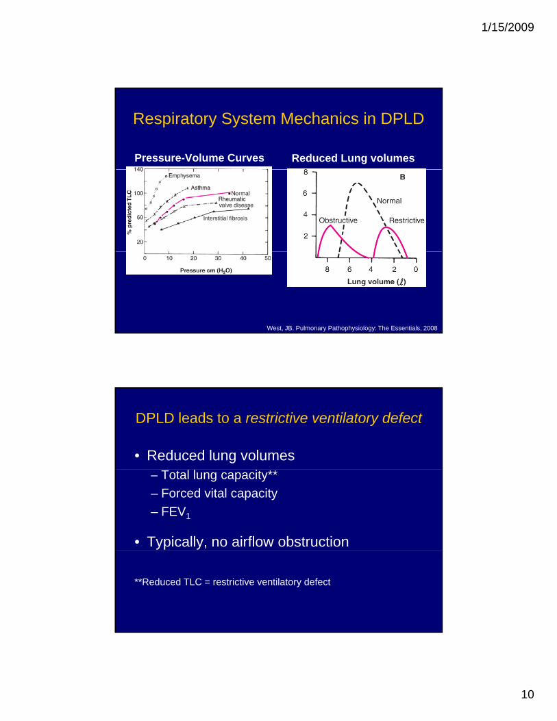
Respiratory System Mechanics in DPLD
Pressure-Volume Curves Reduced Lung volumes
West, JB. Pulmonary Pathophysiology: The Essentials, 2008
DPLD leads to a restrictive ventilatory defect
• Reduced lung volumes– Total lung capacity**– Forced vital capacity– FEV1
• Typically, no airflow obstruction
**Reduced TLC = restrictive ventilatory defect
1/15/2009
11
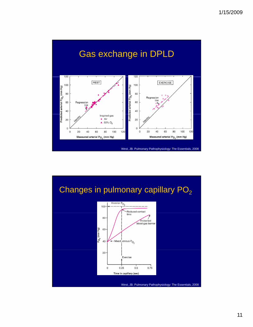
Gas exchange in DPLD
West, JB. Pulmonary Pathophysiology: The Essentials, 2008
Changes in pulmonary capillary PO2
West, JB. Pulmonary Pathophysiology: The Essentials, 2008
1/15/2009
12
DPLD leads to impaired gas exchange
• V/Q mismatch• Diffusion impairment only with exercise• Shunt does not play a role
What about ventilation and vascular changes?
• Alveolar hyperventilation– Hypoxemia– Abnormal mechanics and load
• Vascular disease is common– Intimal hyperplasiaIntimal hyperplasia– Medial hypertrophy– Pulmonary hypertension is typically not severe

1/15/2009
13
Clinical Manifestations of DPLD
DPLDs share many clinical features
SimilaritiesDyspnea
Differencesf• Dyspnea
– progressive– exertional
• Cough– non-productive
• Bibasilar crackles
• Extrapulmonary findings– sarcoidosis– connective tissue disease
• Pattern on lung CT
• Histopathology
• Restrictive ventilatory defect
• Impaired gas exchange
• Abnormal lung imaging
1/15/2009
14
Case
Case• 54 year old man comes to see you because of two
years of dyspneayea s o dysp ea– First, while mowing his lawn – Then, more dyspneic than his wife in the gym– Now dyspneic with most activities at home.
• Dry cough (no sputum) and occasional joint pains. • No wheezing or hemoptysis.• No fever or chills• No fever or chills. • No chest pain, orthopnea, PND, or edema. • No rash, visual changes, Raynaud’s phenomenon,
dysphagia, or heartburn
1/15/2009
15
Case
• Past medical historyOsteoarthritis– Osteoarthritis
– Hypercholesterolemia
• Past surgical history– None
• Medications– Simvastatin, multivitamin, acetaminophen
• No known drug allergies
Case• Family history
– No lung diseaseNo lung disease– Mother 85 yo – alive and well– Father died at 74 with heart failure– Sister with ovarian cancer
• Social history– Smoked one pack per day for 35 years (35 packyears). p p y y ( p y )
Quit 3 years ago– No alcohol or drug use– No pets, humidifiers, or hot tubs– Real estate agent. No military or construction work
1/15/2009
16
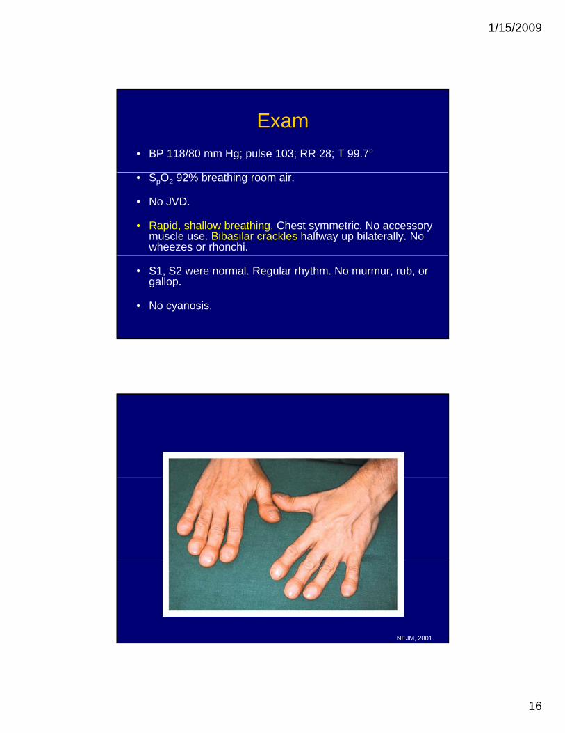
Exam• BP 118/80 mm Hg; pulse 103; RR 28; T 99.7°
• SpO2 92% breathing room air.
• No JVD.
• Rapid, shallow breathing. Chest symmetric. No accessory muscle use. Bibasilar crackles halfway up bilaterally. No wheezes or rhonchi.
• S1, S2 were normal. Regular rhythm. No murmur, rub, or gallop.
• No cyanosis.
NEJM, 2001
1/15/2009
17
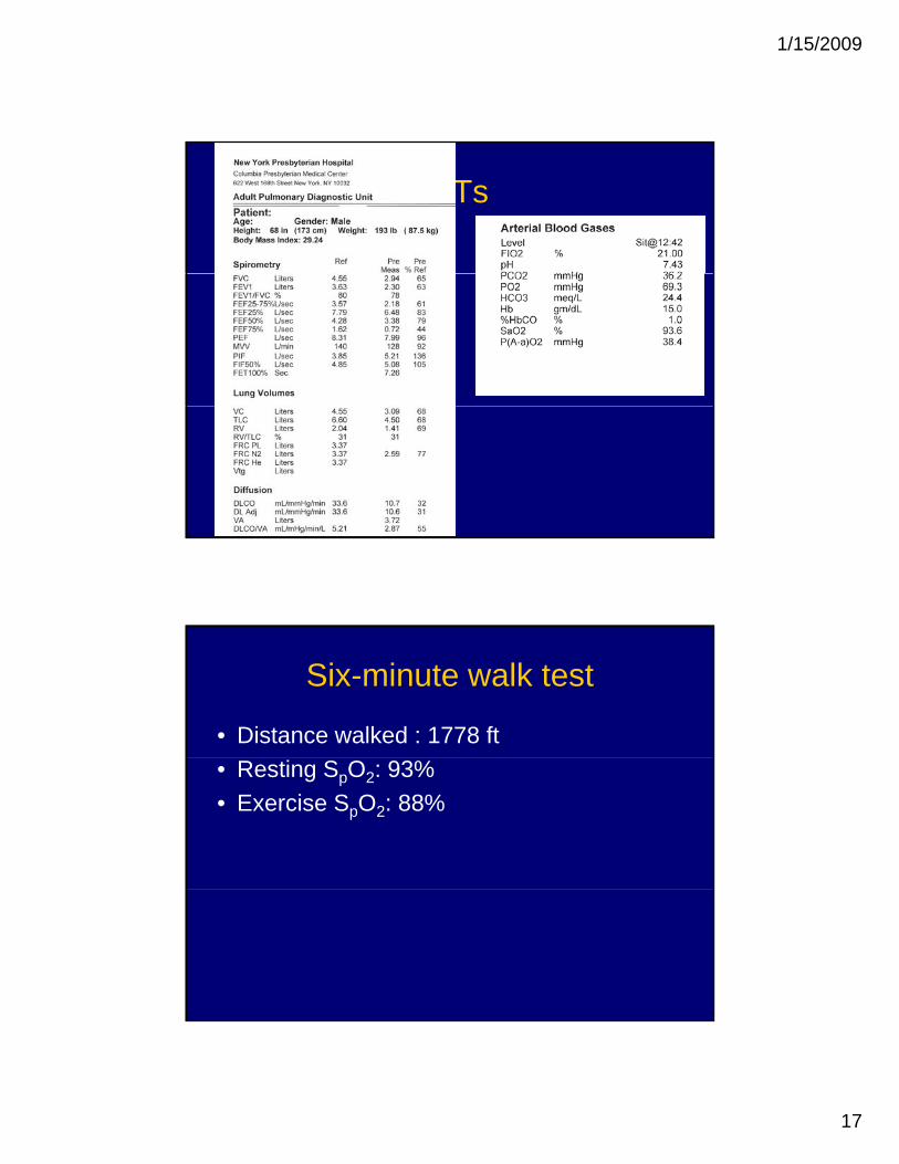
PFTs
Six-minute walk test
• Distance walked : 1778 ft • Resting SpO2: 93%• Exercise SpO2: 88%
1/15/2009
18
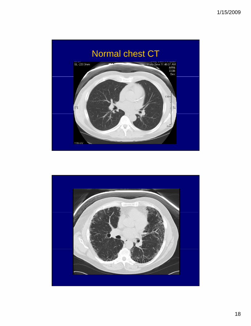
Normal chest CT
1/15/2009
19
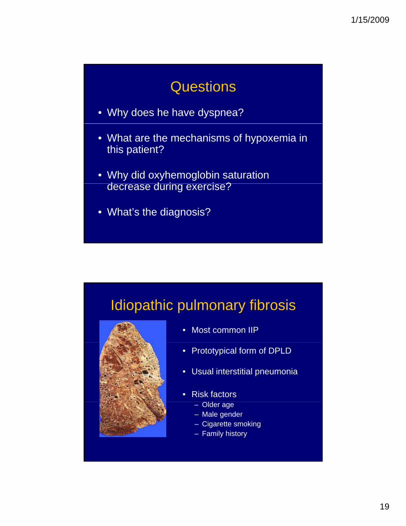
Questions
• Why does he have dyspnea?
• What are the mechanisms of hypoxemia in this patient?
• Why did oxyhemoglobin saturation d d i i ?decrease during exercise?
• What’s the diagnosis?
Idiopathic pulmonary fibrosis• Most common IIP
• Prototypical form of DPLD
• Usual interstitial pneumonia
• Risk factorsOld– Older age
– Male gender– Cigarette smoking– Family history
1/15/2009
20
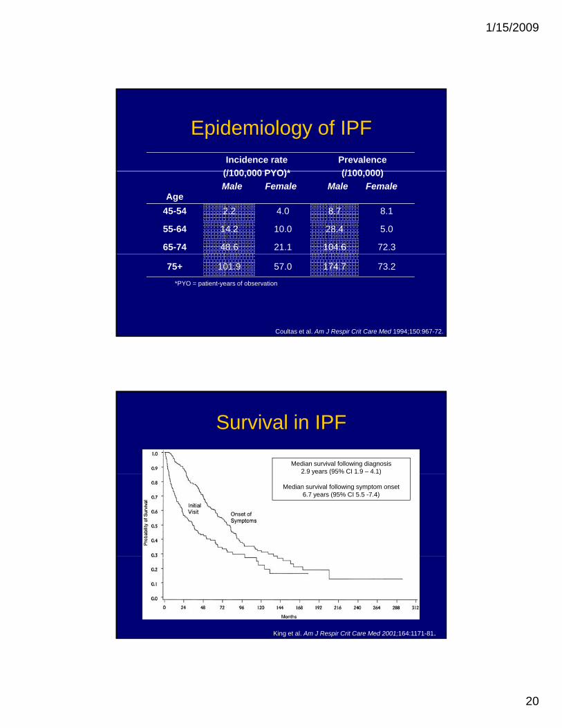
Epidemiology of IPFIncidence rate
(/100 000 PYO)*Prevalence (/100 000)
Age
(/100,000 PYO)*Male Female
(/100,000)Male Female
45-54 2.2 4.0 8.7 8.1
55-64 14.2 10.0 28.4 5.0
65-74 48.6 21.1 104.6 72.3
75+ 101.9 57.0 174.7 73.2
Coultas et al. Am J Respir Crit Care Med 1994;150:967-72.
*PYO = patient-years of observation
Survival in IPF
Median survival following diagnosis2.9 years (95% CI 1.9 – 4.1)y ( % )
Median survival following symptom onset6.7 years (95% CI 5.5 -7.4)
King et al. Am J Respir Crit Care Med 2001;164:1171-81.
1/15/2009
21
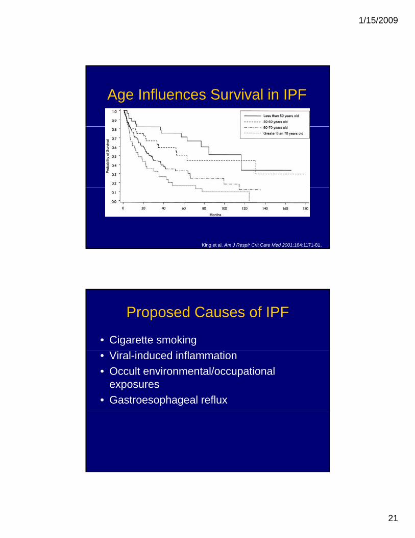
Age Influences Survival in IPF
King et al. Am J Respir Crit Care Med 2001;164:1171-81.
Proposed Causes of IPF
• Cigarette smoking• Viral-induced inflammation• Occult environmental/occupational
exposures• Gastroesophageal reflux
1/15/2009
22
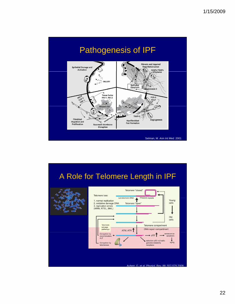
Pathogenesis of IPF
Selman, M. Ann Int Med. 2001
A Role for Telomere Length in IPF
Aubert, G. et al. Physiol. Rev. 88: 557-579 2008;
1/15/2009
23
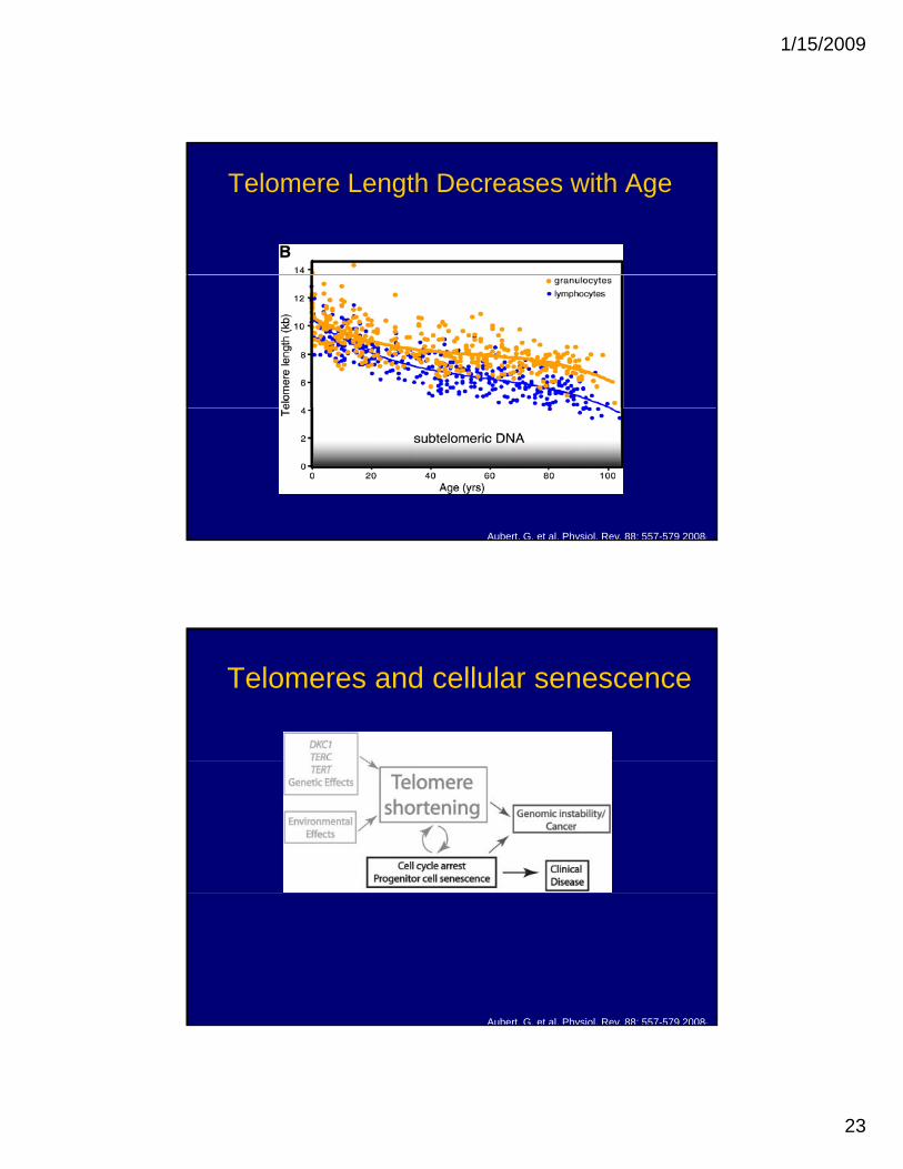
Telomere Length Decreases with Age
Aubert, G. et al. Physiol. Rev. 88: 557-579 2008;
Telomeres and cellular senescence
Aubert, G. et al. Physiol. Rev. 88: 557-579 2008;
1/15/2009
24
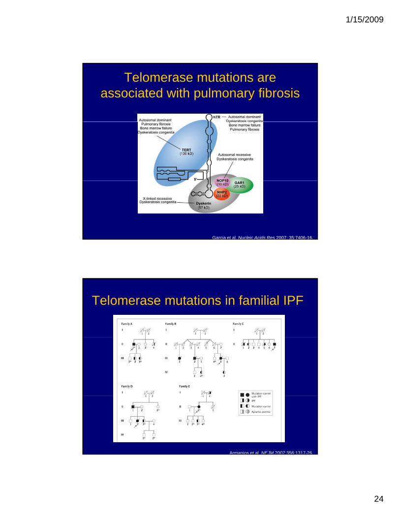
Telomerase mutations are associated with pulmonary fibrosis
Garcia et al. Nucleic Acids Res 2007: 35;7406-16.
Telomerase mutations in familial IPF
Armanios et al. NEJM 2007;356:1317-26.
1/15/2009
25
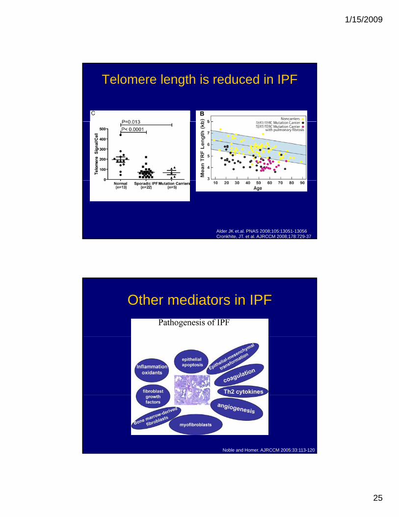
Telomere length is reduced in IPF
Alder JK et.al. PNAS 2008;105:13051-13056Cronkhite, JT. et al. AJRCCM 2008;178:729-37
Other mediators in IPF
Noble and Homer. AJRCCM 2005:33:113-120
1/15/2009
26
What about other DPLDs?
• Injurious triggers– Autoimmune mediated inflammation– Drug-induced injury– Radiation-induced injury– Eosinophil degranulation– Hypersensitivity reactionHypersensitivity reaction
Management of DPLD
• Biopsy often required to make a diagnosis– Surgical lung biopsy– Transbronchial lung biopsy (less useful)
• Oxygen therapy• Pulmonary rehabilitation
1/15/2009
27
Treatment of DPLD
• Injury avoidance– Inhaled agents– Offending drug
• Anti-inflammatory therapy– Treat underlying inflammatory diseases
Trial of corticosteroids for documented– Trial of corticosteroids for documented parenchymal inflammation
– Steroid-sparing agents• Lung transplantation

![Interstitial lung disease (ILD), or diffuse parenchymal lung disease … · 2018-10-28 · Interstitial lung disease (ILD), or diffuse parenchymal lung disease (DPLD),[[1] is a group](https://img.dokumen.tips/doc/110x75/5e7d31d2ec5074254471c7d0/interstitial-lung-disease-ild-or-diffuse-parenchymal-lung-disease-2018-10-28.jpg)