Embed Size (px)
Citation preview

ORIGINAL PAPER
Rieko Hamaoka á Yuji Yaginuma á Tomoaki TakahashiJunichi Fujii á Masahiko Koizumi á Han Geuk SeoYutaka Hatanaka á Kaoru Hashizume á Kunio IiJun-ichiro Miyagawa á Toshiaki HanafusaYuji Matsuzawa á Mutsuo Ishikawa á Naoyuki Taniguchi
Different expression patterns of nitric oxide synthase isozymesin various gynecological cancers
Received: 29 July 1998 /Accepted: 15 December 1998
Abstract The expression of nitric oxide synthase (NOS)in human gynecological cancers, including ovariancancers, uterocervical cancers, and endometrial cancersfor example, was examined by the reverse transcriptase/polymerase chain reaction, coupled with Southern hy-bridization and by immunohistochemistry. Nitric oxidesynthase II (NOS II), an inducible form, was expressedin more than 90% of the cancers. Nitric oxide synthase I(NOS I), a neuronal form, was expressed in 58% of allthe ovarian cancers, in which the serous type is foundmore frequently (5 out of 7) than the mucinous type(2 out of 6), and in all clear-cell cancers. The frequencyof NOS I expression in uterocervical cancers andendometrial cancers was relatively low. Nitric oxidesynthase III (NOS III), an endothelial form, was detectedin 25% of ovarian and 33% of endometrial cancers,while no expression was detected in uterocervical can-cers. In terms of cancer types, all clear-cell adenocarci-
nomas and most of the serous-type adenocarcinomasexpressed both NOS I and NOS II, while most uterinesquamous carcinomas and endometrial adenocarcino-mas expressed only NOS II. However, there was nocorrelation between the frequency of NOS expressionand patients' age or the clinical stage of the disease.Since NO increases vascular permeability and blood¯ow, the high frequency of NOS expression in gyneco-logical cancers may serve to stimulate and promotetumor growth.
Key words Nitric oxide á Nitric oxide synthase áOvarian cancer á Uterocervical cancer áEndometrial cancer
Abbreviations NO nitric oxide á NOS nitric oxidesynthase á NOS I neuronal NOS á NOS II inducibleNOS á NOS III endothelial NOS á RT-PCR reversetranscription/polymerase chain reaction
Introduction
NO is involved in a variety of physiological functionsin the nervous, immune, and cardiovascular systems(Nathan 1992; Moncada and Higgs 1993). Nitric oxidesynthase (NOS) converts L-arginine to L-citrulline andgenerates NO (Bredt and Snyde 1992; Xie et al. 1992;Evans et al. 1992). NOS can be classi®ed into three iso-forms: NOS I, NOS II and NOS III. NOS I, a neuronalform, is expressed mainly in neuronal cells, and the NOproduced functions as a neurotransmitter. NOS IIImainly exists in endothelial cells and regulates bloodpressure. Both isozymes are constitutively expressed andtheir activities are Ca2+-dependent. NOS II activity isCa2+-independent and is induced in various types of cellsby in¯ammatory cytokines, lipopolysaccharids, and otherstimuli. Recently it has also been reported that NO reg-ulates ovulatory processes (Shukovski and Tsafriri 1994).
J Cancer Res Clin Oncol (1999) 125:321 ± 326 Ó Springer-Verlag 1999
R. Hamaoka á T. Takahashi á J. Fujii á M. KoizumiH.G. Seo á Y. Hatanaka á N. Taniguchi (&)Department of Biochemistry, Osaka University Medical School,2-2 Yamadaoka, Suita, Osaka 565-0871, JapanTel.: +81-6-6879-3420; Fax: +81-6-6879-3429E-mail: [email protected]
R. Hamaoka á J.-I. Miyagawa á T. Hanafusa á Y. MatsuzawaSecond Department of Internal Medicine,Osaka University Medical School, 2-2 Yamadaoka,Suita, Osaka 565-0871, Japan
Y. Yaginuma á T. Takahashi á M. IshikawaDepartment of Obstetrics and Gynecology,Asahikawa Medical College, 5-3-11, Nishi-kagura-yonsen,Asahikawa, Hokkaido 078-8307, Japan
Y. Hatanaka á K. HashizumeDako Japan, Nishinotouin-higashiiru, Shijo-dori,Shimogyo-ku, Kyoto 600-8327, Japan
K. IiThe First Department of Pathology, School of Medicine,The University of Tokushima, 3-18-15 Kuramoto,Tokushima, Tokushima 770-0042, Japan

NO produced by NOS II accumulates to high levelsand may cause cellular dysfunction (Beckman et al.1990) and the mutation of DNA (Felley-Bosco et al.1995). The presence of NOS II has been reported inseveral human cancers, including breast cancer (Thom-sen et al. 1995), colon cancer (Jenkins et al. 1994), lungcancer (Fujimoto et al. 1997) and rat colon cancer in-duced by azoxymethane (Takahashi et al. 1997). Whilethe tumoricidal activity of NO has been reported forsome types of cancer or cell lines (Stuehr and Nathan1989), a stimulatory e�ect on tumor growth has alsobeen reported (Doi et al. 1996). Thomsen et al. (1994)reported that poorly di�erentiated ovarian cancersexhibit higher Ca2+-dependent NOS activity than well-di�erentiated cancer tissues and attributed this to ex-pression of NOS I and NOS II as judged by immunoblotanalysis. However, they did not mention its frequency orrelation to tumor types because they measured onlyeight cancer samples. In order to examine further theparticipation and roles of NO in human gynecologicalcancers, it is of importance to clarify these points. In thiscommunication we describe analyses by reverse trans-criptase/polymerase chain reaction (RT-PCR) coupledwith Southern blotting of all three NOS isozymes in 63cases of gynecological cancers. In parallel experiments,histochemical studies and enzyme assays were also car-ried out for some of the samples.
Materials and methods
Tissues samples
A variety of tissue specimens were obtained from patients who hadnot received chemotherapy or radiation therapy. The specimensinvolved 24 cases of ovarian cancer, 12 uterocervical cancers, and27 endometrial cancers; 22 uninvolved tissues from cervical andendometrial cancer were also studied. In this experiment, normalovarian tissues were not examined because it was di�cult to collectsamples that had not been treated with either chemotherapy orradiation therapy. The tissue samples were stored at )80°C untilused for RT-PCR analysis.
RT-PCR
Total RNA from cancer tissues was isolated by the guanidiniumthiocyanate extraction method (Chomczynski and Sacchi 1987).The ®rst-strand cDNA was synthesized in a reaction volume of16 ll containing 5 lg total RNA and 0.5 lg oligo (dT) primer,using a commercial kit according to the manufacturer's recom-mended protocols (Gibco BRL). The PCR was carried out withone-tenth of the cDNA, using 50 pmol each oligonucleotideprimer. A PCR ampli®cation kit (Takara Biochemicals) was usedfor the reaction. Speci®c primers were designed and synthesized inorder to distinguish all three NOS isozymes. For NOS II (Shermanet al. 1993) the sequences were 5¢-GCAAGCCCAAGGTCTATGTT-3¢ (bases 3148±3167, sense) and 5¢-TGTCCTTCTTCGCCTCGTAA-3¢(bases 3398±3417, antisense), amplifying a 270-bp product.Sequences for the constitutive NOS III (Janssens et al. 1992) were5¢-GAAGAGGAAGGAGTCCAGTA-3¢ (bases 1930±1949, sense)and 5¢-GACTTGCTGCTTTGCAGGTT-3¢ (bases 2348±2367, an-tisense) amplifying a 438-bp product, and for the constitutive NOSI (Nakane et al. 1993) 5¢-TTCCGAAGCTTCTGGCAACA-3¢(bases 4208±4227, sense) and 5¢-ACTCAGATCTAAGGCGGT
TG-3¢ (bases 4657±4676, antisense) amplifying a 469-bp product.The conditions for each PCR were 94°C for 1 min, 55°C for 2 minand 72°C for 3 min, in the presence of 1 mM MgCl2 for 35 cycles.In all cases, RNA samples were tested for their ability to generatePCR signals by using Cu, Zn superoxide dismutase primers as apositive control. The sequences for these primers were 5¢-CA-GTGAAGGTGTGGGGAAGC-3¢ (bases 83±102, sense) and 5¢-CCAGCGTTTCCTGTCTTTGT-3¢ (bases 403±422, antisense)(Hallewell et al. 1985), amplifying a 340-bp product. Total RNAfrom the human cell line DLD-1, stimulated with a mixture ofcytokines and the rat cell line PC-12, was used for RT-PCR tomake speci®c NOS probes for NOS II and NOS I respectively. Fora NOS III probe, a cDNA fragment cloned from a human fetalliver was used, as described in an earlier report (Seo et al. 1995).
Southern blotting
The RT-PCR products were analyzed by Southern hybridization.The PCR products were separated by electrophoresis on a 2%agarose gel, and denatured in alkaline solution (0.5 M NaOH and1.5 M NaCl). After neutralization in 0.5 M TRIS/HCl, pH 7.4, thesamples were transferred to nylon membranes, and cross-linked bybaking at 80°C. Prehybridization was performed in the bu�ercontaining 0.9 M NaCl, 60 mM NaH2PO4, 6 mM, Na2EDTA,0.1% Ficol, 0.1% polyvinylpyrrolidone, 0.1% bovine serum albu-min and 50% formamide at 42°C for 4 h. The hybridization bu�erwas made by supplementing the prehybridization bu�er with 10%dextran sulfate. The membranes were hybridized with the 32P-labeled NOS isozyme-speci®c probes for 24 h at 42°C and thenwashed twice at 55°C with 2 ´ SSC (1 ´ SSC: 150 mM NaCl and15 mM sodium citrate, pH 7.5) containing 0.1% sodium dodecylsulfate, with 0.2 ´ SSC at 55°C and with 0.2 ´ SSC at 60°C for15 min each. The Kodak X-AR ®lms were exposed for 15 min to24 h with an intensifying screen at )80°C.
Sodium dodecyl sulfate/polyacrylamide gel electrophoresis(SDS-PAGE) and immunoblotting
To con®rm the reactivity of the anti-rat NOS I antibody to thehuman brain, we performed immunoblot analyses. Protein con-centrations of human and rat brain homogenates were determinedwith a BCA kit (Pierce) using bovine serum albumin as a standard.Samples containing 20 lg homogenates were electrophoresed on a7.5% SDS gel and transferred to a nitrocellulose membrane byTrans-blot (Bio-Rad). After blocking in 4% skim milk overnight,the blot was incubated for 2 h at room temperature with a 1:1000dilution of an anti-(rat NOS I) antibody (Seo et al. 1994). Afterthree 30-min washes of the nitrocellulose membrane, the blot wasincubated for 2 h with 1:1000 diluted, peroxidase-conjugated anti-(goat IgG) (Cappel) at room temperature. The chemiluminescencemethod was employed to amplify the signal, using an enhancedchemiluminescence kit (Amersham).
Antibodies and immunohistochemistry
For the immunohistochemical study, the following antibodies wereused: the anti-(rat NOS I) antibody (Seo et al. 1994) describedabove and an anti-(mouse NOS II) antibody, the cross-reactivity ofwhich to human NOS II had been con®rmed (purchased fromCalbiochem, La Jolla, Calif.). Anti-(human NOS III) antibody wasraised against puri®ed human NOS III expressed in a baculovirus/Sf21 insect cell system (Seo et al. 1995). A three-step indirect systemusing the labeled streptavidin-biotin method was employed as fol-lows. All para�n-embedded tissue sections were treated with 3%hydrogen peroxide to block endogenous peroxidase activity, andreacted with the antibodies speci®c for each isozyme. They werethen incubated sequentially with a mixture of biotinylated goatanti-(rabbit Ig) and anti-(mouse Ig) (Dako), and then a peroxidase-conjugated streptavidin (Dako) for 10 min each. After each step,
322
sections were washed three times in TRIS-bu�ered saline, pH 7.6,for 5 min. Immunostaining was carried out by reaction with thechromogen 3,5-diaminobenzidine (Dako) for 3 min. Finally, thesamples were counterstained with Mayer's hematoxylin for 1 min.
Results
Evaluation of the expressionof all three NOS isoforms by RT-PCR
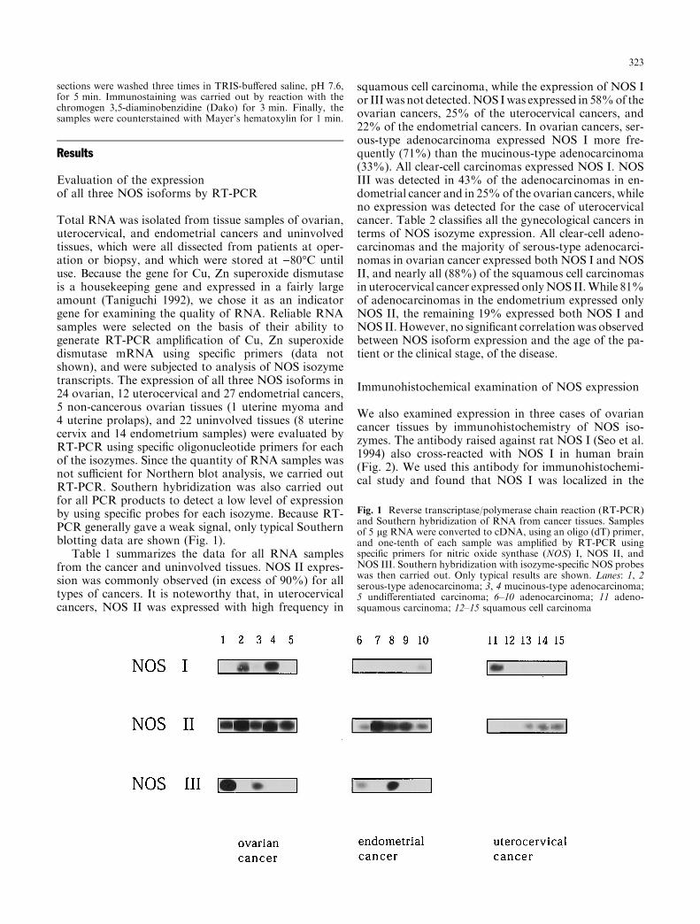
Total RNA was isolated from tissue samples of ovarian,uterocervical, and endometrial cancers and uninvolvedtissues, which were all dissected from patients at oper-ation or biopsy, and which were stored at )80°C untiluse. Because the gene for Cu, Zn superoxide dismutaseis a housekeeping gene and expressed in a fairly largeamount (Taniguchi 1992), we chose it as an indicatorgene for examining the quality of RNA. Reliable RNAsamples were selected on the basis of their ability togenerate RT-PCR ampli®cation of Cu, Zn superoxidedismutase mRNA using speci®c primers (data notshown), and were subjected to analysis of NOS isozymetranscripts. The expression of all three NOS isoforms in24 ovarian, 12 uterocervical and 27 endometrial cancers,5 non-cancerous ovarian tissues (1 uterine myoma and4 uterine prolaps), and 22 uninvolved tissues (8 uterinecervix and 14 endometrium samples) were evaluated byRT-PCR using speci®c oligonucleotide primers for eachof the isozymes. Since the quantity of RNA samples wasnot su�cient for Northern blot analysis, we carried outRT-PCR. Southern hybridization was also carried outfor all PCR products to detect a low level of expressionby using speci®c probes for each isozyme. Because RT-PCR generally gave a weak signal, only typical Southernblotting data are shown (Fig. 1).
Table 1 summarizes the data for all RNA samplesfrom the cancer and uninvolved tissues. NOS II expres-sion was commonly observed (in excess of 90%) for alltypes of cancers. It is noteworthy that, in uterocervicalcancers, NOS II was expressed with high frequency in
squamous cell carcinoma, while the expression of NOS Ior IIIwas not detected.NOS Iwas expressed in 58%of theovarian cancers, 25% of the uterocervical cancers, and22% of the endometrial cancers. In ovarian cancers, ser-ous-type adenocarcinoma expressed NOS I more fre-quently (71%) than the mucinous-type adenocarcinoma(33%). All clear-cell carcinomas expressed NOS I. NOSIII was detected in 43% of the adenocarcinomas in en-dometrial cancer and in 25%of the ovarian cancers, whileno expression was detected for the case of uterocervicalcancer. Table 2 classi®es all the gynecological cancers interms of NOS isozyme expression. All clear-cell adeno-carcinomas and the majority of serous-type adenocarci-nomas in ovarian cancer expressed both NOS I and NOSII, and nearly all (88%) of the squamous cell carcinomasin uterocervical cancer expressed onlyNOS II.While 81%of adenocarcinomas in the endometrium expressed onlyNOS II, the remaining 19% expressed both NOS I andNOS II.However, no signi®cant correlationwas observedbetween NOS isoform expression and the age of the pa-tient or the clinical stage, of the disease.
Immunohistochemical examination of NOS expression
We also examined expression in three cases of ovariancancer tissues by immunohistochemistry of NOS iso-zymes. The antibody raised against rat NOS I (Seo et al.1994) also cross-reacted with NOS I in human brain(Fig. 2). We used this antibody for immunohistochemi-cal study and found that NOS I was localized in the
Fig. 1 Reverse transcriptase/polymerase chain reaction (RT-PCR)and Southern hybridization of RNA from cancer tissues. Samplesof 5 lg RNA were converted to cDNA, using an oligo (dT) primer,and one-tenth of each sample was ampli®ed by RT-PCR usingspeci®c primers for nitric oxide synthase (NOS) I, NOS II, andNOS III. Southern hybridization with isozyme-speci®c NOS probeswas then carried out. Only typical results are shown. Lanes: 1, 2serous-type adenocarcinoma; 3, 4 mucinous-type adenocarcinoma;5 undi�erentiated carcinoma; 6±10 adenocarcinoma; 11 adeno-squamous carcinoma; 12±15 squamous cell carcinoma
323

cytosol of cancer cells (Fig. 3a). The antibody againstNOS II clearly stained the cytosol of cancer cells andmacrophages (Fig. 3b). Although immunoreactivity toNOS III was very weak or undetectable in cancer cells(Fig. 3c), vascular endothelial cells in the connectivetissue space of the tumor showed positive immunore-activity to NOS III (Fig. 3d).
Discussion
The role and functions of NO and its derivatives aremultiple. Nitroso compounds derived from nitrite and
nitrate, which are incorporated into the body via dietaryintake or from NO produced in the body by NOS, es-pecially under in¯ammatory conditions, are well-knowncarcinogens (Nguyen et al. 1992). It is also known thatNO functions as a bactericidal and tumoricidal agent(Takahashi et al. 1997). Thus the actual role of NOappears to depend largely on the amount produced andthe conditions under which it is produced. Thomsen et al.(1994) reported the expression of NOS I or NOS IIisozymes in gynecological cancer. Recently, several linesof evidence have suggested that NOS is expressed incancer as well as in normal rat tissues, and may regulateovarian functions (Chun et al. 1995; Van Voorhis et al.
Table 1 Frequency of nitricoxide synthase (NOS) isozymeexpression in gynecologicalcancers
Tissue NOS I NOS II NOS III
Ovarian cancer 14/24 (58%) 23/24 (96%) 6/24 (25%)Serous-type adenocarcinoma 5/7 7/7 3/7Mucinous-type adenocarcinoma 2/6 6/6 1/6Clear-cell adenocarcinoma 5/5 5/5 1/5Undi�erentiated carcinoma 1/3 3/3 0/3Sertoli cell tumor 1/1 1/1 1/1Yolk sac tumor 0/1 1/1 0/1Immature teratoma 0/1 0/1 0/1
Uterocervical cancer 3/12 (25%) 10/12 (83%) 0/12 (0%)Adenosquamous carcinoma 2/2 1/2 0/2Adenocarcinoma 1/1 1/1 0/1Squamous cell carcinoma + adenocarcinoma 0/1 1/1 0/1Squamous cell carcinoma 0/8 7/8 0/8
Endometrial cancer 6/27 (22%) 26/27 (96%) 9/27 (33%)Sarcoma 0/1 1/1 0/1Carcinosarcoma 1/2 2/2 0/2Adenocarcinoma 4/21 20/21 9/21Adenoacanthoma 0/1 1/1 0/1Adenosquamous carcinoma 1/1 1/1 0/1Mixed carcinoma 0/1 1/1 0/1
Uninvolved tissue 7/22 (32%) 20/22 (91%) 3/22 (14%)Cervix 2/8 6/8 0/8Endometrium 5/14 14/14 3/14
Table 2 Classi®cation of gyne-cological cancers in terms ofNOS isozyme expression.A Expresses both NOS I and II,B expresses only NOS I,C expresses only NOS II,D expresses no NOS
Tissue A B C D
Ovarian cancer 14/24 (58%) 0 8/24 (33%) 2/24 (8%)Serous-type adenocarcinoma 5 (71%) 0 2 (29%) 0Mucinous-type adenocarcinoma 2 (33%) 0 4 (67%) 0Clear-cell adenocarcinoma 5 (100%) 0 0 0Undi�erentiated carcinoma 1 (33%) 0 2 (67%) 0Sertoli cell tumor 1 0 0 0Yolk sac tumor 0 0 0 1Immature teratoma 0 0 0 1
Uterocervical cancer 2/12 (17%) 1/12 (8%) 8/12 (67%) 1/12 (8%)Adenosquamous carcinoma 1 1 0 0Adenocarcinoma 1 0 0 0Squamous cell carcinoma +
adenocarcinoma0 0 1 0
Squamous cell carcinoma 0 0 7 (88%) 1
Endometrial cancer 6/27 (22%) 0 21/27 (78%) 0Sarcoma 0 0 1 0Carcinosarcoma 1 0 1 0Adenocarcinoma 4 (19%) 0 17 (81%) 0Adenoacanthoma 0 0 1 0Adenosquamous carcinoma 1 0 0 0Mixed carcinoma 0 0 1 0
324

1995). Van Voorhis et al. (1995) demonstrated that NOSII expression in ovarian follicles and NOS III expressionin ovarian blood vessels are regulated by gonadotoropinin rats. It has also been suggested that NO is related toovulation in the rat in vivo (Shukovski and Tsafriri1994). However, NOS I has not been detected in eitherimmature or mature rat ovaries.
In this study, we examined the expression of NOSisoforms in many di�erent tissue types of ovarian, ut-erocervical, and endometrial cancers. NOS II was ex-pressed in nearly all the cancer tissues examined. Doiet al. (1996) reported that NO formed in a solid tumorcan be involved in enhanced vascular permeability andincreased blood ¯ow and, hence sustain tumor growth insuch cases. Thus, a high frequency of NOS II expressionin gynecological cancer may support tumor growth via asimilar mechanism. NOS I expression was detected at alower frequency in endometrial cancer than in ovarianand uterocervical cancers. In ovarian cancers, it wasexpressed in serous-type adenocarcinoma more fre-quently than in mucinous-type adenocarcinoma, andsamples that expressed NOS I also expressed NOS II.
Fig. 2 Immunostaining of brain tissues demonstrating that theantibody raised against rat NOS I cross-reacts with NOS I in humanbrain
Fig. 3a±d Immunohistochemical detection of NOS isozymes in semi-serial sections from ovarian serous-type adenocarcinoma tissue, usingspeci®c antibodies against NOS I (a), NOS II (b) and NOS III (c, d).Higher magni®cation of the area indicated by the asterisk (*) showedthat vascular endothelial cells in the connective tissue space stronglyexpressed this isozyme (d, arrow). Original magni®cation: a,b, c ´150,d ´ 380
325

However, the function of NOS I in cancer cells is un-known. Expression of NOS III was detected in 43% ofadenocarcinomas in endometrial cancer and in 25% ofovarian cancers, but was not detected in uterocervicalcancer, even when analyzed by RT-PCR/Southernblotting (Fig. 1, Table 1).
We also examined the immunohistochemical local-ization of NOS I, NOS II and NOS III in three samplesof ovarian cancer tissues (Fig. 3). The antibody againstNOS I and II clearly bound to cancer cells in ovariancancer tissue (Fig. 3a and b). NOS III was detectedmainly in the blood vessels in cancerous tissue, and onlyslightly in the cancer cells themselves (Fig. 3c, d).
In summary, we report the frequent expression ofNOS isozymes in common gynecological cancers.However the actual role of NO produced from eachNOS isozyme in these cancers remains ambiguous andfurther clari®cation is necessary.
Acknowledgements This work was supported, in part, by Grants-in-Aid for Scienti®c Research (A) and (B) from the Ministry ofEducation, Science, Sports, and Culture, Japan, the Japan Foun-dation for Aging, and a Research Grant (9A-1) for Nervous andMental Disorders from the Ministry of Health and Welfare, Japan.
References
Beckman JS, Beckman TN, Chen J, Marshall PA, Freeman BA(1990) Apparent hydroxyl radical production by peroxynitrite:implications for endothelial injury from nitric oxide and su-peroxide. Proc Natl Acad Sci USA 87:1620±1624
Bredt DS, Snyder SH (1992) Nitric oxide, a novel neuronal mes-senger. Neuron, 8:3±11
Chomczynski P, Sacchi N (1987) Single-step method of RNAisolation by acid guanidinium thiocyanate-phenol-chloroformextraction. Anal Biochem 162:156±159
Chun SY, Eisenhauer KM, Kubo M, Hsueh AJ (1995) Interleukin-1b suppresses apoptosis in rat ovarian follicles by increasingnitric oxide production. Endocrinology 136:3120±3127
Doi K, Akaike T, Horie H, Noguchi Y, Fujii S, Beppu T, OgawaM, Maeda H (1996) Excessive production of nitric oxide in ratsolid tumor and its implication in rapid tumor growth. Cancer77 (Suppl):1598±1604
Evans T, Carpenter A, Cohen J (1992) Puri®cation and a distinctiveform of endotoxin-induced nitric oxide synthase from rat liver.Proc Natl Acad Sci USA 89:5361±5365
Felley-Bosco E, Mirkovitch J, Ambs S, Mace K, Pfeifer A, KeeferLK, Harris, CC (1995) Nitric oxide and ethylnitrosourea; rel-ative mutagenicity in the p53 tumor suppressor and hypoxan-thine-phosphoribosyltransferase genes. Carcinogenesis 16:2069±2074
FujimotoH,AndoY,Yamashita T, TerazakiH, TanakaY, Sasaki J,Matsumoto, M, Suga M, Ando M (1997) Nitric oxide synthaseactivity in human lung cancer. Jpn J Cancer Res 88:1190±1198
Hallewell RA, Masiarz FR, Najarian RC, Puma JP, Quiroga MR,Randolph A, Sanchez-Pescador R, Scandella CJ, Smith B,
Steimer KS, Mullenbach GT (1985) Human Cu/Zn superoxidedismutase cDNA isolation of clones synthesising high levels ofactive or inactive enzyme from an expression library. NucleicAcids Res 13:2017±2034
Janssens SP, Shimouchi A, Quertermous T, Bloch DB, Bloch KD(1992) Cloning and expression of a cDNA encoding humanendothelium-derived relaxing factor/nitric oxide synthase.J Biol Chem 267:14519±14522
Jenkins DC, Charles IG, Baylis SA, Lelchuk R, Radomski MW,Moncada S (1994) Human colon cancer cell lines show a diversepattern of nitric oxide synthase gene expression and nitric oxidegeneration. Br J Cancer 70:847±849
Moncada S, Higgs A (1993) The L-arginine-nitric oxide pathway.N Engl J Med 329:2002±2012
Nakane M, Schmidt HH, Pollock JS, Forstermann U, Murad F(1993) Cloned human brain nitric oxide synthase is highly ex-pressed in skeletal muscle. FEBS Lett 316:175±180
Nathan C (1992) Nitric oxide as a secretory product of mammaliancells. FASEB J 6:3051±3064
Nguyen T, Brunson D, Crespi CL, Penman BW, Wishnok JS,Tannenbaum SR (1992) DNA damage and mutation in humancells exposed to nitric oxide in vitro. Proc Natl Acad Sci USA89:3030±3034
Seo HG, Tatsumi H, Fujii J, Nishikawa A, Suzuki K, Kangawa K,Taniguchi N (1994) Nitric oxide synthase from rat colorectum:Puri®cation, peptide sequencing partial PCR cloning, andimmunohistochemistry. J Biochem 115:602±607
Seo HG, Fujii J, Soejima H, Niikawa N, Taniguchi N (1995) Hemerequirement for production of active endothelial nitric oxidesynthase in baculovirus-infected insect cells. Biochem BiophysRes Commun 208:10±18
Sherman PA, Laubach VE, Reep BR, Wood ER (1993) Puri®ca-tion and cDNA sequence of an inducible nitric oxide synthasefrom a human tumor cell line. Biochemistry 32:11600±11605
Shukovski L, Tsafriri A (1994) The involvement of nitric oxidein the ovulatory process in the rat. Endocrinology 135:2287±2290
Stuehr DJ, Nathan CF (1989) Nitric oxide. A macrophage productresponsible for cytostasis and respiratory inhibition in tumortarget cell. J Exp Med 169:1543±1555
Takahashi M, Fukuda K, Ohata T, Sugimura T, Wakabayash K(1997) Increased expression of inducible and endothelial con-stitutive nitric oxide synthases in rat colon tumors induced byazoxymethane. Cancer Res 57:1233±1237
Taniguchi N (1992) Clinical signi®cances of superoxide dismutases;changes in aging, diabetes, ischemia, and cancer. Adv ClinChem 29:1±59
Thomsen LL, Lawton FG, Knowles RG, Beesley JE, Riveros-Moreno V, Moncada S (1994) Nitric oxide synthase activity inhuman gynecological cancer. Cancer Res 54:1352±1354
Thomsen LL, Miles DW, Happer®eld L, Bobrow LG, KnowlesRG, Moncada S (1995) Nitric oxide synthase activity in humanbreast cancer. Br J Cancer 72:41±44
Van Voorhis BJ, Moore K, Strijbos PJ, Nelson S, Baylis SA,Grzybicki D, Weiner CP (1995) Expression and localization ofinducible and endothelial nitric oxide synthase in the rat ovary.E�ects of gonadotropin stimulation in vivo. J Clin Invest96:2719±2726
Xie QW, Cho HJ, Calaycay J, Mumford RA, Swiderek KM, LeeTD, Ding A, Troso T, Nathan C (1992) Cloning and charac-terization of inducible nitric oxide synthase from mouse mac-rophages. Science 256:225±228
326