Embed Size (px)
Citation preview
702 American Family Physician www.aafp.org/afp Volume 90, Number 10 ◆ November 15, 2014
Diagnosis and Management of Tinea InfectionsJOHN W. ELY, MD, MSPH; SANDRA ROSENFELD, MD; and MARY SEABURY STONE, MD University of Iowa Carver College of Medicine, Iowa City, Iowa
The term tinea means fungal infec-tion, whereas dermatophyte refers to the fungal organisms that cause tinea. Tinea is usually followed by
a Latin term that designates the involved site, such as tinea corporis and tinea pedis (Table 1). Tinea versicolor (now called pity-riasis versicolor) is not caused by derma-tophytes but rather by yeasts of the genus Malassezia. Tinea unguium is more com-monly known as onychomycosis. Dermato-phytes are usually limited to involvement of hair, nails, and stratum corneum, which are inhospitable to other infectious agents. Der-matophytes include three genera: Tricho-phyton, Microsporum, and Epidermophyton.
The most common infections in prepu-bertal children are tinea corporis and tinea capitis, whereas adolescents and adults are more likely to develop tinea cruris, tinea pedis, and tinea unguium (onychomycosis). Tinea infections can be difficult to diagnose and treat. In one survey, tinea was the skin condition most likely to be misdiagnosed by primary care physicians.1
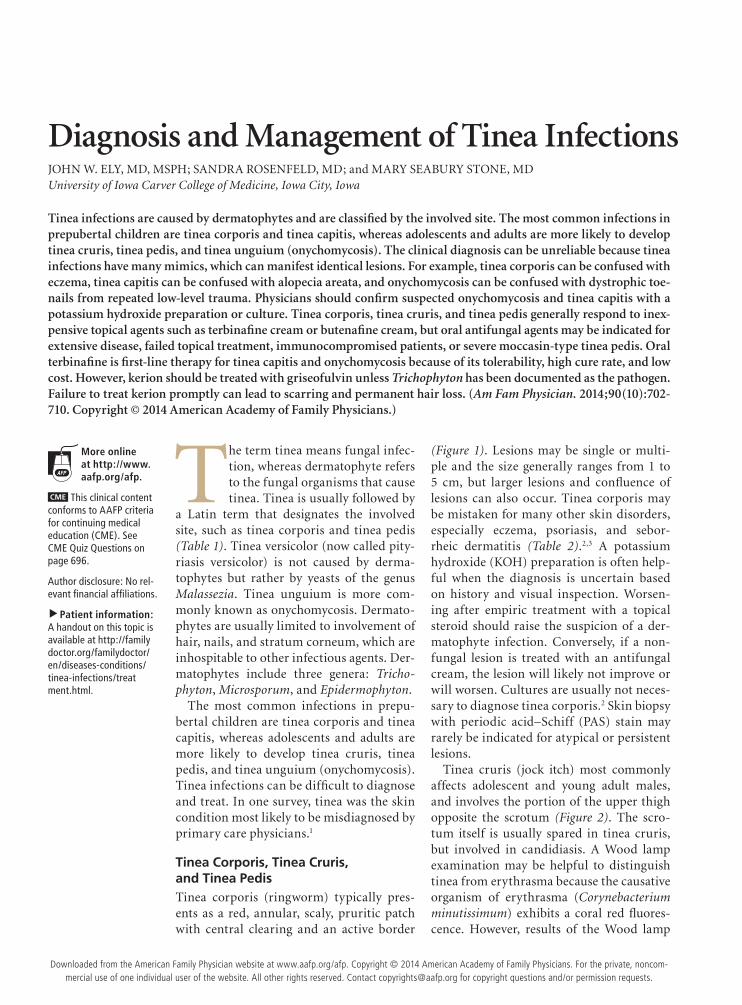
Tinea Corporis, Tinea Cruris, and Tinea PedisTinea corporis (ringworm) typically pres-ents as a red, annular, scaly, pruritic patch with central clearing and an active border
(Figure 1). Lesions may be single or multi-ple and the size generally ranges from 1 to 5 cm, but larger lesions and confluence of lesions can also occur. Tinea corporis may be mistaken for many other skin disorders, especially eczema, psoriasis, and sebor-rheic dermatitis (Table 2).2,3 A potassium hydroxide (KOH) preparation is often help-ful when the diagnosis is uncertain based on history and visual inspection. Worsen-ing after empiric treatment with a topical steroid should raise the suspicion of a der-matophyte infection. Conversely, if a non-fungal lesion is treated with an antifungal cream, the lesion will likely not improve or will worsen. Cultures are usually not neces-sary to diagnose tinea corporis.2 Skin biopsy with periodic acid–Schiff (PAS) stain may rarely be indicated for atypical or persistent lesions.
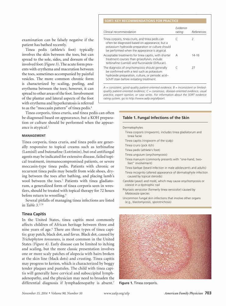
Tinea cruris (jock itch) most commonly affects adolescent and young adult males, and involves the portion of the upper thigh opposite the scrotum (Figure 2). The scro-tum itself is usually spared in tinea cruris, but involved in candidiasis. A Wood lamp examination may be helpful to distinguish tinea from erythrasma because the causative organism of erythrasma (Corynebacterium minutissimum) exhibits a coral red fluores-cence. However, results of the Wood lamp
Tinea infections are caused by dermatophytes and are classified by the involved site. The most common infections in prepubertal children are tinea corporis and tinea capitis, whereas adolescents and adults are more likely to develop tinea cruris, tinea pedis, and tinea unguium (onychomycosis). The clinical diagnosis can be unreliable because tinea infections have many mimics, which can manifest identical lesions. For example, tinea corporis can be confused with eczema, tinea capitis can be confused with alopecia areata, and onychomycosis can be confused with dystrophic toe-nails from repeated low-level trauma. Physicians should confirm suspected onychomycosis and tinea capitis with a potassium hydroxide preparation or culture. Tinea corporis, tinea cruris, and tinea pedis generally respond to inex-pensive topical agents such as terbinafine cream or butenafine cream, but oral antifungal agents may be indicated for extensive disease, failed topical treatment, immunocompromised patients, or severe moccasin-type tinea pedis. Oral terbinafine is first-line therapy for tinea capitis and onychomycosis because of its tolerability, high cure rate, and low cost. However, kerion should be treated with griseofulvin unless Trichophyton has been documented as the pathogen. Failure to treat kerion promptly can lead to scarring and permanent hair loss. (Am Fam Physician. 2014;90(10):702-710. Copyright © 2014 American Academy of Family Physicians.)
More online at http://www.aafp.org/afp.
CME This clinical content conforms to AAFP criteria for continuing medical education (CME). See CME Quiz Questions on page 696.
Author disclosure: No rel-evant financial affiliations.
▲
Patient information: A handout on this topic is available at http://family doctor.org/familydoctor/en/diseases-conditions/tinea-infections/treat ment.html.
Downloaded from the American Family Physician website at www.aafp.org/afp. Copyright © 2014 American Academy of Family Physicians. For the private, noncom-mercial use of one individual user of the website. All other rights reserved. Contact [email protected] for copyright questions and/or permission requests.
Tinea Infections
November 15, 2014 ◆ Volume 90, Number 10 www.aafp.org/afp American Family Physician 703
examination can be falsely negative if the patient has bathed recently.
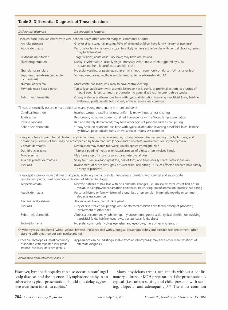
Tinea pedis (athlete’s foot) typically involves the skin between the toes, but can spread to the sole, sides, and dorsum of the involved foot (Figure 3). The acute form pres-ents with erythema and maceration between the toes, sometimes accompanied by painful vesicles. The more common chronic form is characterized by scaling, peeling, and erythema between the toes; however, it can spread to other areas of the foot. Involvement of the plantar and lateral aspects of the foot with erythema and hyperkeratosis is referred to as the “moccasin pattern” of tinea pedis.4
Tinea corporis, tinea cruris, and tinea pedis can often be diagnosed based on appearance, but a KOH prepara-tion or culture should be performed when the appear-ance is atypical.2
MANAGEMENT
Tinea corporis, tinea cruris, and tinea pedis are gener-ally responsive to topical creams such as terbinafine (Lamisil) and butenafine (Lotrimin), but oral antifungal agents may be indicated for extensive disease, failed topi-cal treatment, immunocompromised patients, or severe moccasin-type tinea pedis. Patients with chronic or recurrent tinea pedis may benefit from wide shoes, dry-ing between the toes after bathing, and placing lamb’s wool between the toes.5 Patients with tinea gladiato-rum, a generalized form of tinea corporis seen in wres-tlers, should be treated with topical therapy for 72 hours before return to wrestling.6
Several pitfalls of managing tinea infections are listed in Table 3.2,7,8
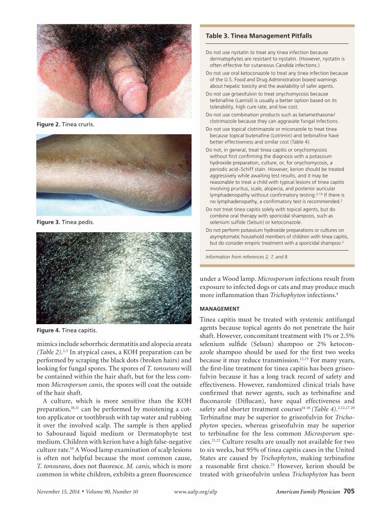
Tinea CapitisIn the United States, tinea capitis most commonly affects children of African heritage between three and nine years of age.4 There are three types of tinea capi-tis: gray patch, black dot, and favus. Black dot, caused by Trichophyton tonsurans, is most common in the United States (Figure 4). Early disease can be limited to itching and scaling, but the more classic presentation involves one or more scaly patches of alopecia with hairs broken at the skin line (black dots) and crusting. Tinea capitis may progress to kerion, which is characterized by boggy tender plaques and pustules. The child with tinea capi-tis will generally have cervical and suboccipital lymph-adenopathy, and the physician may need to broaden the differential diagnosis if lymphadenopathy is absent.7
Table 1. Fungal Infections of the Skin
Dermatophytes
Tinea corporis (ringworm), includes tinea gladiatorum and tinea faciei
Tinea capitis (ringworm of the scalp)
Tinea cruris (jock itch)
Tinea pedis (athlete’s foot)
Tinea unguium (onychomycosis)
Tinea manuum (commonly presents with “one-hand, two-feet” involvement)
Tinea barbae (beard infection in male adolescents and adults)
Tinea incognito (altered appearance of dermatophyte infection caused by topical steroids)
Candida (yeast) and mold, which may cause onychomycosis or coexist in a dystrophic nail
Pityriasis versicolor (formerly tinea versicolor) caused by Malassezia species
Uncommon fungal skin infections that involve other organs (e.g., blastomycosis, sporotrichosis)
SORT: KEY RECOMMENDATIONS FOR PRACTICE
Clinical recommendationEvidence rating References
Tinea corporis, tinea cruris, and tinea pedis can often be diagnosed based on appearance, but a potassium hydroxide preparation or culture should be performed when the appearance is atypical.
C 2
Acceptable treatments for tinea capitis, with shorter treatment courses than griseofulvin, include terbinafine (Lamisil) and fluconazole (Diflucan).
A 14-16
The diagnosis of onychomycosis should generally be confirmed with a test such as potassium hydroxide preparation, culture, or periodic acid–Schiff stain before initiating treatment.
C 27
A = consistent, good-quality patient-oriented evidence; B = inconsistent or limited-quality patient-oriented evidence; C = consensus, disease-oriented evidence, usual practice, expert opinion, or case series. For information about the SORT evidence rating system, go to http://www.aafp.org/afpsort.
Figure 1. Tinea corporis.
704 American Family Physician www.aafp.org/afp Volume 90, Number 10 ◆ November 15, 2014
However, lymphadenopathy can also occur in nonfungal scalp disease, and the absence of lymphadenopathy in an otherwise typical presentation should not delay aggres-sive treatment for tinea capitis.9
Many physicians treat tinea capitis without a confir-matory culture or KOH preparation if the presentation is typical (i.e., urban setting and child presents with scal-ing, alopecia, and adenopathy).2,7,8 The most common
Table 2. Differential Diagnosis of Tinea Infections
Differential diagnosis Distinguishing features
Tinea corporis (annular lesions with well-defined, scaly, often reddish margins; commonly pruritic)
Annular psoriasis Gray or silver scale; nail pitting; 70% of affected children have family history of psoriasis2
Atopic dermatitis Personal or family history of atopy; less likely to have active border with central clearing; lesions may be lichenified
Erythema multiforme Target lesions; acute onset; no scale; may have oral lesions
Fixed drug eruption Dusky; erythematous; usually single, nonscaly lesion; most often triggered by sulfa, acetaminophen, ibuprofen, or antibiotic use
Granuloma annulare No scale, vesicles, or pustules; nonpruritic; smooth; commonly on dorsum of hands or feet
Lupus erythematosus (subacute cutaneous)
Sun-exposed areas; multiple annular lesions; female-to-male ratio 3:13
Nummular eczema More confluent scale; less likely to have central clearing
Pityriasis rosea herald patch Typically an adolescent with a single lesion on neck, trunk, or proximal extremity; pruritus of herald patch is less common; progression to generalized rash in one to three weeks
Seborrheic dermatitis Greasy scale on erythematous base with typical distribution involving nasolabial folds, hairline, eyebrows, postauricular folds, chest; annular lesions less common
Tinea cruris (usually occurs in male adolescents and young men; spares scrotum and penis)
Candidal intertrigo Involves scrotum; satellite lesions; uniformly red without central clearing
Erythrasma Red-brown; no active border; coral red fluorescence with a Wood lamp examination
Inverse psoriasis Red and sharply demarcated; may have other signs of psoriasis such as nail pitting
Seborrheic dermatitis Greasy scale on erythematous base with typical distribution involving nasolabial folds, hairline, eyebrows, postauricular folds, chest; annular lesions less common
Tinea pedis (rare in prepubertal children; erythema, scale, fissures, maceration; itching between toes extending to sole, borders, and occasionally dorsum of foot; may be accompanied by tinea manuum [“one-hand, two-feet” involvement] or onychomycosis)
Contact dermatitis Distribution may match footwear; usually spares interdigital skin
Dyshidrotic eczema “Tapioca pudding” vesicles on lateral aspects of digits; often involves hands
Foot eczema May have atopic history; usually spares interdigital skin
Juvenile plantar dermatosis Shiny taut skin involving great toe, ball of foot, and heel; usually spares interdigital skin
Psoriasis Involvement of other sites; gray or silver scale; nail pitting; 70% of affected children have family history of psoriasis2
Tinea capitis (one or more patches of alopecia, scale, erythema, pustules, tenderness, pruritus, with cervical and suboccipital lymphadenopathy; most common in children of African heritage)
Alopecia areata Discrete patches of hair loss with no epidermal changes (i.e., no scale); total loss of hair or fine miniature hair growth; exclamation point hairs; no crusting; no inflammation; possible nail pitting
Atopic dermatitis Personal history or family history of atopy; less often annular; lymphadenopathy uncommon; alopecia less common
Bacterial scalp abscess Alopecia less likely; hair pluck is painful
Psoriasis Gray or silver scale; nail pitting; 70% of affected children have family history of psoriasis2; involvement of other sites
Seborrheic dermatitis Alopecia uncommon; lymphadenopathy uncommon; greasy scale; typical distribution involving nasolabial folds, hairline, eyebrows, postauricular folds, chest
Trichotillomania No scale; commonly involves eyelashes and eyebrows; hairs of varying lengths
Onychomycosis (discolored [white, yellow, brown], thickened nail with subungual keratinous debris and possible nail detachment; often starting with great toe but can involve any nail)
Other nail dystrophies, most commonly associated with repeated low-grade trauma, psoriasis, or lichen planus
Appearance can be indistinguishable from onychomycosis; may have other manifestations of alternate diagnosis
Information from references 2 and 3.
November 15, 2014 ◆ Volume 90, Number 10 www.aafp.org/afp American Family Physician 705
mimics include seborrheic dermatitis and alopecia areata (Table 2).2,3 In atypical cases, a KOH preparation can be performed by scraping the black dots (broken hairs) and looking for fungal spores. The spores of T. tonsurans will be contained within the hair shaft, but for the less com-mon Microsporum canis, the spores will coat the outside of the hair shaft.
A culture, which is more sensitive than the KOH preparation,10,11 can be performed by moistening a cot-ton applicator or toothbrush with tap water and rubbing it over the involved scalp. The sample is then applied to Sabouraud liquid medium or Dermatophyte test medium. Children with kerion have a high false-negative culture rate.10 A Wood lamp examination of scalp lesions is often not helpful because the most common cause, T. tonsurans, does not fluoresce. M. canis, which is more common in white children, exhibits a green fluorescence
under a Wood lamp. Microsporum infections result from exposure to infected dogs or cats and may produce much more inflammation than Trichophyton infections.4
MANAGEMENT
Tinea capitis must be treated with systemic antifungal agents because topical agents do not penetrate the hair shaft. However, concomitant treatment with 1% or 2.5% selenium sulfide (Selsun) shampoo or 2% ketocon-azole shampoo should be used for the first two weeks because it may reduce transmission.12,13 For many years, the first-line treatment for tinea capitis has been griseo-fulvin because it has a long track record of safety and effectiveness. However, randomized clinical trials have confirmed that newer agents, such as terbinafine and fluconazole (Diflucan), have equal effectiveness and safety and shorter treatment courses14-16 (Table 4).2,12,17-20
Terbinafine may be superior to griseofulvin for Tricho-phyton species, whereas griseofulvin may be superior to terbinafine for the less common Microsporum spe-cies.21,22 Culture results are usually not available for two to six weeks, but 95% of tinea capitis cases in the United States are caused by Trichophyton, making terbinafine a reasonable first choice.23 However, kerion should be treated with griseofulvin unless Trichophyton has been
Table 3. Tinea Management Pitfalls
Do not use nystatin to treat any tinea infection because dermatophytes are resistant to nystatin. (However, nystatin is often effective for cutaneous Candida infections.)
Do not use oral ketoconazole to treat any tinea infection because of the U.S. Food and Drug Administration boxed warnings about hepatic toxicity and the availability of safer agents.
Do not use griseofulvin to treat onychomycosis because terbinafine (Lamisil) is usually a better option based on its tolerability, high cure rate, and low cost.
Do not use combination products such as betamethasone/clotrimazole because they can aggravate fungal infections.
Do not use topical clotrimazole or miconazole to treat tinea because topical butenafine (Lotrimin) and terbinafine have better effectiveness and similar cost (Table 4).
Do not, in general, treat tinea capitis or onychomycosis without first confirming the diagnosis with a potassium hydroxide preparation, culture, or, for onychomycosis, a periodic acid–Schiff stain. However, kerion should be treated aggressively while awaiting test results, and it may be reasonable to treat a child with typical lesions of tinea capitis involving pruritus, scale, alopecia, and posterior auricular lymphadenopathy without confirmatory testing.2,7,8 If there is no lymphadenopathy, a confirmatory test is recommended.2
Do not treat tinea capitis solely with topical agents, but do combine oral therapy with sporicidal shampoos, such as selenium sulfide (Selsun) or ketoconazole.
Do not perform potassium hydroxide preparations or cultures on asymptomatic household members of children with tinea capitis, but do consider empiric treatment with a sporicidal shampoo.2
Information from references 2, 7, and 8.
Figure 2. Tinea cruris.
Figure 3. Tinea pedis.
Figure 4. Tinea capitis.
706 American Family Physician www.aafp.org/afp Volume 90, Number 10 ◆ November 15, 2014
documented as the pathogen.2,17 Failure to treat kerion promptly can lead to scarring and permanent hair loss.2
The child with tinea capitis should return for clini-cal assessment at the completion of therapy or sooner
if indicated, but follow-up cultures are usually unnec-essary if there is clinical improvement. Once treatment has started, the child may return to school, but for 14 days should not share combs, brushes, helmets, hats, or
Table 4. Treatment of Tinea Capitis and Tinea Unguium
Common treatment options Route Dosage forms Standard dosage Estimated drug cost*
U.S. Food and Drug Administration approval? Laboratory monitoring
Cure rates† Duration of treatment
Tinea capitis
Griseofulvin‡ Oral Suspension: 125 mg per 5 mL
Tablets (scored): 125 mg, 250 mg
Microsize (Grifulvin V suspension): 20 to 25 mg per kg per day; single daily dose or two divided doses (maximum: 1 g per day)
Ultramicrosize (Gris-Peg tablets): 10 to 15 mg per kg per day; single daily dose or two divided doses (maximum: 750 mg per day)
Microsize: $44 ($165) for 300 mL of 125-mg-per-5-mL solution
Ultramicrosize: $263 ($430) for 60 250-mg tablets
Yes, for children older than two years
No baseline testing in absence of liver disease
If required for longer than eight weeks, ALT, AST, bilirubin, and creatinine measurements and CBC every eight weeks2,17
92%18 Six to 12 weeks (continue for two weeks after symptoms and signs have resolved)2
Terbinafine (Lamisil)§
Oral Tablets: 250 mg
Granule packets: 125 mg, 187.5 mg
< 25 kg (55 lb): 125 mg once daily
25 to 35 kg (55 lb to 78 lb): 187.5 mg once daily
> 35 kg (78 lb): 250 mg once daily
Tablets: $4 ($660) for 30 250-mg tablets
Granules more expensive
Yes, for persons four years and older
Baseline ALT and AST measurement
CBC at six weeks for courses lasting longer than six weeks
94%18 Six weeks; longer for Microsporum infections
Assume Trichophyton unless culture reveals Microsporum
Fluconazole (Diflucan)||
Oral Tablets: 50 mg, 100 mg, 150 mg, 200 mg
Suspension: 10 mg per mL, 40 mg per mL
Daily dosing: 6 mg per kg per day for three to six weeks
or
Weekly dosing: 6 mg per kg once weekly
Tablets: $100 for 30 150-mg tablets ($1,185 for 90 50-mg tablets)
Suspension: $33 ($290) for 35 mL of 40-mg-per-mL suspension
Not for this indication
Approved for children older than six months for other indications
Baseline ALT, AST, and creatinine measurement and CBC
84%18 Three to six weeks for daily dosing
Eight to 12 weeks for weekly dosing
Itraconazole (Sporanox)¶
Oral Solution: 10 mg per mL
Capsules: 100 mg
Capsules: 5 mg per kg daily for four to six weeks
or
Solution: 3 mg per kg daily for four to six weeks
or
Pulse therapy with capsules: 5 mg per kg daily for one week each month for two to three months
or
Pulse therapy with oral solution: 3 mg per kg daily for one week each month for two to three months
Maximum: 500 mg per day
Solution: NA ($265) for 150 mL of 10-mg-per-mL solution
Capsules: $102 ($590) for 30 100-mg capsules
No Baseline ALT and AST measurement 86%18 Four to six weeks
Tinea unguium (onychomycosis)
Ciclopirox (Penlac)
Topical Bottle: 6.6 mL Apply daily to affected nail and adjacent skin; remove with alcohol every seven days
$16 ($540) for one bottle Yes, for persons older than 12 years
None 7%19 48 weeks
Terbinafine§ Oral Tablets: 250 mg
Granule packets: 125 mg, 187.5 mg
10 to 20 kg (22 to 44 lb): 62.5 mg daily
20 to 40 kg (44 to 89 lb): 125 mg daily
40 kg (89 lb) or more and adults: 250 mg daily
Tablets: $4 ($660) for 30 250-mg tablets
Granules more expensive
Not for this indication
Approved for children older than four years for tinea capitis
Baseline ALT and AST measurement, CBC
ALT and AST measurement, CBC at six weeks
76%20 Six weeks for fingernails; 12 weeks for toenails
Fluconazole|| Oral Tablets: 50 mg, 100 mg, 150 mg, 200 mg
Suspension: 10 mg per mL, 40 mg per mL
Children: 3 to 6 mg per kg once weekly
Adults: 150 to 300 mg once weekly
Tablets: $100 for 30 150-mg tablets ($1,185 for 90 50-mg tablets)
Suspension: $33 ($290) for 35 mL of 40-mg-per-mL suspension
Not for this indication
Approved for adults and children older than six months for other indications
Baseline ALT, AST, alkaline phosphatase, and creatinine measurements, CBC
No repeat needed for once weekly therapy
48%20 12 to 16 weeks for fingernails; 18 to 26 weeks for toenails
ALT = alanine transaminase; AST = aspartate transaminase; CBC = complete blood count; NA = not available.
*—Estimated retail price based on information obtained at http://www.goodrx.com. Accessed June 20, 2014. Generic price listed first; brand price listed in parentheses.†—Clinical cure rates rather than mycological cure rates, unless otherwise noted.‡—Griseofulvin is bitter tasting. It should be taken with whole milk or peanut butter to improve absorption. Recent reports of resistance may favor alterna-tives for uncomplicated tinea capitis.2 Griseofulvin remains the drug of choice for kerion and for tinea capitis caused by Microsporum species.2,17 Adverse effects include nausea, headache, urticaria, and rash. Parents should be asked to report symptoms of hepatic toxicity (e.g., abdominal pain, anorexia, nau-sea, vomiting, jaundice). Cross-sensitivity with penicillin may occur. Adjunctive topical treatment with 2% ketoconazole shampoo or 1% or 2.5% selenium sulfide (Selsun) shampoo should be used. Shampoo should be applied for 5 to 10 minutes three times a week for two to four weeks.§—Sprinkle granules on pudding, mashed potatoes, or ice cream. Adverse effects include nausea, abdominal pain, headache, nasopharyngitis, rash (gener-ally mild and transient), and elevated transaminase levels. Do not use if history of liver disease. Do not use terbinafine for kerion. Instead use griseofulvin.2,17
||—Has many drug interactions. Do not use if history of liver disease. Patients should be asked to report symptoms of hepatic toxicity (e.g., abdominal pain, anorexia, nausea, vomiting, jaundice). For tinea capitis, adjunctive topical treatment with 2% ketoconazole shampoo or 1% or 2.5% selenium sulfide shampoo should be used. Shampoo should be applied for five to 10 minutes three times a week for two to four weeks. Do not use fluconazole for kerion. Instead use griseofulvin.2,17
¶—The capsules must be given with food. The solution must be given on an empty stomach.12 Adverse effects include nausea and abdominal pain (gen-erally mild and transient), and elevated transaminase levels. Do not use if history of liver disease. Patients should be asked to report symptoms of hepatic toxicity (e.g., abdominal pain, anorexia, nausea, vomiting, jaundice). For tinea capitis, adjunctive topical treatment with 2% ketoconazole shampoo or 1% or 2.5% selenium sulfide shampoo should be used. Shampoo should be applied for five to 10 minutes three times a week for two to four weeks. Do not use itraconazole for kerion. Instead use griseofulvin.2,17
Information from references 2, 12, and 17 through 20.
November 15, 2014 ◆ Volume 90, Number 10 www.aafp.org/afp American Family Physician 707
pillowcases, or participate in sports that involve head-to-head contact, such as wrestling.2,17 Household members should be clinically evaluated but not necessarily tested for tinea capitis.17 Many experts recommend treating all
asymptomatic close contacts with a sporicidal shampoo, such as 2.5% selenium sulfide or 2% ketoconazole, for two to four weeks.2 If children do not improve, par-ents should be asked about adherence to the treatment
Table 4. Treatment of Tinea Capitis and Tinea Unguium
Common treatment options Route Dosage forms Standard dosage Estimated drug cost*
U.S. Food and Drug Administration approval? Laboratory monitoring
Cure rates† Duration of treatment
Tinea capitis
Griseofulvin‡ Oral Suspension: 125 mg per 5 mL
Tablets (scored): 125 mg, 250 mg
Microsize (Grifulvin V suspension): 20 to 25 mg per kg per day; single daily dose or two divided doses (maximum: 1 g per day)
Ultramicrosize (Gris-Peg tablets): 10 to 15 mg per kg per day; single daily dose or two divided doses (maximum: 750 mg per day)
Microsize: $44 ($165) for 300 mL of 125-mg-per-5-mL solution
Ultramicrosize: $263 ($430) for 60 250-mg tablets
Yes, for children older than two years
No baseline testing in absence of liver disease
If required for longer than eight weeks, ALT, AST, bilirubin, and creatinine measurements and CBC every eight weeks2,17
92%18 Six to 12 weeks (continue for two weeks after symptoms and signs have resolved)2
Terbinafine (Lamisil)§
Oral Tablets: 250 mg
Granule packets: 125 mg, 187.5 mg
< 25 kg (55 lb): 125 mg once daily
25 to 35 kg (55 lb to 78 lb): 187.5 mg once daily
> 35 kg (78 lb): 250 mg once daily
Tablets: $4 ($660) for 30 250-mg tablets
Granules more expensive
Yes, for persons four years and older
Baseline ALT and AST measurement
CBC at six weeks for courses lasting longer than six weeks
94%18 Six weeks; longer for Microsporum infections
Assume Trichophyton unless culture reveals Microsporum
Fluconazole (Diflucan)||
Oral Tablets: 50 mg, 100 mg, 150 mg, 200 mg
Suspension: 10 mg per mL, 40 mg per mL
Daily dosing: 6 mg per kg per day for three to six weeks
or
Weekly dosing: 6 mg per kg once weekly
Tablets: $100 for 30 150-mg tablets ($1,185 for 90 50-mg tablets)
Suspension: $33 ($290) for 35 mL of 40-mg-per-mL suspension
Not for this indication
Approved for children older than six months for other indications
Baseline ALT, AST, and creatinine measurement and CBC
84%18 Three to six weeks for daily dosing
Eight to 12 weeks for weekly dosing
Itraconazole (Sporanox)¶
Oral Solution: 10 mg per mL
Capsules: 100 mg
Capsules: 5 mg per kg daily for four to six weeks
or
Solution: 3 mg per kg daily for four to six weeks
or
Pulse therapy with capsules: 5 mg per kg daily for one week each month for two to three months
or
Pulse therapy with oral solution: 3 mg per kg daily for one week each month for two to three months
Maximum: 500 mg per day
Solution: NA ($265) for 150 mL of 10-mg-per-mL solution
Capsules: $102 ($590) for 30 100-mg capsules
No Baseline ALT and AST measurement 86%18 Four to six weeks
Tinea unguium (onychomycosis)
Ciclopirox (Penlac)
Topical Bottle: 6.6 mL Apply daily to affected nail and adjacent skin; remove with alcohol every seven days
$16 ($540) for one bottle Yes, for persons older than 12 years
None 7%19 48 weeks
Terbinafine§ Oral Tablets: 250 mg
Granule packets: 125 mg, 187.5 mg
10 to 20 kg (22 to 44 lb): 62.5 mg daily
20 to 40 kg (44 to 89 lb): 125 mg daily
40 kg (89 lb) or more and adults: 250 mg daily
Tablets: $4 ($660) for 30 250-mg tablets
Granules more expensive
Not for this indication
Approved for children older than four years for tinea capitis
Baseline ALT and AST measurement, CBC
ALT and AST measurement, CBC at six weeks
76%20 Six weeks for fingernails; 12 weeks for toenails
Fluconazole|| Oral Tablets: 50 mg, 100 mg, 150 mg, 200 mg
Suspension: 10 mg per mL, 40 mg per mL
Children: 3 to 6 mg per kg once weekly
Adults: 150 to 300 mg once weekly
Tablets: $100 for 30 150-mg tablets ($1,185 for 90 50-mg tablets)
Suspension: $33 ($290) for 35 mL of 40-mg-per-mL suspension
Not for this indication
Approved for adults and children older than six months for other indications
Baseline ALT, AST, alkaline phosphatase, and creatinine measurements, CBC
No repeat needed for once weekly therapy
48%20 12 to 16 weeks for fingernails; 18 to 26 weeks for toenails
ALT = alanine transaminase; AST = aspartate transaminase; CBC = complete blood count; NA = not available.
*—Estimated retail price based on information obtained at http://www.goodrx.com. Accessed June 20, 2014. Generic price listed first; brand price listed in parentheses.†—Clinical cure rates rather than mycological cure rates, unless otherwise noted.‡—Griseofulvin is bitter tasting. It should be taken with whole milk or peanut butter to improve absorption. Recent reports of resistance may favor alterna-tives for uncomplicated tinea capitis.2 Griseofulvin remains the drug of choice for kerion and for tinea capitis caused by Microsporum species.2,17 Adverse effects include nausea, headache, urticaria, and rash. Parents should be asked to report symptoms of hepatic toxicity (e.g., abdominal pain, anorexia, nau-sea, vomiting, jaundice). Cross-sensitivity with penicillin may occur. Adjunctive topical treatment with 2% ketoconazole shampoo or 1% or 2.5% selenium sulfide (Selsun) shampoo should be used. Shampoo should be applied for 5 to 10 minutes three times a week for two to four weeks.§—Sprinkle granules on pudding, mashed potatoes, or ice cream. Adverse effects include nausea, abdominal pain, headache, nasopharyngitis, rash (gener-ally mild and transient), and elevated transaminase levels. Do not use if history of liver disease. Do not use terbinafine for kerion. Instead use griseofulvin.2,17
||—Has many drug interactions. Do not use if history of liver disease. Patients should be asked to report symptoms of hepatic toxicity (e.g., abdominal pain, anorexia, nausea, vomiting, jaundice). For tinea capitis, adjunctive topical treatment with 2% ketoconazole shampoo or 1% or 2.5% selenium sulfide shampoo should be used. Shampoo should be applied for five to 10 minutes three times a week for two to four weeks. Do not use fluconazole for kerion. Instead use griseofulvin.2,17
¶—The capsules must be given with food. The solution must be given on an empty stomach.12 Adverse effects include nausea and abdominal pain (gen-erally mild and transient), and elevated transaminase levels. Do not use if history of liver disease. Patients should be asked to report symptoms of hepatic toxicity (e.g., abdominal pain, anorexia, nausea, vomiting, jaundice). For tinea capitis, adjunctive topical treatment with 2% ketoconazole shampoo or 1% or 2.5% selenium sulfide shampoo should be used. Shampoo should be applied for five to 10 minutes three times a week for two to four weeks. Do not use itraconazole for kerion. Instead use griseofulvin.2,17
Information from references 2, 12, and 17 through 20.
Tinea Infections
708 American Family Physician www.aafp.org/afp Volume 90, Number 10 ◆ November 15, 2014
regimen. The scalp should also be cultured to identify the organism and immunocompromise should be con-sidered. A second treatment course with the same or a different agent is reasonable if the diagnosis is confirmed.
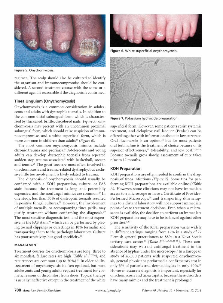
Tinea Unguium (Onychomycosis)Onychomycosis is a common consideration in adoles-cents and adults with dystrophic toenails. In addition to the common distal subungual form, which is character-ized by thickened, brittle, discolored nails (Figure 5), ony-chomycosis may present with an uncommon proximal subungual form, which should raise suspicion of immu-nocompromise, and a white superficial form, which is more common in children than adults24 (Figure 6).
The most common onychomycosis mimics include chronic trauma and psoriasis.25 Adolescents and young adults can develop dystrophic toenails from repeated sudden-stop trauma associated with basketball, soccer, and tennis.26 The great toes are most often involved in onychomycosis and trauma-related dystrophy, but exclu-sive little toe involvement is likely related to trauma.
The diagnosis of onychomycosis should usually be confirmed with a KOH preparation, culture, or PAS stain because the treatment is long and potentially expensive, and the nonfungal mimics are common.27 In one study, less than 50% of dystrophic toenails resulted in positive fungal cultures.28 However, the involvement of multiple toenails, or accompanying tinea pedis, may justify treatment without confirming the diagnosis.29 The most sensitive diagnostic test, and the most expen-sive, is the PAS stain,30 which can be performed by plac-ing toenail clippings or curettings in 10% formalin and transporting them to the pathology laboratory. Culture has poor sensitivity, but good specificity.30
MANAGEMENT
Treatment courses for onychomycosis are long (three to six months), failure rates are high (Table 42,12,17-20), and recurrences are common (up to 50%).31 In older adults, treatment of onychomycosis is often optional, but most adolescents and young adults request treatment for cos-metic reasons or discomfort from shoes. Topical therapy is usually ineffective except in the treatment of the white
superficial form. However, some patients resist systemic treatment, and ciclopirox nail lacquer (Penlac) can be offered together with information about its low cure rate. Oral fluconazole is an option,32 but for most patients oral terbinafine is the treatment of choice because of its superior effectiveness,33 tolerability, and low cost.31,34-38 Because toenails grow slowly, assessment of cure takes nine to 12 months.
KOH PreparationKOH preparations are often needed to confirm the diag-nosis of tinea infections (Figure 7). Some tips for per-forming KOH preparations are available online (eTable A). However, some clinicians may not have immediate access to a microscope or have a Certificate of Provider-Performed Microscopy,39 and transporting skin scrap-ings to a distant laboratory will not support immediate point-of-care treatment decisions. Even when a micro-scope is available, the decision to perform an immediate KOH preparation may have to be balanced against other priorities.1,40
The sensitivity of the KOH preparation varies widely in different settings, ranging from 12% in a study of 27 Flemish general practitioners to 88% in a Nova Scotia tertiary care center 41 (Table 510,11,29,30,41-48). These con-siderations may warrant antifungal treatment in the absence of hyphae under the microscope.2 In a European study of 45,000 patients with suspected onychomyco-sis, general physicians performed a confirmatory test in only 3% of patients and dermatologists in only 40%.40 However, accurate diagnosis is important, especially for onychomycosis and tinea capitis, because these disorders have many mimics and the treatment is prolonged.
Figure 5. Onychomycosis.
Figure 6. White superficial onychomycosis.
Figure 7. Potassium hydroxide preparation.
Tinea Infections
November 15, 2014 ◆ Volume 90, Number 10 www.aafp.org/afp American Family Physician 709
The first Choosing Wisely recommendation from the American Academy of Dermatology is, “Don’t prescribe oral antifungal therapy for suspected nail fungus with-out confirmation of fungal infection.”27 Clinicians who want to confirm the diagnosis of tinea infections before prescribing therapy have several options: (1) send the skin scrapings in a test tube to an off-site laboratory; (2) if feasible, perform the KOH preparation during the patient visit; or (3) substitute a test that involves less phy-sician time, such as a culture or, in the case of onychomy-cosis, a PAS stain of nail clippings.
Data Sources: A PubMed search was completed using the MeSH head-ing “Tinea”[Majr] and including meta-analyses, guidelines, randomized controlled trials, and reviews. Also searched were Essential Evidence Plus, the Cochrane Database of Systematic Reviews, and UpToDate. Finally, we performed multiple targeted searches in PubMed and reference lists of previously retrieved studies to fill in remaining information gaps, such as the performance characteristics of laboratory tests used to diagnose fun-gal infections. Search dates: October 16, 2013, through July 16, 2014.
The Authors
JOHN W. ELY, MD, MSPH, is a professor emeritus in the Department of Family Medicine at the University of Iowa Carver College of Medicine in Iowa City.
SANDRA ROSENFELD, MD, is a clinical assistant professor in the Department of Family Medicine at the University of Iowa Carver College of Medicine.
MARY SEABURY STONE, MD, is a professor in the Depart-ments of Dermatology and Pathology at the University of Iowa Carver College of Medicine.
Address correspondence to John W. Ely, MD, MSPH, Uni-versity of Iowa Carver College of Medicine, 200 Hawkins Dr., 01291-D PFP, Iowa City, IA 52242 (e-mail: [email protected]). Reprints are not available from the authors.
REFERENCES
1. Pariser RJ, Pariser DM. Primary care physicians’ errors in handling cuta-neous disorders. A prospective survey. J Am Acad Dermatol. 1987;17 (2 pt 1):239-245.
2. Kelly BP. Superficial fungal infections. Pediatr Rev. 2012;33(4):e22-e37.
3. Durosaro O, Davis MD, Reed KB, et al. Incidence of cutaneous lupus erythematosus, 1965-2005: a population-based study. Arch Dermatol. 2009;145(3):249-253.
4. Moriarty B, Hay R, Morris-Jones R. The diagnosis and management of tinea. BMJ. 2012;345:e4380.
5. Athlete’s foot, ringworm of the feet. In: Pickering LK, Baker CJ, Kimber-lin DW, et al. Red Book: 2012 Report of the Committee on Infectious Diseases. Elk Grove Village, Ill.: American Academy of Pediatrics; 2012. http://www.r2library.com/Resource/detail/158110703X/ch0003s0338 (subscription required). Accessed February 26, 2014.
6. Tinea corporis, ringworm of the body. In: Pickering LK, Baker CJ, Kim-berlin DW, et al. Red Book: 2012 Report of the Committee on Infectious Diseases. Elk Grove Village, Ill.: American Academy of Pediatrics; 2012. http://www.r2library.com/resource/detail/158110703X/ch0003s0336 (subscription required). Accessed December 12, 2013.
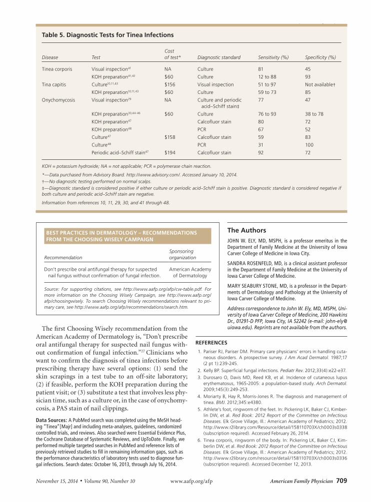
Table 5. Diagnostic Tests for Tinea Infections
Disease TestCost of test* Diagnostic standard Sensitivity (%) Specificity (%)
Tinea corporis Visual inspection41 NA Culture 81 45
KOH preparation41,42 $60 Culture 12 to 88 93
Tina capitis Culture10,11,43 $156 Visual inspection 51 to 97 Not available†KOH preparation10,11,43 $60 Culture 59 to 73 85
Onychomycosis Visual inspection29 NA Culture and periodic acid–Schiff stain‡
77 47
KOH preparation30,44-46 $60 Culture 76 to 93 38 to 78
KOH preparation47 Calcofluor stain 80 72
KOH preparation48 PCR 67 52
Culture47 $158 Calcofluor stain 59 83
Culture48 PCR 31 100
Periodic acid–Schiff stain47 $194 Calcofluor stain 92 72
KOH = potassium hydroxide; NA = not applicable; PCR = polymerase chain reaction.
*—Data purchased from Advisory Board. http://www.advisory.com/. Accessed January 10, 2014.†—No diagnostic testing performed on normal scalps.‡—Diagnostic standard is considered positive if either culture or periodic acid–Schiff stain is positive. Diagnostic standard is considered negative if both culture and periodic acid–Schiff stain are negative.
Information from references 10, 11, 29, 30, and 41 through 48.
BEST PRACTICES IN DERMATOLOGY – RECOMMENDATIONS FROM THE CHOOSING WISELY CAMPAIGN
RecommendationSponsoring organization
Don’t prescribe oral antifungal therapy for suspected nail fungus without confirmation of fungal infection.
American Academy of Dermatology
Source: For supporting citations, see http://www.aafp.org/afp/cw-table.pdf. For more information on the Choosing Wisely Campaign, see http://www.aafp.org/afp/choosingwisely. To search Choosing Wisely recommendations relevant to pri-mary care, see http://www.aafp.org/afp/recommendations/search.htm.
Tinea Infections
710 American Family Physician www.aafp.org/afp Volume 90, Number 10 ◆ November 15, 2014
7. Hubbard TW. The predictive value of symptoms in diagnosing childhood tinea capitis. Arch Pediatr Adolesc Med. 1999;153(11):1150-1153.
8. Lorch Dauk KC, Comrov E, Blumer JL, et al. Tinea capitis: predictive value of symptoms and time to cure with griseofulvin treatment. Clin Pediatr (Phila). 2010;49(3):280-286.
9. Williams JV, Eichenfield LF, Burke BL, et al. Prevalence of scalp scaling in prepubertal children. Pediatrics. 2005;115(1):e1-e6.
10. Gan VN, Petruska M, Ginsburg CM. Epidemiology and treatment of tinea capitis: ketoconazole vs. griseofulvin. Pediatr Infect Dis J. 1987; 6(1):46-49.
11. Ginsburg CM, Gan VN, Petruska M. Randomized controlled trial of intra-lesional corticosteroid and griseofulvin vs. griseofulvin alone for treatment of kerion. Pediatr Infect Dis J. 1987;6(12):1084-1087.
12. Ali S, Graham TA, Forgie SE. The assessment and management of tinea capitis in children. Pediatr Emerg Care. 2007;23(9):662-665.
13. Chen C, Koch LH, Dice JE, et al. A randomized, double-blind study comparing the efficacy of selenium sulfide shampoo 1% and ciclopirox shampoo 1% as adjunctive treatments for tinea capitis in children. Pedi-atr Dermatol. 2010;27(5):459-462.
14. Deng S, Hu H, Abliz P, et al. A random comparative study of terbin-afine versus griseofulvin in patients with tinea capitis in Western China. Mycopathologia. 2011;172(5):365-372.
15. Elewski BE, Cáceres HW, DeLeon L, et al. Terbinafine hydrochloride oral granules versus oral griseofulvin suspension in children with tinea capi-tis: results of two randomized, investigator-blinded, multicenter, inter-national, controlled trials. J Am Acad Dermatol. 2008;59(1):41-54.
16. González U, Seaton T, Bergus G, et al. Systemic antifungal therapy for tinea capitis in children. Cochrane Database Syst Rev. 2007;(4):CD004685.
17. Tinea capitis, ringworm of the scalp. In: Pickering LK, Baker CJ, Kimber-lin DW, et al. Red Book: 2012 Report of the Committee on Infectious Diseases. Elk Grove Village, Ill.: American Academy of Pediatrics; 2012. http://www.r2library.com/Resource/detail/158110703X/ch0003s0335 (subscription required). Accessed December 12, 2013.
18. Gupta AK, Adam P, Dlova N, et al. Therapeutic options for the treatment of tinea capitis caused by Trichophyton species: griseofulvin versus the new oral antifungal agents, terbinafine, itraconazole, and fluconazole. Pediatr Dermatol. 2001;18(5):433-438.
19. Gupta AK, Fleckman P, Baran R. Ciclopirox nail lacquer topical solution 8% in the treatment of toenail onychomycosis. J Am Acad Dermatol. 2000;43(4 suppl):S70-S80.
20. Gupta AK, Ryder JE, Johnson AM. Cumulative meta-analysis of systemic antifungal agents for the treatment of onychomycosis. Br J Dermatol. 2004;150(3):537-544.
21. Gupta A, Simpson F. Device-based therapies for onychomycosis treat-ment. Skin Therapy Lett. 2012;17(9):4-9.
22. Tey HL, Tan AS, Chan YC. Meta-analysis of randomized, controlled trials comparing griseofulvin and terbinafine in the treatment of tinea capitis. J Am Acad Dermatol. 2011;64(4):663-670.
23. Foster KW, Ghannoum MA, Elewski BE. Epidemiologic surveillance of cutaneous fungal infection in the United States from 1999 to 2002. J Am Acad Dermatol. 2004;50(5):748-752.
24. de Berker D. Childhood nail diseases. Dermatol Clin. 2006;24(3):355-363.
25. de Berker D. Clinical practice. Fungal nail disease. N Engl J Med. 2009;360(20):2108-2116.
26. Allevato MA. Diseases mimicking onychomycosis. Clin Dermatol. 2010; 28(2):164-177.
27. American Academy of Dermatology. Five things physicians and patients should question. http://www.choosingwisely.org/doctor-patient-lists/american-academy-of-dermatology/. Accessed June 20, 2014.
28. Summerbell RC, Kane J, Krajden S. Onychomycosis, tinea pedis and tinea manuum caused by non-dermatophytic filamentous fungi. Myco-ses. 1989;32(12):609-619.
29. Garcia-Doval I, Cabo F, Monteagudo B, et al. Clinical diagnosis of toenail onychomycosis is possible in some patients: cross-sectional diagnostic study and development of a diagnostic rule. Br J Dermatol. 2010;163(4): 743-751.
30. Haghani I, Shokohi T, Hajheidari Z, et al. Comparison of diagnostic methods in the evaluation of onychomycosis. Mycopathologia. 2013; 175(3-4):315-321.
31. Sigurgeirsson B, Olafsson JH, Steinsson JB, et al. Long-term effective-ness of treatment with terbinafine vs itraconazole in onychomycosis: a 5-year blinded prospective follow-up study. Arch Dermatol. 2002; 138(3):353-357.
32. Scher RK, Breneman D, Rich P, et al. Once-weekly fluconazole (150, 300, or 450 mg) in the treatment of distal subungual onychomycosis of the toenail. J Am Acad Dermatol. 1998;38(6 pt 2):S77-S86.
33. Volk B, Tiu A, St Anna L. Clinical Inquiry: which oral antifungal works best for toenail onychomycosis? J Fam Pract. 2013;62(2):100-101.
34. Crawford F, Young P, Godfrey C, et al. Oral treatments for toenail ony-chomycosis: a systematic review. Arch Dermatol. 2002;138(6):811-816.
35. Epstein E. How often does oral treatment of toenail onychomycosis pro-duce a disease-free nail? An analysis of published data. Arch Dermatol. 1998;134(12):1551-1554.
36. Evans EG, Sigurgeirsson B; The LION Study Group. Double blind, ran-domised study of continuous terbinafine compared with intermittent itraconazole in treatment of toenail onychomycosis. BMJ. 1999;318 (7190):1031-1035.
37. Gupta AK, Cooper EA, Lynde CW. The efficacy and safety of terbinafine in children. Dermatol Clin. 2003;21(3):511-520.
38. Gupta AK, Cooper EA, Paquet M. Recurrences of dermatophyte toenail onychomycosis during long-term follow-up after successful treatments with mono- and combined therapy of terbinafine and itraconazole. J Cutan Med Surg. 2013;17(3):201-206.
39. Centers for Medicare & Medicaid Services. Interpretive guidelines for laboratories. Appendix C: survey procedures and interpretive guide-lines for laboratories and laboratory services. http://www.cms.gov/Regulations-and-Guidance/Legislation/CLIA/Interpretive_Guidelines_for_Laboratories.html. Accessed January 9, 2014.
40. Effendy I, Lecha M, Feuilhade de Chauvin M, et al.; European Onycho-mycosis Observatory. Epidemiology and clinical classification of onycho-mycosis. J Eur Acad Dermatol Venereol. 2005;19(suppl 1):8-12.
41. Lousbergh D, Buntinx F, Piérard G. Diagnosing dermatomycosis in gen-eral practice. Fam Pract. 1999;16(6):611-615.
42. Haldane DJ, Robart E. A comparison of calcofluor white, potassium hydroxide, and culture for the laboratory diagnosis of superficial fungal infection. Diagn Microbiol Infect Dis. 1990;13(4):337-339.
43. Fathi HI, al-Samarai AM. Tinea capitis in Iraq: laboratory results. East Mediterr Health J. 2000;6(1):138-148.
44. Litz CE, Cavagnolo RZ. Polymerase chain reaction in the diagnosis of onychomycosis: a large, single-institute study. Br J Dermatol. 2010; 163(3):511-514.
45. Shemer A, Trau H, Davidovici B, et al. Nail sampling in onychomycosis: comparative study of curettage from three sites of the infected nail. J Dtsch Dermatol Ges. 2007;5(12):1108-1111.
46. Souza PR, Vettorato G, Pinto GM, et al. Concordance between direct microscopy and fungical culture for the diagnostic of feet’s onychomy-cosis. An Bras Dermatol. 2012;87(1):157-159.
47. Weinberg JM, Koestenblatt EK, Tutrone WD, et al. Comparison of diag-nostic methods in the evaluation of onychomycosis. J Am Acad Derma-tol. 2003;49(2):193-197.
48. Garg J, Tilak R, Singh S, et al. Evaluation of pan-dermatophyte nested PCR in diagnosis of onychomycosis. J Clin Microbiol. 2007;45(10): 3443-3445.
Tinea Infections
November 15, 2014 ◆ Volume 90, Number 10 www.aafp.org/afp American Family Physician
eTable A. Tips for KOH Preparation
Obtaining the sample
The scraping should be taken with a #15 scalpel blade or the edge of a glass slide. The scraped scale should fall onto a microscope slide or into a test tube. False-negative KOH preparations often result from inadequate scrapings.
A tinea capitis sample for KOH preparation can be taken by scraping the black dots (hairs broken off at the skin line).
For suspected onychomycosis, consider a periodic acid–Schiff stain of nail clippings instead of KOH preparation.
Because the scrapings will easily blow off the slide, shield it from drafts or apply KOH preparation to the slide before transport.
Preparing the slide
Place two drops of 10% or 20% KOH on the scrapings, followed by a coverslip. Alternatively, place a coverslip over the dry scrapings and a drop or two of KOH next to the coverslip and allow it to run under the coverslip. KOH dissolves squamous cells but leaves the fungal elements intact.
Heat the slide with a match or alcohol lamp. The match may leave a smoky deposit on the slide. Avoid boiling the KOH, but the slide should be hot enough to be uncomfortable to the dorsum of the hand, usually three to four seconds over the flame. Skin scrapings and hair can be examined under the microscope immediately. Toenail curettings should wait at least 10 minutes to several hours before examination.
After heating the slide, tap down the coverslip to compress the sample and separate the hyphae from the squamous cells.
Examining the slide under the microscope
Adjust the light filter and drop the condenser to achieve a low light level and increased refraction.
Scan the slide under low power, and use high power to confirm hyphae in suspicious areas.
False-negative results on KOH preparations are common and are usually caused by inadequate material on the slide. False-positive results can occur from misinterpretation of hair shafts or clothing fibers, which are often larger than hyphae, not segmented, and not branching. The borders between squamous cells can also be mistaken for hyphae.
The shelf life of a bottle of KOH is at least five years. KOH can damage microscope lenses. Therefore, use an old microscope, and avoid spills and excess KOH on the slide.
KOH = potassium hydroxide.
Information from Kelly BP. Superficial fungal infections. Pediatr Rev. 2012; 33(4):e22-e37.
Downloaded from the American Family Physician website at www.aafp.org/afp. Copyright © 2014 American Academy of Family Physicians. For the private, non-commercial use of one individual user of the website. All other rights reserved. Contact [email protected] for copyright questions and/or permission requests.


![SCIENCE CHINA Life Sciences - Springer · tions, such as tinea capitis, tinea corporis, tinea inguinalis, tinea manus, tinea unguium and tinea pedis [1–3]. Unlike](https://img.dokumen.tips/doc/110x75/5d1b54ac88c993283c8ce38a/science-china-life-sciences-springer-tions-such-as-tinea-capitis-tinea-corporis.jpg)


![Ringworm Disease- Causes, Diagnosis and Treatment: AMYCOT ... · Tinea capitis, Tinea pedis, and . Tinea unguium . or onychomycosis [1]. Ringworm is the most common type of fungal](https://img.dokumen.tips/doc/110x75/5ca4e5e688c993a3308c5db0/ringworm-disease-causes-diagnosis-and-treatment-amycot-tinea-capitis.jpg)