Embed Size (px)
Citation preview
JPMI VOL. 30 NO. 2 133
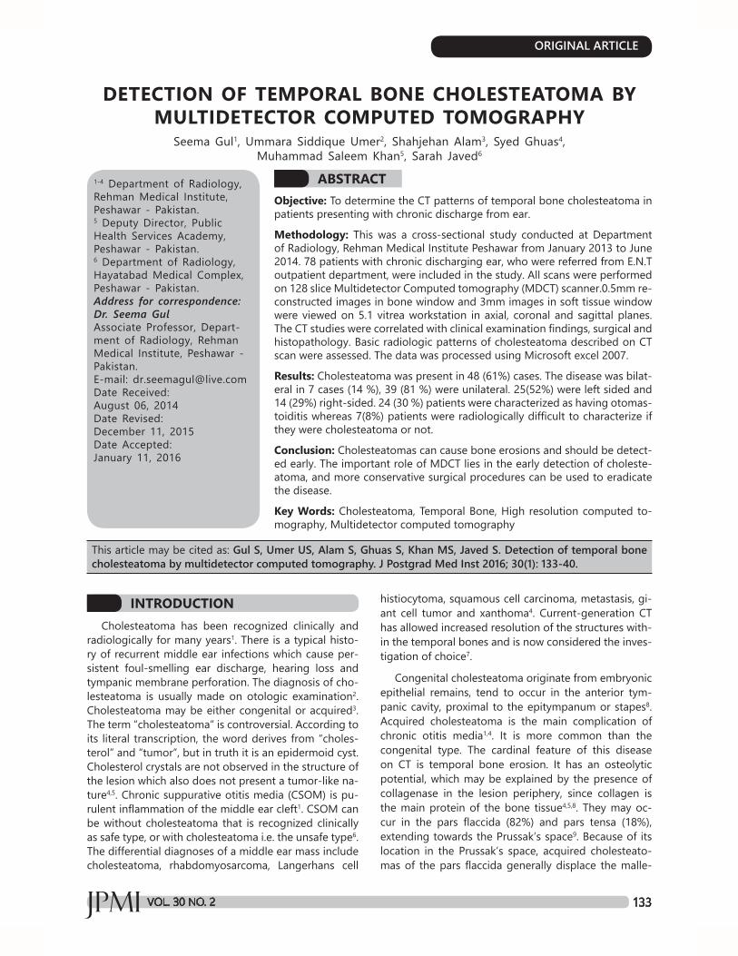
DETECTION OF TEMPORAL BONE CHOLESTEATOMA BY MULTIDETECTOR COMPUTED TOMOGRAPHY
Seema Gul1, Ummara Siddique Umer2, Shahjehan Alam3, Syed Ghuas4, Muhammad Saleem Khan5, Sarah Javed6
ORIGINAL ARTICLE
INTRODUCTIONCholesteatoma has been recognized clinically and
radiologically for many years1. There is a typical histo-ry of recurrent middle ear infections which cause per-sistent foul-smelling ear discharge, hearing loss and tympanic membrane perforation. The diagnosis of cho-lesteatoma is usually made on otologic examination2. Cholesteatoma may be either congenital or acquired3. The term “cholesteatoma” is controversial. According to its literal transcription, the word derives from “choles-terol” and “tumor”, but in truth it is an epidermoid cyst. Cholesterol crystals are not observed in the structure of the lesion which also does not present a tumor-like na-ture4,5. Chronic suppurative otitis media (CSOM) is pu-rulent inflammation of the middle ear cleft1. CSOM can be without cholesteatoma that is recognized clinically as safe type, or with cholesteatoma i.e. the unsafe type6. The differential diagnoses of a middle ear mass include cholesteatoma, rhabdomyosarcoma, Langerhans cell
histiocytoma, squamous cell carcinoma, metastasis, gi-ant cell tumor and xanthoma4. Current-generation CT has allowed increased resolution of the structures with-in the temporal bones and is now considered the inves-tigation of choice7.
Congenital cholesteatoma originate from embryonic epithelial remains, tend to occur in the anterior tym-panic cavity, proximal to the epitympanum or stapes8. Acquired cholesteatoma is the main complication of chronic otitis media1,4. It is more common than the congenital type. The cardinal feature of this disease on CT is temporal bone erosion. It has an osteolytic potential, which may be explained by the presence of collagenase in the lesion periphery, since collagen is the main protein of the bone tissue4,5,8. They may oc-cur in the pars flaccida (82%) and pars tensa (18%), extending towards the Prussak’s space9. Because of its location in the Prussak’s space, acquired cholesteato-mas of the pars flaccida generally displace the malle-
This article may be cited as: Gul S, Umer US, Alam S, Ghuas S, Khan MS, Javed S. Detection of temporal bone cholesteatoma by multidetector computed tomography. J Postgrad Med Inst 2016; 30(1): 133-40.
ABSTRACTObjective: To determine the CT patterns of temporal bone cholesteatoma in patients presenting with chronic discharge from ear.
Methodology: This was a cross-sectional study conducted at Department of Radiology, Rehman Medical Institute Peshawar from January 2013 to June 2014. 78 patients with chronic discharging ear, who were referred from E.N.T outpatient department, were included in the study. All scans were performed on 128 slice Multidetector Computed tomography (MDCT) scanner.0.5mm re-constructed images in bone window and 3mm images in soft tissue window were viewed on 5.1 vitrea workstation in axial, coronal and sagittal planes. The CT studies were correlated with clinical examination findings, surgical and histopathology. Basic radiologic patterns of cholesteatoma described on CT scan were assessed. The data was processed using Microsoft excel 2007.
Results: Cholesteatoma was present in 48 (61%) cases. The disease was bilat-eral in 7 cases (14 %), 39 (81 %) were unilateral. 25(52%) were left sided and 14 (29%) right-sided. 24 (30 %) patients were characterized as having otomas-toiditis whereas 7(8%) patients were radiologically difficult to characterize if they were cholesteatoma or not.
Conclusion: Cholesteatomas can cause bone erosions and should be detect-ed early. The important role of MDCT lies in the early detection of choleste-atoma, and more conservative surgical procedures can be used to eradicate the disease.
Key Words: Cholesteatoma, Temporal Bone, High resolution computed to-mography, Multidetector computed tomography
1-4 Department of Radiology, Rehman Medical Institute, Peshawar - Pakistan.5 Deputy Director, Public Health Services Academy, Peshawar - Pakistan.6 Department of Radiology, Hayatabad Medical Complex, Peshawar - Pakistan.Address for correspondence:Dr. Seema GulAssociate Professor, Depart-ment of Radiology, Rehman Medical Institute, Peshawar - Pakistan.E-mail: [email protected] Received:August 06, 2014Date Revised:December 11, 2015Date Accepted:January 11, 2016
DETECTION OF TEMPORAL BONE CHOLESTEATOMA BY MULTIDETECTOR COMPUTED TOMOGRAPHY
JPMI VOL. 30 NO. 2 134
us head and the body of the incus medially. From the Prussak’s space, the mass can easily extend itself pos-teriorly in the superior incudal space towards the pos-terolateral portion of the attic, and then, through the aditus ad antrum, towards the antrum and the mastoid air cells10. The diagnosis of cholesteatoma is based on clinical evaluation (otoscopy), where retraction of the tympanic membrane with pars flaccida perforation and a whitish mass in the middle ear are observed11. The ability of Multidetector Computed Tomography (MDCT) to predict accurately the status of the struc-tures of the temporal bone represents a major advance in delineating pathology prior to surgical exploration of ears with cholesteatoma12. MDCT is considered the imaging method of choice in the evaluation of middle ear cholesteatoma. Magnetic resonance imaging (MRI) has gained importance in the evaluation of complicat-ed cholesteatoma and in the postoperative follow-up of patients to evaluate residual or recurrent choleste-atoma4,5. Role played by preoperative CT imaging is evaluation of lesion extent (attic, antrum and mastoid); detection of complications, such as bone lysis and cere-bromeningeal complications; and detection of anatom-ical variations (jugular dehiscence and lateralized sinus). Special attention should be given to the tympanic sinus and to the facial recess, since they are locations whose visualization is difficult during the surgery, and are fre-quent sites of residual disease.
METHODOLOGYThis was a cross-sectional study conducted at De-
partment of Radiology, Rehman Medical Institute Pe-shawar from January 2013 to June2014. We retrospec-tively collected 78 patients with chronic discharging ear, who were referred from E.N.T outpatient depart-ment. Clinically patients were with either a marginal tympanic membrane perforation or a tympanic mem-brane inadequately visualized owing to the presence of granulations or squamous debris in the external au-ditory meatus. All scans were performed on 128 slice Multidetector Computed tomography (MDCT) scanner in the Department of Radiology, Rehman Medical In-stitute Peshawar.0.5mm reconstructed images in bone window and 3mm images in soft tissue window were viewed on 5.1 vitrea workstation in axial, coronal and sagittal planes. Non-contrast CT was adequate. Intrave-nous contrast medium was only given in those patients with suspicion of intracranial extension. The data was processed using Microsoft excel 2007. The CT findings were compared with clinical examination findings, sur-gical and histopathological results. The pre-operative CT scans were reported to assess characteristic choles-teatoma findings, location and extension of soft tissues in middle ear, integrity of scutum, erosion of the ossic-ular chain, integrity of the thin bony septum of facial nerve canal, the semicircular canals and the tegmen,
extension of cholesteatoma outside the middle ear, in-tegrity of mastoid air cells, trabeculae, as well as rela-tionship and proximity of soft tissues with the tympanic membrane were assessed.
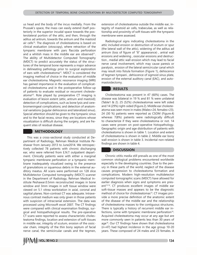
Radiological signs indicating cholesteatoma in the attic included erosion or destruction of scutum or spur (the lateral wall of the attic), widening of the aditus ad antrum (loss of figure of “8” appearance) , antral wall erosions and widening , ossicular erosions and destruc-tion , medial attic wall erosion which may lead to facial nerve canal involvement, which may cause paresis or paralysis , erosion of the lateral semicircular canal which may result into fistula formation (Figure 1), dehiscence of tegmen tympani , dehiscence of sigmoid sinus plate, erosion of the external auditory canal (EAC), and auto-mastoidectomy.
RESULTSCholesteatoma was present in 47 (60%) cases. The
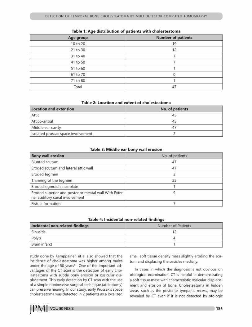
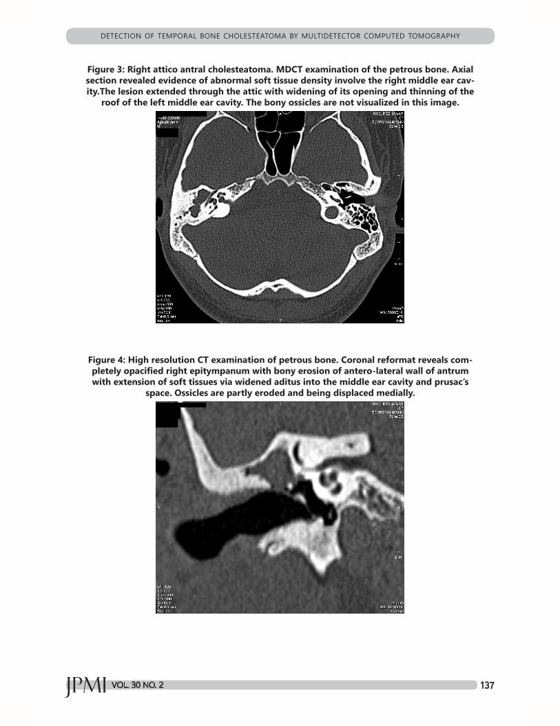
disease was bilateral in 19 % and 81 % were unilateral (Table1 & 2). 25 (52%) cholesteatomas were left sided and 14 (29%) right-sided (Figure.2). Middle ear choleste-atoma was seen more in males (Males=24, females=23). 24 (30 %) patients were reported as CSOM (Figure.4) whereas 7(8%) patients were radiologically difficult to characterize if they were cholesteatoma or not. 14 cases were proven on post-operative histopathology. Geographic origin and age distribution of patients with cholesteatoma is shown in table 1. Location and extent of cholesteatoma is shown in table 2. Middle ear bony wall erosion is shown in table 3. Incidental non-related findings are shown in table 4.
DISCUSSIONChronic otitis media still prevails as one of the most
common otological problems encountered worldwide especially in the developing countries. Due to the pen-ury in these parts of the world, neglect of the disease causes progression to cholesteatoma formation and complications. Modern high-resolution multidetector computed tomographic scans (MDCT) have allowed for earlier diagnosis when signs and symptoms are pres-ent13,14. CT produces excellent images of middle ear soft-tissue masses and appears to be the diagnostic method of choice for cholesteatomas15. CT images pro-vide a more precise definition of the anatomic extent of the disease of the middle ear and the relationship of cholesteatoma masses to the contiguous structures. There is typically a history of recurrent middle ear in-fections, some with tympanic membrane perforation16. Acquired cholesteatoma may occur at any age but are more commonly seen in patients less than 30 years of age16. Our CT findings have shown that cholesteatoma (n=47) had highest incidence in the age group 10-20 years. These composed of 24 males and 23 females. A
JPMI VOL. 30 NO. 2 135
DETECTION OF TEMPORAL BONE CHOLESTEATOMA BY MULTIDETECTOR COMPUTED TOMOGRAPHY
Table 1: Age distribution of patients with cholesteatoma Age group Number of patients
10 to 20 1921 to 30 1231 to 40 741 to 50 751 to 60 161 to 70 071 to 80 1
Total 47
Table 2: Location and extent of cholesteatomaLocation and extension No. of patientsAttic 45Attico-antral 45Middle ear cavity 47Isolated prussac space involvement 2
Table 3: Middle ear bony wall erosionBony wall erosion No. of patientsBlunted scutum 47Eroded scutum and lateral attic wall 47Eroded tegmen 2Thinning of the tegmen 25Eroded sigmoid sinus plate 1Eroded superior and posterior meatal wall With Exter-nal auditory canal involvement
9
Fistula formation 7
Table 4: Incidental non-related findingsIncidental non-related findings Number of PatientsSinusitis 12Polyp 4Brain infarct 1
study done by Kemppainen et al also showed that the incidence of cholesteatoma was higher among males under the age of 50 years8 . One of the important ad-vantages of the CT scan is the detection of early cho-lesteatoma with subtle bony erosion or ossicular dis-placement. This early detection by CT scan with the use of a simple noninvasive surgical technique (atticotomy) can preserve hearing. In our study, early Prussak’s space cholesteatoma was detected in 2 patients as a localized
small soft tissue density mass slightly eroding the scu-tum and displacing the ossicles medially.
In cases in which the diagnosis is not obvious on otological examination, CT is helpful in demonstrating a soft tissue mass with characteristic ossicular displace-ment and erosion of bone. Cholesteatoma in hidden areas, such as the posterior tympanic recess, may be revealed by CT even if it is not detected by otologic
DETECTION OF TEMPORAL BONE CHOLESTEATOMA BY MULTIDETECTOR COMPUTED TOMOGRAPHY
JPMI VOL. 30 NO. 2 136
Figure 1: 50 years old female with left chronic otomastoiditis. 0.5mm MDCT coronal refor-matted image of the petrous bone. There is evidence of abnormal soft tissue density involv-ing the left middle ear cavity and extending into left external auditory canal. Scutum tip is
preserved.
Figure 2: Coronal reformat of an adult patient with chronic right ear discharge. CT image reveals atico-antral opacification of right temporal bone with involvement of middle ear cavity. The scutum tip is relatively blunt and raised strong suspicion of cholesteatoma.
JPMI VOL. 30 NO. 2 137
DETECTION OF TEMPORAL BONE CHOLESTEATOMA BY MULTIDETECTOR COMPUTED TOMOGRAPHY
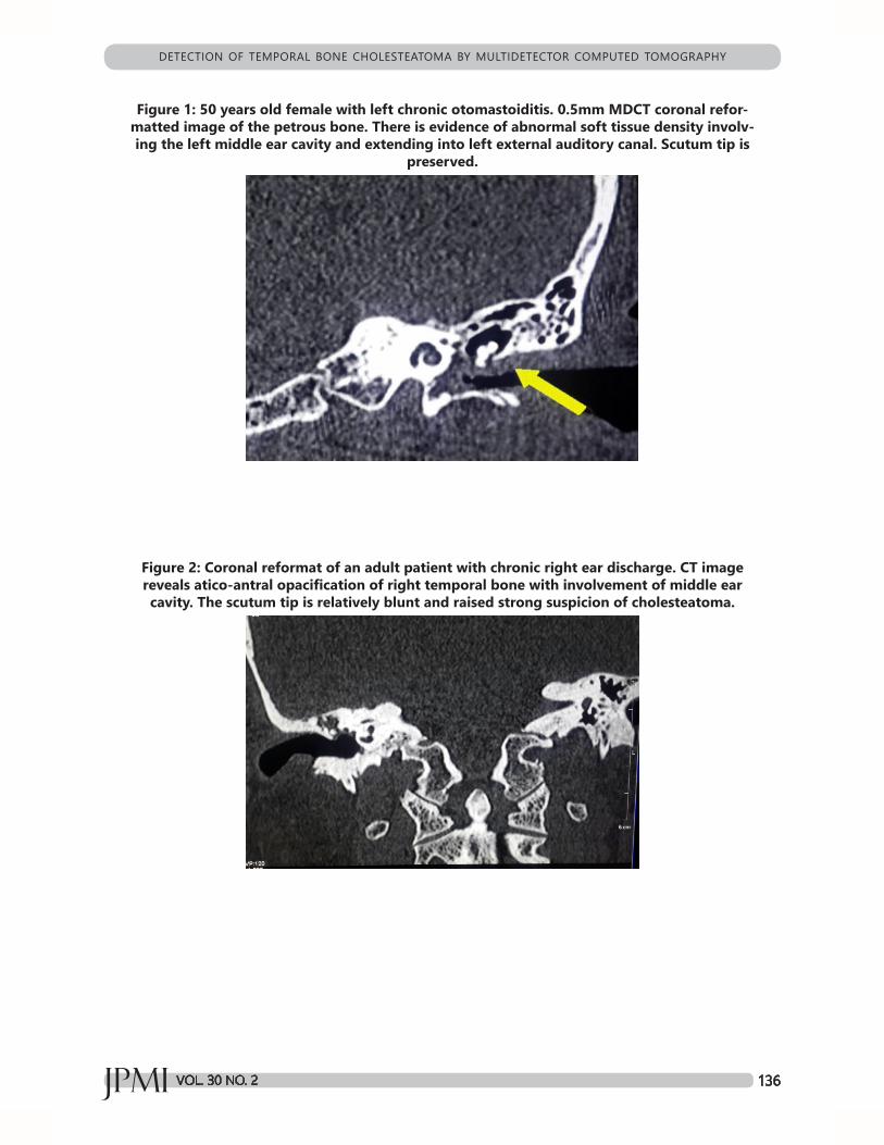
Figure 3: Right attico antral cholesteatoma. MDCT examination of the petrous bone. Axial section revealed evidence of abnormal soft tissue density involve the right middle ear cav-ity.The lesion extended through the attic with widening of its opening and thinning of the
roof of the left middle ear cavity. The bony ossicles are not visualized in this image.
Figure 4: High resolution CT examination of petrous bone. Coronal reformat reveals com-pletely opacified right epitympanum with bony erosion of antero-lateral wall of antrum with extension of soft tissues via widened aditus into the middle ear cavity and prusac’s
space. Ossicles are partly eroded and being displaced medially.
DETECTION OF TEMPORAL BONE CHOLESTEATOMA BY MULTIDETECTOR COMPUTED TOMOGRAPHY
JPMI VOL. 30 NO. 2 138
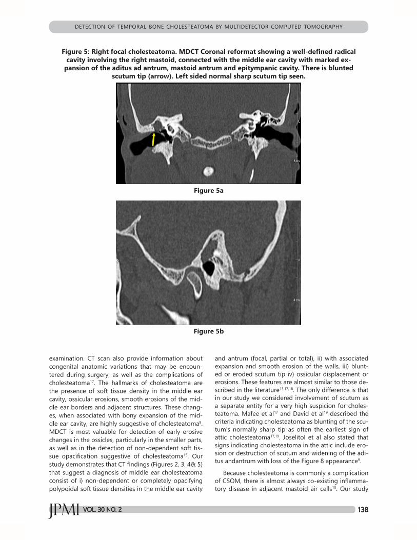
Figure 5: Right focal cholesteatoma. MDCT Coronal reformat showing a well-defined radical cavity involving the right mastoid, connected with the middle ear cavity with marked ex-
pansion of the aditus ad antrum, mastoid antrum and epitympanic cavity. There is blunted scutum tip (arrow). Left sided normal sharp scutum tip seen.
Figure 5a
Figure 5b
examination. CT scan also provide information about congenital anatomic variations that may be encoun-tered during surgery, as well as the complications of cholesteatoma17. The hallmarks of cholesteatoma are the presence of soft tissue density in the middle ear cavity, ossicular erosions, smooth erosions of the mid-dle ear borders and adjacent structures. These chang-es, when associated with bony expansion of the mid-dle ear cavity, are highly suggestive of cholesteatoma9. MDCT is most valuable for detection of early erosive changes in the ossicles, particularly in the smaller parts, as well as in the detection of non-dependent soft tis-sue opacification suggestive of cholesteatoma15. Our study demonstrates that CT findings (Figures 2, 3, 4& 5) that suggest a diagnosis of middle ear cholesteatoma consist of i) non-dependent or completely opacifying polypoidal soft tissue densities in the middle ear cavity
and antrum (focal, partial or total), ii) with associated expansion and smooth erosion of the walls, iii) blunt-ed or eroded scutum tip iv) ossicular displacement or erosions. These features are almost similar to those de-scribed in the literature13,17,18. The only difference is that in our study we considered involvement of scutum as a separate entity for a very high suspicion for choles-teatoma. Mafee et al17 and David et al19 described the criteria indicating cholesteatoma as blunting of the scu-tum’s normally sharp tip as often the earliest sign of attic cholesteatoma17,19. Joselitol et al also stated that signs indicating cholesteatoma in the attic include ero-sion or destruction of scutum and widening of the adi-tus andantrum with loss of the Figure 8 appearance9.
Because cholesteatoma is commonly a complication of CSOM, there is almost always co-existing inflamma-tory disease in adjacent mastoid air cells13. Our study
JPMI VOL. 30 NO. 2 139
DETECTION OF TEMPORAL BONE CHOLESTEATOMA BY MULTIDETECTOR COMPUTED TOMOGRAPHY
demonstrates this in all patients. CSOM without cho-lesteatoma is indicated on CT when air cells appear opaque by soft tissue or fluid densities but maintain their normal trabecular pattern or whenever there is obliteration of mastoid antrum and periantral cells by increased reactive bone formation/sclerosis17. In our series, 24 patients had these CT features. These were confidently labeled as CSOM. Complete opacification of the middle ear with no bony destruction makes radio-logic differentiation of cholesteatoma from middle ear effusions and granulation tissue difficult, if not impossi-ble13,18. The presence of an air-fluid level or a soft tissue (fluid) mass in the dependent portion of the middle ear would render support to a diagnosis of effusion. When CT features of cholesteatoma present with an air fluid level, possibility of infected cholesteatoma should be considered13. One of our patients had these features. In our series, non-dependent, homogeneous and pol-ypoidal soft tissue densities were present in the mas-toid antrum and middle ear cavity. In some of our cases (n=7), soft tissues occupied all spaces at the time of CT study with antral expansion, abutting the ossicular chain and bulging into the external auditory meatus. These patients were considered as having equivocal features with possibility of CSOM and + possibility of associated cholesteatoma. Many of the patients have both gran-ulation tissue and cholesteatoma, which cannot be ra-diographically distinguished17. Our study showed that all patients with cholesteatoma had at least one of the CT criteria indicating cholesteatoma, and 47 patients showed all features of radiological findings of choles-teatoma. Chee et al concluded in their series of 36 pa-tients that 34 patients (94.4%) had been correctly diag-nosed by CT12. Joselito et al reported in their series of 64 patients that the analysis of the preoperative CT scan correlated with the surgical findings and histopatholog-ic reports with a high degree of accuracy (96.8%)9. In our study, 14 cases were later on proven on biopsy. Rest of the CT diagnosed cholesteatoma cases were either operated in another center or didn’t come for follow up visit. Ossicular chain erosion occurred in 8 patients. The literature presents higher frequency, with sensitivity ranging from 80% to 100%21. El-Essawy et al22 in a series of 32 cases concluded that temporal bone complica-tions including bone erosion and cavity formation were seen in all patients with cholesteatoma (100%) which is similar to our study; sclerosis of the mastoid and ossic-ular destruction were seen in 93.81% of patients where-as in our study ossicular chain destruction was seen in 17% of cases. Palva and fellows concluded in their study that the labyrinthine fistula may occur in 10% of pa-tients with chronic ear infection due to cholesteatoma23. Our study did not detect any labyrinthine fistula on CT images. Joselito et al stated that preoperative demon-stration of facial nerve canal involvement was often dif-ficult not only because of the small size of the facial
nerve canal but also due to its oblique orientation and the presence of developmental dehiscence, particularly when abutted by the soft tissue9. CT analysis shows ana-tomic structure implicated in cholesteatoma damages20.Preoperative computed tomography is necessary for the diagnosis and the evaluation of chronic middle ear cholesteatoma in order to show extending lesion and to detect complications20. Cholesteatoma can be accu-rately diagnosed by CT scan. Mafee et al reported in his series of 48 patients with cholesteatoma that 46 of them (96%) were diagnosed correctly using preopera-tive CT scans17. MDCT is highly accurate to demonstrate the presence of abnormal tissue in the middle ear and mastoid, with sensitivity ranging between 70% to 96% 8,9. But one of its limitations is that it cannot define if the tissue is inflammatory, fibrotic or cholesteatoma. How-ever, the presence of bone erosion of structures such as scutum, ossicular chain, tympanic tegmen, bone laby-rinth and lateral wall of the attic, is strongly indicative of cholesteatoma. At times, the differential diagnosis be-tween CSOM and cholesteatoma cannot be accurate-ly done with MDCT, since the radiological findings are very similar and many times overlap each other. In such cases, histopathological workup is necessary4.
CONCLUSIONPatients with chronic discharging ear cholesteato-
ma should be scanned using MDCT scanner as many relevant structures are best seen in multiplanar images and it is helpful in diagnosing characteristic findings of cholesteatomas. MDCT is a unique method of detection of early cholesteatoma as well as detection of choles-teatoma in hidden areas. CT scanning serves as a road map to assist the surgeon during cholesteatoma sur-gery. With the more prevalent use of CT, considerable morbidity may be avoided.
RECOMMENDATIONWe recommend that the study should be done on
large number of patients with a proper follow up as well as at multiple centers.
ACKNOWLEDGMENTWe thank Arshad for his meticulous computer typing
skills and valuable assistance in helping with data entry and compilation.
REFERENCES1. Anwar K, Said M, Zada B, Din G, Khan IA. Cholesteatoma
and the Destruction of Middle Ear and Mastoid Cavities in Ottorhoea. Pak J Med Health Sci 2012; 6:51-4.
2. Gomaa MA, Abdel Karim AR, Abdel Ghany HS, Elhiny AA, Sadek AA. Evaluation of temporal bone cholesteatoma and the correlation between High Resolution Computed Tomography and surgical finding. Clin Med Insights Ear
DETECTION OF TEMPORAL BONE CHOLESTEATOMA BY MULTIDETECTOR COMPUTED TOMOGRAPHY
JPMI VOL. 30 NO. 2 140
15. Mafee MF, Kumar A, Yannias DA, Valvassori GE, Apple-baum EL. Computed tomography of the middle ear in the evaluation of cholesteatomas and other soft-tissue mass-es: comparison with pluridirectional tomography. Radiol-ogy1983; 148:465-72.
16. Kemppainen HO, Puhakka HJ, Laippala PJ, Sipilä MM, Manninen MP, Karma PH. Epidemiology and aetiology of middle ear cholesteatoma. Acta Otolaryngol 1999; 119:568-72.
17. Maffe MF, Levin BC, Applebaum EL, Campos M, James CF. Cholesteatoma of the middle ear and mastoid. A compar-ison of CT scan and operative findings. Otolaryngol Clin North Am 1988; 21:265–93.
18. Swartz JD, Goodman RS, Russel KB, Marlowe FI, Wolfson RJ. High resolution computed tomography of the middle ear and mastoid. Part II: Tubotympanic disease. Radiolo-gy1983; 148:455-9.
19. David C, Lia D, Thomas R, Bergeron M. Contemporary ra-diologic imaging in the evaluation of middle ear-attic-an-tral complex cholesteatoma. Otolaryngol Clin North Am 1989; 22:897–909.
20. Sethom A1, Akkari K, Dridi I, Tmimi S, Mardassi A, Benzarti S, et al. Preoperative CT Scan in middle ear cholesteato-ma. Tunis Med 2011; 89:248-53.
21. Banerjee A, Flood LM, Yates P, Clifford K. Computed to-mography in suppurative ear disease: Does it influence management. J Laryngol Otol 2003; 117:454–8.
22. El-Essawy S, El-Nahas M, El-Shewahy H, Ghoniem MR. Complicated middle ear cholesteatoma, a CT study. Egypt J Radiol Nucl Med 1992; 23:161–70.
23. Palva T. The pathogenesis and treatment of cholesteato-ma. Acta Otolaryngol 1990; 109:323–30.
CONTRIBUTORSSG conceived the idea, planned the study, and
drafted the manuscript. USU, SA, SG and SJ helped acquisition of data and did statistical analysis. MSK drafted and critically revised the manuscript. All au-thors contributed significantly to the submitted man-uscript.
Nose Throat 2013; 6: 21–8.
3. Ahn SH, Kim YW, Baik SK, Hwang JY, Lee IW. Usefulness of Computed Tomography Hounsfield Unit Measurement for Diagnosis of Congenital Cholesteatoma. J Korean Soc Radiol 2014; 70:153-8.
4. Nemzek WR, Swartz JD. Temporal bone: inflammatory disease. In: Som PM, Curtin HD. Head and neck imaging. 4th ed. St Louis: Mosby; 2003:1184-99.
5. Corrales CE, Blevins NH. Imaging for evaluation of cho-lesteatoma: current concepts and future directions. Curr Opin Otolaryngol Head Neck Surg 2013;21:461-7.
6. Kesser BW, Friedman RA. Infectious and Inflammatory Disorders. In: Seiden AM, Tami TA, Pensak ML, Cotton RT, Gluckman JL. Otorhinolaryngology:The Essentials. New York, NY: Thieme; 2001:44–58.
7. Ghiasi S. Labyrinthine Fistula in Chronic Otitis Media with Cholesteatoma. J Pak Med Assoc 2011; 352:61.
8. Fatterpekar GM, Doshi AH, Dugar M, Delman BN, Naidich TP, Som PM. Role of 3D CT in the evaluation of the tem-poral bone. Radiographics 2006; 26:117-32.
9. Gaurano JL, Joharjy IA. Middle ear cholesteatoma: charac-teristic CT findings in 64 patients. Ann Saudi Med 2004; 24:442-7.
10. Razek AA, Huang BY. Lesions of the petrous apex: classifi-cation and findings at CT and MR imaging. Radiographics 2012; 32:151-73.
11. De Foer B, Vercruysse JP, Pouillon M, Somers T, Cassel-man JW, Offeciers E. Value of high-resolution computed tomography and magnetic resonance imaging in the de-tection of residual cholesteatomas in primary bony oblit-erated mastoids. Am J Otolaryngol 2007;28:230-4.
12. Chee NC, Tan TY. The value of preoperative high resolu-tion CT scans in cholesteatoma surgery. Singapore Med J 2001; 2:155–9.
13. Liu DP, Bergeron RT. Contemporary radiologic imaging in the evaluation of middle ear-attic-antral complex choles-teatomas. Otolaryngol Clin North Am 1989; 22:897-909.
14. Viswanathal B, Naseeruddin K. Neurotologic Complica-tions of Chronic Otitis Media with Cholesteatoma. J Neu-rol Epidemiol 2013; 1:20-30.