Embed Size (px)
Citation preview
Journal of Clinical Virology 43 (2008) 260–265
Contents lists available at ScienceDirect
Journal of Clinical Virology
journa l homepage: www.e lsev ier .com/ locate / j cv
Detection of Epstein-Barr virus in T-cell prolymphocytic leukemia cells in vitro
Ke Lana,b, Masanao Murakamia, Tathagata Choudhuria, Donald E. Tsai c,Stephen J. Schusterc, Mariusz A. Wasikd, Erle S. Robertsona,∗
a Department of Microbiology and the Tumor Virology Program of Abramson Comprehensive Cancer Center, University of Pennsylvania Medical School,201E Johnson Pavilion, 3610 Hamilton Walk, Philadelphia, PA 19104, United Statesb Institut Pasteur of Shanghai, Chinese Academy of Sciences, South Chongqing Road 225, Shanghai 200025, PR Chinac Division of Hematology–Oncology, Hospital of University of Pennsylvania, 15 Penn Tower, 3400 Spruce Street, Philadelphia, PA 19104, United Statesd Department of Pathology and Laboratory Medicine, University of Pennsylvania Medical School, 3610 Hamilton Walk, Philadelphia, PA 19104, United States
a r t i c l e i n f o
Article history:Received 2 February 2008Received in revised form 20 June 2008Accepted 17 July 2008
Keywords:Epstein-Barr virusT-cell prolymphocytic leukemiaInfection
a b s t r a c t
Background: Epstein-Barr virus (EBV) is closely associated with the development of a number of tumors.During latent infection, EBV continuously expresses a number of viral genes which are essential for celltransformation and maintenance of the malignant phenotype of EBV-related tumors. There has beenno previous link between EBV and T-cell prolymphocytic leukemia (T-PLL), a distinctive form of leukemiaderived from T-cells at an intermediate stage of differentiation between a cortical thymocyte and a matureperipheral blood T-cell.Objective: To determine if EBV was present in the T-PLL cells collected.Study design: T-PLL cells were isolated from the peripheral blood of a patient diagnosed with T-PLL andcontinuously cultured for about 1 year. The existence of EBV in these cells was detected using multiplestrategies including PCR, Western blotting, immunofluorescent assay and flow cytometry analysis.Results: The EBV genome was present in these T-PLL cells by PCR analysis across multiple sites in the viral
genome. In addition, these T-PLL cells expressed a number of EBV latent antigens. The EBV oncoproteinsLMP1, EBNA1 and EBNA3C were expressed in the majority of the infected cells.Conclusion: This report suggests a potential link between EBV infection and T-PLL and provides newentia
1
cbrcttLEoaREm
dE
tlcaodek1
1d
information about the pot
. Introduction
Epstein-Barr virus (EBV) was first reported as the etiologi-al agent of Burkitt’s lymphoma in 1964.6 Since then, EBV haseen associated with many human pathologies including nasopha-yngeal carcinoma which is a highly prevalent and aggressiveancer in Southeast Asia, infectious mononucleosis, and AIDS- andransplant-related B cell lymphomas.18 EBV has also been linkedo T-cell lymphomas, Hodgkin’s lymphoma, and NK leukemia orGL leukemia,32,33 supporting the concept of a wide spectrum ofBV-associated disorders. The EBV genome encodes for a numberf latent proteins such as EBNA1–3, LMP 1, 2 and others which
re selectively expressed during different types of viral latency.27ecent studies have demonstrated that EBNA2, EBNA-LP, EBNA3A,BNA3C and LMP1 are important for cell transformation.27 Further-ore, different strategies are utilized by these latent antigens to
∗ Corresponding author. Tel.: +1 215 746 0114/6; fax: +1 215 898 9557.E-mail address: [email protected] (E.S. Robertson).
stAodTrcF
386-6532/$ – see front matter © 2008 Elsevier B.V. All rights reserved.oi:10.1016/j.jcv.2008.07.018
l contribution of EBV in the initiation or maintenance of T-PLL.© 2008 Elsevier B.V. All rights reserved.
rive virus-associated transformation or carcinogenesis among theBV-associated disorders.
Although EBV is linked to a number of different types of tumors,he association between this virus and T-cell prolymphocyticeukemia (T-PLL) has not been previously explored. T-PLL, formerlyategorized as T-cell chronic lymphocytic leukemia, is a rare andggressive hematologic malignancy.2,3,8,29,30 It is a distinctive typef leukemia that is derived from T-cells at the intermediate stage ofifferentiation between a cortical thymocyte and a mature periph-ral blood T-cell.8,29,30 Patients with T-PLL often have a complexaryotype with recurrent alterations that involve chromosomes 14,1, and 8. Rearrangements of chromosome 14q32.1 through inver-ion [inv(14)(q11;q32)] and translocation [t(14;14)(q11;q32)] arehe most common cytogenetic abnormalities reported in T-PLL.4,5
s a result of these rearrangements, TCL1, a gene that is physi-logically expressed in CD4/CD8 double-negative thymocytes, is
eregulated.16,17,31 Typically, the TCL1 locus rearranges with the-cell receptor (TCR) �/� chain locus in the above mentioned rear-angements, but it can also rearrange with the TCR-� chain locus onhromosome 7 in another translocation [t(7;14)(q35;q32.1)].16,21,22urthermore, several patients with mature T-cell leukemias have
ical Virology 43 (2008) 260–265 261
trlParnsbTmtr
itromatItftwg
2
2
plwcascT
2
mpawmpawe
2
p2bhpv
Table 1Primers used for PCR analysis
Amplified region Primers sequences Primer co-ordinates(NC 007605)
BamHI C fragment 5′GCAGGGCTCGCAAAGTATAG3′ 11095–118555′TGCGGAAGTGACACCAAATA3′
BamHI E fragment 5′TACTGCCACCAGTACCACAACA3′ 99939–100091
B
PdDcsf7Rua
2
puipfacv(e
2
wslabaaa
2
maaacca4
K. Lan et al. / Journal of Clin
he translocation [t(X;14)(q28;q11)], which results in the rear-angement of the MTCP1 gene (a member of the TCL1 gene family)ocated at Xq28.15,26 Therefore, chromosomal rearrangements in T-LL juxtapose TCL1 and MTCP1 to the TCR loci and lead to theirctivation. Recent genomic analyses of the 14q32.1 breakpointegion has revealed three additional genes, TCL1b, TNG1 (T-CL1eighboring gene 1), and TNG2. These have an expression patternimilar to TCL1 in that they are not expressed in normal T-cells,ut are expressed in T-PLL cell lines and cells from patients with-PLL.7,23,28 Activation of TCL1 through hypomethylation of its pro-oter has also been described.34 These molecular events related to
he above chromosomal aberrations are believed to play a casualole in the pathogenesis of T-PLL.
EBV infects the vast majority (>90%) of healthy adults and cannfect T-cells in vivo.33 In addition, the infection of T-cells appearso convey an increased risk of malignancy such as lymphoma. Thisaised the possibility that EBV might also contribute to the devel-pment of another hematologic malignancy, T-PLL. A 70 years oldale T-PLL patient was observed with an increased titer of EBV
ntigens, suggesting that there was a high level of EBV viremia andhat EBV infection might be associated with this rare malignancy.n this report, we sought to determine if EBV was present in theumor cells obtained from this EBV-positive patient with T-PLL. Weound that EBV DNA can be continuously detected in the culturedumor cells and that the majority of the essential EBV latent genesere expressed in these cells suggesting a latency pattern that is
eared toward growth of the latently infected cells.
. Methods
.1. Patient
The study patient was a 70 years old male diagnosed at the Hos-ital of the University of Pennsylvania with T-cell prolymphocytic
eukemia (T-PLL) (IRB#706586). He had an increase in the EBV titerhich was determined by detection of the antibody against viral
apsid antigen (VCA). The diagnosis was primarily based on FACSnalysis of the bone marrow aspirate as well as a peripheral bloodample. The combined clinical, morphologic (absence of flowerells) and immunophenotypic features were also consistent with-cell prolymphocytic leukemia.
.2. Lymphocyte isolation and cell culture
Primary lymphocytes were harvested from peripheral bloodononuclear cells using a Lymphoprep gradient as described
reviously.14 The enriched lymphocyte population was washednd plated at 50,000 cells in 150 �l of complete medium perell in a 96-well tissue culture plate. The cells were grown andaintained in RPMI 1640 medium (Hyclone, Inc., Logan, UT) sup-
lemented with 2 mM l-glutamine, 5 �g of gentamicin per ml,nd 10% heat-inactivated fetal bovine serum. Ninety-six-well platesere incubated at 37 ◦C with 5% CO2. The plates were treated once
very 8 days with a fresh 100 �l of complete medium.
.3. DNA lysate preparation and PCR analysis
Approximately 50,000 T-PLL cells derived from the patient’seripheral blood cells were collected and were centrifuged at
000 rpm for 5 min. The cell pellet was washed with phosphate-uffered saline (PBS) twice and resuspended in 50 �l of 0.2× PBS,eated to 95 ◦C for 15 min, and then switched to 56 ◦C for 1 h withroteinase K treatment (10 mg/ml). The enzyme was then inacti-ated at 95 ◦C for 30 min. A 5-�l portion of the lysate was used forFfi0wt
5′GGCCGACATTCTCCAAGATAA3′
amHI W fragment 5′CCAGACAGCAGCCAATTGTC3′ 17374–175025′GGTAGAAGACCCCCTCTTAC3′
CR amplification of EBV-specific regions for 40 cycles, and stan-ard protocols were followed using primers indicated in Table 1.NA was also prepared from the same number of control BJABells. PCR was performed by heating at 95 ◦C for 5 min and sub-equently for 40 cycles, each cycle including 95 ◦C for 1 min, 56 ◦Cor 1 min, and 72 ◦C for 2 min, followed by further incubation at2 ◦C for 10 min, in a PTC-100 programmable thermal cycler (MJesearch, Inc., Waltham, MA). Five microliters aliquots of PCR prod-cts were then assayed by electrophoresis on a 2.0% ME-agarose gelnd visualized by ethidium bromide.
.4. Immunofluorescence assay
Immunofluorescence analyses were performed as describedreviously.12 One million T-PLL cells were collected and briefly fixedsing a 1:1 methanol and acetone mix ice cold at −20 ◦C, blocked
n the appropriate serum, and then incubated with the pre-clearedatient serum or specific primary antibody for EBNA3C and LMP1or 1 h. Cells were washed and then further incubated with theppropriate secondary antibody conjugated to fluorescein isothio-yanate at 1:1000 dilutions in PBS for 1 h. Slides were washed andisualized with an Olympus XI70 inverted fluorescence microscopeOlympus, Inc., Melville, NY) and photographed using a digital Pix-lFly camera and software (Cooke, Inc., Warren, MI).
.5. Western blotting
T-PLL from the patient and an EBV-negative cell BJAB controlere lysed with 300 �l of reporter lysis buffer (Promega, Inc., Madi-
on, WI), respectively, and 40 �l of this lysate was used. Briefly,ysates were boiled in SDS-loading buffer, fractionated by SDS-PAGEnd transferred to a 0.45 mm nitrocellulose membrane. The mem-ranes were then probed with patient serum absorbed against B cellntigens or EBV-specific antibodies followed by incubation withppropriate infrared-tagged secondary antibodies and viewed onn Odyssey imager (LiCor, Inc., Lincoln, NE).
.6. FACS analysis
For the determination of the expression of CD3 positive cells, oneillion cells from patient samples, negative controls (B-cell) and
ntibody controls were incubated with rabbit polyclonal PE-taggednti-CD3 antibody (2 mg/ml in PBS containing 1% BSA) for 30 mint room temperature. After thoroughly washing with 1× PBS theells were incubated with FITC-conjugated anti LMP1 mouse mono-lonal antibody for 30 min. Cells were then washed and analyzed onflow cytometer (Becton Dickinson, San Diego, CA) equipped with88 nm Argon laser light source and a 623-nm band pass filter for
ITC fluorescence. For determination of LMP-1 signals, cells werexed in 1% paraformaldehyde followed by permeabilization with.05% Triton X 100 at room temperature. The cells were washedith 1× PBS 3 times at 5 min intervals. In this fixing procedurehe cytoplasm remains intact and the LMP1 antibody was allowed
2 ical Virology 43 (2008) 260–265
teDaaAd
3
3
ropCTashTce
Towcfiaopcp
Table 2Immunophenotyping Results
wP
3
gcpGcpp
FataHd
62 K. Lan et al. / Journal of Clin
o gain access to the intracellular compartment. A total of 10,000vents were acquired for analysis using CellQuest software (Bectonickinson, San Diego, CA). Cells were gated as described earlier,13
nd histogram plots of PE fluorescence (x-axis) versus counts (y-xis) were shown as a fraction of logarithmic fluorescence intensity.dot plot of PE fluorescence (x-axis) versus FITC (y-axis) was also
etermined as a fraction of logarithmic fluorescence intensity.
. Results
.1. Growth of T-cell prolymphocytic leukemia cells in vitro
The diagnosis was based on the FACS analysis of the bone mar-ow aspirate as well as the peripheral blood sample. The resultsf FACS on the bone marrow demonstrated an aberrant expandedopulation (70% of total events) of CD2, CD3, CD5, CD7, CD8, andD25 T-cells. These cells were CD1a, CD4, CD16, CD30, CD56, CD57,dT and gamma-delta negative. In addition, B-cells were essentiallybsent (Table 2). The FACS performed on peripheral blood demon-trated an essentially identical immuno-profile and the markedlyigh CD7 intensity was not reduced in comparison to other pan-antigens. The combined clinical, morphologic (absence of flower
ells) and immuno-phenotypic (CD8 positivity, without loss of CD7xpression) features were consistent with T-PLL (Table 2).
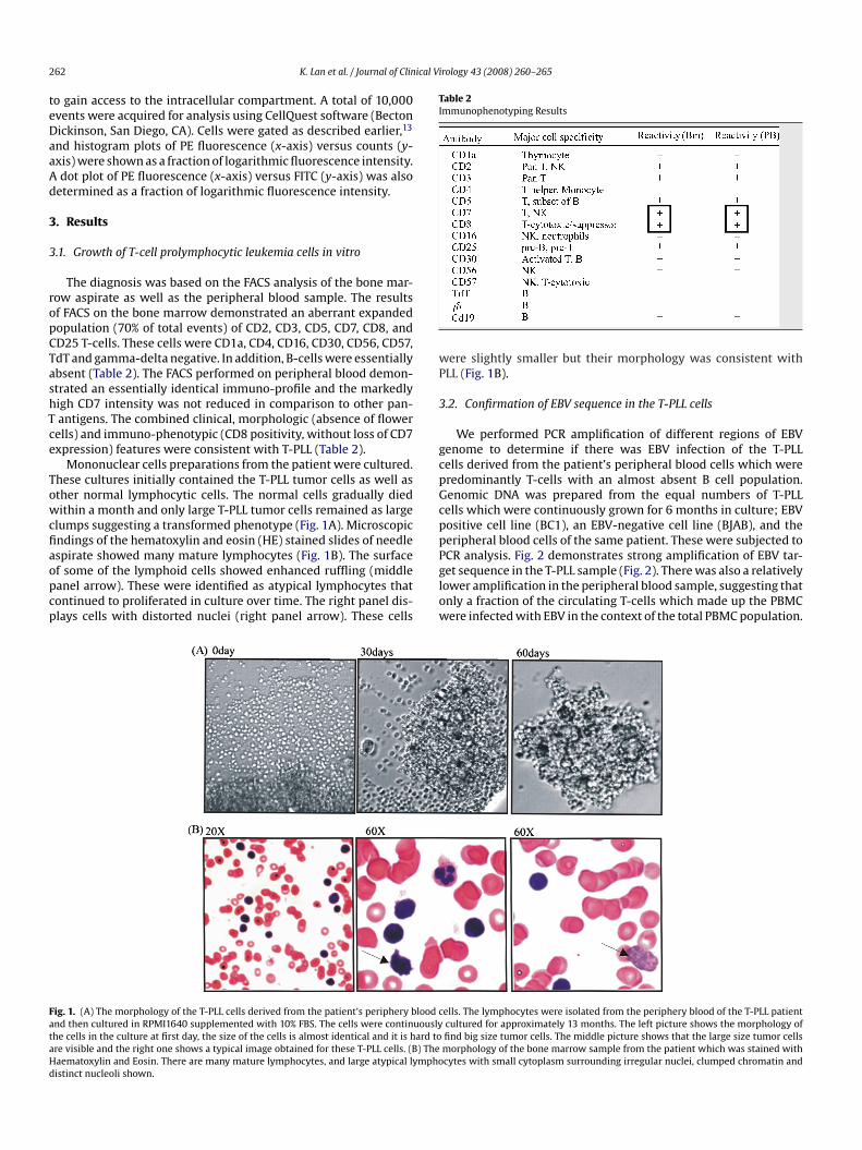
Mononuclear cells preparations from the patient were cultured.hese cultures initially contained the T-PLL tumor cells as well asther normal lymphocytic cells. The normal cells gradually diedithin a month and only large T-PLL tumor cells remained as large
lumps suggesting a transformed phenotype (Fig. 1A). Microscopicndings of the hematoxylin and eosin (HE) stained slides of needle
spirate showed many mature lymphocytes (Fig. 1B). The surfacef some of the lymphoid cells showed enhanced ruffling (middleanel arrow). These were identified as atypical lymphocytes thatontinued to proliferated in culture over time. The right panel dis-lays cells with distorted nuclei (right panel arrow). These cellsPglow
ig. 1. (A) The morphology of the T-PLL cells derived from the patient’s periphery blood cnd then cultured in RPMI1640 supplemented with 10% FBS. The cells were continuouslyhe cells in the culture at first day, the size of the cells is almost identical and it is hard tore visible and the right one shows a typical image obtained for these T-PLL cells. (B) Theaematoxylin and Eosin. There are many mature lymphocytes, and large atypical lymphoistinct nucleoli shown.
ere slightly smaller but their morphology was consistent withLL (Fig. 1B).
.2. Confirmation of EBV sequence in the T-PLL cells
We performed PCR amplification of different regions of EBVenome to determine if there was EBV infection of the T-PLLells derived from the patient’s peripheral blood cells which wereredominantly T-cells with an almost absent B cell population.enomic DNA was prepared from the equal numbers of T-PLLells which were continuously grown for 6 months in culture; EBVositive cell line (BC1), an EBV-negative cell line (BJAB), and theeripheral blood cells of the same patient. These were subjected to
CR analysis. Fig. 2 demonstrates strong amplification of EBV tar-et sequence in the T-PLL sample (Fig. 2). There was also a relativelyower amplification in the peripheral blood sample, suggesting thatnly a fraction of the circulating T-cells which made up the PBMCere infected with EBV in the context of the total PBMC population.ells. The lymphocytes were isolated from the periphery blood of the T-PLL patientcultured for approximately 13 months. The left picture shows the morphology offind big size tumor cells. The middle picture shows that the large size tumor cellsmorphology of the bone marrow sample from the patient which was stained withcytes with small cytoplasm surrounding irregular nuclei, clumped chromatin and
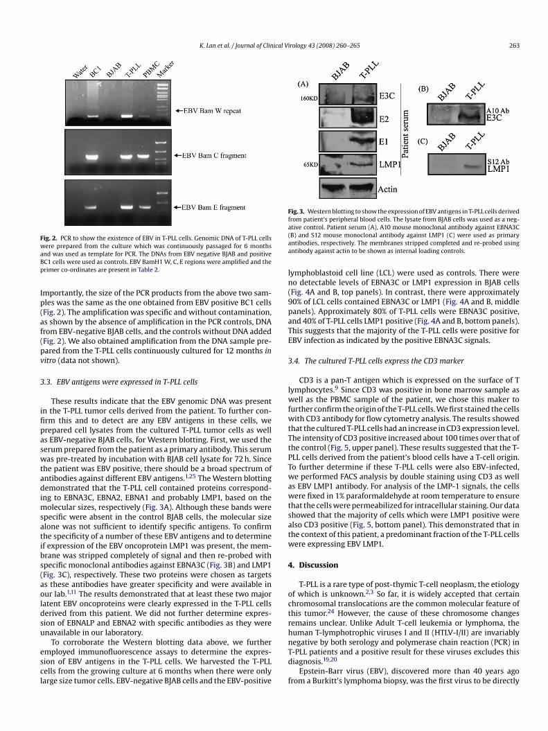
K. Lan et al. / Journal of Clinical Virology 43 (2008) 260–265 263
Fig. 2. PCR to show the existence of EBV in T-PLL cells. Genomic DNA of T-PLL cellswaBp
Ip(af(pv
3
ifipaswtadimsatibs(aoldsu
escl
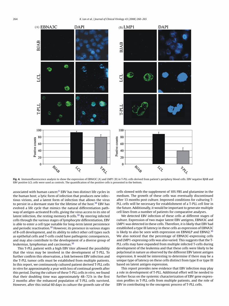
Fig. 3. Western blotting to show the expression of EBV antigens in T-PLL cells derivedfrom patient’s peripheral blood cells. The lysate from BJAB cells was used as a neg-a(aa
ln(9paTE
3
lwfwtTtPTwawtsatw
4
octrh
ere prepared from the culture which was continuously passaged for 6 monthsnd was used as template for PCR. The DNAs from EBV negative BJAB and positiveC1 cells were used as controls. EBV BamH1 W, C, E regions were amplified and therimer co-ordinates are present in Table 2.
mportantly, the size of the PCR products from the above two sam-les was the same as the one obtained from EBV positive BC1 cellsFig. 2). The amplification was specific and without contamination,s shown by the absence of amplification in the PCR controls, DNArom EBV-negative BJAB cells, and the controls without DNA addedFig. 2). We also obtained amplification from the DNA sample pre-ared from the T-PLL cells continuously cultured for 12 months initro (data not shown).
.3. EBV antigens were expressed in T-PLL cells
These results indicate that the EBV genomic DNA was presentn the T-PLL tumor cells derived from the patient. To further con-rm this and to detect are any EBV antigens in these cells, werepared cell lysates from the cultured T-PLL tumor cells as wells EBV-negative BJAB cells, for Western blotting. First, we used theerum prepared from the patient as a primary antibody. This serumas pre-treated by incubation with BJAB cell lysate for 72 h. Since
he patient was EBV positive, there should be a broad spectrum ofntibodies against different EBV antigens.1,25 The Western blottingemonstrated that the T-PLL cell contained proteins correspond-
ng to EBNA3C, EBNA2, EBNA1 and probably LMP1, based on theolecular sizes, respectively (Fig. 3A). Although these bands were
pecific were absent in the control BJAB cells, the molecular sizelone was not sufficient to identify specific antigens. To confirmhe specificity of a number of these EBV antigens and to determinef expression of the EBV oncoprotein LMP1 was present, the mem-rane was stripped completely of signal and then re-probed withpecific monoclonal antibodies against EBNA3C (Fig. 3B) and LMP1Fig. 3C), respectively. These two proteins were chosen as targetss these antibodies have greater specificity and were available inur lab.1,11 The results demonstrated that at least these two majoratent EBV oncoproteins were clearly expressed in the T-PLL cellserived from this patient. We did not further determine expres-ion of EBNALP and EBNA2 with specific antibodies as they werenavailable in our laboratory.
To corroborate the Western blotting data above, we furthermployed immunofluorescence assays to determine the expres-ion of EBV antigens in the T-PLL cells. We harvested the T-PLLells from the growing culture at 6 months when there were onlyarge size tumor cells. EBV-negative BJAB cells and the EBV-positive
nTd
f
tive control. Patient serum (A), A10 mouse monoclonal antibody against EBNA3CB) and S12 mouse monoclonal antibody against LMP1 (C) were used as primaryntibodies, respectively. The membranes stripped completed and re-probed usingntibody against actin to be shown as internal loading controls.
ymphoblastoid cell line (LCL) were used as controls. There wereo detectable levels of EBNA3C or LMP1 expression in BJAB cellsFig. 4A and B, top panels). In contrast, there were approximately0% of LCL cells contained EBNA3C or LMP1 (Fig. 4A and B, middleanels). Approximately 80% of T-PLL cells were EBNA3C positive,nd 40% of T-PLL cells LMP1 positive (Fig. 4A and B, bottom panels).his suggests that the majority of the T-PLL cells were positive forBV infection as indicated by the positive EBNA3C signals.
.4. The cultured T-PLL cells express the CD3 marker
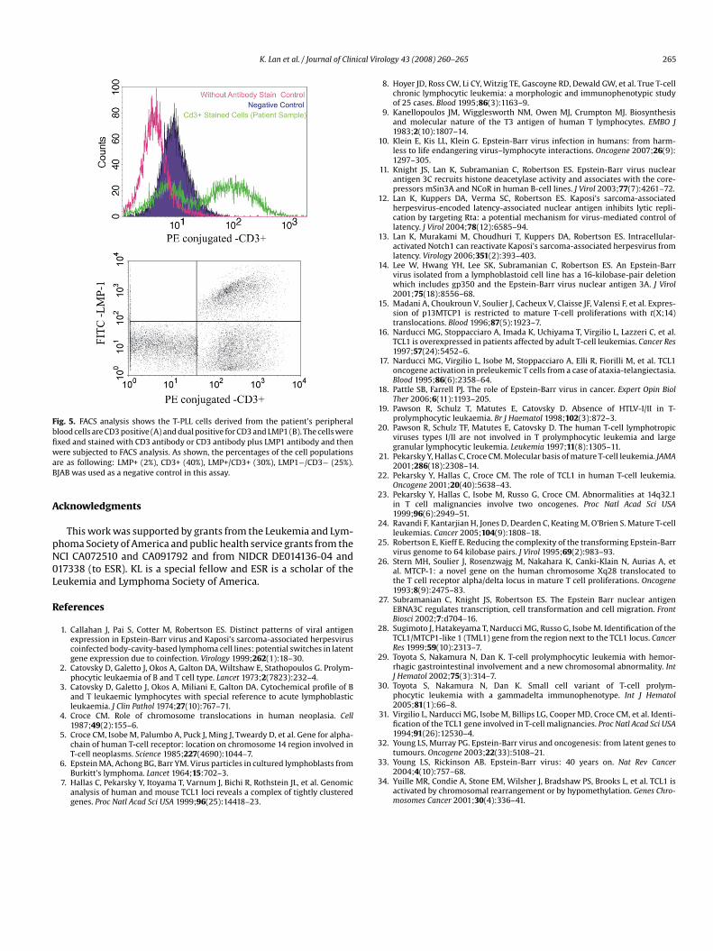
CD3 is a pan-T antigen which is expressed on the surface of Tymphocytes.9 Since CD3 was positive in bone marrow sample as
ell as the PBMC sample of the patient, we chose this maker tourther confirm the origin of the T-PLL cells. We first stained the cellsith CD3 antibody for flow cytometry analysis. The results showed
hat the cultured T-PLL cells had an increase in CD3 expression level.he intensity of CD3 positive increased about 100 times over that ofhe control (Fig. 5, upper panel). These results suggested that the T-LL cells derived from the patient’s blood cells have a T-cell origin.o further determine if these T-PLL cells were also EBV-infected,e performed FACS analysis by double staining using CD3 as well
s EBV LMP1 antibody. For analysis of the LMP-1 signals, the cellsere fixed in 1% paraformaldehyde at room temperature to ensure
hat the cells were permeabilized for intracellular staining. Our datahowed that the majority of cells which were LMP1 positive werelso CD3 positive (Fig. 5, bottom panel). This demonstrated that inhe context of this patient, a predominant fraction of the T-PLL cellsere expressing EBV LMP1.
. Discussion
T-PLL is a rare type of post-thymic T-cell neoplasm, the etiologyf which is unknown.2,3 So far, it is widely accepted that certainhromosomal translocations are the common molecular feature ofhis tumor.24 However, the cause of these chromosome changesemains unclear. Unlike Adult T-cell leukemia or lymphoma, theuman T-lymphotrophic viruses I and II (HTLV-I/II) are invariably
egative by both serology and polymerase chain reaction (PCR) in-PLL patients and a positive result for these viruses excludes thisiagnosis.19,20Epstein-Barr virus (EBV), discovered more than 40 years agorom a Burkitt’s lymphoma biopsy, was the first virus to be directly
264 K. Lan et al. / Journal of Clinical Virology 43 (2008) 260–265
F P1 (BE is pre
atttewlciaoaal
tftIitt2H
cmaPtc
cLeiWaPdpeub
ig. 4. Immunofluorescence analysis to show the expression of EBNA3C (A) and LMBV positive LCL cells were used as controls. The quantification of the positive cells
ssociated with human cancer.6 EBV has two distinct life cycles inhe human host; a lytic form of infection that produces new infec-ious virions, and a latent form of infection that allows the viruso persist in a dormant state for the lifetime of the host.10 EBV hasvolved a life cycle that mimics the natural differentiation path-ay of antigen-activated B cells, giving the virus access to its site of
atent infection, the resting memory B cells.10 By steering infectedells through the various stages of lymphocyte differentiation, EBVs able to enter a cell type suitable for long-term latent persistencend periodic reactivation.10 However, its presence in various stagesf B-cell development, and its ability to infect other cell types suchs epithelial cells and T-cells could have pathogenic consequences,nd may also contribute to the development of a diverse group ofeukemias, lymphomas and carcinomas.10
This T-PLL patient with a high EBV titer allowed the possibilityhat the virus may be involved in the development of T-PLL. Tourther confirm this observation, a link between EBV infection andhe T-PLL tumor cells must be established from multiple patients.n this report, we continuously cultured patient derived T-PLL cells
n vitro for approximately a year with loss of continual growth afterhis period. During the culture of these T-PLL cells in vitro, we foundhat their doubling time was approximately 48–72 h in the firstmonths after the enhanced population of T-PLL cells survived.owever, after this initial 60 days in culture the growth rate of the
afsE
) in T-PLL cells derived from patient’s periphery blood cells. EBV negative BJAB andsented in the bottom.
ells slowed with the supplement of 10% FBS and glutamine in theedium. The growth of these cells was eventually discontinued
fter 13 months post culture. Improved conditions for culturing T-LL cells will be necessary for establishment of a T-PLL cell line inhe future. Additionally, it would be important to generate multipleell lines from a number of patients for comparative analyses.
We detected EBV infection of these cells at different stages ofulture. Expression of two major latent EBV antigens, EBNA3C andMP1 was detected in these cells. Therefore, it is likely that EBV hadstablished a type III latency in these cells as expression of EBNA3Cs likely to also be seen with expression on EBNALP and EBNA2.10
e also noticed that the percentage of EBNA3C-expressing cellsnd LMP1-expressing cells was also varied. This suggests that the T-LL cells may have expanded from multiple infected T-cells duringevelopment of the leukemia and that these cells were likely to beolyclonal in nature as observed by the different EBV latent antigenxpression. It would be interesting to determine if there may be anique type of latency on these cells distinct from type II or type IIIased on latent antigen expression.
This report provides new evidence that EBV infection may playrole in development of T-PLL. Additional effort will be needed to
urther focus on the systemic characterization of EBV gene expres-ion profiles in T-PLL cells from multiple patients, and the role ofBV in contributing to the oncogenic process of T-PLL cells.
K. Lan et al. / Journal of Clinical Vi
Fig. 5. FACS analysis shows the T-PLL cells derived from the patient’s peripheralblood cells are CD3 positive (A) and dual positive for CD3 and LMP1 (B). The cells werefixed and stained with CD3 antibody or CD3 antibody plus LMP1 antibody and thenwaB
A
pN0L
R
ere subjected to FACS analysis. As shown, the percentages of the cell populationsre as following: LMP+ (2%), CD3+ (40%), LMP+/CD3+ (30%), LMP1−/CD3− (25%).JAB was used as a negative control in this assay.
cknowledgments
This work was supported by grants from the Leukemia and Lym-homa Society of America and public health service grants from theCI CA072510 and CA091792 and from NIDCR DE014136-04 and17338 (to ESR). KL is a special fellow and ESR is a scholar of theeukemia and Lymphoma Society of America.
eferences
1. Callahan J, Pai S, Cotter M, Robertson ES. Distinct patterns of viral antigenexpression in Epstein-Barr virus and Kaposi’s sarcoma-associated herpesviruscoinfected body-cavity-based lymphoma cell lines: potential switches in latentgene expression due to coinfection. Virology 1999;262(1):18–30.
2. Catovsky D, Galetto J, Okos A, Galton DA, Wiltshaw E, Stathopoulos G. Prolym-phocytic leukaemia of B and T cell type. Lancet 1973;2(7823):232–4.
3. Catovsky D, Galetto J, Okos A, Miliani E, Galton DA. Cytochemical profile of Band T leukaemic lymphocytes with special reference to acute lymphoblasticleukaemia. J Clin Pathol 1974;27(10):767–71.
4. Croce CM. Role of chromosome translocations in human neoplasia. Cell1987;49(2):155–6.
5. Croce CM, Isobe M, Palumbo A, Puck J, Ming J, Tweardy D, et al. Gene for alpha-chain of human T-cell receptor: location on chromosome 14 region involved in
T-cell neoplasms. Science 1985;227(4690):1044–7.6. Epstein MA, Achong BG, Barr YM. Virus particles in cultured lymphoblasts fromBurkitt’s lymphoma. Lancet 1964;15:702–3.
7. Hallas C, Pekarsky Y, Itoyama T, Varnum J, Bichi R, Rothstein JL, et al. Genomicanalysis of human and mouse TCL1 loci reveals a complex of tightly clusteredgenes. Proc Natl Acad Sci USA 1999;96(25):14418–23.
rology 43 (2008) 260–265 265
8. Hoyer JD, Ross CW, Li CY, Witzig TE, Gascoyne RD, Dewald GW, et al. True T-cellchronic lymphocytic leukemia: a morphologic and immunophenotypic studyof 25 cases. Blood 1995;86(3):1163–9.
9. Kanellopoulos JM, Wigglesworth NM, Owen MJ, Crumpton MJ. Biosynthesisand molecular nature of the T3 antigen of human T lymphocytes. EMBO J1983;2(10):1807–14.
10. Klein E, Kis LL, Klein G. Epstein-Barr virus infection in humans: from harm-less to life endangering virus–lymphocyte interactions. Oncogene 2007;26(9):1297–305.
11. Knight JS, Lan K, Subramanian C, Robertson ES. Epstein-Barr virus nuclearantigen 3C recruits histone deacetylase activity and associates with the core-pressors mSin3A and NCoR in human B-cell lines. J Virol 2003;77(7):4261–72.
12. Lan K, Kuppers DA, Verma SC, Robertson ES. Kaposi’s sarcoma-associatedherpesvirus-encoded latency-associated nuclear antigen inhibits lytic repli-cation by targeting Rta: a potential mechanism for virus-mediated control oflatency. J Virol 2004;78(12):6585–94.
13. Lan K, Murakami M, Choudhuri T, Kuppers DA, Robertson ES. Intracellular-activated Notch1 can reactivate Kaposi’s sarcoma-associated herpesvirus fromlatency. Virology 2006;351(2):393–403.
14. Lee W, Hwang YH, Lee SK, Subramanian C, Robertson ES. An Epstein-Barrvirus isolated from a lymphoblastoid cell line has a 16-kilobase-pair deletionwhich includes gp350 and the Epstein-Barr virus nuclear antigen 3A. J Virol2001;75(18):8556–68.
15. Madani A, Choukroun V, Soulier J, Cacheux V, Claisse JF, Valensi F, et al. Expres-sion of p13MTCP1 is restricted to mature T-cell proliferations with t(X;14)translocations. Blood 1996;87(5):1923–7.
16. Narducci MG, Stoppacciaro A, Imada K, Uchiyama T, Virgilio L, Lazzeri C, et al.TCL1 is overexpressed in patients affected by adult T-cell leukemias. Cancer Res1997;57(24):5452–6.
17. Narducci MG, Virgilio L, Isobe M, Stoppacciaro A, Elli R, Fiorilli M, et al. TCL1oncogene activation in preleukemic T cells from a case of ataxia-telangiectasia.Blood 1995;86(6):2358–64.
18. Pattle SB, Farrell PJ. The role of Epstein-Barr virus in cancer. Expert Opin BiolTher 2006;6(11):1193–205.
19. Pawson R, Schulz T, Matutes E, Catovsky D. Absence of HTLV-I/II in T-prolymphocytic leukaemia. Br J Haematol 1998;102(3):872–3.
20. Pawson R, Schulz TF, Matutes E, Catovsky D. The human T-cell lymphotropicviruses types I/II are not involved in T prolymphocytic leukemia and largegranular lymphocytic leukemia. Leukemia 1997;11(8):1305–11.
21. Pekarsky Y, Hallas C, Croce CM. Molecular basis of mature T-cell leukemia. JAMA2001;286(18):2308–14.
22. Pekarsky Y, Hallas C, Croce CM. The role of TCL1 in human T-cell leukemia.Oncogene 2001;20(40):5638–43.
23. Pekarsky Y, Hallas C, Isobe M, Russo G, Croce CM. Abnormalities at 14q32.1in T cell malignancies involve two oncogenes. Proc Natl Acad Sci USA1999;96(6):2949–51.
24. Ravandi F, Kantarjian H, Jones D, Dearden C, Keating M, O’Brien S. Mature T-cellleukemias. Cancer 2005;104(9):1808–18.
25. Robertson E, Kieff E. Reducing the complexity of the transforming Epstein-Barrvirus genome to 64 kilobase pairs. J Virol 1995;69(2):983–93.
26. Stern MH, Soulier J, Rosenzwajg M, Nakahara K, Canki-Klain N, Aurias A, etal. MTCP-1: a novel gene on the human chromosome Xq28 translocated tothe T cell receptor alpha/delta locus in mature T cell proliferations. Oncogene1993;8(9):2475–83.
27. Subramanian C, Knight JS, Robertson ES. The Epstein Barr nuclear antigenEBNA3C regulates transcription, cell transformation and cell migration. FrontBiosci 2002;7:d704–16.
28. Sugimoto J, Hatakeyama T, Narducci MG, Russo G, Isobe M. Identification of theTCL1/MTCP1-like 1 (TML1) gene from the region next to the TCL1 locus. CancerRes 1999;59(10):2313–7.
29. Toyota S, Nakamura N, Dan K. T-cell prolymphocytic leukemia with hemor-rhagic gastrointestinal involvement and a new chromosomal abnormality. IntJ Hematol 2002;75(3):314–7.
30. Toyota S, Nakamura N, Dan K. Small cell variant of T-cell prolym-phocytic leukemia with a gammadelta immunophenotype. Int J Hematol2005;81(1):66–8.
31. Virgilio L, Narducci MG, Isobe M, Billips LG, Cooper MD, Croce CM, et al. Identi-fication of the TCL1 gene involved in T-cell malignancies. Proc Natl Acad Sci USA1994;91(26):12530–4.
32. Young LS, Murray PG. Epstein-Barr virus and oncogenesis: from latent genes to
tumours. Oncogene 2003;22(33):5108–21.33. Young LS, Rickinson AB. Epstein-Barr virus: 40 years on. Nat Rev Cancer2004;4(10):757–68.
34. Yuille MR, Condie A, Stone EM, Wilsher J, Bradshaw PS, Brooks L, et al. TCL1 isactivated by chromosomal rearrangement or by hypomethylation. Genes Chro-mosomes Cancer 2001;30(4):336–41.
![EC0804-PLL [Modo de compatibilidad]€¦ · (PLL) 1 Capítulo 4 Lazos enganchados en fase. PLL Aplicaciones de los PLL Síntesis de frecuencia Partiendo de un oscilador patrón (f0),](https://img.dokumen.tips/doc/110x75/5e8e438d8741af3761030a0b/ec0804-pll-modo-de-compatibilidad-pll-1-captulo-4-lazos-enganchados-en-fase.jpg)