Embed Size (px)
Citation preview
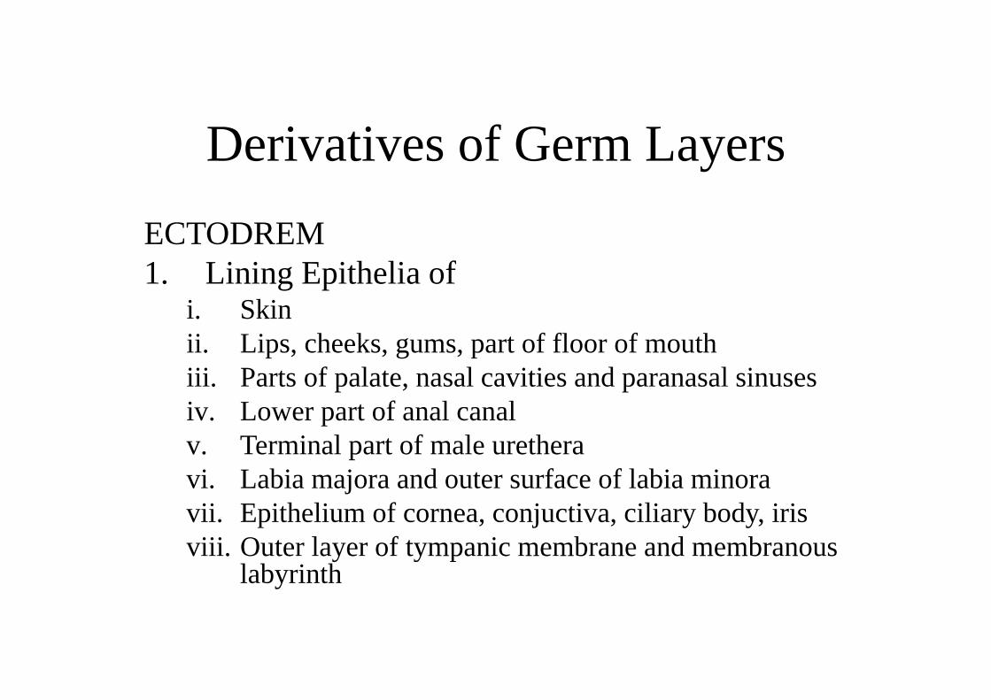
Derivatives of Germ LayersECTODREM1. Lining Epithelia of
i. Skinii. Lips, cheeks, gums, part of floor of mouthiii. Parts of palate, nasal cavities and paranasal sinusesiv. Lower part of anal canalv. Terminal part of male uretheravi. Labia majora and outer surface of labia minoravii. Epithelium of cornea, conjuctiva, ciliary body, irisviii. Outer layer of tympanic membrane and membranous
labyrinth

ECTODERM (contd.):
2. Glands– Exocrine – Sweet glands, sebaceous glands
Parotid, Mammary and lacrimal3. Other derivatives
i. Hairii. Nailsiii. Enamel of teethiv. Lens of eye; musculature of irisv. Nervous system
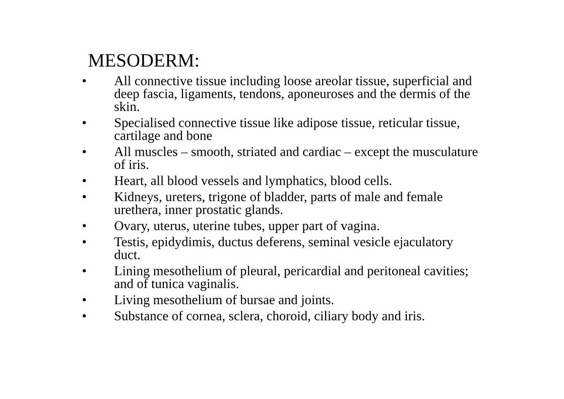
MESODERM:• All connective tissue including loose areolar tissue, superficial and
deep fascia, ligaments, tendons, aponeuroses and the dermis of the skin.
• Specialised connective tissue like adipose tissue, reticular tissue, cartilage and bone
• All muscles – smooth, striated and cardiac – except the musculature of iris.
• Heart, all blood vessels and lymphatics, blood cells.• Kidneys, ureters, trigone of bladder, parts of male and female
urethera, inner prostatic glands.• Ovary, uterus, uterine tubes, upper part of vagina.• Testis, epidydimis, ductus deferens, seminal vesicle ejaculatory
duct.• Lining mesothelium of pleural, pericardial and peritoneal cavities;
and of tunica vaginalis.• Living mesothelium of bursae and joints.• Substance of cornea, sclera, choroid, ciliary body and iris.
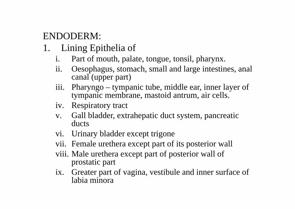
ENDODERM:1. Lining Epithelia of
i. Part of mouth, palate, tongue, tonsil, pharynx.ii. Oesophagus, stomach, small and large intestines, anal
canal (upper part)iii. Pharyngo – tympanic tube, middle ear, inner layer of
tympanic membrane, mastoid antrum, air cells.iv. Respiratory tractv. Gall bladder, extrahepatic duct system, pancreatic
ductsvi. Urinary bladder except trigonevii. Female urethera except part of its posterior wallviii. Male urethera except part of posterior wall of
prostatic partix. Greater part of vagina, vestibule and inner surface of
labia minora
ENDODERM (contd.)
2. Glandsi. Endocrine: Thyroid, parathyroid,
thymus, islets of Langerhansii. Exocrine : Live, pancreas, glands in G.I.T., prostatic
glands and its female homologues
Placenta
•Primary site of nutrient and gasexchange between mother and foetus
•feto-maternal organ


Trophoblast
Cytotrophoblast
Syncytiotrophoblast
Lacunar stage
Start of uteroplacental circulation
Formation of primary Villi
Initially villi cover whole surface-villi at embryonic pole disappear-chorionic laeve (smooth)-villi at embryonic pole expand--chorionic frondosum (bushy)


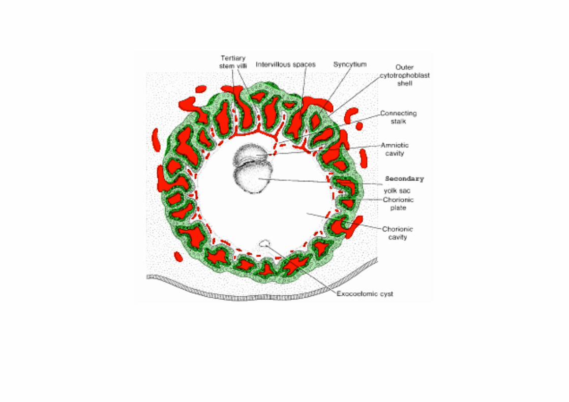
Formation of Secondary Villi
Formation of Tertiary Villi
Foetal contribution- Chorionic frondosum
Maternal contribution- Decidua basalis
Decidua which is shed off in labour
1.decidua capsularis--covers abembryonic pole
2. decidua basalis--covers embryonic pole
3. decidua parietalis-- rest of uterine wall
•Decidua capsularis disappears•Chorionic laeve adheres to decidua parietalis; uterine
cavity obliterates.•Amnion increases in size rapidly; amnion fuses to chorion -chorionic cavity obliterates-amniochorionicmembrane formed
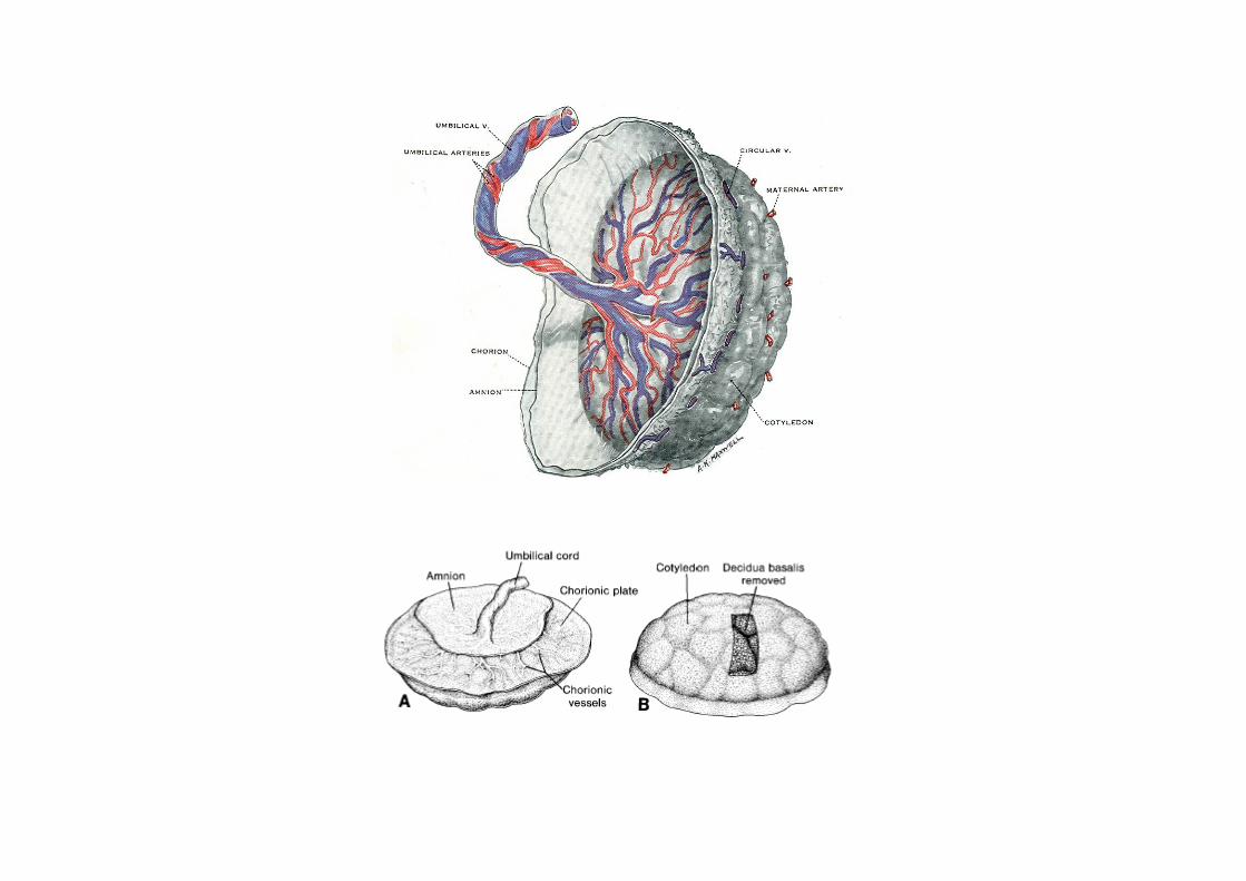
•Decidua sends septa into intervillous space-these septa are incomplete-divide the maternal surface into compartments-
cotyledons


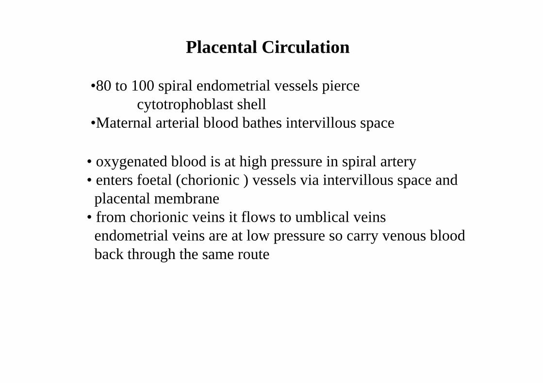
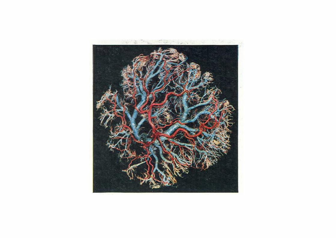
Placental Circulation
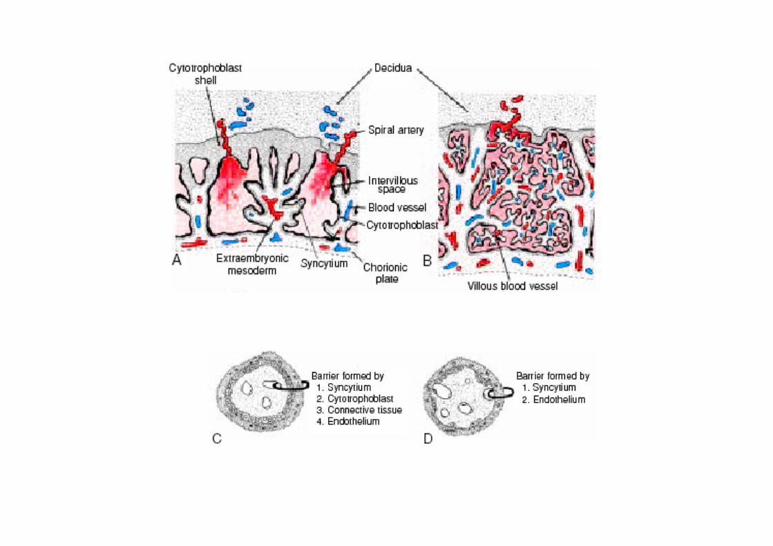
•80 to 100 spiral endometrial vessels pierce cytotrophoblast shell
•Maternal arterial blood bathes intervillous space
• oxygenated blood is at high pressure in spiral artery• enters foetal (chorionic ) vessels via intervillous space and placental membrane
• from chorionic veins it flows to umblical veinsendometrial veins are at low pressure so carry venous bloodback through the same route


Placental membrane
Placental membrane initially composed of•Endothelium of fetal vessel•connective tissue (extra embryonic mesoderm)•syncytiotrophoblast•cytotrophoblast
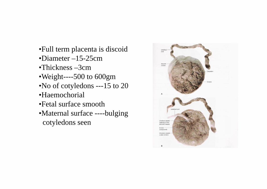
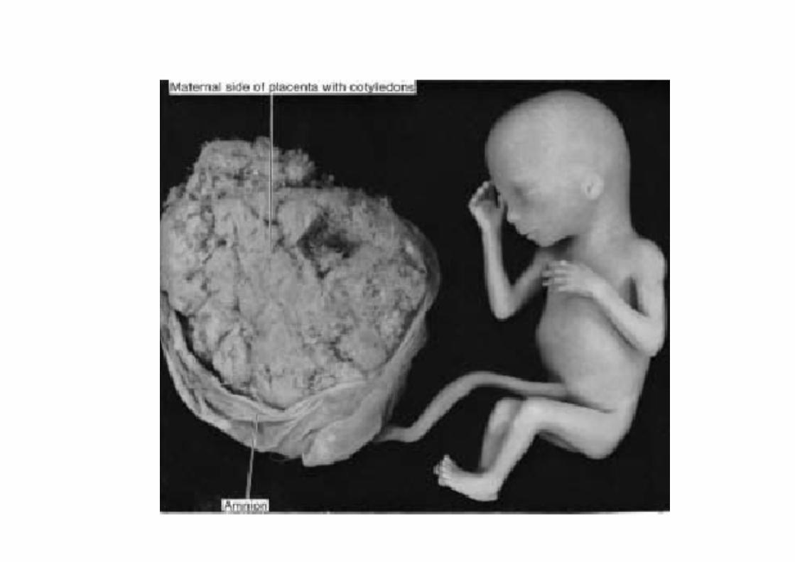
•Full term placenta is discoid •Diameter –15-25cm•Thickness –3cm•Weight----500 to 600gm•No of cotyledons ---15 to 20•Haemochorial•Fetal surface smooth•Maternal surface ----bulging
cotyledons seen
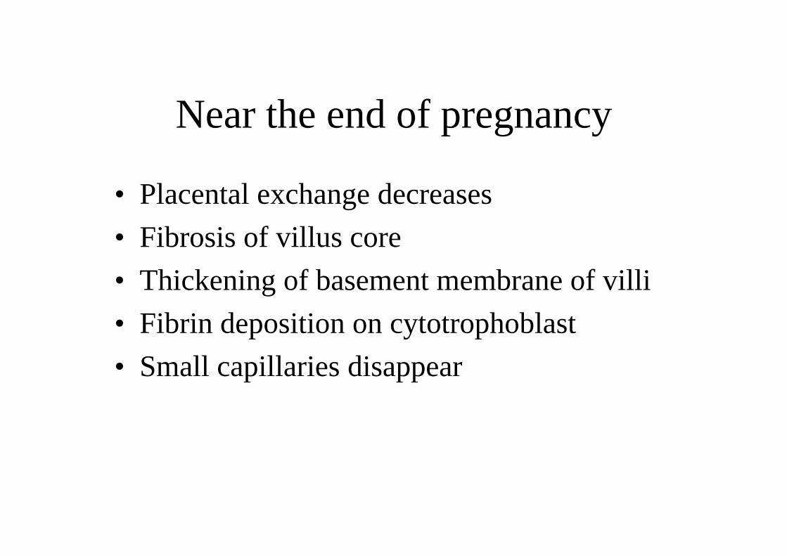
Near the end of pregnancy
• Placental exchange decreases• Fibrosis of villus core• Thickening of basement membrane of villi• Fibrin deposition on cytotrophoblast• Small capillaries disappear

Amniotic Cavity
• clear watery fluid in amniotic cavity• secreted by amniotic cells and maternal
blood• provides protective cushion• Volume 30 ml at 10 weeks
- 450ml at 20 weeks-1000 ml at 37 weeks
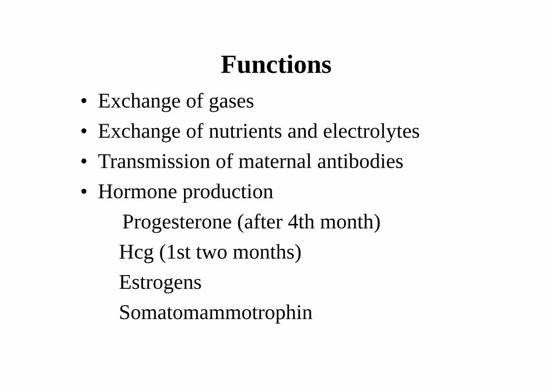
Functions• Exchange of gases• Exchange of nutrients and electrolytes• Transmission of maternal antibodies• Hormone production
Progesterone (after 4th month)Hcg (1st two months)EstrogensSomatomammotrophin

Umbilical Ring
• Comprises of-• connecting stalk with allantois and
umbilical vessels• yolk stalk with vitelline vessels• canal connecting intra and extraembryonic
cavity

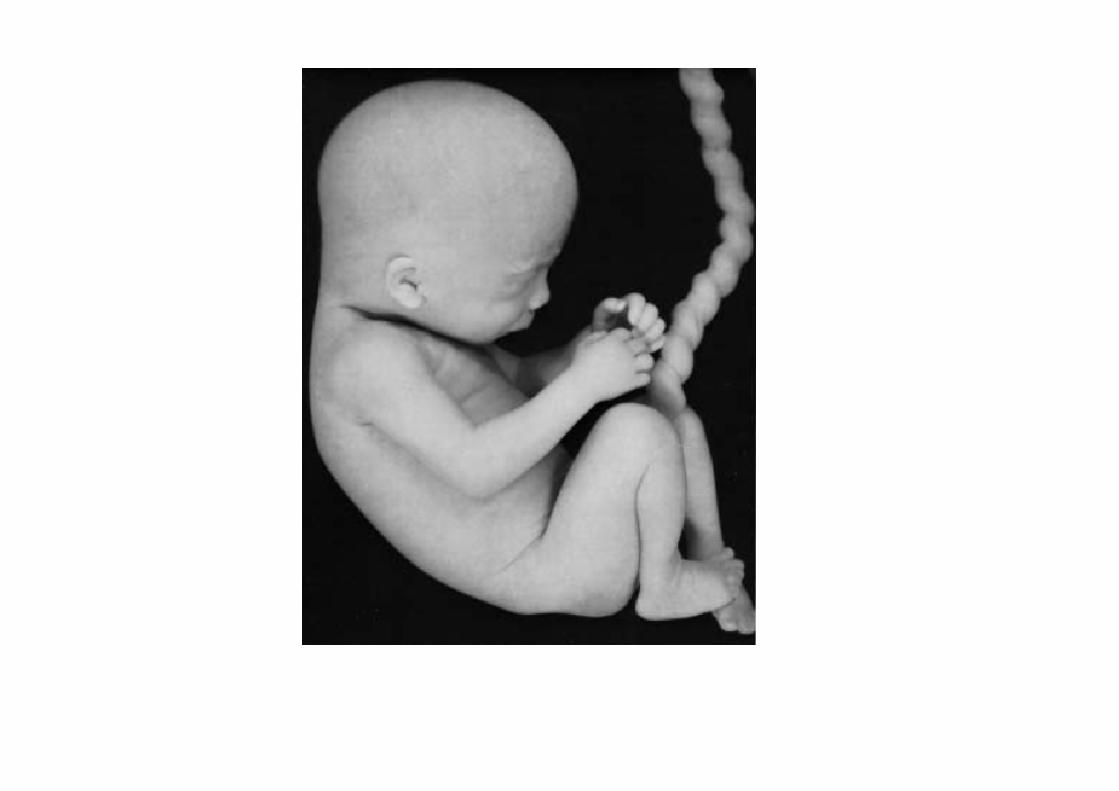
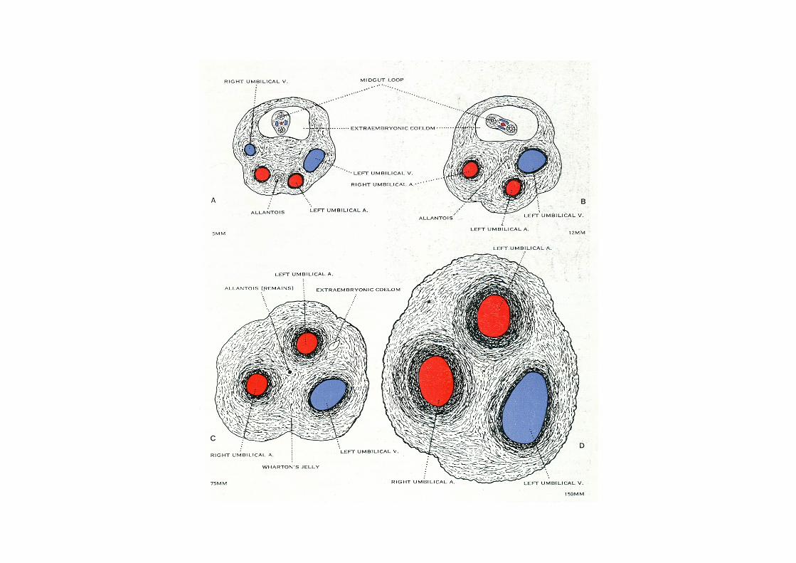
Umbilical Cord
• It forms when amnion envelops umbilical ring structures.
• yolk sac obliterates by third month.• loops of intestine may enter umbilical ring.• allantois, vitelline duct and vessels disappear.• Wharton's jelly now protects umbilical vessels.• it is rich in proteoglycans.


![WELCOME [gmch.gov.in]](https://img.dokumen.tips/doc/110x75/616a5d6311a7b741a351b2cf/welcome-gmchgovin.jpg)