Embed Size (px)
Citation preview
CYTOPLASMIC METACHROMASIA IN CULTURED SKIN FIBROBLASTS FROM PARENTS OF CHILDREN WITH CYSTIC FIBROSIS
Susan P. McManus and Joseph Masterson
Section of Medical Genetics, Department of Pathology, University College, Dublin 2.
Summary
T HE cytoplasmic metachromasia phenomenon with Toluidine Blue 0 in cultured skin fibroblasts was studied in 12 parents of children with cystic fibrosis and in 12 controls. Positive findings were recorded in 10 of the
CF parents and in one of the controls. The average culture time required for development of maximum metachromasia in positive cases was six weeks. The possible significance of the ametachromatic CF parents in relation to the suggested genetic heterogeneity of cystic fibrosis is discussed.
Introduction The diagnosis of cystic fibrosis (CF) can in many instances be made on
the basis of clinical signs and symptoms. When only clinical suspicion exists, however, and especially when genetic prognosis must be based on an unequivocal diagnosis, a reliable laboratory diagnostic test is indispens- able. Such a test should ideally distinguish between homozygote affected, heterozygote and homozygote normal, not alone because a firm diagnosis of cystic fibrosis may be sought but also because the identification of the unaffected CF heterozygote may be desirable. Since, on average, two out of three of the unaffected sibs of a CF propositus are CF heterozygotes and since in Caucasian populations approximately four to five per cent of their poten- tial marriage partners are also heterozygotes, efficient detection of the carrier state would form the basis of accurate premarital genetic counsel- ing.
Some studies (Danks et al., 1965; Anderson et al., 1967) have indicated the existence of a fertility advantage in the grandparents of CF children. In nearly all (96 per cent) grandparental matings, on either side of the family of the affected child, either the grandmother or the grandfather is heterozy- gote (disregarding illegitimacy) and the probability is half for either sex. Precise detection of the CF heterozygote in the grandparental generation would permit comparison of family sizes from both types of mating, with a view to determining if the heterozygote fertility advantage is peculiar to one or other sex. We are at present investigating this possibility.
Finally the concurrence of cystic fibrosis and a partial chromosome deletion in the child of chromosomally normal parents would provide strong evidence for assignment of the CF locus to the deleted segment if only one of the parents were shown to be heterozygous for the CF gene (Smith et al., 1968). A reliable test for the heterozygous state would also clearly be essential in these circumstances.
In 1968 Danes and Bearn reported the observation that cultured skin
227
228 IRISH JOURNAL OF MEDICAL SCIENCE
fibroblasts from CF patients and heterozygotes showed cytoplasmic meta- chromatic staining with Toluidine Blue 0. They further reported the absence of this phenomenon (apart from a very occasional positive cell) in the cultured fibroblasts of normal individuals. Cytoplasmic metachromasia has also been seen in skin cultures from patients with various mucopolysac- charidcses and from heterozygotes for the relevant genes (Danes and Bearn, 1966). Taysi et al. (1969) reported that in their CF study 27 per cent of the controls showed cytoplasmic metachromasia similar to that seen in cultures from both CF patients and carriers. The present investigation was designed to evaluate the cytoplasmic metachromasia phenomenon as a means of distinguishing between CF heterozygotes and normal controls.
Materials and Methods
Split thickness skin biopsies were taken without anaesthesia from the extensor surface of the forearm of 12 healthy parents of children with cystic fibrosis, i.e. obligatory CF heterozygotes. All were in the age range 25 to 40 years. Control skin samples were also taken from 12 accident hospital admissions matched for age and sex. Test and control biopsies were set up in parallel in Leighton culture tubes as detailed elsewhere (McManus and Masterson, 1973). Each culture resulted in 16 monolayers one week after the first subculture. Twelve of these were reserved for histochemical studies and four were again subcultured, resulting in a further 16 mono- layers seven days later. This process was repeated in every case for 10 subcultures in all.
For histochemical preparation the coverslips bearing cell monolayers were first washed in Earle's balanced salt solution, fixed in methanol for five minutes and air dried. After drying they were placed in 0.1 per cent Tolui- dine Blue 0 (Nat. Am. Div. N.Y., C.I. 925) in 30 per cent methanol for five minutes. The preparations were then cleared in acetone for one minute, ace- tone-xylene for two minutes and finally in pure xylene for two minutes. Per- manent preparations for photomicrography were mounted in Permount.
At each subculture stage in all cases 1,000 cells were examined for metachromasia under oil immersion at a magnification of 1000 diameters. Metachromatic cells were classified according to the criteria of Danes and Bearn (19s Three distinct classes of cells were defined as follows :
Class I Vesicular cytoplasmic metachromasia. Class II Generalised cytoplasmic metachromasia (metachromatic vesic-
les and granules in the cytoplasm). Class III No cytoplasmic metachromasia.
Successive subcultures were examined until a percentage of metachromatic cells was observed which did not increase in three subsequent subcultures. This was then taken as the maximum percentage of metachromatic cells for the relevant specimen. A culture from a CF heterozygote was considered ametachromatic when it did not exhibit marked cytoplasmic metachromasia (at least 10 per cent of cells) after 10 subcultures, i.e. approximately four months in culture.
CYTOPLASMIC METACHROMASIA IN CULTURED SKIN FIBROBLASTS 229
R e s u l t s
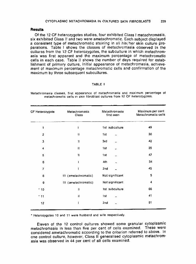
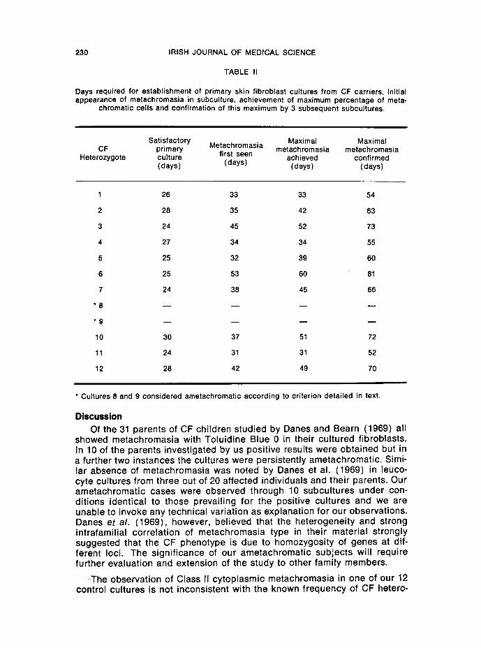
Of the 12 CF heterozygotes studies, four exh ib i ted Class I metachromasia, six exh ib i ted Class II and two were ametachromat ic . Each subject d isp layed a consis tent type of metachromat ic s ta in ing in aJI h i s / he r skin cul ture pre- parat ions. Table I shows the c lasses of metachromas ia observed in the cul tures f rom the 12 CF heterozygotes, the subcu l ture in wh ich metachrom- asia was f i rst apparent and the max imum percentage of metachromat ic cel ls in each case. Table II shows the number of days required for estab- l ishment of p r imary culture, in i t ia l appearance of metachromasia , achieve- ment of max imum percentage metachromat ic ce l ls and conf i rmat ion of the max imum by three subsequent subcul tures.
TABLE I
Metachromasia classes, first appearance of metachromasia and maximum percentage of metachromatic cells in skin fibroblast cultures from 12 CF heterozygotes.
CF Heterozygote Metachromasia Metachromasia Class first seen
Maximum per cent Metachromatic cells
1 I 1 st subculture 49
2 II 1st ,, 30
3 II 3rd ,, 42
4 II 1st ,, 35
5 II 1st ,, 47
6 I 4th ,, 34
7 I 2nd ,, 45
8 ~11 (ametachromatic) Not significant 5
9 Ill (ametachromatic) Not significant 4
�9 10 II 1st subculture 66
�9 11 II 1st ,, 41
12 I 2nd ,, 51
* Heterozygotes 10 and 11 were husband and wife respectively.
Eleven of the 12 control cu l tures showed some granu lar cy top lasmic metachromasia in less than f ive per cent of cel ls examined. These were considered ametachromat i c accord ing to the cr i ter ion referred to above. In one control cul ture, however, Class II genera l ised cy top lasmic metachrom- asia was observed in 44 per cent of al l cel ls examined.
230 IRISH JOURNAL OF MEDICAL SCIENCE
TABLE II
Days required for establishment of primary skin fibroblast cultures from CF carriers, initial appearance of metachromasia in subculture, achievement of maximum percentage of meta-
chromatic cells and confirmation of this maximum by 3 subsequent subcultures.
Satisfactory Metachromasia Maximal Maximal CF primary metachromasia metachromasia
Heterozygote culture first seen achieved confirmed (days) (days) (days) (days)
1 26 33 33 54
2 28 35 42 63
3 24 45 52 73
4 27 34 34 55
5 25 32 39 60
6 25 53 60 81
7 24 38 45 66
~ 8 . . . .
. g . . . .
10 30 37 51 72
11 24 31 31 52
12 28 42 49 70
" Cultures 8 and 9 considered ametachromatic according to criterion detailed in text.
Discussion Of the 31 parents of CF children studied by Danes and Bearn (1969) all
showed metachromasia with Toluidine Blue 0 in their cul tured fibroblasts. In 10 of the parents investigated by us posit ive results were obtained but in a further two instances the cultures were persistently ametachromatic. Simi- lar absence of metachromasia was noted by Danes et al. (1969) in leuco- cyte cultures from three out of 20 affected individuals and their parents. Our ametachromat ic cases were observed through 10 subcul tures under con- dit ions identical to those prevail ing for the posit ive cultures and we are unable to invoke any technical variation as explanation for our observations. Danes et al. (1969), however, bel ieved that the heterogenei ty and strong intrafamilial correlat ion of metachromasia type in their material strongly suggested that the CF phenotype is due to homozygosity of genes at dif- ferent loci. The signi f icance of our ametachromat ic subjects will require further evaluation and extension of the study to other family members.
T h e observation of Class II cytoplasmic metachromasia in one of our 12 control cultures is not inconsistent with the known f requency of CF hetero-
CYTOPLASMIC METACHROMASIA IN CULTURED SKIN FIBROBLASTS 231
zygotes in the general population. Furthermore it must be appreciated that while metachromasia with Toluidine Blue 0 is a useful marker in CF patients and heterozygotes, the reaction is non-specific and can be observed in cul- tured fibroblasts in a number of genetic metabolic disorders. We feel, nevertheless, that intrafamilial investigation of this phenomenon in cystic fibrosis and its correlation with other relevant laboratory diagnostic pro- cedures will help to elucidate the possibility of genetic heterogeneity in this condition.
The authors wish to express their gratitude to Dr. Edward Tempeny for his kind clinical co-operation and to the CF parents and controls for their cheerful participation in this study. The investigation reported here was supported by a grant-in-aid from the Medical Research Council of Ireland.
References
Anderson, C. M., Allan, J. and Johansen, P. C. 1967. Comments on the possible existence and nature of a heterozygote advantage in cystic fibrosis. Bibl. Paed., 86, 381.
Danes, B. S. and Beam, A. G. 1966. Hurler's Syndrome: A genetic study in cell culture. J. Exp. Med., 123, 1.
Danes, B. S. and Bearn, A. G. 1968. A genetic cell marker in cystic fibrosis of the pancreas. Lancet, i, 1061.
Danes, B. S: and Beam, A. G. 1969. Cystic fibrosis of the pancreas--a study in cell cul- ture. J, Exp. Med., 129, 775.
Danes, B. S., Foley, K. M., Dillon, S. D. and Beam, A. G. 1969. Genetic study of cystic fibrosis of the pancreas using white blood cell cultures. Nature, 222, 685.
Danks, D. M., Allan, J. and Anderson, C. M. 1965. A genetic study of fibrocystic disease of the pancreas. Ann. Hum. Genet., 28, 323.
McManus, S. P. and Masterson, J. 1973. Diagnostic skin fibroblast culture. Irish J. Med. Sci., 142, 358.
Smith, D. W., Docter, J. M., Ferrier, P. E., Frias, J. L. and Spock, A. 1968. Possible local- ization of the gene for cystic fibrosis of the pancreas to the short arm of chromosome 5. Lancet, ii, 309.
Taysi, K., Kistenmacher, M. L. and Punnett, H. H. 1969. Limitations of metachromasia as a diagnostic aid in paediatrics. New Eng. J. Med., 281, 1108.

![Impaired Stimulation D-24-hydroxylase in Fibroblasts ... · tosol from cultured skin fibroblasts (10) and to assess retention of [3H]1,25-(OH)2D3 in fibroblast nuclei fol-lowing binding](https://img.dokumen.tips/doc/110x75/5f040b207e708231d40c09b4/impaired-stimulation-d-24-hydroxylase-in-fibroblasts-tosol-from-cultured-skin.jpg)