Embed Size (px)
Citation preview
2903
Cyclodextrin–polysaccharide-based, in situ-gelled system forocular antifungal deliveryAnxo Fernández-Ferreiro1,2, Noelia Fernández Bargiela1, María Santiago Varela3,Maria Gil Martínez3, Maria Pardo4, Antonio Piñeiro Ces3, José Blanco Méndez1,Miguel González Barcia2, Maria Jesus Lamas2 and Francisco.J. Otero-Espinar*1,5
Full Research Paper Open Access
Address:1Pharmacy and Pharmaceutical Technology Department, Faculty ofPharmacy, University of Santiago de Compostela (USC), PrazaSeminario de Estudos Galegos s/n, Santiago de Compostela, 1570,Spain, 2Pharmacy Department, Xerencia de Xestión Integrada deSantiago de Compostela, SERGAS, Travesía Choupana s/n,Santiago de Compostela, 15706, Spain, 3Ophthalmology Department,Hospital de Conxo, Xerencia de Xestión Integrada de Santiago deCompostela, SERGAS, Rua Ramón Baltar s/n, Santiago deCompostela, 15706, Spain, 4Grupo Obesidomica, Instituto deInvestigación Sanitaria (IDIS-ISCIII), SERGAS, Travesía daChoupana s/n, Santiago de Compostela, 15706, Spain and 5IndustrialPharmacy Institute, Faculty of Pharmacy, University of Santiago deCompostela (USC), Praza Seminario de Estudos Galegos s/n,Santiago de Compostela, 15701, Spain
Email:Francisco.J. Otero-Espinar* - [email protected]
* Corresponding author
Keywords:cyclodextrins; eye drops; fluconazole; hydroxypropyl-β-cyclodextrin;sulfobutylether-β-cyclodextrin
Beilstein J. Org. Chem. 2014, 10, 2903–2911.doi:10.3762/bjoc.10.308
Received: 01 August 2014Accepted: 21 November 2014Published: 08 December 2014
This article is part of the Thematic Series "Superstructures withcyclodextrins: Chemistry and applications II".
Guest Editor: G. Wenz
© 2014 Fernández-Ferreiro et al; licensee Beilstein-Institut.License and terms: see end of document.
AbstractFluconazole was studied with two different hydrophilic cyclodextrins (hydroxypropyl-β-cyclodextrin (HPBCD) and sulfobutyl
ether-β-cyclodextrin (SBECD)) for the formation of inclusion complexes. HPBCD and SBECD showed low cell cytotoxicity in
human keratocytes as assessed by the label-free xCELLigence system for real-time monitoring. The fluconazole–HPBCD complex
was incorporated into an ion-sensitive ophthalmic gel composed of the natural polysaccharides gellan gum and κ-carrageenan. This
system showed good bioadhesive properties and effective control of fluconazole release.
2903
IntroductionFungal keratitis is a serious disease that can lead to loss of
vision. Candida albicans is one of the most widespread fungal
pathogens involved in fungal keratitis [1,2]. The most common
antifungal treatments include the use of polyenes and azoles;
however, a significant treatment failure rate exists with these
pharmacological agents [3]. Specifically, azole-based medica-
Beilstein J. Org. Chem. 2014, 10, 2903–2911.
2904
tions, which include imidazoles (e.g., miconazole, clotrimazole,
and ketoconazole) and triazoles (e.g., fluconazole, itraconazole,
posaconazole, and voriconazole), inhibit ergosterol synthesis in
the cell wall [4].
Fluconazole is considered to be a good therapeutic option for
most eye fungal infections; however, there are resistant species
that require higher concentrations than usual [5]. Unfortunately,
antifungal eye drops have low ocular bioavailability due to their
poor water solubility and known effective eye clearance mecha-
nisms [6]. Thus, after administration of volumes in the range of
25–50 µL, a rapid drainage occurs from the front of the eye.
Additionally, due to the low volume that the eye can accommo-
date, an overflowing of the solution takes place. In most
commercial formulations, the administered volume is about
30 µL, which is the mean volume of the human conjunctiva sac.
However, it has been shown that after a single blink, only 10 µL
of the solution remains [7].
In this context, pharmaceutical researchers have paid special
attention to the development of ophthalmic drug delivery
systems during the last decade. Different strategies have been
studied to prolong the ocular surface–drug permanence time, to
increase drug corneal permeability and also to delay drug elimi-
nation [8,9]. Hence, the in situ formation of polymer-based
delivery systems has received increased attention due to their
manageability and prolonged ocular residence time, which
improves patient compliance and comfort [10]. Specifically,
stimuli-responsive polymer hydrogels have recently attracted
special attention as drug delivery systems due to their capacity
to respond to changing environmental conditions such as ion
composition, pH, temperature, presence of chemicals, or
changes on electrical field, which result in the modification of
its structure and properties [11]. Gellan gum (GG) and
carrageenans are polysaccharides that can be used to enhance
ion-sensitive hydrogels. Gellan gum is a linear anionic
heteropolysaccharide with high molecular mass formed by a
tetrasaccharide repeating unit of glucose, glucuronic acid and
rhamnose in the ratio 2:1:1. Similarly, κ-carrageenan (CK) is
also an anionic heteropolysaccharide with high molecular mass
formed by repeating units of sulfated and nonsulfated galactose
and anhydrogalactose joined by β-1→4 and α-1→3 glycosidic
linkages. Aqueous solutions of both polysaccharides have char-
acteristic properties related to their temperature dependence and
cation-induced gelation [12].
A challenge in the design of bioadhesive ion-sensitive hydro-
gels is the incorporation of drugs with poor aqueous solubility.
Cyclodextrins (CDs) are useful pharmaceutical excipients that
aid in the formulation of poorly aqueous soluble drugs in a wide
range of delivery devices from the classical dosage forms to the
newest drug carriers [13]. Their spatial structure together with
the great variety of substituents can provide versatile pharma-
ceutical advantages. 2-Hydroxypropyl-β-cyclodextrin (HPBCD)
and sulfobutyl ether-β-cyclodextrin (SBECD) are two chemi-
cally modified cyclodextrins frequently used as vehicles to
improve drug solubility. The aim of this work is to obtain an
ophthalmic drug delivery system for the release of fluconazole
(FC) based on the use of ion-sensitive bioadhesive hydrogels.
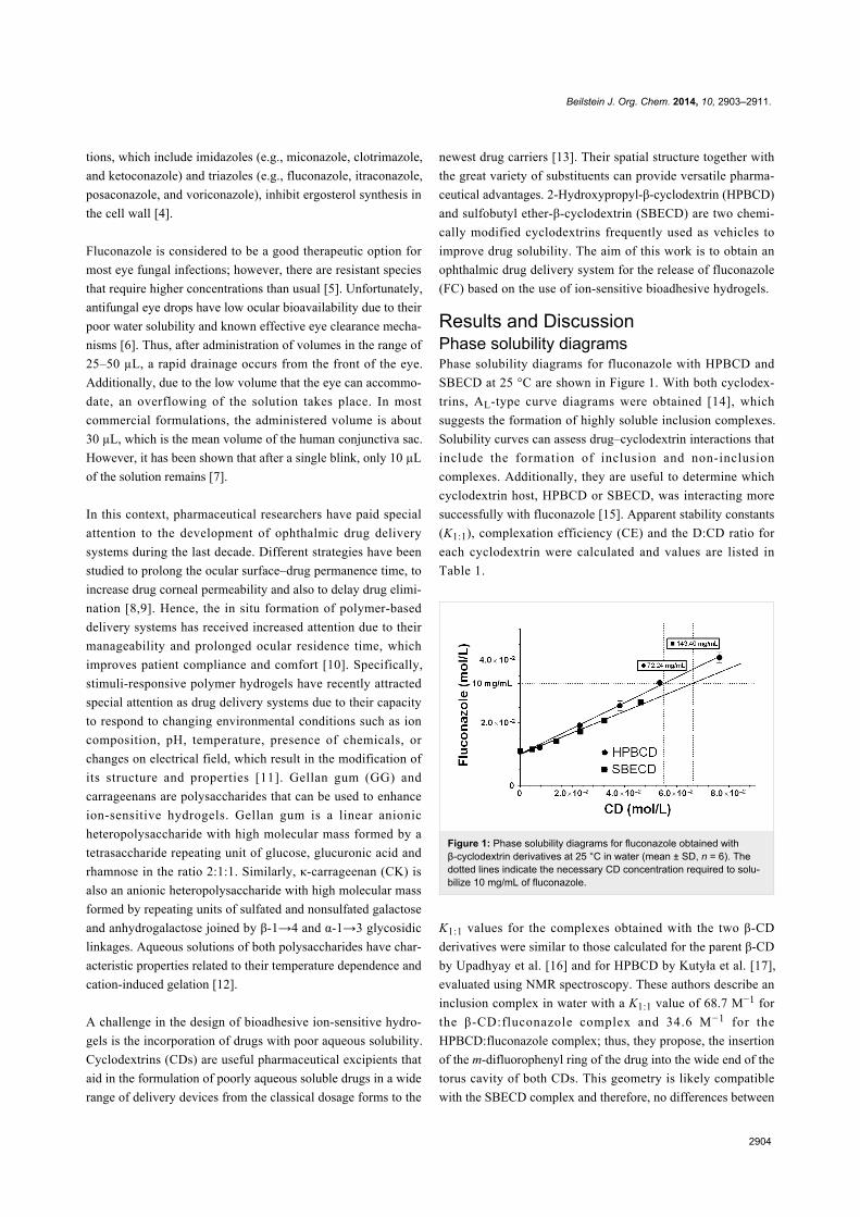
Results and DiscussionPhase solubility diagramsPhase solubility diagrams for fluconazole with HPBCD and
SBECD at 25 °C are shown in Figure 1. With both cyclodex-
trins, AL-type curve diagrams were obtained [14], which
suggests the formation of highly soluble inclusion complexes.
Solubility curves can assess drug–cyclodextrin interactions that
include the formation of inclusion and non-inclusion
complexes. Additionally, they are useful to determine which
cyclodextrin host, HPBCD or SBECD, was interacting more
successfully with fluconazole [15]. Apparent stability constants
(K1:1), complexation efficiency (CE) and the D:CD ratio for
each cyclodextrin were calculated and values are listed in
Table 1.
Figure 1: Phase solubility diagrams for fluconazole obtained withβ-cyclodextrin derivatives at 25 °C in water (mean ± SD, n = 6). Thedotted lines indicate the necessary CD concentration required to solu-bilize 10 mg/mL of fluconazole.
K1:1 values for the complexes obtained with the two β-CD
derivatives were similar to those calculated for the parent β-CD
by Upadhyay et al. [16] and for HPBCD by Kutyła et al. [17],
evaluated using NMR spectroscopy. These authors describe an
inclusion complex in water with a K1:1 value of 68.7 M−1 for
the β-CD:fluconazole complex and 34.6 M−1 for the
HPBCD:fluconazole complex; thus, they propose, the insertion
of the m-difluorophenyl ring of the drug into the wide end of the
torus cavity of both CDs. This geometry is likely compatible
with the SBECD complex and therefore, no differences between
Beilstein J. Org. Chem. 2014, 10, 2903–2911.
2905
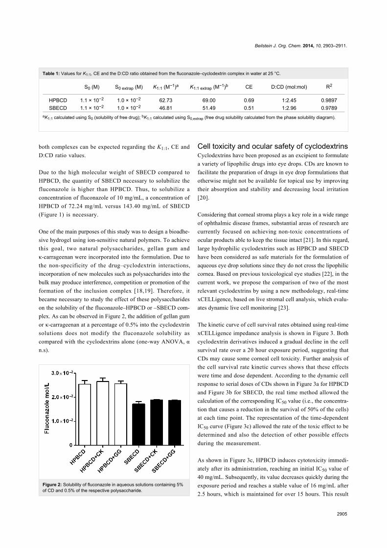
Table 1: Values for K1:1, CE and the D:CD ratio obtained from the fluconazole–cyclodextrin complex in water at 25 °C.
S0 (M) S0 extrap (M) K1:1 (M−1)a K1:1 extrap (M−1)b CE D:CD (mol:mol) R2
HPBCD 1.1 × 10⁻² 1.0 × 10⁻² 62.73 69.00 0.69 1:2.45 0.9897SBECD 1.1 × 10⁻² 1.0 × 10⁻² 46.81 51.49 0.51 1:2.96 0.9789
aK1:1 calculated using S0 (solubility of free drug); bK1:1 calculated using S0,extrap (free drug solubility calculated from the phase solubility diagram).
both complexes can be expected regarding the K1:1, CE and
D:CD ratio values.
Due to the high molecular weight of SBECD compared to
HPBCD, the quantity of SBECD necessary to solubilize the
fluconazole is higher than HPBCD. Thus, to solubilize a
concentration of fluconazole of 10 mg/mL, a concentration of
HPBCD of 72.24 mg/mL versus 143.40 mg/mL of SBECD
(Figure 1) is necessary.
One of the main purposes of this study was to design a bioadhe-
sive hydrogel using ion-sensitive natural polymers. To achieve
this goal, two natural polysaccharides, gellan gum and
κ-carrageenan were incorporated into the formulation. Due to
the non-specificity of the drug–cyclodextrin interactions,
incorporation of new molecules such as polysaccharides into the
bulk may produce interference, competition or promotion of the
formation of the inclusion complex [18,19]. Therefore, it
became necessary to study the effect of these polysaccharides
on the solubility of the fluconazole–HPBCD or –SBECD com-
plex. As can be observed in Figure 2, the addition of gellan gum
or κ-carrageenan at a percentage of 0.5% into the cyclodextrin
solutions does not modify the fluconazole solubility as
compared with the cyclodextrins alone (one-way ANOVA, α
n.s).
Figure 2: Solubility of fluconazole in aqueous solutions containing 5%of CD and 0.5% of the respective polysaccharide.
Cell toxicity and ocular safety of cyclodextrinsCyclodextrins have been proposed as an excipient to formulate
a variety of lipophilic drugs into eye drops. CDs are known to
facilitate the preparation of drugs in eye drop formulations that
otherwise might not be available for topical use by improving
their absorption and stability and decreasing local irritation
[20].
Considering that corneal stroma plays a key role in a wide range
of ophthalmic disease frames, substantial areas of research are
currently focused on achieving non-toxic concentrations of
ocular products able to keep the tissue intact [21]. In this regard,
large hydrophilic cyclodextrins such as HPBCD and SBECD
have been considered as safe materials for the formulation of
aqueous eye drop solutions since they do not cross the lipophilic
cornea. Based on previous toxicological eye studies [22], in the
current work, we propose the comparison of two of the most
relevant cyclodextrins by using a new methodology, real-time
xCELLigence, based on live stromal cell analysis, which evalu-
ates dynamic live cell monitoring [23].
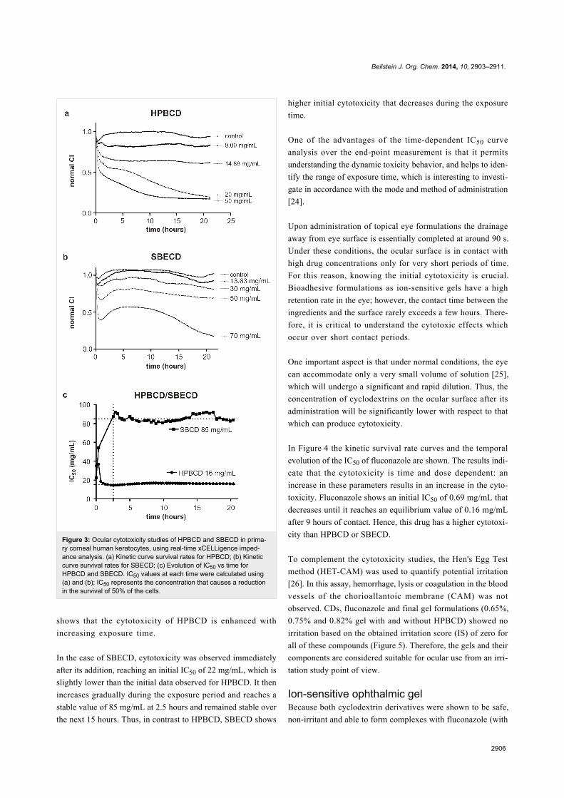
The kinetic curve of cell survival rates obtained using real-time
xCELLigence impedance analysis is shown in Figure 3. Both
cyclodextrin derivatives induced a gradual decline in the cell
survival rate over a 20 hour exposure period, suggesting that
CDs may cause some corneal cell toxicity. Further analysis of
the cell survival rate kinetic curves shows that these effects
were time and dose dependent. According to the dynamic cell
response to serial doses of CDs shown in Figure 3a for HPBCD
and Figure 3b for SBECD, the real time method allowed the
calculation of the corresponding IC50 value (i.e., the concentra-
tion that causes a reduction in the survival of 50% of the cells)
at each time point. The representation of the time-dependent
IC50 curve (Figure 3c) allowed the rate of the toxic effect to be
determined and also the detection of other possible effects
during the measurement.
As shown in Figure 3c, HPBCD induces cytotoxicity immedi-
ately after its administration, reaching an initial IC50 value of
40 mg/mL. Subsequently, its value decreases quickly during the
exposure period and reaches a stable value of 16 mg/mL after
2.5 hours, which is maintained for over 15 hours. This result
Beilstein J. Org. Chem. 2014, 10, 2903–2911.
2906
Figure 3: Ocular cytotoxicity studies of HPBCD and SBECD in prima-ry corneal human keratocytes, using real-time xCELLigence imped-ance analysis. (a) Kinetic curve survival rates for HPBCD; (b) Kineticcurve survival rates for SBECD; (c) Evolution of IC50 vs time forHPBCD and SBECD. IC50 values at each time were calculated using(a) and (b); IC50 represents the concentration that causes a reductionin the survival of 50% of the cells.
shows that the cytotoxicity of HPBCD is enhanced with
increasing exposure time.
In the case of SBECD, cytotoxicity was observed immediately
after its addition, reaching an initial IC50 of 22 mg/mL, which is
slightly lower than the initial data observed for HPBCD. It then
increases gradually during the exposure period and reaches a
stable value of 85 mg/mL at 2.5 hours and remained stable over
the next 15 hours. Thus, in contrast to HPBCD, SBECD shows
higher initial cytotoxicity that decreases during the exposure
time.
One of the advantages of the time-dependent IC50 curve
analysis over the end-point measurement is that it permits
understanding the dynamic toxicity behavior, and helps to iden-
tify the range of exposure time, which is interesting to investi-
gate in accordance with the mode and method of administration
[24].
Upon administration of topical eye formulations the drainage
away from eye surface is essentially completed at around 90 s.
Under these conditions, the ocular surface is in contact with
high drug concentrations only for very short periods of time.
For this reason, knowing the initial cytotoxicity is crucial.
Bioadhesive formulations as ion-sensitive gels have a high
retention rate in the eye; however, the contact time between the
ingredients and the surface rarely exceeds a few hours. There-
fore, it is critical to understand the cytotoxic effects which
occur over short contact periods.
One important aspect is that under normal conditions, the eye
can accommodate only a very small volume of solution [25],
which will undergo a significant and rapid dilution. Thus, the
concentration of cyclodextrins on the ocular surface after its
administration will be significantly lower with respect to that
which can produce cytotoxicity.
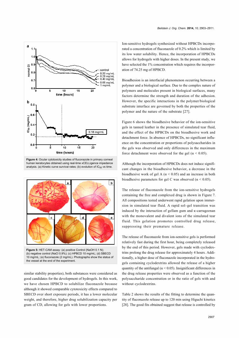
In Figure 4 the kinetic survival rate curves and the temporal
evolution of the IC50 of fluconazole are shown. The results indi-
cate that the cytotoxicity is time and dose dependent: an
increase in these parameters results in an increase in the cyto-
toxicity. Fluconazole shows an initial IC50 of 0.69 mg/mL that
decreases until it reaches an equilibrium value of 0.16 mg/mL
after 9 hours of contact. Hence, this drug has a higher cytotoxi-
city than HPBCD or SBECD.
To complement the cytotoxicity studies, the Hen's Egg Test
method (HET-CAM) was used to quantify potential irritation
[26]. In this assay, hemorrhage, lysis or coagulation in the blood
vessels of the chorioallantoic membrane (CAM) was not
observed. CDs, fluconazole and final gel formulations (0.65%,
0.75% and 0.82% gel with and without HPBCD) showed no
irritation based on the obtained irritation score (IS) of zero for
all of these compounds (Figure 5). Therefore, the gels and their
components are considered suitable for ocular use from an irri-
tation study point of view.
Ion-sensitive ophthalmic gelBecause both cyclodextrin derivatives were shown to be safe,
non-irritant and able to form complexes with fluconazole (with
Beilstein J. Org. Chem. 2014, 10, 2903–2911.
2907
Figure 4: Ocular cytotoxicity studies of fluconazole in primary cornealhuman keratocytes obtained using real-time xCELLigence impedanceanalysis. (a) Kinetic curve survival rates; (b) evolution of IC50 vs time.
Figure 5: HET-CAM assay: (a) positive Control (NaOH 0.1 N);(b) negative control (NaCl 0.9%); (c) HPBCD 10 mg/mL; (d) SBECD10 mg/mL; (e) fluconazole (2 mg/mL). Photographs show the status ofthe vessel at the end of the experiment.
similar stability properties), both substances were considered as
good candidates for the development of hydrogels. In this work,
we have chosen HPBCD to solubilize fluconazole because
although it showed comparable cytotoxicity effects compared to
SBECD over short exposure periods, it has a lower molecular
weight, and therefore, higher drug solubilization capacity per
gram of CD, allowing for gels with lower proportions.
Ion-sensitive hydrogels synthesized without HPBCDs incorpo-
rated a concentration of fluconazole of 0.2% which is limited by
its low water solubility. Hence, the incorporation of HPBCDs
allows for hydrogels with higher doses. In the present study, we
have selected the 1% concentration which requires the incorpor-
ation of 74.25 mg of HPBCD.
Bioadhesion is an interfacial phenomenon occurring between a
polymer and a biological surface. Due to the complex nature of
polymers and molecules present in biological surfaces, many
factors determine the strength and duration of the adhesion.
However, the specific interactions in the polymer/biological
substrate interface are governed by both the properties of the
polymer and the nature of the substrate [27].
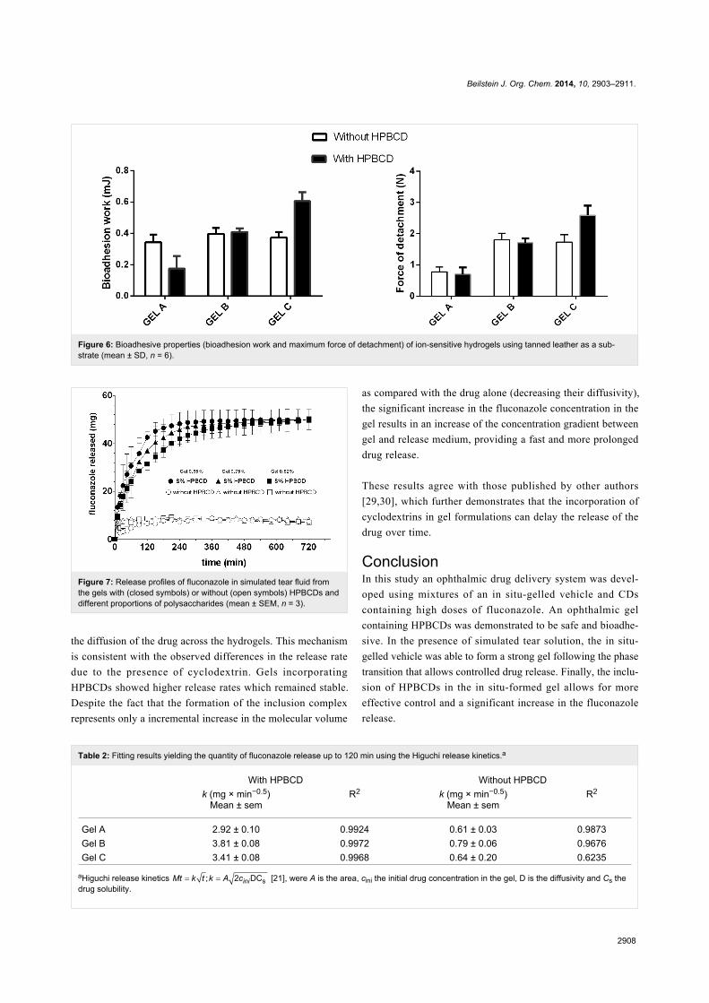
Figure 6 shows the bioadhesive behavior of the ion-sensitive
gels in tanned leather in the presence of simulated tear fluid,
and the effect of the HPBCDs on the bioadhesive work and
detachment force. In absence of HPBCDs, no significant influ-
ence on the concentration or proportions of polysaccharides in
the gels was observed and only differences in the maximum
force detachment were observed for the gel (α < 0.05).
Although the incorporation of HPBCDs does not induce signifi-
cant changes in the bioadhesive behavior, a decrease in the
bioadhesive work of gel A (α < 0.05) and an increase in both
bioadhesive parameters for gel C was observed (α < 0.05).
The release of fluconazole from the ion-sensitive hydrogels
containing the free and complexed drug is shown in Figure 7.
All compositions tested underwent rapid gelation upon immer-
sion in simulated tear fluid. A rapid sol–gel transition was
induced by the interaction of gellam gum and κ-carrageenan
with the monovalent and divalent ions of the simulated tear
fluid. This gelation promotes controlled drug release,
suppressing their premature release.
The release of fluconazole from ion-sensitive gels is performed
relatively fast during the first hour, being completely released
by the end of this period. However, gels made with cyclodex-
trins prolong the drug release for approximately 4 hours. Addi-
tionally, a higher dose of fluconazole incorporated in the hydro-
gels containing cyclodextrins allowed the release of a higher
quantity of the antifungal (α < 0.05). Insignificant differences in
the drug release properties were observed as a function of the
polysaccharide concentration or in the ratio of gels with and
without cyclodextrins.
Table 2 shows the results of the fitting to determine the quan-
tity of fluconazole release up to 120 min using Higuchi kinetics
[28]. The good fits obtained suggest that release is controlled by
Beilstein J. Org. Chem. 2014, 10, 2903–2911.
2908
Figure 6: Bioadhesive properties (bioadhesion work and maximum force of detachment) of ion-sensitive hydrogels using tanned leather as a sub-strate (mean ± SD, n = 6).
Table 2: Fitting results yielding the quantity of fluconazole release up to 120 min using the Higuchi release kinetics.a
With HPBCD Without HPBCDk (mg × min−0.5)
Mean ± semR2 k (mg × min−0.5)
Mean ± semR2
Gel A 2.92 ± 0.10 0.9924 0.61 ± 0.03 0.9873Gel B 3.81 ± 0.08 0.9972 0.79 ± 0.06 0.9676Gel C 3.41 ± 0.08 0.9968 0.64 ± 0.20 0.6235
aHiguchi release kinetics [21], were A is the area, cini the initial drug concentration in the gel, D is the diffusivity and Cs thedrug solubility.
Figure 7: Release profiles of fluconazole in simulated tear fluid fromthe gels with (closed symbols) or without (open symbols) HPBCDs anddifferent proportions of polysaccharides (mean ± SEM, n = 3).
the diffusion of the drug across the hydrogels. This mechanism
is consistent with the observed differences in the release rate
due to the presence of cyclodextrin. Gels incorporating
HPBCDs showed higher release rates which remained stable.
Despite the fact that the formation of the inclusion complex
represents only a incremental increase in the molecular volume
as compared with the drug alone (decreasing their diffusivity),
the significant increase in the fluconazole concentration in the
gel results in an increase of the concentration gradient between
gel and release medium, providing a fast and more prolonged
drug release.
These results agree with those published by other authors
[29,30], which further demonstrates that the incorporation of
cyclodextrins in gel formulations can delay the release of the
drug over time.
ConclusionIn this study an ophthalmic drug delivery system was devel-
oped using mixtures of an in situ-gelled vehicle and CDs
containing high doses of fluconazole. An ophthalmic gel
containing HPBCDs was demonstrated to be safe and bioadhe-
sive. In the presence of simulated tear solution, the in situ-
gelled vehicle was able to form a strong gel following the phase
transition that allows controlled drug release. Finally, the inclu-
sion of HPBCDs in the in situ-formed gel allows for more
effective control and a significant increase in the fluconazole
release.
Beilstein J. Org. Chem. 2014, 10, 2903–2911.
2909
ExperimentalMaterialsIn this study, the following substances were used: Sulfobutyl
ether-β-cyclodextrin (SBECD, Captisol®, average degree of
substitution for the sulfobutyl group: 6.6, average MW 2179)
was a generous gift from Cydex Inc. (Lenexa, KS); 2-hydrox-
ypropyl-β-cyclodextrin (HPBCD, Kleptose HPBCD® with a
molar substitution of 0.65 and MW 1399 Da) was a generous
gift from Roquette-Laisa (España); fluconazole (FC) was
purchased from ACOFARMA (España); gellam gum (GG,
Kelcogel CG-LA, CPKelco); and κ-carrageenan (CK, Genugel®
carrageenan CG-130, CPKelco).
Phase solubility diagramDrug–cyclodextrin interactions can be studied by a variety of
techniques, most of which examine the behavior of mixtures of
the cyclodextrin (host) and drug species (guest) in solution. A
more accurate method for the determination of the efficiency of
the cyclodextrin solubility is to determine their complexation
efficiency (CE) [31], that is, the concentration ratio between
cyclodextrin in a complex and free cyclodextrin. The CE is
calculated from the slope of the phase solubility diagram, and is
independent of both S0 and S0,extrap, and thus more reliable
when the influences of polymers (which are contained in an
ophthalmic gel) on the solubility are being investigated.
A phase solubility diagram technique was employed to esti-
mate the stability constants of fluconazole, HPBCD and
SBECD. Solubility measurements were carried out according to
the method of Higuchi and Connors [14] and by following the
protocols previously described by Anguiano-Igea et al. [15].
Excess amounts of fluconazole were added to a series of
aqueous solutions of increasing concentrations of cyclodextrin.
These solutions were shaken in an orbital shaking bath (VWR)
at 25 °C and 70 rpm for 7 days in order to reach equilibrium.
After equilibrium was attained, an aliquot was centrifuged for
0.5 h at 12,500 rpm (SIGMA 2-16P) and 1 mL aliquots of the
supernatant were diluted 100 times. The concentration of
fluconazole in each sample was determined using a spectropho-
tometer diode array (Hewlett Packard 8452A, λ = 260 nm). The
final values presented for the fluconazole solubility measure-
ments are the means of three replicate measurements.
The phase solubility diagrams were obtained by plotting the
mean solubility against the cyclodextrin concentration. The
apparent stability constant, assuming the formation of a
cyclodextrin inclusion complex with a 1:1 stoichiometry (K1:1),
was calculated from the slope and the drug solubility (S0) or the
intercept (S0,extrap) from linear regions obtained by least squares
regression using the following equation:
(1)
The parameter complexation efficiency CE is given by:
(2)
In addition, the D:CD ratio can be calculated using the CE
according to:
(3)
In order to study the influence of the presence of the two poly-
saccharides on the complex solubility, the drug solubility in an
aqueous solution of gellam gum or κ-carrageenan at 0.5% and
HPBCD or SBECD at 5% was determined by the same method-
ology described above.
Cell toxicity and ocular safety of CDsTo determinate the ocular toxicity of the cyclodextrins used in
the gels, both varieties were tested in primary corneal human
keratocytes (HCK) and Hen's Egg Test.
Isolation of human keratocytesThe study was conducted according to our institution’s guide-
lines and the declaration of Helsinki. To isolate human kerato-
cytes, a modification of the method proposed by Ramke et al
was used [32].
Cell cytotoxicity assayCell cytotoxicity was assessed by using the label-free xCELLi-
gence system (ACEA Biosciences, San Diego, CA) which
allows for real-time monitoring. Using this platform, the cell
index (CI) was the parameter used to represent the cell status
based on the measured electrical impedance [23,33].
Briefly, 3000 cells/well (E-plates, 16 wells) were seeded and in-
cubated for 24 hours until the CI reached a range of 1.0–1.2,
indicating about 60% cell confluence. At that point, the cell
culture medium was aspirated to perform cell treatment with the
CDs diluted in culture medium of different concentrations. The
data obtained was represented in dose response curves.
The IC50 values represent the concentration of CDs producing a
50% reduction of CI and the concentration of CDs producing a
50% reduction as compared to the negative control (culture
medium). The IC50 values were obtained directly from the dose
response curves, which has been previously described [34].
Beilstein J. Org. Chem. 2014, 10, 2903–2911.
2910
The Hen's Egg Test (HET)The HET-chorioallantoic membrane (HET-CAM) is used to
quantify the potential membrane irritation, providing an alter-
native to the Draize methodology [35]. Briefly, freshly fertilised
white leghorn eggs, with a weight of 50–60 g, were incubated at
37.5 °C and a relative humidity of 62 ± 7.5% for 9 days in an
incubator with an automatic rotating device. After removing the
egg shell covering the air cell with an electric drill and cutting
through the inner egg membranes, 0.3 mL of test substance was
applied (fluconazole: 2 mg/mL, CDs: 10 mg/mL and final gel
formulations: 0.65%, 0.75% and 0.82% gel with and without
HPBCD) was applied onto the vasculated chorioallantoic
membranes (CAM) of at least three eggs. The CAM, the blood
vessels (including the capillary system), and the albumen were
all observed over time (300 s) under a stereomicroscope
(Olympus SZ-STN) and scored for effects (hemorrhage, coagu-
lation and partial lysis). Sodium hydroxide 0.1 N served as posi-
tive control and sodium chloride 0.9% as a negative control.
Both cyclodextrins were placed in the CAM for determination
of irritation score (IS) by the methodology based on described
in protocol No. 96 from INVITTOX [26].
Preparation of ophthalmic gelsIon-sensitive hydrogels were prepared using three different
concentrations of polysaccharides. Gel A was prepared using a
0.82% (w/v) of a mixture of GG and CK in 4:1 ratio; Gel B
using 0.75% (w/v) of a mixture of GG and CK in ratio 2:1, and
Gel C using a 0.65% (w/v) of a mixture of GG and CK in ratio
1:1. The concentrations and ratios of the polymer were selected
in function of a previous work (unpublished data). The solu-
tions were prepared by dispersing in warm distilled water
(55 °C) and stirring for 24 hours.
Incorporation of drug and CD–drug complexes intoophthalmic gelsFluconazole was incorporate at the gel at a concentration of
2 mg/mL by dispersion of the solid drug into the gel under
magnetic stirring. To incorporate the fluconazole–CD complex,
cyclodextrin were previously dissolved in distilled water. Next
fluconazole (10 mg/mL) was dissolved in the CD solution.
Finally the polymers were incorporates in the different concen-
trations and ratios to obtain the gels.
Characterization of bioadhesionBioadhesive capacity was determined by measuring the
maximum detachment force and the bioadhesion work
employing a TA-XT Plus texture analyzer (TA Instruments,
Newcastle, UK) using tanned leather as substrate. The substrate
was selected because tanned leather is a good model for deter-
mining bioadhesion [36]. Tanned leather cylinders of 2 cm
diameter were adhered on the upper and the lower support by
using double layer paper and 0.2 mL of the sample was
deposited on the lower support. Then a compression/extension
stage using compression and extension rates of 1 mm/s (main-
taining an applied contact force of 0.5 N over 300 s) was
applied. Force versus elongation was recorded and maximum
force and bioadhesion work was calculated.
In vitro release of fluconazole from ophthalmic ion-sensitive gelThe in vitro release of fluconazole–CDs from the in situ-gelled
systems was studied using a membraneless model using an
no. 2 USP automated dissolution testing apparatus consisting of
a Prolabo Dissolutest fitted with a Hewlett Packard 8452A
diode array spectrophotometer. Similar membraneless methods
using a modified dissolution testing apparatus and high volumes
of simulated tear fluid was implemented for another study in
order to characterize in vitro drug release of ophthalmic gels
[37,38]. These models were demonstrated as more useful than
membrane models (i.e., using Franz cells) as they mimic the
clearance effect and dilution of the tear in the eye surface.
Hydrogels (5 g) were placed in open, 4 cm diameter, glass
containers which were placed in the bottom of a beaker filled
with 400 mL of simulated tear fluid at 37 °C and 75 rpm. The in
situ gel formation occurred during the sol–gel transition upon
contact with the simulated tear fluid.
Statistical analysisThe statistical analysis was made using the software, Stat-
graphics Centurion XVI (Stat Point Technologies, Inc.). The
solubility analysis was performed using a one-way ANOVA
analysis and the Student–Newman–Keuls method as multiple
comparison tests. The bioadhesion work and maximum detach-
ment force were analyzed by a two-way ANOVA analysis using
the concentration of polymers and the presence of CDs as inde-
pendent factors and the Student–Newman–Keuls method was
used as a multiple comparison test. Finally, release study
analysis was developed by applying a two-way ANOVA to the
volume of fluconazole released using time and formulation as
independent factors. Again, the Student–Newman–Keuls
method was used as a multiple comparison test.
AcknowledgementsThis work was supported by Fundación Española de Farmacia
Hospitalaria and Fundación Mutua Madrileña.
References1. Miller, D. Expert Opin. Pharmacother. 2013, 14, 543–560.
doi:10.1517/14656566.2013.775248
Beilstein J. Org. Chem. 2014, 10, 2903–2911.
2911
2. Keay, L. J.; Gower, E. W.; Iovieno, A.; Oechsler, R. A.; Alfonso, E. C.;Matoba, A.; Colby, K.; Tuli, S. S.; Hammersmith, K.; Cavanagh, D.;Lee, S. M.; Irvine, J.; Stulting, R. D.; Mauger, T. F.; Schein, O. D.Ophthalmology 2011, 118, 920–926. doi:10.1016/j.ophtha.2010.09.011
3. Mravii, I.; Dekaris, I.; Gabri, N.; Romac, I.; Glavota, V.; Mlinari, E. InKeratitis; Srinivasan, M., Ed.; InTech, 2012.
4. Kathiravan, M. K.; Salake, A. B.; Chothe, A. S.; Dudhe, P. B.;Watode, R. P.; Mukta, M. S.; Gadhwe, S. Bioorg. Med. Chem. 2012,20, 5678–5698. doi:10.1016/j.bmc.2012.04.045
5. Pfaller, M. A.; Diekema, D. J.; Sheehan, D. J. Clin. Microbiol. Rev.2006, 19, 435–447. doi:10.1128/CMR.19.2.435-447.2006
6. Gratieri, T.; Gelfuso, G. M.; de Freitas, O.; Rocha, E. M.;Lopez, R. F. V. Eur. J. Pharm. Biopharm. 2011, 79, 320–327.doi:10.1016/j.ejpb.2011.05.006
7. Pawar, P.; Kashyap, H.; Malhotra, S.; Sindhu, R. BioMed Res. Int.2013, No. 341218. doi:10.1155/2013/341218
8. Achouri, D.; Alhanout, K.; Piccerelle, P.; Andrieu, V.Drug Dev. Ind. Pharm. 2013, 39, 1599–1617.doi:10.3109/03639045.2012.736515
9. Kushwaha, S. K. S.; Saxena, P.; Rai, A. K. Int. J. Pharm. Invest. 2012,2, 54–60. doi:10.4103/2230-973X.100036
10. Liu, Z.; Li, J.; Nie, S.; Liu, H.; Ding, P.; Pan, W. Int. J. Pharm. 2006,315, 12–17. doi:10.1016/j.ijpharm.2006.01.029
11. Chen, X.; Li, W.; Zhong, W.; Lu, Y.; Yu, T. J. Appl. Polym. Sci. 1997,65, 2257–2262.doi:10.1002/(SICI)1097-4628(19970912)65:11<2257::AID-APP23>3.0.CO;2-Z
12. Coviello, T.; Matricardi, P.; Marianecci, C.; Alhaique, F.J. Controlled Release 2007, 119, 5–24.doi:10.1016/j.jconrel.2007.01.004
13. Otero-Espinar, F. J.; Blanco-Méndez, J. Curr. Top. Med. Chem. 2014,14, 463–464. doi:10.2174/1568026613666131219122902
14. Higuchi, T.; Connors, K. A. Phase-Solubility Techniques. Advances inAnalytical Chemistry and Instrumentation; Interscience: New York,1965; pp 117–212.
15. Anguiano-Igea, S.; Otero-Espinar, F. J.; Vila-Jato, J. L.;Blanco-Méndez, J. Eur. J. Pharm. Sci. 1997, 5, 215–221.doi:10.1016/S0928-0987(97)00277-7
16. Upadhyay, S. K.; Kumar, G. Chem. Cent. J. 2009, 3, No. 9.doi:10.1186/1752-153X-3-9
17. Kutyła, M. J.; Lambert, L. K.; Davies, N. M.; McGeary, R. P.;Shaw, P. N.; Ross, B. P. Int. J. Pharm. 2013, 444, 175–184.doi:10.1016/j.ijpharm.2013.01.005
18. Nogueiras-Nieto, L.; Alvarez-Lorenzo, C.; Sandez-Macho, I.;Concheiro, A.; Otero-Espinar, F. J. J. Phys. Chem. B 2009, 113,2773–2782. doi:10.1021/jp809806w
19. Jansook, P.; Loftsson, T. Int. J. Pharm. 2009, 379, 32–40.doi:10.1016/j.ijpharm.2009.06.005
20. Loftsson, T.; Stefánsson, E. Acta Ophthalmol. Scand. 2002, 80,144–150. doi:10.1034/j.1600-0420.2002.800205.x
21. Kilic, C.; Girotti, A.; Rodriguez-Cabello, J. C.; Hasirci, V. Biomater. Sci.2014, 2, 318–329. doi:10.1039/c3bm60194c
22. Niles, A. L.; Moravec, R. A.; Riss, T. L. Expert Opin. Drug Discovery2008, 3, 655–669.
23. Xing, J. Z.; Zhu, L.; Gabos, S.; Xie, L. Toxicol. In Vitro 2006, 20,995–1004. doi:10.1016/j.tiv.2005.12.008
24. Chen, H.; Cui, L.; Jiang, X.-Y.; Pang, Y.-Q.; Tang, G.-L.; Hou, H.-W.;Jiang, J.-H.; Hu, Q.-Y. Food Chem. Toxicol. 2012, 50, 612–618.doi:10.1016/j.fct.2011.11.040
25. Kumar, S.; Karki, R.; Meena, M.; Prakash, T.; Rajeswari, T.; Goli, D.J. Adv. Pharm. Technol. Res. 2011, 2, 192–194.doi:10.4103/2231-4040.85540
26. Hen’s Egg Test on the Chorioallantoic Membrane (HET-CAM)INVITTOX no. 96.http://www.vitrotox.com/uploadfile/UploadFile/2008121382926916.pdf(accessed Oct 9, 2014).
27. Vallejo Diaz, B. M.; Perilla, J. E. Rev. Colomb. Cienc. Quim.-Farm.2008, 37, 33–61.
28. Siepmann, J.; Peppas, N. A. Int. J. Pharm. 2011, 418, 6–12.doi:10.1016/j.ijpharm.2011.03.051
29. Otero Espinar, F. J.; Torres-Labandeira, J. J.; Alvarez-Lorenzo, C.;Blanco-Méndez, J. J. Drug Delivery Sci. Technol. 2010, 20, 289–301.
30. Bibby, D. C.; Davies, N. M.; Tucker, I. G. Int. J. Pharm. 2000, 197,1–11. doi:10.1016/S0378-5173(00)00335-5
31. Loftsson, T.; Hreinsdóttir, D.; Másson, M. Int. J. Pharm. 2005, 302,18–28. doi:10.1016/j.ijpharm.2005.05.042
32. Ramke, M.; Lam, E.; Meyer, M.; Knipper, A.; Heim, A. Mol. Vision2013, 19, 614–622.
33. Xing, J. Z.; Zhu, L.; Jackson, J. A.; Gabos, S.; Sun, X.-J.; Wang, X.-b.;Xu, X. Chem. Res. Toxicol. 2005, 18, 154–161. doi:10.1021/tx049721s
34. Ceriotti, L.; Ponti, J.; Broggi, F.; Kob, A.; Drechsler, S.; Thedinga, E.;Colpo, P.; Sabbioni, E.; Ehret, R.; Rossi, F. Sens. Actuators, B 2007,123, 769–778. doi:10.1016/j.snb.2006.10.024
35. Fernández-Ferreiro, A.; González Barcia, M.; Gil Martínez, M.;Blanco Mendez, J.; Lamas Díaz, M. J.; Otero Espinar, F. J.Farm. Hosp. 2014, 38, 300–304. doi:10.7399/FH.2014.38.4.7343
36. Blanco-Fuente, H.; Anguiano-Igea, S.; Otero-Espinar, F. J.;Blanco-Méndez, J. Int. J. Pharm. 1996, 142, 169–174.doi:10.1016/0378-5173(96)04665-0
37. Lin, H.-R.; Sung, K. C. J. Controlled Release 2000, 69, 379–388.doi:10.1016/S0168-3659(00)00329-1
38. Wu, H.; Liu, Z.; Peng, J.; Li, L.; Li, N.; Li, J.; Pan, H. Int. J. Pharm.2011, 410, 31–40. doi:10.1016/j.ijpharm.2011.03.007
License and TermsThis is an Open Access article under the terms of the
Creative Commons Attribution License
(http://creativecommons.org/licenses/by/2.0), which
permits unrestricted use, distribution, and reproduction in
any medium, provided the original work is properly cited.
The license is subject to the Beilstein Journal of Organic
Chemistry terms and conditions:
(http://www.beilstein-journals.org/bjoc)
The definitive version of this article is the electronic one
which can be found at:
doi:10.3762/bjoc.10.308