Embed Size (px)
Citation preview
Current Concepts inHepatobil iary Surgery
Harry W. Boothe Jr, DVM, MS
KEYWORDS
� Hepatobiliary surgery � Hepatic mass lesions � Liver biopsy � Partial hepatectomy� Cholecystectomy � Cholecystoenterostomy � Choledochotomy
KEY POINTS
� Knowledge of the anatomy of the liver and biliary tract helps minimize complicationsassociated with hepatobiliary surgery.
� Information important for planning partial hepatectomies to treat hepatic masses includesdistribution of mass lesions; histologic diagnosis; and patient oncotic, blood typing/cross-matching, and coagulation status.
� Goals of extrahepatic biliary surgery include confirmation of the underlying diseaseprocess (eg, biliary mucocele, cholecystitis, and bile duct obstruction; trauma; or leakage)establishment of a patent biliary system, and minimization of perioperative complications.
� Veterinary patients undergoing either extensive liver resection or correction of biliary tractobstruction or leakage tend to have an extensive list of risk factors associated with theprimary condition and the surgical procedure.
INTRODUCTION AND ANATOMIC CONSIDERATIONS
Hepatobiliary surgery in dogs and cats may be used to investigate or treat variousconditions of the liver and biliary tract including persistent hepatic disease, hepaticabscessation, hepatic mass lesions, gallbladder mucocele, cholecystitis, biliaryleakage, and extrahepatic biliary obstruction. Surgical procedures performed includehepatic biopsy, partial hepatectomy, cholecystotomy, cholecystectomy, cholecys-toenterostomy, and choledochotomy. Although liver transplantation is not currentlyperformed clinically in dogs and cats, information gleaned from its use in researchdogs has provided valuable information to the clinical veterinary surgeon.1 Knowledgeof the anatomy of the liver and biliary tract helps minimize complications associatedwith hepatobiliary surgery.
The author has nothing to disclose.Wilford and Kate Bailey Small Animal Teaching Hospital, Department of Clinical Sciences,College of Veterinary Medicine, 1220 Wire Road, Auburn, AL 36849-5540, USAE-mail address: [email protected]
Vet Clin Small Anim 45 (2015) 463–475http://dx.doi.org/10.1016/j.cvsm.2015.01.001 vetsmall.theclinics.com0195-5616/15/$ – see front matter � 2015 Elsevier Inc. All rights reserved.
Boothe Jr464
The liver is the largest gland in the body and has exocrine (bile) and endocrine func-tion. It is divided into four lobes (left, right, quadrate, and caudate), four sublobes, andtwo processes by deep fissures. The left hepatic lobe, comprised of the left lateral andmedial sublobes, which may be joined by a bridge of liver tissue dorsally, forms nearlyone-half of the total liver mass. The right hepatic lobe is smaller than the left and hasthe right lateral and medial sublobes. The right lateral lobe is often fused to the rightmedial lobe and the caudate process of the caudate lobe. The right medial lobe isvariably fused to the quadrate lobe. The quadrate lobe lies almost on the midline,and its lateral aspect forms one side of the gallbladder fossa. The caudate lobe iscomposed of the caudate and papillary processes and the connecting isthmus. Theisthmus is located between the dorsally located caudal vena cava and the moreventral portal vein. The caudate process forms the most caudal portion of the liver,whereas the papillary process lies in the lesser curvature of the stomach.2 From asurgical perspective, the liver may be grouped into three subdivisions: left (left lateraland medial lobes) comprising approximately 44% of liver volume, central (quadrateand right medial lobes), and right (right lateral and caudate lobes), each comprisingabout 28% of liver volume.The portal vein provides the functional blood supply to the liver. It divides into left
and right branches in the dog, with the left branch supplying the central and leftdivisions. The feline portal vein divides into right, left, and central branches. The hepat-ic artery provides nutritional supply to hepatic parenchyma and bile ducts.1 Eachcanine sublobe is supplied by a single hepatic artery and at least one lobar portal vein.3
The biliary system begins at the hepatic canaliculi, with up to eight hepatic ducts,although three or four hepatic ducts was more commonly observed, joining to formthe bile duct.4 The initial hepatic duct to enter the bile duct usually is the right medialhepatic duct.4 The gallbladder is connected to the bile duct via the cystic duct, whichtends to be greater than 5 mm long in most dogs.4 After passing intramurally within theduodenum for approximately 2 cm, the bile duct opens approximately 3 to 6 cmaborad to the pylorus.
LIVER BIOPSY CONSIDERATIONSIndications and Contraindications
Diagnosis of most liver diseases requires histopathologic examination of liver tissue.5
Diffuse liver diseases may be sampled randomly, but focal lesions require carefulselective sampling.5 Ideally, the patient’s coagulation status shouldbe assessedbeforea liver biopsy is performed.5 Significant bleeding complications have been observed indogs and cats with thrombocytopenia (platelets <80� 103/mL) undergoing ultrasound-guided liver biopsies.6 The livermaybeevaluated via fine-needle aspiration (cytology) orbiopsy (histopathology). Ultrasound-guided fine-needle aspirations for cytologic exam-ination of the liver have been shown to have serious limitationswhen used to identify theprimary diseaseprocess in dogsandcatswith clinical evidenceof liver disease.7Hepat-ic cytologic samples are more reliable for diffuse hepatic disease, especially neoplasia,and less reliable for inflammation, necrosis, and hyperplasia.8
Technique
Liver biopsies are performed frequently and use various techniques in dogs and cats,including needle core, laparoscopic, and surgical biopsy. An ideal liver biopsy shouldbe of proper size and taken from a location that represents the primary liver patho-logy.5 Samples from multiple lobes are often preferred. In addition to tissues for histo-pathology, samples may also be obtained for microbiologic testing or quantification of
Current Concepts in Hepatobiliary 465
copper or other metals.5,9 Comparison of needle and wedge hepatic biopsy tech-niques has been made.10 Ultrasound-guided percutaneous techniques using a needlecore biopsy are one option for sampling the liver. Findings in needle biopsy samplestaken with ultrasound guidance or at laparotomy concurred with the definitive diag-nosis in 48% of dogs and cats in an earlier study.10
Laparoscopic liver biopsies may be obtained from grossly abnormal areas of theliver, particularly near the periphery of the liver lobes. Advantages of laparoscopicbiopsy over laparotomy sampling include lower patient morbidity and decreasedinfection rate, postoperative pain, and hospitalization time.5,11
Surgical methods of sampling the liver include ligature (suture) fracture or guillotinetechnique and biopsy punch technique. Surgical liver biopsies should be taken earlyduring the laparotomy to minimize hepatocellular changes from prolonged anesthesiaor manipulation of intestine.12 Advantages of surgical biopsy techniques includeenhanced exposure and ability to manipulate tissues, obtain large sample sizes,and monitor biopsy sites for bleeding.5 Hepatic biopsies obtained via laparotomyare the largest of any of the methods described and should provide adequate tissuefor various analyses.5,9
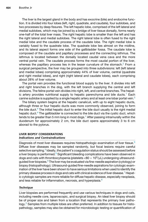
The ligature (suture) fracture or guillotine technique is performed on the periphery ofa liver lobe, with variably sized samples obtained (Fig. 1). Use of a pretied ligating loopto obtain liver biopsies was found to be versatile and safe in dogs.13 The biopsy punchtechnique results in collection of partial-thickness samples (ie, less than half the thick-ness of the lobe) of liver, usually from its ventral surface.3 Lesions located away fromthe periphery of the liver may be sampled using the biopsy punch. Hemostasis is pro-vided by suture (ligature fracture technique), omental coverage of the biopsy site(either surgical biopsy technique), electrocoagulation (either technique), or gelatinsponge (either technique).
Complications
Hemorrhage is the most frequently described complication, although abscessation ofan hepatic biopsy site has been reported and observed by the author.14
HEPATIC ABSCESSATION
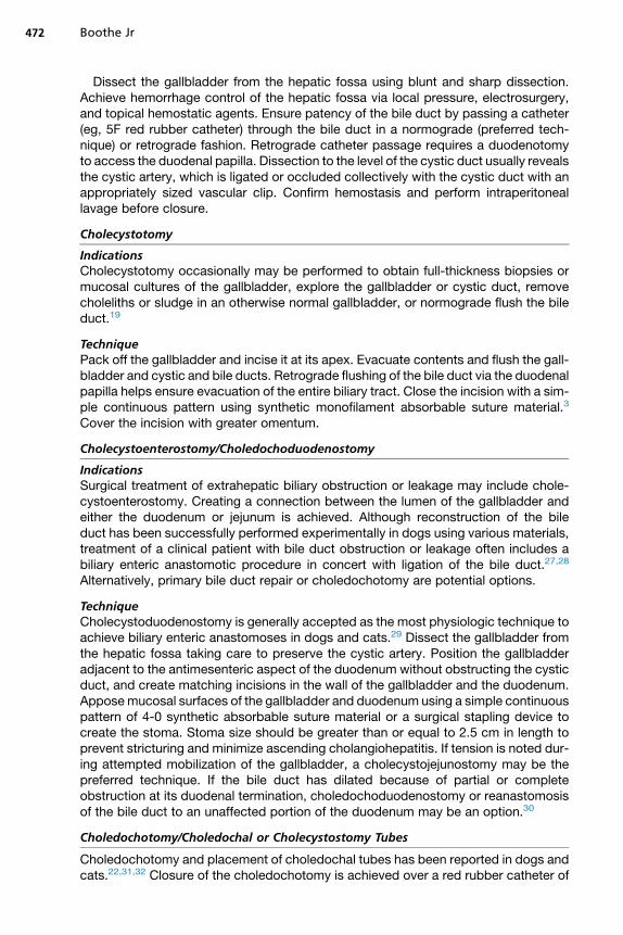
Hepatic abscessation in dogs or cats is reported relatively uncommonly, with middle-aged to older dogs and cats usually being described.3,14,15 Abdominal ultrasono-graphy is a relatively sensitive tool for diagnosis. Solitary hepatic abscessation may
Fig. 1. A biopsy of the liver is obtained using the ligature fracture (guillotine) method. Theisolated portion of liver distal to the suture material (2-0 PDS) is excised using scissors.
Boothe Jr466
be more common in dogs than in cats.14 Microbiologic sampling often yields variableisolates, although Escherichia coli is frequently found.
Technique
Diagnostic and treatment principles include complete evaluation to determine anyconcurrent disease process (eg, neoplasia) and the extent and number of abscesses,use of appropriate broad-spectrum antimicrobials, and possibly surgery. Surgicalintervention involves partial hepatectomy or drainage procedures (eg, omentalization).Hepatic abscessation in dogs does not seem to have an anatomic site predilection orto be associated with neoplasia, whereas cats have their right hepatic lobes morecommonly affected. Multiple abscess sites and concurrent hepatobiliary neoplasiaare more likely in cats.15
HEPATIC MASS LESIONS
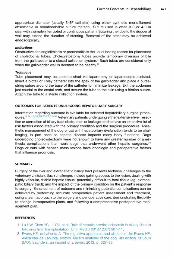
Primary hepatic neoplasia is reported to occur in 0.6% to 2.6% of dogs and 1.5% to2.3% of cats, with biliary neoplasia seen less frequently.3 Four general types of pri-mary hepatobiliary neoplasia are described: (1) hepatocellular, (2) cholangiocellular,(3) neuroendocrine, and (4) mesenchymal. Primary hepatic neoplasms in dogs canbe classified and differentiated using immunohistochemical stains as markers repre-sentative of hepatocytic and cholangiocytic lineages.16 Metastatic tumors of the liverare more common than primary tumors, with approximately 30% of dogs having he-patic metastatic tumors. The presence of hepatic masses may be noted on abdominalultrasonography, although advanced imaging (eg, computed tomography or MRI) pro-vides information to help discern tumor location, distribution, and potentially differen-tiate malignant from benign masses.3 Determination of specific tissue types isfacilitated by fine-needle aspiration of cells or needle core, laparoscopic, or surgicalbiopsy. Reported rates for correct diagnoses of hepatic masses range from up to50% for fine-needle aspiration to 70% for needle core samples.3
Hepatocellular Tumors
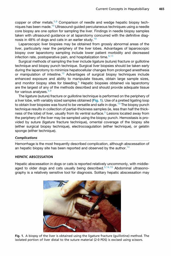
Hepatocellular tumors are the most common primary hepatic tumor of dogs, repre-senting 50% to 70% of all nonhematopoietic neoplasms (Fig. 2). Three forms of hepa-tocellular tumors are described: (1) massive (61%), (2) nodular (29%), and (3) diffuse(10%). Metastasis is more common with nodular or diffuse forms (93%) than withthe massive form (36%). Anatomic distribution of lesions with the massive form is
Fig. 2. Omental adhesions are dissected from this massive hepatocellular carcinoma presentin the left lateral lobe of a 12-year-old Labrador retriever.
Current Concepts in Hepatobiliary 467
approximately 67% in left lobes, 15% in central lobes, and 18% in right lobes. Hepa-tocellular tumors with hepatic progenitor cellular characteristics tend to be poorlydifferentiated and aggressive in behavior.16 Hepatocellular tumors are reported lesscommonly in cats, representing less than 25% of primary hepatic neoplasms. Hepa-tocellular adenomas are more common than carcinomas in cats, whereas hepatocel-lular carcinomas in dogs are seen twice as frequently as are hepatocellular adenomas.Surgical resection is the preferred treatment of massive hepatocellular carcinoma in
dogs, generally carrying a favorable prognosis.3 Because of likely incomplete surgicalresection and high metastatic rate, surgery is not a good option for nodular or diffuseforms of hepatocellular carcinoma in dogs.3 Surgical challenges presented by centralor right-sided masses are usually greater than those presented by left-sided masses.
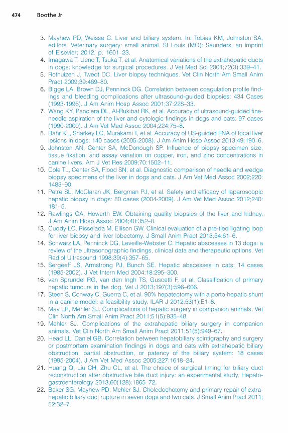
Cholangiocellular (Bile Duct) Tumors
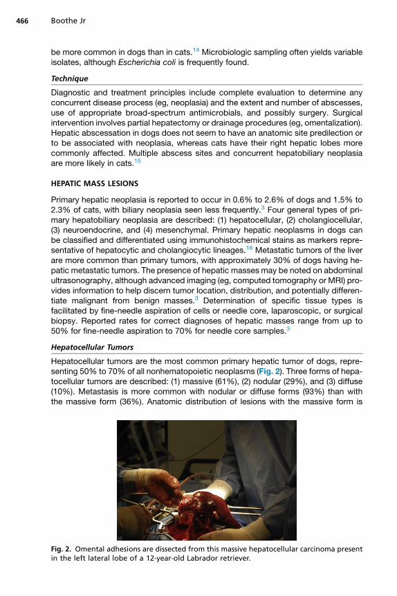
Cholangiocellular tumors account for approximately 30% of primary hepatic tumors indogs. Cholangiocellular carcinomas are thought to be derived from differentiatedmucin-producing cholangiocytes, normally present in larger bile ducts. Bile ducttumors tend to exhibit infiltrative growth, vascular invasion, and intrahepatic or distantmetastasis.16 Most canine cholangiocellular carcinomas are intrahepatic in location.Massive and nodular types occur with relative similar frequency, with diffuse typesbeing less common. Bile duct tumors are the most common primary hepatic neoplasmin cats, with the benign adenoma, biliary cystadenoma, being about twice as commonas cholangiocellular carcinomas (Fig. 3).3 Benign bile duct tumors in cats have a betterprognosis than malignant forms.
Neuroendocrine Tumors
Neuroendocrine tumors account for approximately 15% of canine and 4% of felineprimary hepatic tumors.3 They are thought to be derived from pre-existing neuroendo-crine cells in the biliary epithelium.16 Neuroendocrine carcinomas are aggressivetumors and are associated with a poor prognosis.3 Diffuse liver involvement and peri-toneal carcinomatosis are frequent features of canine neuroendocrine carcinoma.More feline neuroendocrine carcinomas are extrahepatic in location, with involvementof the bile ducts or gallbladder being observed.3
Mesenchymal Tumors
A variety of mesenchymal tumors of the liver in dogs and cats have been described.They account for approximately 10% of primary hepatic neoplasms in dogs andcats.3 Primary hepatic hemangiosarcoma may be seen in dogs and cats, but less
Fig. 3. Partial hepatectomy of the left medial lobe in an 18-year-old domestic shorthair cat(DSH) revealed biliary cystadenomas.
Boothe Jr468
commonly than themetastatic form from spleen or other organs.3 Other primary hepat-ic mesenchymal tumors include leiomyosarcoma, osteosarcoma, and fibrosarcoma.
PERIOPERATIVE CONSIDERATIONS FOR PARTIAL HEPATECTOMY
Although research dogs have been shown to tolerate extended hepatectomy, with upto 90% of the hepatic mass being excised, partial hepatectomy in clinical patientsusually involves removal of one or two hepatic lobes.17 Information important for plan-ning partial or complete hepatic lobectomies includes distribution of mass lesions; his-tologic diagnosis; and patient oncotic, blood typing/cross-matching, and coagulationstatus. Using a team approach to provide patient care in the perioperative periodseemingly has beneficial effects. The team usually consists of two experienced sur-geons and one anesthetist. Planning by the team should include having appropriatefluid and blood products available, proper patient instrumentation for anesthetic moni-toring, presurgical calculation of the trigger point of blood loss for administration ofblood products, having special surgical equipment available, and an immediate post-operative patient management strategy. Having specific information about location ofthe mass (ie, left, right, or central) helps make presurgical planning more accurate andappropriate.
Indications
Partial hepatectomy is performed for smaller, more peripherally located lesions. Com-plete hepatic lobectomy is technically easier to perform on the left hepatic lobes,because of their more accessible hilus.
Technique
Adequate exposure is essential to success of hepatic lobectomy surgery. Extension ofthe ventral midline abdominal approach through or along the xiphoid process andthrough the ventral diaphragm into the thoracic cavity or paracostally on the affectedside may improve access to the affected hepatic lobe. Assess extent of adhesions tosurrounding tissues and proximity of the mass to the hilus. Transect ligamentousattachments to the affected lobe. Ligate branches of the portal and hepatic veinsand hepatic artery and hepatic ducts to the affected hepatic lobes. When central lobesare affected, confirm the location of the portal vein branches to the right and left divi-sions, because these branches need to be preserved. Confirm the proximity of thecaudal vena cava and bile duct to the affected lobes. Initiate parenchymal dissection,using fingers or suction tip as close to the hilus as required to achieve a grossly normalmargin of hepatic tissue, if possible. Achieve hemostasis of the exposed hepatic pa-renchyma before final transection of the mass, because traction on the affected lobeenhances visibility of the excision site. No significant bleeding should be noted fromthe cut surface of the liver after partial or complete hepatic lobectomy.3 Confirmpatency of the bile duct and portal venous and hepatic arterial branches to remaininghepatic lobes. Lavage the peritoneal cavity with warm saline before closure to removedislodged blood clots. Postoperative management should include appropriate anal-gesic administration, attention to blood volume and oncotic status, and conscientiousantimicrobial therapy.
Complications and Management
Possible complications of partial or complete hepatic lobectomy include hemorrhageand trauma, including occlusion, of the biliary tract or portal vasculature to the remain-ing liver. Hemorrhage is a common and occasionally life-threatening complication of
Current Concepts in Hepatobiliary 469
hepatic surgery.18 Hepatic vascular anatomy presents challenges in hemostasis.18
Most of the blood flow to the liver is via large, thin-walled branches of the portalvein. Additionally the right liver lobes are adhered to a lengthy section of the caudalvena cava.18 Dissection around the caudal vena cava or portal branches may resultin brisk hemorrhage. Fracture or incision of hepatic tissue causes parenchymalbleeding that is challenging to control, even with stapling or vessel sealing equip-ment.18 Options for achieving hemostasis include one or more of the following: directpressure; use of topical hemostatic agents; hemostatic clips; ligations; stapling equip-ment (eg, thoracoabdominal stapler with vascular cartridge); and electrosurgicaldevices, including vessel sealing systems.Direct pressure is the simplest technique to address parenchymal hemorrhage
during liver surgery.18 Pressure with a moistened laparotomy sponge can be appliedto the traumatized liver surface for several minutes. Slowly remove the sponge toavoid clot disturbance.18 Various topical hemostatic agents (eg, gelatin sponge,oxidized regenerated cellulose) can help achieve hemorrhage control from the hepaticparenchyma. Hemostatic clips are more easily placed than ligatures, because theyrequire less dissection and are easier to place in deep, confined locations.18 Properselection of clip size and good application technique are essential to avoid clipdislodgement.18 Length of the compressed clip should be two to three times thediameter of the vessel.18 Direct vascular ligation placement (eg, on the lobar portaland hepatic veins and hepatic artery) is more versatile and effective than is an encir-cling ligature around the base of the liver. Encircling ligatures are only recommendedfor use in small dogs and cats and for removal of left hepatic lobes.18
Use of stapling equipment is usually an efficient process, although challenges associ-atedwith its use inpartial hepatectomysurgery include limitedaccessibility of thearea tobe stapled and dimensions (width and thickness) of the hepatic tissue to be divided.Stapler use does not require blunt dissection of hepatic tissue or isolation of specificlobar vessels and hepatic ducts.18 A variety of electrosurgical units may be used duringpartial hepatectomies. Bipolar or monopolar handpieces may have applicability to he-patic lobectomies. Vessel sealing systems (eg, LigaSure [Covidien, Minneapolis, MN])are effectiveonarteries up to 5-mmdiameter andveinsup to 7-mmdiameter.18Collagenand elastin are effectively melted, creating a permanent seal after a single application.18
Other vessel sealing technologies used in hepatic surgery include an ultrasound-activated scalpel or LASER [Aesculight, LLC, Woodinville, WA] energy systems.
EXTRAHEPATIC BILIARY SURGERY AND POTENTIAL COMPLICATIONS
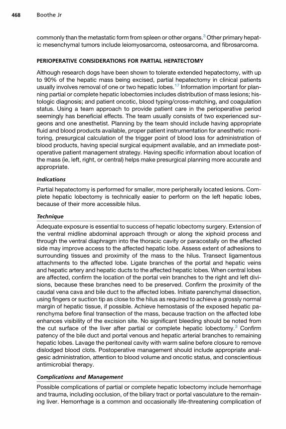
Goals of extrahepatic biliary surgery include confirmation of the underlying diseaseprocess (eg, biliary mucocele, cholecystitis, and bile duct obstruction, trauma, orleakage), establishment of a patent biliary system, and minimization of perioperativecomplications.18 Confirming the extent (partial vs complete) and the cause of biliaryobstruction in dogs and cats is challenging and frequently involves multiple diagnosticmodalities. Although information gained from serum biochemical testing results andabdominal ultrasonography is helpful in assessing the extrahepatic biliary tract,hepatobiliary scintigraphy may be needed to differentiate biliary obstruction from he-patocellular disease or damage and determine whether biliary tract dilation indicates aresolved or ongoing obstruction.19,20 Fashioning a rational plan to treat biliary obstruc-tion is best accomplished by knowing the cause, extent, and likely duration of theobstruction (Fig. 4). Information from experimental dogs suggests that delaying pri-mary surgical repair of an obstructed bile duct for at least 10 days after onset ofobstruction may be appropriate because of wound healing considerations.21
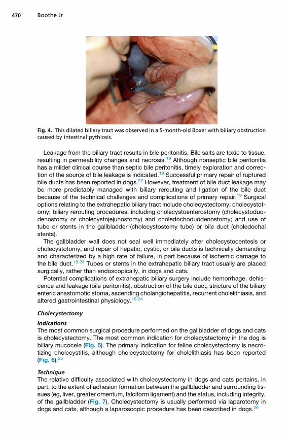
Fig. 4. This dilated biliary tract was observed in a 5-month-old Boxer with biliary obstructioncaused by intestinal pythiosis.
Boothe Jr470
Leakage from the biliary tract results in bile peritonitis. Bile salts are toxic to tissue,resulting in permeability changes and necrosis.19 Although nonseptic bile peritonitishas a milder clinical course than septic bile peritonitis, timely exploration and correc-tion of the source of bile leakage is indicated.19 Successful primary repair of rupturedbile ducts has been reported in dogs.22 However, treatment of bile duct leakage maybe more predictably managed with biliary rerouting and ligation of the bile ductbecause of the technical challenges and complications of primary repair.19 Surgicaloptions relating to the extrahepatic biliary tract include cholecystectomy; cholecystot-omy; biliary rerouting procedures, including cholecystoenterostomy (cholecystoduo-denostomy or cholecystojejunostomy) and choledochoduodenostomy; and use oftube or stents in the gallbladder (cholecystostomy tube) or bile duct (choledochalstents).The gallbladder wall does not seal well immediately after cholecystocentesis or
cholecystotomy, and repair of hepatic, cystic, or bile ducts is technically demandingand characterized by a high rate of failure, in part because of ischemic damage tothe bile duct.19,23 Tubes or stents in the extrahepatic biliary tract usually are placedsurgically, rather than endoscopically, in dogs and cats.Potential complications of extrahepatic biliary surgery include hemorrhage, dehis-
cence and leakage (bile peritonitis), obstruction of the bile duct, stricture of the biliaryenteric anastomotic stoma, ascending cholangiohepatitis, recurrent cholelithiasis, andaltered gastrointestinal physiology.19,24
Cholecystectomy
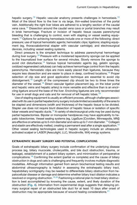
IndicationsThe most common surgical procedure performed on the gallbladder of dogs and catsis cholecystectomy. The most common indication for cholecystectomy in the dog isbiliary mucocele (Fig. 5). The primary indication for feline cholecystectomy is necro-tizing cholecystitis, although cholecystectomy for cholelithiasis has been reported(Fig. 6).25
TechniqueThe relative difficulty associated with cholecystectomy in dogs and cats pertains, inpart, to the extent of adhesion formation between the gallbladder and surrounding tis-sues (eg, liver, greater omentum, falciform ligament) and the status, including integrity,of the gallbladder (Fig. 7). Cholecystectomy is usually performed via laparotomy indogs and cats, although a laparoscopic procedure has been described in dogs.26
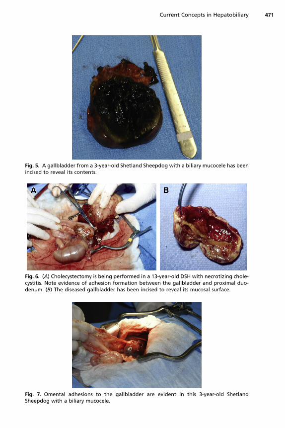
Fig. 5. A gallbladder from a 3-year-old Shetland Sheepdog with a biliary mucocele has beenincised to reveal its contents.
Fig. 6. (A) Cholecystectomy is being performed in a 13-year-old DSH with necrotizing chole-cystitis. Note evidence of adhesion formation between the gallbladder and proximal duo-denum. (B) The diseased gallbladder has been incised to reveal its mucosal surface.
Fig. 7. Omental adhesions to the gallbladder are evident in this 3-year-old ShetlandSheepdog with a biliary mucocele.
Current Concepts in Hepatobiliary 471
Boothe Jr472
Dissect the gallbladder from the hepatic fossa using blunt and sharp dissection.Achieve hemorrhage control of the hepatic fossa via local pressure, electrosurgery,and topical hemostatic agents. Ensure patency of the bile duct by passing a catheter(eg, 5F red rubber catheter) through the bile duct in a normograde (preferred tech-nique) or retrograde fashion. Retrograde catheter passage requires a duodenotomyto access the duodenal papilla. Dissection to the level of the cystic duct usually revealsthe cystic artery, which is ligated or occluded collectively with the cystic duct with anappropriately sized vascular clip. Confirm hemostasis and perform intraperitoneallavage before closure.
Cholecystotomy
IndicationsCholecystotomy occasionally may be performed to obtain full-thickness biopsies ormucosal cultures of the gallbladder, explore the gallbladder or cystic duct, removecholeliths or sludge in an otherwise normal gallbladder, or normograde flush the bileduct.19
TechniquePack off the gallbladder and incise it at its apex. Evacuate contents and flush the gall-bladder and cystic and bile ducts. Retrograde flushing of the bile duct via the duodenalpapilla helps ensure evacuation of the entire biliary tract. Close the incision with a sim-ple continuous pattern using synthetic monofilament absorbable suture material.3
Cover the incision with greater omentum.
Cholecystoenterostomy/Choledochoduodenostomy
IndicationsSurgical treatment of extrahepatic biliary obstruction or leakage may include chole-cystoenterostomy. Creating a connection between the lumen of the gallbladder andeither the duodenum or jejunum is achieved. Although reconstruction of the bileduct has been successfully performed experimentally in dogs using various materials,treatment of a clinical patient with bile duct obstruction or leakage often includes abiliary enteric anastomotic procedure in concert with ligation of the bile duct.27,28
Alternatively, primary bile duct repair or choledochotomy are potential options.
TechniqueCholecystoduodenostomy is generally accepted as the most physiologic technique toachieve biliary enteric anastomoses in dogs and cats.29 Dissect the gallbladder fromthe hepatic fossa taking care to preserve the cystic artery. Position the gallbladderadjacent to the antimesenteric aspect of the duodenum without obstructing the cysticduct, and create matching incisions in the wall of the gallbladder and the duodenum.Apposemucosal surfaces of the gallbladder and duodenum using a simple continuouspattern of 4-0 synthetic absorbable suture material or a surgical stapling device tocreate the stoma. Stoma size should be greater than or equal to 2.5 cm in length toprevent stricturing and minimize ascending cholangiohepatitis. If tension is noted dur-ing attempted mobilization of the gallbladder, a cholecystojejunostomy may be thepreferred technique. If the bile duct has dilated because of partial or completeobstruction at its duodenal termination, choledochoduodenostomy or reanastomosisof the bile duct to an unaffected portion of the duodenum may be an option.30
Choledochotomy/Choledochal or Cholecystostomy Tubes
Choledochotomy and placement of choledochal tubes has been reported in dogs andcats.22,31,32 Closure of the choledochotomy is achieved over a red rubber catheter of
Current Concepts in Hepatobiliary 473
appropriate diameter (usually 5–8F catheter) using either synthetic monofilamentabsorbable or nonabsorbable suture material. Suture used is often 3-0 or 4-0 insize, with a simple interrupted or continuous pattern. Suturing the tube to the duodenalwall may extend the duration of stenting. Removal of the stent may be achievedendoscopically.
IndicationsObstructive cholangiolithiasis or pancreatitis is the usual inciting reason for placementof choledochal tubes. Cholecystostomy tubes provide temporary diversion of bilefrom the gallbladder to a closed collection system.3 Such tubes are considered onlywhen the gallbladder wall is deemed to be healthy.3
TechniqueTube placement may be accomplished via laparotomy or laparoscopic-assisted.Insert a pigtail or Foley catheter into the apex of the gallbladder and place a purse-string suture around the base of the catheter to minimize leakage. Exit the abdomenjust caudal to the costal arch, and secure the tube to the skin using a friction suture.Attach the tube to a sterile collection system.
OUTCOMES FOR PATIENTS UNDERGOING HEPATOBILIARY SURGERY
Information regarding outcome is available for selected hepatobiliary surgical proce-dures.3,14,15,18,19,25,29,31–34 Veterinary patients undergoing either extensive liver resec-tion or correction of biliary tract obstruction or leakage tend to have an extensive list ofrisk factors associated with the primary condition and the surgical procedure. Anes-thetic management of the dog or cat with hepatobiliary dysfunction tends to be chal-lenging, in part because hepatic disease impacts many body functions. Dogsundergoing cholecystectomy were not shown to have any greater number of anes-thesia complications than were dogs that underwent other hepatic surgeries.35
Dogs or cats with hepatic mass lesions have oncologic and perioperative factorsthat influence prognosis.
SUMMARY
Surgery of the liver and extrahepatic biliary tract presents technical challenges to theveterinary clinician. Such challenges include gaining access to the lesion; dealing withhighly vascular, friable hepatic tissue; potentially difficult-to-heal tissue (eg, extrahe-patic biliary tract); and the impact of the primary condition on the patient’s responseto surgery. Enhancement of outcome and minimizing potential complications can beachieved by performing accurate preoperative patient assessment and treatment,using a team approach to the surgery and perioperative care, demonstrating flexibilityto change intraoperative plans, and following a comprehensive postoperative man-agement plan.
REFERENCES
1. Lu HW, Chen YB, Li YM, et al. Role of hepatic arterial ischaemia in biliary fibrosisfollowing liver transplantation. Chin Med J 2010;123(7):907–11.
2. Evans HE, deLahunta A. The digestive apparatus and abdomen. In: Evans HE,Alexander de Lahunta, editors. Miller’s anatomy of the dog. 4th edition. St Louis(MO): Saunders, an imprint of Elsevier; 2013. p. 327–33.
Boothe Jr474
3. Mayhew PD, Weisse C. Liver and biliary system. In: Tobias KM, Johnston SA,editors. Veterinary surgery: small animal. St Louis (MO): Saunders, an imprintof Elsevier; 2012. p. 1601–23.
4. Imagawa T, Ueno T, Tsuka T, et al. Anatomical variations of the extrahepatic ductsin dogs: knowledge for surgical procedures. J Vet Med Sci 2001;72(3):339–41.
5. Rothuizen J, Twedt DC. Liver biopsy techniques. Vet Clin North Am Small AnimPract 2009;39:469–80.
6. Bigge LA, Brown DJ, Penninck DG. Correlation between coagulation profile find-ings and bleeding complications after ultrasound-guided biopsies: 434 Cases(1993-1996). J Am Anim Hosp Assoc 2001;37:228–33.
7. Wang KY, Panciera DL, Al-Rukibat RK, et al. Accuracy of ultrasound-guided fine-needle aspiration of the liver and cytologic findings in dogs and cats: 97 cases(1990-2000). J Am Vet Med Assoc 2004;224:75–8.
8. Bahr KL, Sharkey LC, Murakami T, et al. Accuracy of US-guided FNA of focal liverlesions in dogs: 140 cases (2005-2008). J Am Anim Hosp Assoc 2013;49:190–6.
9. Johnston AN, Center SA, McDonough SP. Influence of biopsy specimen size,tissue fixation, and assay variation on copper, iron, and zinc concentrations incanine livers. Am J Vet Res 2009;70:1502–11.
10. Cole TL, Center SA, Flood SN, et al. Diagnostic comparison of needle and wedgebiopsy specimens of the liver in dogs and cats. J Am Vet Med Assoc 2002;220:1483–90.
11. Petre SL, McClaran JK, Bergman PJ, et al. Safety and efficacy of laparoscopichepatic biopsy in dogs: 80 cases (2004-2009). J Am Vet Med Assoc 2012;240:181–5.
12. Rawlings CA, Howerth EW. Obtaining quality biopsies of the liver and kidney.J Am Anim Hosp Assoc 2004;40:352–8.
13. Cuddy LC, Risselada M, Ellison GW. Clinical evaluation of a pre-tied ligating loopfor liver biopsy and liver lobectomy. J Small Anim Pract 2013;54:61–6.
14. Schwarz LA, Penninck DG, Leveille-Webster C. Hepatic abscesses in 13 dogs: areview of the ultrasonographic findings, clinical data and therapeutic options. VetRadiol Ultrasound 1998;39(4):357–65.
15. Sergeeff JS, Armstrong PJ, Bunch SE. Hepatic abscesses in cats: 14 cases(1985-2002). J Vet Intern Med 2004;18:295–300.
16. van Sprundel RG, van den Ingh TS, Guscetti F, et al. Classification of primaryhepatic tumours in the dog. Vet J 2013;197(3):596–606.
17. Steen S, Conway C, Guerra C, et al. 90% hepatectomy with a porto-hepatic shuntin a canine model: a feasibility study. ILAR J 2012;53(1):E1–8.
18. May LR, Mehler SJ. Complications of hepatic surgery in companion animals. VetClin North Am Small Anim Pract 2011;51(5):935–48.
19. Mehler SJ. Complications of the extrahepatic biliary surgery in companionanimals. Vet Clin North Am Small Anim Pract 2011;51(5):949–67.
20. Head LL, Daniel GB. Correlation between hepatobiliary scintigraphy and surgeryor postmortem examination findings in dogs and cats with extrahepatic biliaryobstruction, partial obstruction, or patency of the biliary system: 18 cases(1995-2004). J Am Vet Med Assoc 2005;227:1618–24.
21. Huang Q, Liu CH, Zhu CL, et al. The choice of surgical timing for biliary ductreconstruction after obstructive bile duct injury: an experimental study. Hepato-gastroenterology 2013;60(128):1865–72.
22. Baker SG, Mayhew PD, Mehler SJ. Choledochotomy and primary repair of extra-hepatic biliary duct rupture in seven dogs and two cats. J Small Anim Pract 2011;52:32–7.
Current Concepts in Hepatobiliary 475
23. Geng L, Luo D, Zhang HC, et al. Microvessel density at different levels of normalor injured bile duct in dogs and its surgical implications. Hepatobiliary PancreatDis Int 2011;10:83–7.
24. Sato M, Shibata C, Kikuchi D, et al. Effects of biliary and pancreatic juice diver-sion into the ileum on gastrointestinal motility and gut hormone secretion inconscious dogs. Surgery 2010;148:1012–9.
25. Eich CS, Ludwig LL. The surgical treatment of cholelithiasis in cats: a study ofnine cases. J Am Anim Hosp Assoc 2002;38:290–6.
26. Mayhew PD. Advanced laparoscopic procedures (hepatobiliary, endocrine) indogs and cats. Vet Clin North Am Small Anim Pract 2009;39(5):925–39.
27. Nau P, Liu J, Ellison EC, et al. Novel reconstruction of the extrahepatic biliary treewith a biosynthetic absorbable graft. HPB (Oxford) 2011;13:573–8.
28. Shi J, Lv Y, Yu L, et al. Interest of a new biodegradable stent coated with pacli-taxel on anastomotic wound healing after biliary reconstruction. Eur J Gastroen-terol Hepatol 2013;25(12):1415–23.
29. Morrison S, Prostredny J, Roa D. Retrospective study of 28 cases of cholecysto-duodenostomy performed using endoscopic gastrointestinal anastomosis sta-pling equipment. J Am Anim Hosp Assoc 2008;44(1):10–8.
30. Breznock EM. Surgical procedures of the hepatobiliary system. In: Bojrab MJ,editor. Current techniques in small animal surgery. 4th edition. Baltimore (MD):Williams & Wilkins; 1998. p. 298–308.
31. Mayhew PD, Richardson RW, Mehler SJ, et al. Choledochal tube stenting fordecompression of the extrahepatic portion of the biliary tract in dogs: 13 cases(2002-2005). J Am Vet Med Assoc 2006;228(8):1209–14.
32. Son TT, Thompson L, Serrano S, et al. Surgical intervention in the management ofsevere acute pancreatitis in cats: 8 cases (2003-2007). J Vet Emerg Crit Care2010;20(4):426–35.
33. Papazoglou L, Mann FA, Wagner-Mann C. Long-term survival of dogs after chol-ecystoenterostomy: a retrospective study of 15 cases (1981-2005). J Am AnimHosp Assoc 2008;44:67–74.
34. Center SA. Disease of the gallbladder and biliary tree. Vet Clin North Am SmallAnim Pract 2009;39(3):543–98.
35. Burns BR, Hofmeister EH, Brainard BM. Anesthetic complications in dogs under-going hepatic surgery: cholecystectomy versus non-cholecystectomy. VetAnaesth Analg 2014;41:186–90.


![Original Article Application of three dimensional ... · surgery, urology, general surgery (hepatobiliary and gastrointestinal), obstetrics and gynecolo-gy [4], and thoracic surgery](https://img.dokumen.tips/doc/110x75/5f088bac7e708231d4228af6/original-article-application-of-three-dimensional-surgery-urology-general.jpg)