1
April 2000 AGAA751 4052 CARDITIS: ALL H.PYLORI OR AN ASSOCIATION WITH GERD? Deirdre A. McNamara, Sean P. Montague, Paul M. Crotty, Martin M. Buckley, Colm A. O'Morain, Amnch, Dublin, Ireland. Carditis has been associated with H.pylori infection and pan-gastritis. The role of GERD in the aetiology of cardia inflammation is debated. Gastric adenocarcinoma is increasing in prevalence, and the role of cardia intes- tinal metaplasia is unknown. Aim:To determine the prevalence of carditis and cardia I.M. and their disease associations. Methods:Consecutive pa- tients undergoing routine endoscopy in a single university center were recruited. The endoscopic diagnosis was recorded. Biopsies were obtained in all cases as follows. Two each from the antrum, corpus, incisura, cardia and oesophagus (2cm above the squamocolumnar junction) for histology (Sydney classification) and two further antral biopsies for a rapid urease test and culture. Patients were considered H.p positive if any two tests were positive or if culture was positive. Exclusion criteria included, recent consumption of antibiotics or use of antacids or NSAID's, previous gastric or oesophageal surgery. Results:One hundred and fifty four patients were recruited, 61 (40%) were H.p positive and 51 (33%) had endoscopic oesophagitis. The prevalence of carditis and cardia I.M. was 91 (59%) and 15 (lO%)respectively. H.p infection was significantly associated with car- ditis p<0.OO2. GERO alone accounted for 20% of cases with carditis. Of 15 cases of I.M. 3(20%)had H.p infection and 5 (33%) Barrett's disease. Seven cases of cardia 1M had GERO without either endoscoscopic or histological evidence of Barrett's disease and were H.p negative. In all seven the 1M was of immature subtype. Conclusion: Carditis occurs with H.p infection and GERO. I.M of the cardia is uncommon, 10% of cases. In the presence of multi focal atrophic gastritis and H.p infection LM may be found in the cardia in conjunction with other distal sites. Immature type cardia 1M is associated with Barrett's and GERD. Immature I.M in patients with GERD without Barrett's suggests that GERD is primarily responsible for cardia damage in these patients. Carditis and disease associations H.p n=61 GERD n=51 Barretts n··24 Total n=91 4054 CURE OF HEUCOBACTER PYLORI INFECTION IMPROVES GASTRIC ATROPHY WITHOUT REGRESSION OF INTESTINAL METAPLASIA. Ryugo Sato, Toshio Fujioka, Kazunari Murakami, Masaaki Kodama, Toshihiro Kubota, Masaru Nasu, Oita Med Univ, Oita, Japan. Background and Aims: It is well known that Hellicobacter pylori (Hp) plays a major role in the development of atrophic gastritis and intestinal metaplasia (1M) which were associated with gastric carcinogenesis. The aim of this study was to evaluate the histological change of gastric atrophy and 1M after Hp eradication by 5 sites biopsy recommended in the updated Sydney system (USS). Subjects and Methods: Twenty patients with Hp- associated gastritis (12 men, mean age 44± 13 years) were subject. Endo- scopic biopsies were performed before and at 6, 12 and 24 months after successful eradication. Biopsies for histological evaluation were taken with large forceps from the following 5 sites: the lesser curvatures of the antrum (AI) and corpus (B 1), the greater curvatures of the antrum (A2) and corpus (B2), and the incisula angularis (IA). The degree of gastric atrophy was scored from 0 to 3 according to the USS (0: normal, 1: mild, 2: moderate, 3: marked). Results expressed as mean±SD in Table Conclusions: Gastric atrophy was improved histologically at Bland IA after Hp eradication, although 1M was not at any site and period. Reb =t .- '1- 1<1_ - ... , lAt1ol) , 1:,.0 " 1011:1.2 O... At. 008100' Ute.t II MtOJ '1:001 III tol1:101 "'1:1.0- ' ,hl,oo ' ... 1:'.0- .. ...... AI a.tel.1 DoaeLl I.181Jr 1.1 aU M fI.-. Il.IeA7 I' CUea.t ClMU a.tlllllS a aulLO o.1aA1 QJIIIlZ Q.QaU " a.te1.1 IlAaU 1 ...1.1 DoaeU poUI <w-----:l Carditis Cardia I.M 47=51% 3 36=40% 5 17=19% 7 91=59% 15=10% 4053 DOES HELICOBACTER PYLORI ERADICATION MODIFY IN· TESTINAL METAPLASIA EVOLUTION IN THE STOMACH? Mauro G. Ravizza, Renzo Suriani, Fulvio Cappelletti, Pietro Dusio, Hosp Evangelico Valdese TORINO, Torino, Italy; Hosp degli Infirmary, Rivoli, Italy; Hosp Evangelico Valdese, Torino, Italy. Purpose of the study: To evaluate gastric intestinal metaplasia(lM) further evolution in Helicobacter Pylori (HP) positive patients after eradication in a long term follow-up. Subjects and methods: We studied for four years after bacterial eradication 69 HP positive patients affected from ulcerative diseases of the stomach or duodenum, showing antral intestinal metaplasia at histology. Eradication was obtained by OAC short-term therapy and was assessed both performing 13C-UBT one month after eradication and by Giemsa stain in six histological specimen obtained from gastric antrum at least 2 em. away from pylorus during endoscopy. The endoscopy was repeated every year in each patient involved in the study, in order to evaluate 1M evolution and HP eradication persistence. During the entire follow-up histological specimens were examinated from the same pathol- ogist and 1M differentiated as complete (typeI), incomplete (type II) or incomplete solphomucin-positive (type III). Statistical analysis between different groups after each year was performed by x-square method. Results: All patients eradicated at time 0, persisted HP negative during further follow-up. At time 0, 48 cases out of 69 (69,7%) presented type I 1M, 14 cases (20,2%) type II 1M and only 7 cases (10%) type III 1M. 1M presence in histological specimens after one, two, three, four years from eradication are summarized in Table I. Statistical analysis between the three 1M groups after each control showed a significant regression after HP eradication in type I versus type III 1M (p<O.OOI), but no significance in type I versus type II 1M (p=0.284) neither in type II versus type III 1M (p=0.981). Conclusions: After a long period follow-up, HP eradication seems able to affect 1M in gastric antrum by permitting its regression. Otherwise incomplete 1M in gastric antrum. both type II and III, isn't modified significantly after bacterial eradication. 4055 P53·POSITIVE CELLS IN THE GASTRIC MUCOSA BEFORE AND AFTER HEUCOBACTER PYLORI ERADICATION. Kiichi Satoh, Ken Kihira, Hiroshi Kawata, Keiko Fukazawa, Satoshi Kawakami, Yasuhisa Kumakura, Kenkichi Tokumaru, Yumiko Ishino, Toshichika Kojima, Kentaro Sugano, Japan. It has been reported that accumulation of p53 protein in the gastric mucosa decreased 3 months after eradication of Helicobacter pylori (Am J Clin Pathol 1997;108:26-34). We investigated the prevalence of p53-positive cells in the gastric mucosa before and one month after eradication of H. pylori and the relationship between p53 positivity and inflammation and cell proliferation. Methods: The subjects included 24 H. pylori-positive patients. They achieved eradication one month after anti-H. pylori therapy. Biopsies were taken from the greater curvatures of the antrum and middle body. H. pylori status was assessed using culture and tissue section (Gi- emsa stain). Alternate sections were used for examination of gastritis (HE stain) and for immunohistochemistry. Immunostaining of p53 was per- formed using 00-7 antibody (DBS, Pleasanton, CAl, and that of Ki-67 using Mm-I antibody (Immunotech, Marseille, France). Sections were stained by the avidin-biotin method with microwave retrieval. p53 index and Ki-67lavelling index (LI) were calculated by counting DO-7-positive and MIB-l-positive cells in the entire gastric pits longitudinally sectioned and expressing them as a percentage ratio of the total cells in a gastric pit. In the neck regions with and without p53-positive cells, inflammatory cells were counted in the corresponding area (50X50/Lm2) of the sections stained with HE. Results: Table shows the mean (standard error (SE) of p53 index and Ki-67 LI. There was no significant correlation between p53 index and Ki-67 LI before eradication. The number of polymorphonuclear cells (PMNs) was significantly higher in the neck regions with p53-positive cells than in those without (1.9 ± 0.6 vs. 0.4 ± 0.2 in the antrum, and 3.9 ± 1.3 vs. 0.4 ± 0.2 in the body; (mean ± SE». Conclusions: In the gastric mucosa with H. pylori infection, p53-positive cells were found in the neck region heavily infiltrated with PMNs. p53-positive cells disappeared one month after eradication of H. pylori. **p<O.01 ,*p<0.05 versus before eradication (one sample Wilcoxon test). mean (standard error (SEll of p53index and Ki·67 LJ before andafter eradication TABLE 1: 1M POSITIVE PATIENTS AT TIME 0 AND DURING FOLLOW·UP 1M Time a After 1 year After 2 years After3 years After 4 years % % % % % I 48 100 23 48 19 39.5 18 37.5 18 37.5 II 14 100 10 714 10 71.4 8 571 8 57.1 III 7 100 7 100 7 100 6 857 6 857 p53 index (%J Before After Before Antrum 1.1 (0.2) 0.6(0.1)" 38(3) Body 0.9(0.2) 0.2(0.1)'* 40(4) Ki·67 U(%J Aftereradication 29(3)* 23(3)**

Cure of Helicobacter pylori infection improves gastric atrophy without regression of intestinal metaplasia

  • Upload
    masaru

  • View
    214

  • Download
    0

Embed Size (px)

Citation preview

April 2000 AGAA751

4052

CARDITIS: ALL H.PYLORI OR AN ASSOCIATION WITH GERD?Deirdre A. McNamara, Sean P. Montague, Paul M. Crotty, Martin M.Buckley, Colm A. O'Morain, Amnch, Dublin, Ireland.

Carditis has been associated with H.pylori infection and pan-gastritis. Therole of GERD in the aetiology of cardia inflammation is debated. Gastricadenocarcinoma is increasing in prevalence, and the role of cardia intes­tinal metaplasia is unknown. Aim:To determine the prevalence of carditisand cardia I.M. and their disease associations. Methods:Consecutive pa­tients undergoing routine endoscopy in a single university center wererecruited. The endoscopic diagnosis was recorded. Biopsies were obtainedin all cases as follows. Two each from the antrum, corpus, incisura, cardiaand oesophagus (2cm above the squamocolumnar junction) for histology(Sydney classification) and two further antral biopsies for a rapid ureasetest and culture. Patients were considered H.p positive if any two tests werepositive or if culture was positive. Exclusion criteria included, recentconsumption of antibiotics or use of antacids or NSAID's, previous gastricor oesophageal surgery. Results:One hundred and fifty four patients wererecruited, 61 (40%) were H.p positive and 51 (33%) had endoscopicoesophagitis. The prevalence of carditis and cardia I.M. was 91 (59%) and15 (lO%)respectively. H.p infection was significantly associated with car­ditis p<0.OO2. GERO alone accounted for 20% of cases with carditis. Of15 cases of I.M. 3(20%)had H.p infection and 5 (33%) Barrett's disease.Seven cases of cardia 1M had GERO without either endoscoscopic orhistological evidence of Barrett's disease and were H.p negative. In allseven the 1M was of immature subtype. Conclusion: Carditis occurs withH.p infection and GERO. I.M of the cardia is uncommon, 10% of cases. Inthe presence of multi focal atrophic gastritis and H.p infection LM may befound in the cardia in conjunction with other distal sites. Immature typecardia 1M is associated with Barrett's and GERD. Immature I.M in patientswith GERD without Barrett's suggests that GERD is primarily responsiblefor cardia damage in these patients.

Carditis and disease associations

H.p n=61 GERD n=51 Barretts n··24 Total n=91

4054

CURE OF HEUCOBACTER PYLORI INFECTION IMPROVESGASTRIC ATROPHY WITHOUT REGRESSION OF INTESTINALMETAPLASIA.Ryugo Sato, Toshio Fujioka, Kazunari Murakami, Masaaki Kodama,Toshihiro Kubota, Masaru Nasu, Oita Med Univ, Oita, Japan.

Background and Aims: It is well known that Hellicobacter pylori (Hp)plays a major role in the development of atrophic gastritis and intestinalmetaplasia (1M) which were associated with gastric carcinogenesis. Theaim of this study was to evaluate the histological change of gastric atrophyand 1Mafter Hp eradication by 5 sites biopsy recommended in the updatedSydney system (USS). Subjects and Methods: Twenty patients with Hp­associated gastritis (12 men, mean age 44± 13 years) were subject. Endo­scopic biopsies were performed before and at 6, 12 and 24 months aftersuccessful eradication. Biopsies for histological evaluation were taken withlarge forceps from the following 5 sites: the lesser curvatures of the antrum(AI) and corpus (B1), the greater curvatures of the antrum (A2) and corpus(B2), and the incisula angularis (IA). The degree of gastric atrophy wasscored from 0 to 3 according to the USS (0: normal, 1: mild, 2: moderate,3: marked). Results expressed as mean±SD in Table Conclusions: Gastricatrophy was improved histologically at Bland IA after Hp eradication,although 1M was not at any site and period.

Reb =t .- '1- 1<1_-..., lAt1ol) L4~ '~L', 1:,.0

" 1011:1.2 ...,~ o.l~ O...t~At. 008100' .,~ M~ Ute.tII MtOJ ~ ~ '1:001III tol1:101 "'1:1.0- ' ,hl,oo ' ...1:'.0-........AI a.tel.1 DoaeLl I.181Jr 1.1aUM a.te~ fI.-. a.t~ Il.IeA7I' CUea.t ClMU a.tlllllS ~a aulLO o.1aA1 QJIIIlZ Q.QaU

" a.te1.1 IlAaU 1...1.1 DoaeU

• poUI <w-----:lCarditisCardia I.M

47=51%3

36=40%5

17=19%7

91=59%15=10%

4053

DOES HELICOBACTER PYLORI ERADICATION MODIFY IN·TESTINAL METAPLASIA EVOLUTION IN THE STOMACH?Mauro G. Ravizza, Renzo Suriani, Fulvio Cappelletti, Pietro Dusio, HospEvangelico Valdese TORINO, Torino, Italy; Hosp degli Infirmary, Rivoli,Italy; Hosp Evangelico Valdese, Torino, Italy.

Purpose of the study: To evaluate gastric intestinal metaplasia(lM) furtherevolution in Helicobacter Pylori (HP) positive patients after eradication ina long term follow-up. Subjects and methods: We studied for four yearsafter bacterial eradication 69 HP positive patients affected from ulcerativediseases of the stomach or duodenum, showing antral intestinal metaplasiaat histology. Eradication was obtained by OAC short-term therapy and wasassessed both performing 13C-UBT one month after eradication and byGiemsa stain in six histological specimen obtained from gastric antrum atleast 2 em. away from pylorus during endoscopy. The endoscopy wasrepeated every year in each patient involved in the study, in order toevaluate 1M evolution and HP eradication persistence. During the entirefollow-up histological specimens were examinated from the same pathol­ogist and 1M differentiated as complete (typeI), incomplete (type II) orincomplete solphomucin-positive (type III). Statistical analysis betweendifferent groups after each year was performed by x-square method.Results: All patients eradicated at time 0, persisted HP negative duringfurther follow-up. At time 0, 48 cases out of 69 (69,7%) presented type I1M, 14 cases (20,2%) type II 1M and only 7 cases (10%) type III 1M. 1Mpresence in histological specimens after one, two, three, four years fromeradication are summarized in Table I. Statistical analysis between thethree 1Mgroups after each control showed a significant regression after HPeradication in type I versus type III 1M (p<O.OOI),but no significance intype I versus type II 1M (p=0.284) neither in type II versus type III 1M(p=0.981). Conclusions: After a long period follow-up, HP eradicationseems able to affect 1M in gastric antrum by permitting its regression.Otherwise incomplete 1M in gastric antrum. both type II and III, isn'tmodified significantly after bacterial eradication.

4055

P53·POSITIVE CELLS IN THE GASTRIC MUCOSA BEFOREAND AFTER HEUCOBACTER PYLORI ERADICATION.Kiichi Satoh, Ken Kihira, Hiroshi Kawata, Keiko Fukazawa, SatoshiKawakami, Yasuhisa Kumakura, Kenkichi Tokumaru, Yumiko Ishino,Toshichika Kojima, Kentaro Sugano, Japan.

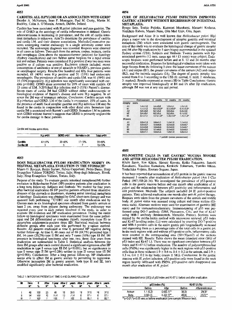
It has been reported that accumulation of p53 protein in the gastric mucosadecreased 3 months after eradication of Helicobacter pylori (Am J ClinPathol 1997;108:26-34). We investigated the prevalence of p53-positivecells in the gastric mucosa before and one month after eradication of H.pylori and the relationship between p53 positivity and inflammation andcell proliferation. Methods: The subjects included 24 H. pylori-positivepatients. They achieved eradication one month after anti-H. pylori therapy.Biopsies were taken from the greater curvatures of the antrum and middlebody. H. pylori status was assessed using culture and tissue section (Gi­emsa stain). Alternate sections were used for examination of gastritis (HEstain) and for immunohistochemistry. Immunostaining of p53 was per­formed using 00-7 antibody (DBS, Pleasanton, CAl, and that of Ki-67using Mm-I antibody (Immunotech, Marseille, France). Sections werestained by the avidin-biotin method with microwave retrieval. p53 indexand Ki-67lavelling index (LI) were calculated by counting DO-7-positiveand MIB-l-positive cells in the entire gastric pits longitudinally sectionedand expressing them as a percentage ratio of the total cells in a gastric pit.In the neck regions with and without p53-positive cells, inflammatory cellswere counted in the corresponding area (50X50/Lm2) of the sectionsstained with HE. Results: Table shows the mean (standard error (SE) ofp53 index and Ki-67 LI. There was no significant correlation between p53index and Ki-67 LI before eradication. The number of polymorphonuclearcells (PMNs) was significantly higher in the neck regions with p53-positivecells than in those without (1.9 ± 0.6 vs. 0.4 ± 0.2 in the antrum, and 3.9 ±1.3 vs. 0.4 ± 0.2 in the body; (mean ± SE». Conclusions: In the gastricmucosa with H. pylori infection, p53-positive cells were found in the neckregion heavily infiltrated with PMNs. p53-positive cells disappeared onemonth after eradication of H. pylori.

**p<O.01 ,*p<0.05 versus before eradication (one sample Wilcoxon test).

mean (standard error (SEll ofp53index and Ki·67 LJ before andafter eradicationTABLE 1:1M POSITIVE PATIENTS AT TIME 0 AND DURING FOLLOW·UP

1M Time a After 1year After 2 years After3 years After4 yearsN° % N° % N° % N° % N° %

I 48 100 23 48 19 39.5 18 37.5 18 37.5II 14 100 10 714 10 71.4 8 571 8 57.1III 7 100 7 100 7 100 6 857 6 857

p53 index (%JBefore After Before

Antrum 1.1 (0.2) 0.6(0.1)" 38(3)Body 0.9(0.2) 0.2(0.1)'* 40(4)

Ki·67 U(%JAftereradication

29(3)*23(3)**