Embed Size (px)
Citation preview
All rights reserved. This book or any parts thereof may not be reproduced in any form without written permission.
1
The following autopsy report is fictional. Each section is written as an actually autopsy report is written and therefore may resemble an existing autopsy report.
AUTOPSY REPORT NAME: Jill Simms
AGE: 31Y AUTOPSY D/T: SEX: F ID
PATH MD: Dale Dolitte
All rights reserved. This book or any parts thereof may not be reproduced in any form without written permission.
2
FINAL DIAGNOSIS: I. Blunt Force Trama II. Cranial Cerebral Injuries
A. Multiple cranial penetrations to the back of the head concentrated on the right rear lobe
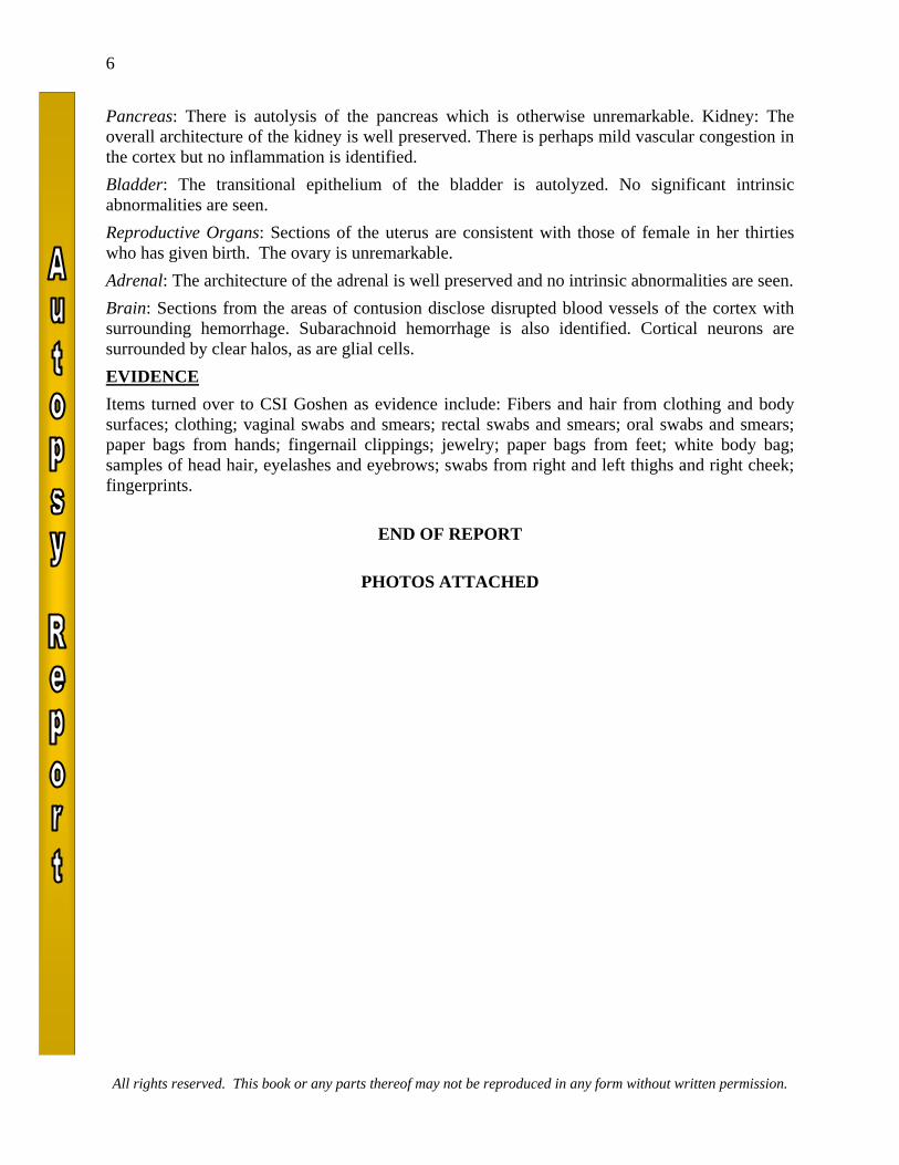
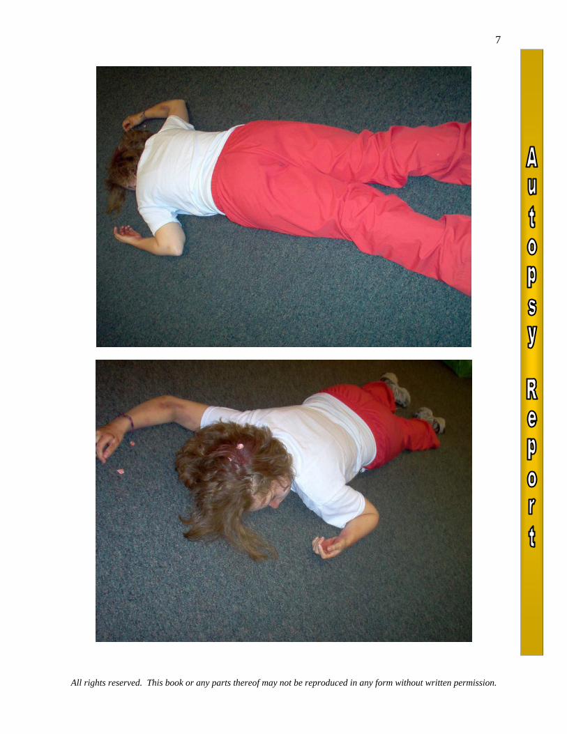
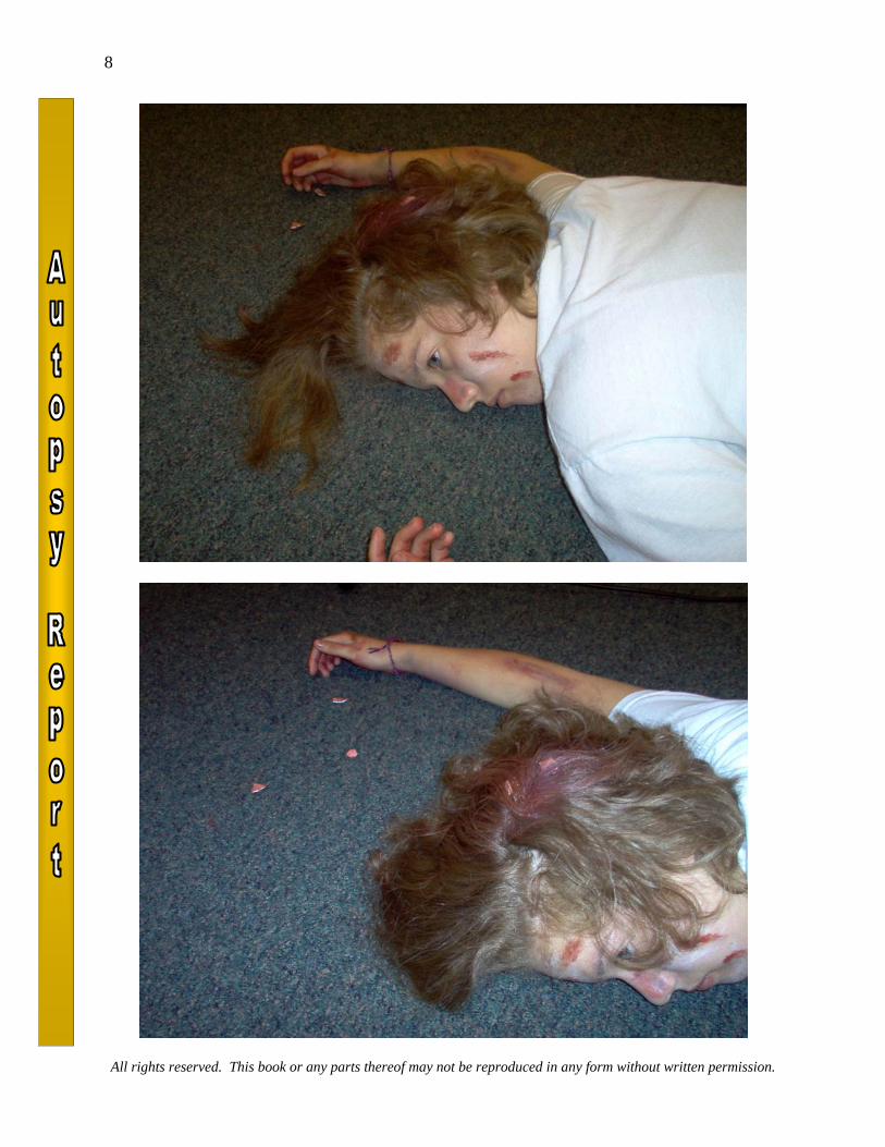
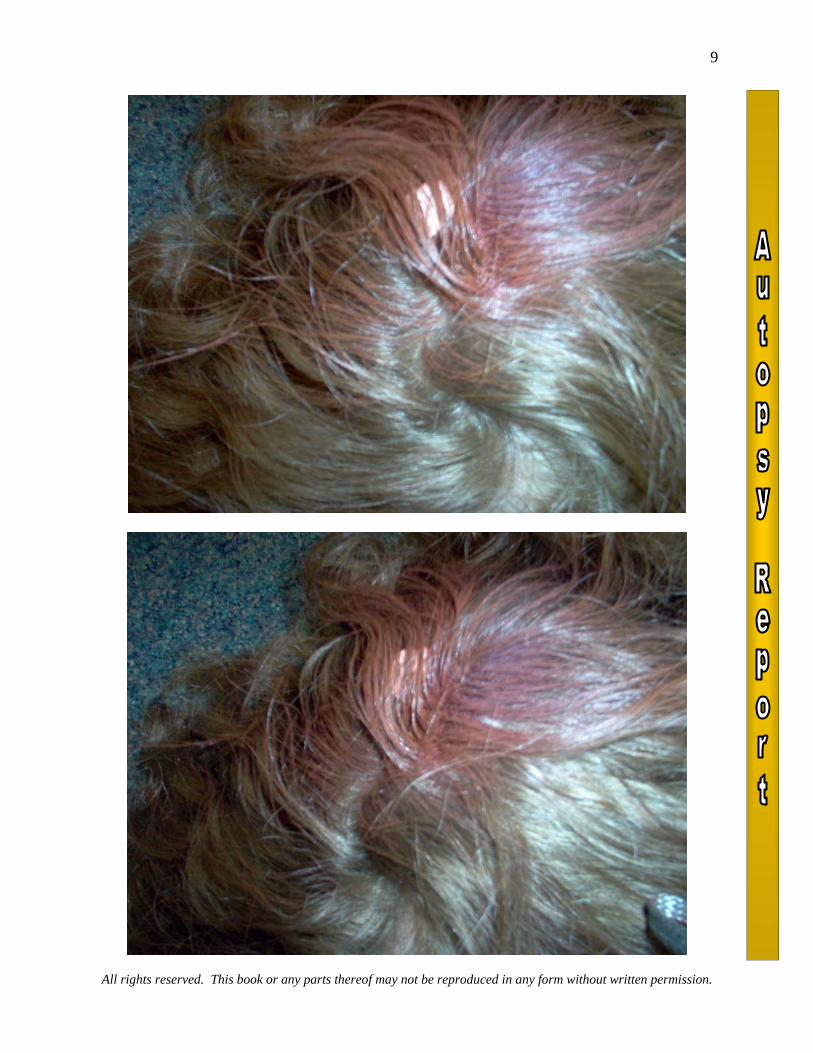
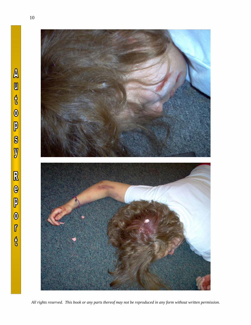
C. Linear fracture on right side of the skull III. Abrasions of Left Cheek IV. Abrasion on Right Fore Arm TOXICOLOGIC STUDIES blood ethanol - 0.06 blood drug screen - no drugs detected CLINICOPATHOLOGIC CORRELATION Cause of death of this thirty-one year old female is blunt force trauma to the cranial The body of this thirty-one year old female was first seen by me after I was called to North Oldham High School in Goshen Kentucky. I arrived approximately 11 a.m. and entered the class room where the decedent's body was located. I viewed the body on the floor of the room. The decedent was lying on her stomach between two tables covered by a sheet. On removing the sheet from the body, the decedent was found to be lying on her stomach with her arms extended out to the sides. The head was turned to the left. A quick examination of the body revealed several scars on her left check and right arm. In addition there was extensive damage to the top and back of the head. Piece of scalp and skull were spray around the area were the body lay. There was also much blood splatter around the room. EXTERNAL EXAM Deceased is clothed in a short sleeved white knit shirt and a nice pair of black slacks. There are no defects noted in the slacks but the upper anterior left sleeve of the shirt contains a dried brown-tan stain measuring 2 x 1 inches. These red areas of staining, measure 0.5 inch in maximum dimension. EXTERNAL EVIDENCE OF INJURY Located just below the left ear at a right angle of the mandible, below the right external auditory canal is a 1 inch x 1 inch area of rust colored abrasion. The remaining left side of the face has a large rounded break and bruising. The top of the skull has a star shaped break with some pieces of skull and scalp missing. This same pattern is present on the back right side of the skull. Livor mortis on this side of the face makes identification difficult. Located on the left side of the chin is a three-sixteenths by one-eighth of an inch area of superficial abrasion. On the posterior aspect of the left shoulder is a poorly demarcated, very
All rights reserved. This book or any parts thereof may not be reproduced in any form without written permission.
3
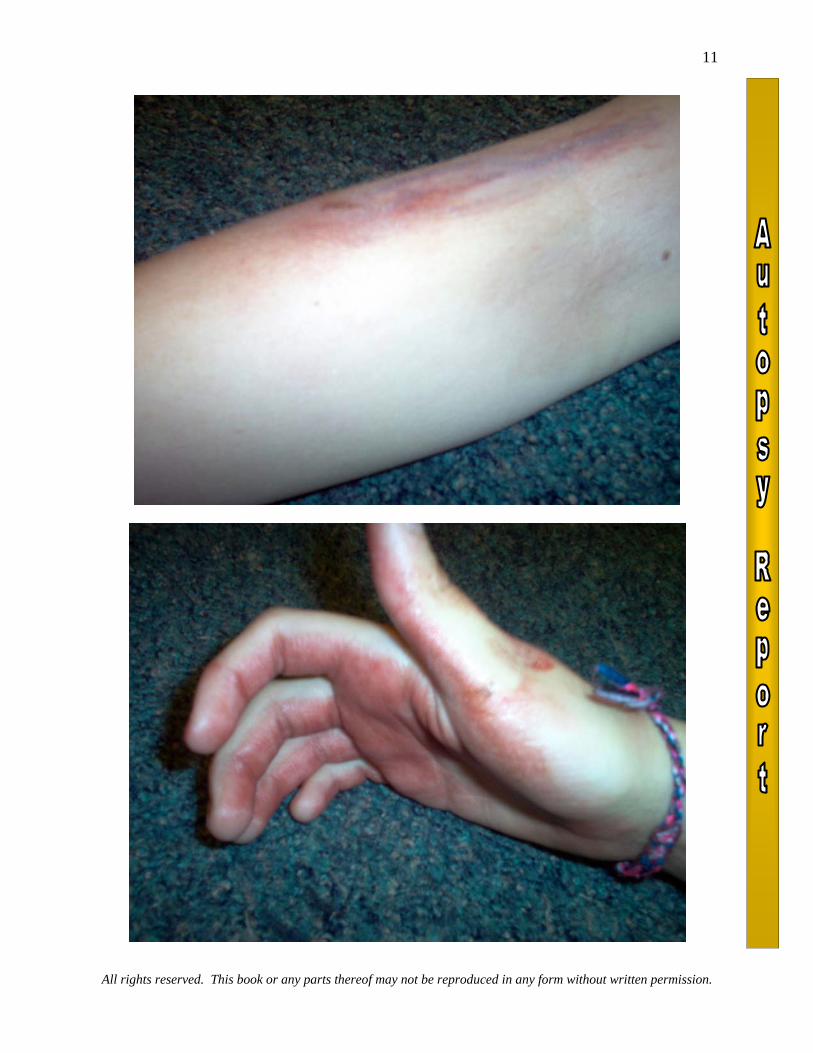
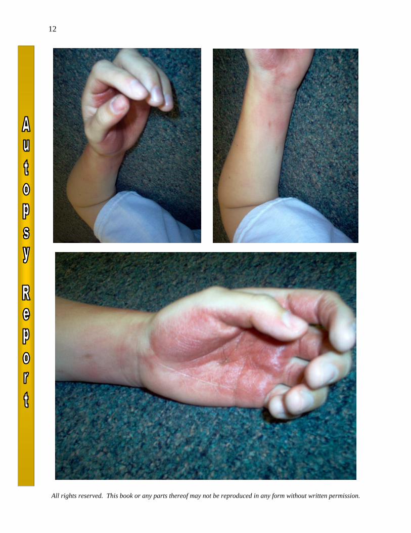
superficial focus of abrasion/contusion which is pale purple in color and measures up to three-quarters by one-half inch in maximum dimension. Several linear aggregates are present on the right forearm between the wrist and elbow. These measure up to one inch in length by one-sixteenth to one-eighth of an inch in width. On the right lateral aspect of the lower back, approximately sixteen and one-quarter inches and seventeen and one-half inches below the level of the top of the head are two dried purple abrasions. The more superior of the two measures one-eighth by one-sixteenth of an inch and the more inferior measures three-sixteenths by one-eighth of an inch. There is smeared blood on the palm side of the right hand but no abrasions with-in the hand its self. REMAINDER OF EXTERNAL EXAMINATION The un-embalmed, well developed nourished Caucasian female body measures 70 inches in length and weighs an estimated 140 pounds. The scalp is covered by long brunette hair which is fixed in burettes, on top of the head. The external auditory canals are patent and free of blood. The eyes are blue and the pupils are dilated. The sclera is red-lined with the left eye containing a significant amount of blood. The nostrils are both patent and contain a small amount of tan mucous material. The teeth are native and in good repair. The tongue is smooth, pink-tan and granular. No buccal mucosal trauma is seen. The frenulum is intact. There is a slight drying artifact of the tip of the tongue. On the left cheek is a pattern of dried saliva and mucous material which does not appear to be hemorrhagic. The neck contains no palpable adenopathy or masses and the trachea and larynx are midline. The chest is symmetrical. Breasts are firm and appear to be implants. The abdomen is flat and contains no scars. No palpable organ omegaly or masses are identified. The external genitalia are unremarkable. Only small amounts of pubic hair are present. The anus is patent. Examination of the extremities is unremarkable. On the ring finger of the right hand is a yellow metal band with a large (approximately 3 karat) diamond. Around the right wrist is a yellow metal bracelet. The fingernails of both hands are painted red and are of sufficient length for clipping. Examination of the back is unremarkable. There is dorsal 2+ to 3+ livor mortis which is non-blanching. Livor mortis is also present on the right side of the face. At the time of the initiation of the autopsy there is mild 2 to 2+rigor mortis of the elbows and shoulders with more advanced 3 to 4+ rigor mortis of the joints of the lower extremities. INTERNAL EXAM The anterior chest musculature is well developed. No sternal or rib fractures are identified. Mediastinum: The mediastinal contents are normally distributed. The 29 gm thymus gland has a normal external appearance. The cut sections are finely lobular and pink-tan. No petechial hemorrhages are seen. The aorta and remainder of the mediastinal structures are unremarkable. Body Cavities: The right and left thoracic cavities contain approximately 5 cc of straw colored fluid. The pleural surfaces are smooth and glistening. The percardial sac contains 3-4 cc of straw colored fluid and the epicardium and pericardium are unremarkable. The abdominal contents are normally distributed and covered by a smooth glistening serosa. No intraabdominal accumulation of fluid or blood is seen. Lungs: The 500 gm right lung and 465 gm left lung have a normal lobar configuration. The intrapulmonary bronchi and vasculature are unremarkable. No evidence of consolidation is seen.
All rights reserved. This book or any parts thereof may not be reproduced in any form without written permission.
4
Heart: The 300 gm heart has a normal external configuration. There are scattered subepicardial petechial hemorrhages over the anterior surface of the heart. The coronary arteries are normal in their distribution and contain no evidence of atherosclerosis. The tan-pink myocardium is homogeneous and contains no areas of fibrosisor infarction. The endocardium is unremarkable. The valve cusps are thin, delicate and pliable and contain no vegetation or thrombosis. The major vessels enter and leave the heart in the normal fashion. The foramen ovaleis closed. Aorta and Vena Cava: The aorta is patent throughout its course as are its major branches. No atherosclerosisis seen. The vena cava is unremarkable. Spleen: The 140 gm spleen has a finely wrinkled purple capsule. Cut sections are homogeneous and disclose readily identifiable red and white pulp. No intrinsic abnormalities are identified. Adrenals: The adrenal glands are of normal size and shape. A golden yellow cortex surmounts a thin brown-tan medullary area. No intrinsic abnormalities are identified. Kidneys: The 60 gm right kidney and 59 gm left kidney have a normal external appearance. The surfaces are smooth and glistening. Cut sections disclose intact corticomedullary architecture. The renal papillae are sharply demarcated. The pelvocaliceal system is lined by gray-white mucosa which is unremarkable. Both uretersare patent throughout their course to the bladder. Liver: The 980 gm liver has a consolidated external appearance. The capsule is rough and exhibits much consolidation. Cut sections disclose an intact lobular architecture with what appears to be evidence of extensive alcohol abuse. This is classical first stage liver disease. Pancreas: The pancreas is of normal size and shape. Cut sections are finely lobular and tan. No intrinsic abnormalities are identified. Bladder: The bladder is contracted and contains no urine. The bladder mucosa is smooth and tan-gray. No intrinsic abnormalities are seen. Genitalia: The upper portions of the vaginal vault contain no abnormalities. The uterus measures 9 X 5 X 4 cm and is unremarkable. The cervical contains no abnormalities. Both fallopian tubes and ovaries are unremarkable by gross examination. Gallbladder: The gallbladder contains 8-10 cc of amber bile. No stones are identified and the mucosa is smooth and velvety. The cystic duct, right and left hepatic duct and common bile duct are patent throughout their course to the duodenum. G.I. Tract: The esophagus is empty. It is lined by gray-white mucosa. The stomach contains a small amount (15-20 cc) of viscous to green to tan colored thick mucous material without particulate matter identified. The gastric mucosa is autolyzed but contains no areas of hemorrhage or ulceration. The proximal portion of the small intestine contains fragmented pieces of yellow to light green-tan apparent vegetable or fruit material which may represent fragments of pineapple. No hemorrhage is identified. The remainder of the small intestine is unremarkable. The large intestine contains soft green fecal material. The appendix is absent. Lymphatic System: Unremarkable Musculoskeletal System: Unremarkable Skull and Brain: Located just below the left ear at a right angle of the mandible, below the right external auditory canal is a 1 inch x 1 inch area of rust colored abrasion. Examination shows a few radial fractures. The remaining left side of the face has a large rounded break and bruising with multiple radial fractures into the skull. The top of the skull has a star shaped break with
All rights reserved. This book or any parts thereof may not be reproduced in any form without written permission.
5
some pieces of skull and scalp missing. Upon close examination the pieces collected at the scene match and complete the skull. This same pattern is present on the back right side of the skull. Located on the left side of the chin is a three-sixteenths by one-eighth of an inch area of superficial abrasion. This area exhibits the same pattern of radial fractures to the bones. On removal of the skull cap there is found to be a thin film of subdural hemorrhage measuring approximately 8-10 cc over the surface of the left cerebral hemisphere and extending around to the back of the cerebral hemisphere. The 2510 gm brain has a normal overall architecture. Mild narrowing of the sulci and flattening of the gyri are seen. Inflammation is present consistant with head trauma. There is a thick film of subarachnoid hemorrhage overlying the entire left cerebral hemisphere. On the left cerebral hemisphere underlying the previously mentioned linear skull fracture is an extensive linear area of purple contusion extending from the left frontal area, posteriorly along the lateral aspect of the parietal region and into the occipital area. This area of contusion measures 20 inches in length with a width of up to 3.75 inches. At the tip of the left temporal lobe is a one-quarter by one-quarter inch similar appearing purple contusion. Only very minimal contusion is present at the tip of the right temporal lobe. This area of contusion measures only one-half inch in maximum dimension. The cerebral vasculature contains no evidence of atherosclerosis. Multiple coronal sections of the cerebral hemispheres, brain stem and cerebellum disclose no additional abnormalities. The areas of previously described contusion are characterized by purple linear streak-like discolorations of the gray matter perpendicular to the surface of the cerebral cortex. These extend approximately 10 mm into the cerebral cortex. Examination of the base of the brain discloses no additional fractures. Neck: Dissection of the neck is performed after removal of the thoracoabdominal organs and the brain. The anterior strap musculature of the neck is serially dissected. Multiplesections of the sternocleidomastoid muscle disclose no hemorrhages. Sections of the remainder of the strap musculature of the neck disclose no evidence of hemorrhage. Examination of the thyroid cartilage, cricoid cartilage and hyoid bone disclose no evidence of fracture or hemorrhage. Multiple cross sections of the tongue disclose no hemorrhage or traumatic injury. The thyroid gland weighs 6 gm and is normal in appearance. Cut sections are finely lobular and red-tan. The trachea and larynx are lined by smooth pink-tan mucosa without intrinsic abnormalities. Myocardium: Sections of the ventricularmy ocardium are composed of interlacing bundles of cardiac muscle fibers. No fibrosis or inflammation is identified. Lungs: The alveolar architecture of the lungs is well preserved. Pulmonary vascular congestion is identified. No intrinsic abnormalities are seen. Spleen: There is mild autolysis of the spleen. Both red-and-white pulp are identifiable. Thyroid: The thyroid gland is composed of normal-appearing follicles. An occasional isolated area of chronic interstitial inflammatory infiltrate is seen. There is also a small fragment of parathyroid tissue. Thymus: The thymus gland retains the usual architecture. The lymphoid material is intact and scattered Hassall's corpuscles are identified. Mild vascular congestion is identified. Trachea: There is mild chronic inflammation in the sub mucosa of the trachea. Liver: The lobular architecture of the liver is well preserved. No inflammation or intrinsic abnormality is identified.
All rights reserved. This book or any parts thereof may not be reproduced in any form without written permission.
6
Pancreas: There is autolysis of the pancreas which is otherwise unremarkable. Kidney: The overall architecture of the kidney is well preserved. There is perhaps mild vascular congestion in the cortex but no inflammation is identified. Bladder: The transitional epithelium of the bladder is autolyzed. No significant intrinsic abnormalities are seen. Reproductive Organs: Sections of the uterus are consistent with those of female in her thirties who has given birth. The ovary is unremarkable. Adrenal: The architecture of the adrenal is well preserved and no intrinsic abnormalities are seen. Brain: Sections from the areas of contusion disclose disrupted blood vessels of the cortex with surrounding hemorrhage. Subarachnoid hemorrhage is also identified. Cortical neurons are surrounded by clear halos, as are glial cells. EVIDENCE Items turned over to CSI Goshen as evidence include: Fibers and hair from clothing and body surfaces; clothing; vaginal swabs and smears; rectal swabs and smears; oral swabs and smears; paper bags from hands; fingernail clippings; jewelry; paper bags from feet; white body bag; samples of head hair, eyelashes and eyebrows; swabs from right and left thighs and right cheek; fingerprints.
END OF REPORT
PHOTOS ATTACHED
All rights reserved. This book or any parts thereof may not be reproduced in any form without written permission.
7
All rights reserved. This book or any parts thereof may not be reproduced in any form without written permission.
8
All rights reserved. This book or any parts thereof may not be reproduced in any form without written permission.
9
All rights reserved. This book or any parts thereof may not be reproduced in any form without written permission.
10
All rights reserved. This book or any parts thereof may not be reproduced in any form without written permission.
11
All rights reserved. This book or any parts thereof may not be reproduced in any form without written permission.
12