Embed Size (px)
Citation preview
Poster produced by Faculty & Curriculum Support (FACS), Georgetown University School of Medicine
Creating Expression Vectors Containing Accessory Proteins to Hirudo verbana Glutamate ReceptorsMandara A. Levine, Peter D. Brodfuehrer
Department of Biology, Bryn Mawr College, 101 N Merion Ave, Bryn Mawr, PA 19010
BACKGROUND
Background: The leech is an excellent model organism, widely utilized in the field of neurosciencebecause its nervous system is amenable to physiological investigation and it exhibits a variety of distinctbehaviors, including swimming, that are easily observed. Previous research has shown that glutamatereceptors mediate all known excitatory synaptic connections in the swim network. One approach tocharacterizing the role of glutamate receptors in swimming is through expressing them in Xenopusoocytes in conjunction with two-electrode voltage clamping. In order to form functional glutamatereceptors, however, several accessory proteins, including SOL1 and STG, which are TransmembraneAMPA receptor Regulatory Proteins (TARPs), must also be expressed in oocytes. Although two putativecoding regions for glutamate receptor subunits and SOL1A have been cloned in the leech, STG andadditional SOL-1 proteins have yet to be identified. Methods: Using the program Geneious, the leechtranscriptome database was searched for putative coding regions homologous to sequences for SOL1and STG in C. elegans. Based on the search results, primers were designed for open reading frames.Using PCR, postulated protein encoding sequences were amplified from an existing leech cDNA library.PCR products were cloned into pGEM-T Easy for sequencing and compared to existing databases. Thesequences were then cloned into the expression vector pXOON for synthesizing mRNA to be expressedin Xenopus oocytes so that glutamate receptors could be accurately studied. Results: Sequences forSTG1, STG2, and SOL1B were isolated, sequenced, and cloned pXOON. Conclusions: Based onBLAST searches, it is likely that these sequences do in fact code for the accessory proteins necessaryfor proper glutamate receptor activity and should facilitate the physiological analysis of glutamatereceptors in Xenopus oocytes, leading to a better understanding of glutamate receptors and their role incontrolling leech swimming.
ABSTRACT RESULTS
REFERENCES
The medicinal leech has often been utilized as a model organism for studying neuronalcontrol of behavior because of its easily manipulated nerve cord containing 21 virtuallyhomogenous segmental ganglion, each comprised of approximately 400 neurons that arefairly large.
Walker, Craig S. et al. Reconstitution of Invertebrate Glutamate Receptor Function Depends on Stargazin-like Proteins. PNAS 103.28 (2006): 10781–10786. PMC.Zheng, Yi et al. SOL-1 is a CUB-domain Protein Required for GLR-1 Glutamate Receptor Function in C. Elegans. Nature 427 (2004): 451-57.Wang et al. Evolutionary Conserved Role for TARPs in the Gating of Glutamate Receptors and Tuning of Synaptic Function. Nature (2008) 59:997–1008.
Hirudo verbana also exhibits several discrete rhythmic motor behaviors, includingswimming. It has been demonstrated that glutamate receptors are involved in mediating theexcitatory synaptic connections in the swim network. However, the cellular mechanism bywhich glutamate receptors generate and maintain swimming is unknown. To investigate thebiophysical properties of these glutamate receptors, they need to be expressed in Xenopusoocytes and studied through two-electrode voltage clamping.
Previous research has shown that glutamate receptors are unable to be fully functionalwhen expressed alone in Xenopus oocytes, and that full function required the co-expression oftwo accessory proteins: SOL1 and STG (Walker et al. 2006). SOL1 is a transmembraneprotein that is essential for full function of glutamate-gated currents. STG is another regulatoryprotein that is also associated with promoting function of glutamate receptors, although itsexact role in this is unknown. These proteins have been characterized in other organisms,such as C. elegans, but not yet in Hirudo verbana .
HYPOTHESIS & EXPERIMENTAL PLANWe hypothesize that the medicinal leech expresses SOL-1 and STG proteins
involved in the function of glutamate receptors.While the overall functions of SOL1 and STG have been characterized in several organisms such as C. elegans, it is unknown if they play a role in the glutamate receptors of the medicinal leech. By carrying out a BLAST search in the Hirudo medicinalis database using the gene sequences from C. elegans, putative genes were located. We cross-referenced with open reading frames in the database in order to create primers and then extracted the sequences in question from a Hirudo verbana cDNA library using PCR. The resulting pieces of DNA were cloned into pGEM-T Easy for sequencing in order to see if they seemed to encode for the accessory proteins in question.
FUTURE DIRECTIONS
CONCLUSIONS1. Hirudo verbana contains genes for Transmembrane AMPA receptor Regulatory Proteins -
SOL1 and STG.2. Putative coding sequences for SOL1A (previous research), SOL1B, and STG1 were
identified and cloned into pXOON expression vector.3. There are two STG1 alleles!
1. Clone full length coding sequence for STG2 into pXOON expresson vector.2. Use the expression vectors with the SOL1 and STG inserts to create mRNA that will then
be injected into Xenopus oocytes along with the glutamate receptors in order to have fullyfunctioning glutamate receptors to study.
Many thanks to Peter Brodfuehrer, Fransheska Clara, and Blanca Lopez for their continuous support and guidance regarding this research. Mandy Levine received summer research support from the Summer Scholars Program at Haverford College.
ACKNOWLEDGMENTS
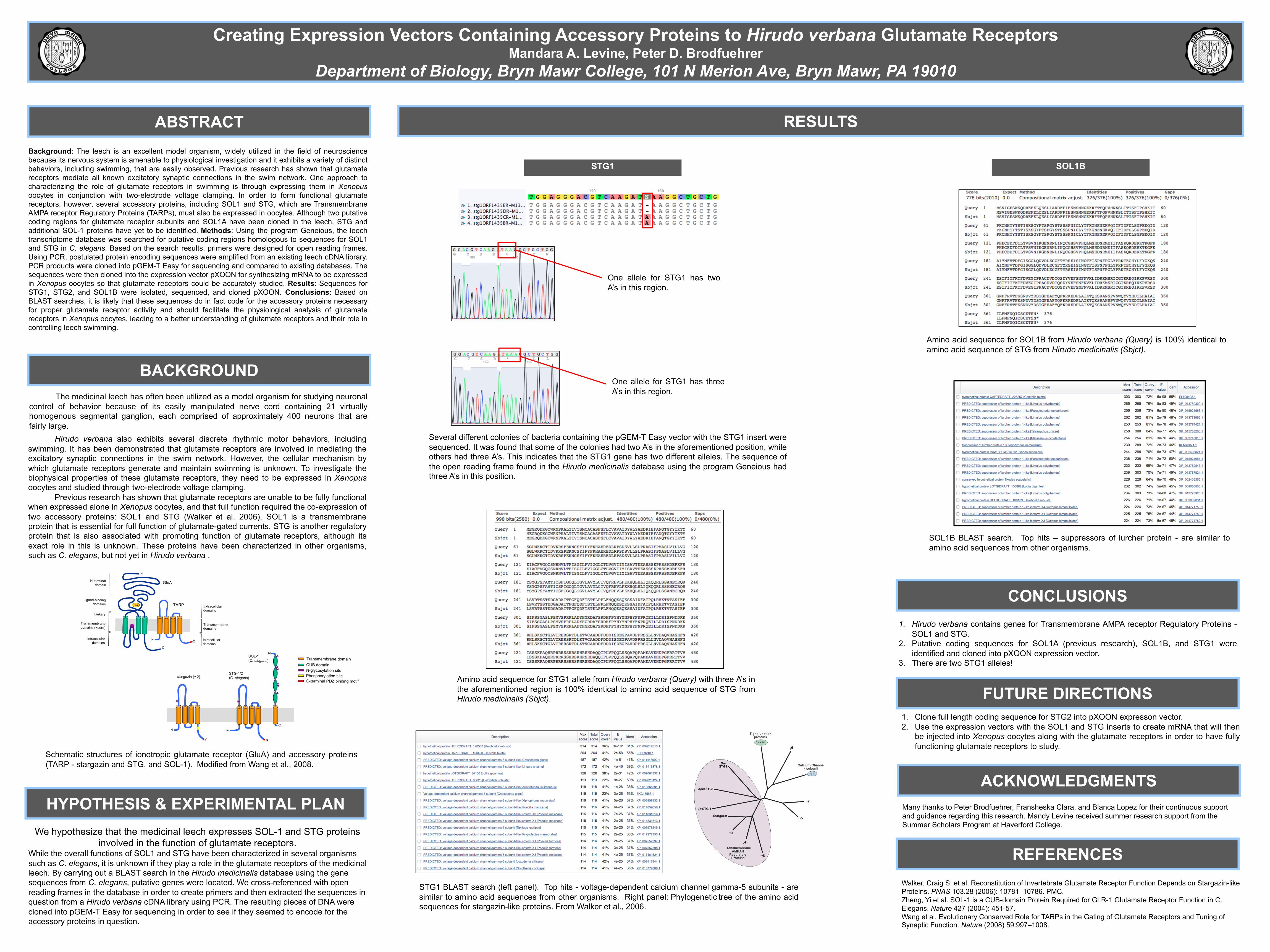
STG1
One allele for STG1 has twoA’s in this region.
One allele for STG1 has threeA’s in this region.
SOL1B
Amino acid sequence for STG1 allele from Hirudo verbana (Query) with three A’s inthe aforementioned region is 100% identical to amino acid sequence of STG fromHirudo medicinalis (Sbjct).
Amino acid sequence for SOL1B from Hirudo verbana (Query) is 100% identical toamino acid sequence of STG from Hirudo medicinalis (Sbjct).
STG1 BLAST search (left panel). Top hits - voltage-dependent calcium channel gamma-5 subunits - aresimilar to amino acid sequences from other organisms. Right panel: Phylogenetic tree of the amino acidsequences for stargazin-like proteins. From Walker et al., 2006.
SOL1B BLAST search. Top hits – suppressors of lurcher protein - are similar toamino acid sequences from other organisms.
Several different colonies of bacteria containing the pGEM-T Easy vector with the STG1 insert weresequenced. It was found that some of the colonies had two A’s in the aforementioned position, whileothers had three A’s. This indicates that the STG1 gene has two different alleles. The sequence ofthe open reading frame found in the Hirudo medicinalis database using the program Geneious hadthree A’s in this position.
alone or coexpressed with SOL-1 (9). Nor were we able to recordcurrents when GLR-2 was also coexpressed (data not shown).Therefore, we reasoned that reconstitution might require asecond auxiliary protein in addition to SOL-1. Based on weaksequence identity to vertebrate stargazin, we identified predictedORFs in the C. elegans (C18D1.4), honey bee (Apis mellifera;XM 397021), and Drosophila (CG33670) genomes. The pre-dicted Apis sequence was incomplete; therefore, we used PCRamplification and RACE to clone the complete ORF from A.mellifera first-strand cDNA.
C. elegans, Apis, and Drosophila encode predicted 366 (CeSTG-1), 397 (Apis STG1), and 447 (Dro STG1) amino acidtransmembrane proteins (Fig. 1A), and share !21% (Ce STG-1),24% (Apis STG1), and 25% (Dro STG1) sequence identity withvertebrate stargazin. Hydropathy analysis identified four puta-tive transmembrane domains. Because no N-terminal signalsequences are evident, we predict that the N and C termini of CeSTG-1, Apis STG1 and Dro STG1 are intracellular. Dro STG1has a predicted 5" region that is longer by !200 aa than the otherstargazin-like proteins. In addition, its 3" region is significantly
shorter (Fig. 1A). These differences may indicate that theparticular clone we have identified is alternatively spliced. Theamino acid sequences of these predicted proteins were analyzedby generating a neighbor-joining tree of related vertebrateproteins, including TARPs, Ca2# channel ! subunits, and claudintight junction proteins (Fig. 1B). Ce STG-1, Apis STG1, and DroSTG1 seem only slightly more related to TARPs than to the Ca2#
channel ! subunits. To determine the cellular expression of CeSTG-1, we used the stg-1 promoter to drive the expression ofGFP (Fig. 2A) or yellow fluorescent protein (YFP) (Fig. 2B) intransgenic worms. GFP and YFP were strongly expressed in thenervous system, with expression apparent in most of the neuronsthat normally express the GLR-1 subunit as indicated by coex-pression of Pglr-1::CFP (cyan f luorescent protein) andPstg-1::YFP (Fig. 2B). We did not observe expression in non-neuronal tissues such as muscle.
GLR-1 Function, but Not Surface Expression, Depends on Ce STG-1 andSOL-1. To attempt to reconstitute GLR-1 receptor activity, weinjected cRNAs encoding C. elegans GLR-1 and auxiliary pro-
Fig. 1. C. elegans STG-1, Drosophila STG1, and Apis STG1 are related to members of the TARP family. (A) The predicted amino acid sequences encoded by C.elegans stg-1, Drosophila stg1, vertebrate stg, and A. mellifera stg1. Amino acids are numbered beginning with the first predicted methionine. For Ce STG-1,bars and asterisks indicate predicted transmembrane domains and putative N-linked glycosylation sites, respectively. Underlined in gray are the potential typeI PDZ-domain-binding sites for vertebrate stargazin and Apis STG1. (B) Phylogenetic tree of the amino acid sequences for stargazin-like proteins (figure modifiedfrom ref. 16).
10782 ! www.pnas.org"cgi"doi"10.1073"pnas.0604482103 Walker et al.
Schematic structures of ionotropic glutamate receptor (GluA) and accessory proteins(TARP - stargazin and STG, and SOL-1). Modified from Wang et al., 2008.