Embed Size (px)
Citation preview

Creating a Novel Origin of Replication throughModulating DNA-Protein InterfacesF. Curtis Hewitt1,2, R. Jude Samulski1,2*
1 Gene Therapy Center, University of North Carolina at Chapel Hill, Chapel Hill, North Carolina, United States of America, 2 Curriculum in Genetics and Molecular Biology,
University of North Carolina at Chapel Hill, Chapel Hill, North Carolina, United States of America
Abstract
Background: While the molecular mechanisms of DNA-protein specificity at the origin of replication have been determinedin many model organisms, these interactions remain unknown in the majority of higher eukaryotes and numerousvertebrate viruses. Similar to many viral origins of replication, adeno-associated virus (AAV) utilizes a cis-acting origin ofreplication and a virus specific Replication protein (Rep) to faithfully carry out self-priming replication. The mechanisms ofAAV DNA replication are generally well understood. However, the molecular basis of specificity between the Rep proteinand the viral origin of replication between different AAV serotypes remains uncharacterized.
Methodology/Principal Findings: By generating a panel of chimeric and mutant origins between two AAV serotypes, wehave mapped two independent DNA-Protein interfaces involved in replicative specificity. In vivo replication assays andstructural modeling demonstrated that three residues in the AAV2 Rep active site are necessary to cleave its cognate origin.An analogous origin (AAV5) possesses a unique interaction between an extended Rep binding element and a 49 aa regionof Rep containing two DNA binding interfaces.
Conclusions/Significance: The elucidation of these structure-function relationships at the AAV origin led to the creation of aunique recombinant origin and compatible Rep protein with properties independent of either parent serotype. This novelorigin may impact the safety and efficacy of AAV as a gene delivery tool. This work may also explain the unique ability ofcertain AAV serotypes to achieve site-directed integration into the human chromosome. Finally, this result impacts thestudy of conserved DNA viruses which employ rolling circle mechanisms of replication.
Citation: Hewitt FC, Samulski RJ (2010) Creating a Novel Origin of Replication through Modulating DNA-Protein Interfaces. PLoS ONE 5(1): e8850. doi:10.1371/journal.pone.0008850
Editor: Maciej Lesniak, The University of Chicago, United States of America
Received October 16, 2009; Accepted December 14, 2009; Published January 22, 2010
Copyright: � 2010 Hewitt, Samulski. This is an open-access article distributed under the terms of the Creative Commons Attribution License, which permitsunrestricted use, distribution, and reproduction in any medium, provided the original author and source are credited.
Funding: This work was supported by National Institutes of Health grants GM0529299, HL066973, HL051818, AI072176 awarded to Dr. Jude Samulski andNational Institute of Allergy and Infectious Diseases grant AI007419 awarded to Curtis Hewitt. The funders had no role in study design, data collection andanalysis, decision to publish, or preparation of the manuscript.
Competing Interests: The authors have declared that no competing interests exist.
* E-mail: [email protected]
Introduction
General understanding of the mechanisms required for function
at origins of replication has grown immensely since the first
prokaryotic origins were characterized. While the DNA-protein
interactions necessary for replication in prokaryotes, lower
eukaryotes, and bacteriophages are generally well understood,
mechanisms employed in the majority of higher eukaryotes and
vertebrate viruses, such as adeno-associated virus (AAV), are still
being determined. The inverted terminal repeats (ITRs) of AAV
and other Parvoviruses act as the origin of replication. These
elements flank the short, single stranded genome and typically
possess a T-shaped secondary structure. The replication strategies
of the genus Dependovirus, specifically those of adeno-associated
virus (AAV), have been well characterized. The viral non-
structural or Replication proteins (Rep) are the only factors
required to interact with the ITR in order to catalyze replication
[1]. The majority of AAV serotypes possess highly conserved
origins of replication with interchangeable DNA-protein interac-
tions. However, the Rep proteins of several serotypes interact
exclusively with their cognate ITR. Discovering the mechanisms
which drive Rep-ITR specificity promises to advance our
understanding of DNA-protein interactions at viral origins of
replication. These findings also promise to shed light on how
eukaryotic and prokaryotic proteins achieve selectivity to DNA
substrates.
The AAV ITRs are critical for nearly every aspect of the viral
life-cycle (Fig. S1). The secondary structure of the ITR is necessary
to prime synthesis of the second strand to allow transcription of the
viral genes [2]. The full length Rep proteins contain a unique N-
terminal DNA binding region which specifically recognizes the
ITR at the 16 nt Rep binding element (RBE) and at the tip of one
of the hairpin stems known as the RBE’ (Fig. 1A) [3,4]. Rep
molecules multimerize on the ITR allowing the C-terminus of
Rep, acting as an ATP-dependent SF3 helicase, to unwind the
ITR and form a putative internal hairpin [1,5]. This hairpin,
(here, termed ‘nicking stem’) contains the terminal resolution site
(trs) where Rep nicks the ITR in a site-specific manner (Fig. 1A)
[6]. This DNA cleavage is required for replication of the closed
ITR and is necessary to initiate subsequent rounds of genomic
replication. Replicated genomes can undergo replication again or
be encapsidated in the presence of the smaller Rep proteins [7].
PLoS ONE | www.plosone.org 1 January 2010 | Volume 5 | Issue 1 | e8850

The ITR sequences of seven human/primate AAV serotypes
have been published. These sequences typically display 80% or
greater nucleotide conservation and segregate into two groups [8].
The AAV2 Rep proteins (Rep2) are able to function on the ITR of
nearly every known AAV serotype except those of AAV5 (ITR5)
[8,9]. Consistently, Rep5 is unable to catalyze replication of ITR2
(Fig. 1B). Replicative specificity between these serotypes does not
exist at the level of binding, as Rep2 and Rep5 can bind
interchangeably to ITR2 or ITR5 [10]. Instead, specificity is
created by the inability of Rep to cleave the ITR of the opposite
serotype. This occurs despite high conservation between the ITR2
and ITR5 sequence, secondary structure, and location of elements
required for Rep interaction (RBE, RBE’, trs, nicking stem, Fig. 1A).
Identification of the elements involved in Rep-ITR specificity
stands to increase the understanding of viral and cellular DNA
binding and endonucleolytic proteins. It is likely that similar
interactions take place in a wide range of viral and cellular
replication and repair pathways. Localization of these elements may
also facilitate the identification of other unique Parvovirus origins of
replication. Here, we demonstrate two unique mechanisms at the
DNA and protein level to achieve Rep-ITR specificity and utilize
these factors to create a novel AAV origin of replication.
Results
Construction and Characterization of Chimeric ITRsPreviously, AAV replicative specificity was postulated to be
driven by the trs sequence [10,11]. Rep2 can nick the ITR2 trs
(AGT/TGG) and the AAVS1 trs of human chromosome 19
(GGT/TGG) [12]. Rep5 nicks only the ITR5 trs (AGTG/TGG).
However, alignment of the ITR2 and ITR5 sequences revealed
several significant sequence and structural differences outside the
trs sequence (Fig. 1A). The spacing between the putative RBE and
the nicking stem was significantly different; three nucleotides (nt)
for ITR2 and 15 nt for ITR5. Additionally, while the trs sequence
is not tightly conserved between ITR2 and ITR5, neither is the
height or overall length of the putative nicking stem.
Here, we utilized a novel method to generate mutant ITRs in
order to determine which portions of the ITR were responsible for
replicative specificity. Previous studies have investigated Rep-ITR
interactions in vitro largely due to the difficulty of synthesizing full
length ITRs for in vivo assays. PCR through the secondary
structure of the ITR is inefficient and sequencing through these
elements typically requires radiolabeled chain-terminator sequenc-
ing [13]. The AAV ITRs are highly recombinogenic and are
frequently mutated even in a plasmid context [14].
In order to address these concerns the ITR was synthesized and
amplified in halves (Fig. S2). Assembly of the halves required the
inclusion of a SfiI site in one of the hairpin arms of the ITR. SfiI
allowed the conservation of the RBE’ sequence [4]. Cloning the
ITR in a DD format required only one ITR per plasmid for
replication [15]. The three core Rep functions necessary for AAV
replication (Rep binding, helicase, and nicking) were analyzed by
the presence or absence of intracellular replication of the plasmid.
This assay provided the ability to quantitate Rep-ITR function in
a physiological setting, removing the concern that highly purified
Figure 1. Cloning and characterization of chimeric ITRs. (A) Sequence and structure of ITR2 (black) and ITR5 (blue) shown with incorporationof SfiI sites for cloning (green). Length in nt of ITR elements indicated above brackets. RBE is boxed. RBE’ is indicated by a hatched circle. Nicking stemis extruded with arrow indicating the nicking site and hatched box indicating the trs. The four initial chimeric ITRs generated are shown (right). (B)Replication assay and quantitation of chimeric Reps. Replication products from the indicated ITR and either Rep2 or Rep5 were analyzed by Southernblot. Monomeric (m) and dimeric (d) replicating species are indicated. The level of replication of each sample was measured by densitometric analysisand compared to wt replication.doi:10.1371/journal.pone.0008850.g001
AAV Replicative Specificity
PLoS ONE | www.plosone.org 2 January 2010 | Volume 5 | Issue 1 | e8850

Rep protein might take on aberrant function in vitro. This system
also avoided concerns that previous in vitro assays used only a
fragment of the ITR or that oligos used to recapitulate the ITR
might not fold correctly.
An alignment of ITR2 and ITR5 (Fig. 1A) revealed several
divergent elements which might infer Rep specificity. The RBE
and hairpins seemed unlikely to impact specificity as Rep2 and
Rep5 have been reported to bind ITR2 and ITR5 interchange-
ably [10]. Additionally, no evidence of Rep interactions with the
portion of the D-element outside the nicking stem has been
presented. Therefore, the spacer and nicking stem elements
appeared to be the most likely candidates for unique interactions
with their cognate Rep protein. This hypothesis was supported by
low homology of these elements between AAV2 and AAV5.
Wt ITRs containing the SfiI site functioned as expected with
Rep2 specific to ITR2 and Rep5 specific to ITR5 (Fig. 1B). Rep2-
ITR2 replicated approximately 2-fold better than Rep5-ITR5,
potentially due to the lower folding energy of ITR5 resulting in
reduced plasmid stability prior to replication. Due to this minor
difference in replicative fidelity, all ITRs replicated with Rep2
were normalized to Rep2-ITR2, while ITRs replicated with Rep5
were normalized to Rep5-ITR5 (Fig. 1B).
In order to confirm that the RBE and hairpin arms played no
role in Rep specificity, we generated a chimeric ITR with ITR5
binding elements and an ITR2 spacer and nicking stem
(ITR5+2SNS). Only Rep2 replicated this ITR, confirming the
determinants of replicative specificity lie in the spacer/nicking
stem elements (Fig. 1B). While ITR5+2SNS replication was not as
efficient as ITR2-Rep2, it was replicated at ITR5-Rep5 levels.
Conversely, Rep5 specifically replicated an ITR comprised of
ITR2 hairpins and hairpin spacer and the ITR5 spacer and
nicking stem (ITR2+5SNS, Fig. 1B). Rep5 replicated this ITR at
wt levels. This data demonstrated that Rep-ITR specificity lies
outside of the ITR binding regions.
Next, we explored whether the nicking stem or the spacing
between the RBE and nicking stem harbored unique interactions
with the Rep protein by creating chimeric ITRs which divided
these individual elements. An ITR with the ITR5 binding
elements and spacer and the ITR2 nicking stem could not be
replicated by either Rep2 or Rep5 (ITR5+2NS, Fig. 1B). The
corresponding chimeric ITR (ITR2 binding elements and spacer
with an ITR5 nicking stem) was replicated by both Rep2 and
Rep5 (ITR2+5NS, Fig. 1B). This disparity suggested that the
spacer and nicking stem play different roles in Rep-ITR specificity
between AAV2 and AAV5.
The Nicking Stem Is Critical for ITR5 SpecificityITR2+5NS established that Rep2 is capable of nicking an ITR
with an ITR5 nicking stem and that Rep-ITR specificity is not
driven exclusively by the trs sequence (Fig. 1B). In order to
determine the flexibility of Rep2 toward mutant nicking stems, we
generated ITR2s containing altered forms of the hairpin (Fig. 2A).
Rep2 is able to replicate an ITR with an ITR5 nicking stem even
though the ITR5 nicking stem contains a different trs sequence, is
one bp shorter, and has two fewer unpaired nucleotides at its tip
(Fig. 2A). The substitution of the ITR5 nicking stem into ITR2
also allowed replication by Rep5.
To determine which element of the ITR2 nicking stem
prevented Rep5 activity, we altered specific portions of the
ITR2 stem. First, one bp at the top of the putative ITR2 nicking
stem was removed to lower the height to that of ITR5 (ITR2-TA).
Removing the T-A bp also resulted in a trs resembling ITR5,
nicking between G/T opposed to T/T. Rep2 continued to
function on this ITR as did Rep5, demonstrating that Rep5 can
tolerate five unpaired nucleotides at the tip of the stem as long as
the stem height and nt sequence are correct. A similar deletion
from the base of the ITR2 nicking stem reduced the height to that
of ITR5 while retaining the ITR2 nicking site (ITR2-GC). Rep2
continued to function efficiently on this ITR while Rep5 activity
was ablated. This data suggested that the inability of Rep5 to
function on ITR2 is primarily the sequence of the trs, specifically
the requirement for a nick to be generated between G/T.
To determine the extent of Rep2 flexibility for different nicking
stems, we created three additional ITR2 mutants. Extending the
nicking stem by one bp at the base had no effect on replication by
Rep2 (ITR2 9 nt). However, a three bp extension was sufficient to
ablate Rep2 function on the ITR (ITR2 11 nt). Surprisingly, Rep2
was able to tolerate a three bp deletion from the base of the stem,
underlining the flexibility of Rep2 with respect to nicking stem
substrates (ITR2 5 nt).
In order to explore the level of flexibility Rep5 possessed toward
non-wt nicking stems, we created a panel of mutant ITR5s
harboring altered nicking stems (Fig. 2C). Curiously, Rep2
replicated none of these ITRs, suggesting an element outside the
ITR5 nicking stem is responsible for preventing Rep2 function. As
in Figure 1B, replacement of the ITR5 nicking stem with that of
ITR2 resulted in the ablation of replication by Rep5, attributable
to the incompatible trs sequence. The addition of one bp at the top
of the ITR5 nicking stem severely decreased the ability of Rep5 to
replicate the ITR (ITR5 +TA, Fig. 2D). This insertion disrupted
the ITR5 trs sequence and increased the size of the stem one bp.
However, the low level of replication by Rep5 on ITR5 +TA
suggests that the entire trs site of ITR2 is necessary to confer Rep2
specificity, not just the presence of a T/T nick site.
The addition of one bp to the base of the ITR5 nicking stem,
preserving the ITR5 trs at the tip, nearly eliminated replication by
Rep5 (ITR5 +GC). Likewise, the removal of one bp from the base
of the ITR5 nicking stem strongly decreased replication by Rep5
(ITR5 6nt, Fig. 2D). This data suggests that Rep5 is sensitive both
to the height of the nicking stem as well as to the sequence of the
trs. Thus, Rep5 is unable to replicate ITR2 because the ITR2
nicking stem is one bp too tall and has an incompatible trs
sequence.
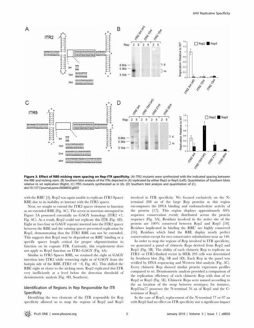
Spacer Length Is Critical for ITR2, Not ITR5While Rep2 can replicate a vector with an ITR5 nicking stem, it
can not replicate wt ITR5 (Fig. 1B). The only difference between
ITR5+2SNS (which Rep2 can replicate) and ITR5+2NS (which
Rep2 can not) is the ITR5 spacer (Fig. 1B). The wt Rep2 spacer is
three nt long while the wt Rep5 spacer is 15 nt long. Thus, we
hypothesized that Rep2 may be sensitive to spacer length. Previous
in vitro data supported this conclusion as insertions into the ITR2
spacer prevented nicking by Rep2 [4].
To explore the effect of spacer length on ITR2 and ITR5, we
generated a series of mutant ITR2s and ITR5s with differing
spacer lengths (Fig. 3A and 3C). An insertion extending the ITR2
spacer to 10 nt ablated replication by Rep2 (ITR2 10nt, Fig. 3B).
Similarly, substitution of the ITR2 spacer with the 15 nt spacer of
ITR5 also ablated replication by Rep2 (ITR2 15nt, Fig. 3B). Rep5
was unable to replicate any of these vectors due to the presence of
the ITR2 stem loop.
Rep5 displayed greater flexibility toward spacer elements of
differing lengths. Replacing the 15 nt ITR5 spacer with that of ITR2
resulted in an ITR which Rep5 retained the ability to replicate at a
reduced level (ITR5 3nt, Fig. 3D). Additionally, the presence of the
three nt spacer allowed Rep2 to function on this ITR. The addition of
six nt to the ITR5 spacer (for a total spacer length of 21nt) resulted in
an ITR capable of being replicated by Rep5 at an efficient level
AAV Replicative Specificity
PLoS ONE | www.plosone.org 3 January 2010 | Volume 5 | Issue 1 | e8850

(ITR5 21nt, Fig. 3D). Replication by Rep5 was effectively abolished
only after the insertion of 15 nt into the spacer (ITR5 30nt, Fig. 3D).
This panel of mutant ITR5s demonstrates the requirement for a three
nt spacer element for Rep2 function.
This data confirmed that the length of the ITR5 spacer was
critical to block Rep2 function. Even small insertions into the
ITR2 spacer were not tolerated by Rep2. Meanwhile, Rep5 is
flexible in regard to spacer length, demonstrating the ability to
function on ITRs with spacers from 3–21 nt.
The ITR5 Spacer Acts as a RBE for Rep5The inability of Rep2 to function on ITRs with spacers longer
than three nt led to the question of why Rep5 was so flexible in this
regard. We hypothesized that Rep5 might specifically bind the
ITR5 spacer just as it binds the RBE. The inability of Rep2 to
bind this sequence would preclude its function on ITR5.
Supporting this hypothesis was a moderately conserved GAGY
Rep binding motif extending throughout the ITR5 spacer
(Fig. 4A). Additionally, as Rep monomers bind every four nt,
the binding of three Rep5 monomers to the 15 nt spacer element
would result in a three nt spacer, similar to that of ITR2
[16].
If Rep5 does bind the loosely conserved GAGY motif, the removal
of that motif from the spacer should abolish Rep5 function. Indeed,
the ITR5 No GAGY mutant could not be replicated by Rep2 or
Rep5 (Fig. 4B). This suggested that the specific sequence of the ITR5
spacer plays an active role in the Rep5-ITR5 interaction. Conversely,
a spacer with a pure GAGY repeat should not disrupt the ability of
Rep5 to function on the ITR. Indeed, Rep5 was able to replicate this
ITR at wt levels (ITR5 GAGY, Fig. 4B). Rep2 was also able to
replicate this ITR efficiently, suggesting the poorly conserved nature
of the GAGY repeat within the ITR5 spacer prevents a critical DNA-
protein interaction with Rep2 necessary for replication.
To explore how the ITR5 spacer functioned as an RBE, we
removed three GAGY repeats from the hairpin side of the RBE
(ITR5 Spacer RBE, Fig. 4A). This essentially shifted the 16 nt RBE
12 nt closer to the nicking stem. Rep5 replicated this ITR efficiently,
confirming the ITR5 spacer acts as a RBE (ITR5 Spacer RBE,
Fig. 4B). The slight reduction in replication fidelity of this ITR with
respect to wt ITR5 may signal the inability Rep to properly interact
Figure 2. Relation of nicking stem height and sequence to Rep-ITR specificity. (A) Sequence of nicking stem in an otherwise ITR2 context. Arrowindicates trs site. Brackets indicate height of putative stems in nt from the base of the stem to the putative nicking site. Predicted DG values for the hairpins arebelow. Southern blot analysis of the ITRs replicated by Rep2 or Rep5 are shown below. (B) Quantitation of the Southern blots relative to wt replication from(A). (C) Same as (A), except nicking stems indicated were used in an ITR5 context. (D) Quantitation of the Southern blots relative to wt replication from (C).doi:10.1371/journal.pone.0008850.g002
AAV Replicative Specificity
PLoS ONE | www.plosone.org 4 January 2010 | Volume 5 | Issue 1 | e8850
with the RBE’ [4]. Rep2 was again unable to replicate ITR5 Spacer
RBE due to its inability to interact with the ITR5 spacer.
Next, we sought to extend the ITR2 spacer element to function
as an extended RBE (Fig. 4C). The seven nt insertion attempted in
Figure 3A possessed essentially no GAGY homology (ITR2 +7,
Fig. 4C). As a result, Rep2 could not replicate this ITR (Fig. 4D).
Eight nt (two four nt GAGY repeats) inserted into the ITR2 spacer
between the RBE and the existing spacer prevented replication by
Rep2, demonstrating that the ITR2 RBE can not be extended.
This suggests that Rep2 may be dependent on RBE’ binding or a
specific spacer length critical for proper oligomerization to
function on its cognate ITR. Curiously, this requirement does
not apply to Rep2 function on ITR5 GAGY (Fig. 4A).
Similar to ITR5 Spacer RBE, we retained the eight nt GAGY
insertion into ITR2 while removing eight nt of GAGY from the
hairpin side of the RBE (ITR2 +8 28, Fig. 4C). This shifted the
RBE eight nt closer to the nicking stem. Rep2 replicated this ITR
very inefficiently at a level below the detection threshold of
densitometric analysis (Fig. 4D, Southern).
Identification of Regions in Rep Responsible for ITRSpecificity
Identifying the two elements of the ITR responsible for Rep
specificity allowed us to map the regions of Rep2 and Rep5
involved in ITR specificity. We focused exclusively on the N-
terminal 208 aa of the large Rep proteins as this region
encompasses the DNA binding and endonucleolytic activity of
the protein [17]. This region displays approximately 60%
sequence conservation evenly distributed across the protein
sequence (Fig. 5A). Residues involved in the active site of the
protein are 100% conserved between Rep2 and Rep5 [18].
Residues implicated in binding the RBE’ are highly conserved
[16]. Residues which bind the RBE display nearly perfect
conservation except for two conservative substitutions near aa 140.
In order to map the regions of Rep involved in ITR specificity,
we generated a panel of chimeric Reps derived from Rep2 and
Rep5 (Fig. 5B). The ability of each chimeric Rep to replicate an
ITR2- or ITR5-flanked vector in HEK 293 cells was determined
by Southern blot (Fig. 5B and 5D). Each Rep in the panel was
verified by DNA sequencing and Western blot analysis (Fig. 5C).
Every chimeric Rep showed similar protein expression profiles
compared to wt. Densitometric analysis provided a comparison of
the replication efficiency of each chimeric Rep with that of wt
Rep2 or Rep5 (Fig. 5E). Chimeric Reps were named according to
the aa location of the swap between serotypes; for instance,
Rep25aa77 possesses the N-terminal 76 aa of Rep2 and the C-
terminus of Rep5.
In the case of Rep5, replacement of the N-terminal 77 or 97 aa
with Rep2 had no effect on ITR specificity nor a significant impact
Figure 3. Effect of RBE-nicking stem spacing on Rep-ITR specificity. (A) ITR2 mutants were synthesized with the indicated spacing betweenthe RBE and nicking stem. (B) Southern blot analysis of the ITRs depicted in (A) replicated by either Rep2 or Rep5 (Left). Quantitation of Southern blotsrelative to wt replication (Right). (C) ITR5 mutants synthesized as in (A). (D) Southern blot analysis and quantitation of (C).doi:10.1371/journal.pone.0008850.g003
AAV Replicative Specificity
PLoS ONE | www.plosone.org 5 January 2010 | Volume 5 | Issue 1 | e8850

on replicative fidelity (Fig. 5D and 5E). Larger pieces of Rep2
substituted onto the N-terminus of Rep5 were sufficient to prevent
efficient replication of ITR5s (Rep25aa116, Rep25aa125, and
Rep25aa141). This suggested that these chimeras possessed
interruptions of a critical region of Rep5 for ITR5 specificity.
Rep2-based chimeras were unable to replicate ITR5s without
the inclusion of the N-terminal 146 aa of Rep5 (Rep52aa146,
Fig. 5D). Rep52aa146 replicated ITR5 at wt levels, as did the
three chimeras with larger portions of Rep5 on the N-terminus
(Rep52aa160, Rep52aa175, Rep52aa207). This mapping reveals
that the critical region for ITR specificity in Rep5 lies between aa
97–146. Surprisingly, the Rep52aa146 clone also functioned
efficiently on ITR2, constituting a Rep capable of replicating
ITR2 and ITR5. This suggested that ITR specificity existed in two
different regions of Rep.
For Rep2, the N-terminal 83 or 109 aa of Rep5 could be
substituted with no effect on ITR specificity or major influence
on replicative fidelity (Rep52aa84 and Rep52aa110, Fig. 5D and
5E). Chimeras including slightly larger portions of Rep5 were
unable to replicate either ITR, again suggesting the interruption
of a domain critical for ITR specificity (Rep52aa126 and
Rep52aa138).
Rep5-based chimeras were unable to replicate ITR2s without
the inclusion of the N-terminal 149 aa of Rep2. However, ITR2
replication was inefficient (25aa149, Fig. 5D and 5e). The
inclusion of larger portions of Rep2 allowed replication of ITR2s
to increase to wt levels (Rep25aa166, Rep25aa216). This data
maps the Rep2 region involved in ITR specificity to aa 110–149.
However, unlike Rep5, this was not the only region which played a
role in ITR specificity. The ability of the Rep52aa146 chimera to
replicate ITR2 and ITR5 vectors demonstrated a second region of
Rep2 between aa 138–160 sufficient to allow replication of ITR2s
even when the other critical region (aa 110–149) was Rep5. The
isolation of two different Rep regions involved in ITR specificity
was consistent with the discovery of two independent elements
governing specificity within the ITR.
Figure 4. The ITR5 spacer acts as a RBE for Rep5. (A) ITR5 mutants were synthesized with the indicated RBE and spacer sequence. Bracketsindicate individual tetranucleotide repeats bound by Rep monomers. Both strands of the wt ITR5 sequence are shown to illustrate conservation withthe GAGY motif (indicated by *). Only one strand shown on others. (B) Southern blot analysis of the ITRs depicted in (A) replicated by either Rep2 orRep5 (Left). Quantitation of Southern blots relative to wt replication (Right). (C) ITR2 mutants were generated with the RBE and spacer sequencesindicated. (D) Southern blot analysis and quantitation for (C).doi:10.1371/journal.pone.0008850.g004
AAV Replicative Specificity
PLoS ONE | www.plosone.org 6 January 2010 | Volume 5 | Issue 1 | e8850

Characterization of Rep Regions Involved in ITRSpecificity
To characterize the Rep domains identified in Figure 5, we
created chimeric Rep proteins which specifically exchanged the
regions implicated in ITR specificity (Fig. 6A). Region 1 existed in
Rep2 from aa 110–149 and in Rep5 from aa97–146. Region 2 lay
within Rep2 from aa 149–187 and Rep5 from aa 146–187. As in
Figure 5, all chimeras were verified by DNA sequencing and
Western blot analysis (Fig. 6B). Chimeras were then assayed for
the ability to replicate ITR2- or ITR5-flanked vectors (Fig. 6C).
Replacing Rep5 region 1 with Rep2 yielded a clone unable to
replicate either vector, suggesting the chimera lacked the ability to
bind the ITR5 spacer or nick the ITR2 nicking stem
(Rep525aa110–148, Fig. 6C). Replacing Rep5 region 2 with that
of Rep2 allowed this chimera to replicate an ITR2 vector,
suggesting region 2 of Rep2 was critical to nick the ITR2 nicking
stem (Rep525aa146–187). The inability of this chimera to
recognize ITR5 is harder to explain as Rep52aa146 could
replicate ITR2 and ITR5 efficiently (Fig. 5B). This result suggests
that Rep2 region 2 makes specific contacts within Rep2 aa 188–
208 which are necessary in order to function on the ITR5 nicking
stem. Replacing regions 1 and 2 of Rep5 with Rep2 resulted in a
Rep chimera which replicated only ITR2s (Rep525aa110–187).
Replacing Rep2 region 1 with Rep5 resulted in replication of
only ITR2s, again demonstrating a connection between Rep2
region 2 and the ITR2 nicking stem (Rep252aa97–146). The lack
of ITR5 replication by Rep252aa97–146 is difficult to explain
based on the Rep52aa146 chimera which replicates ITR2s and
ITR5s efficiently (Fig. 5B). This result suggests that Rep5 region 1
requires specific contacts within the preceding 96 aa of Rep5 in
order to replicate ITR5. Replacing Rep2 region 2 with Rep5
resulted in a chimera unable to replicate either ITR (Re-
p252aa149–187). This chimeric Rep possesses neither Rep2
region 2 (required to nick the ITR2 nicking stem) nor Rep5
region 1 which appears necessary to interact with the ITR5 spacer.
Finally, replacing both Rep2 regions 1 and 2 with Rep5 resulted in
a chimera capable of replicating only ITR5 vectors (Rep252aa97–
187).
The crystal structure of the N-terminal 193 aa of Rep5 complexed
to the RBE allowed the location of these two critical regions to be
Figure 5. Cloning and characterization of chimeric Reps. (A) An alignment of the N-termini of Rep2 and Rep5. (*) represents conserved aminoacids. (: and.) indicates conservative substitutions. Blue indicates residues implicated in RBE binding interactions. Pink indicates residues whichparticipate in the endonucleolytic active site. Green indicates residues implicated in RBE’ binding. (B) Chimeric Reps created and their ability toreplicate ITR2 or ITR5 flanked vectors. Numbers indicate the aa position of the switch from one Rep to the other. (+) indicates the presence ofreplication, (2) indicates the absence. (C) Western blot for expression of the chimeric Reps. (D) Southern blot demonstrating replication of an ITR2 oran ITR5 vector by the chimeric Reps. Note that the ITR5 vector is 500bp larger than the ITR2 vector. (E) Level of replication of the chimeric Repsrelative to wt Rep2 or Rep5.doi:10.1371/journal.pone.0008850.g005
AAV Replicative Specificity
PLoS ONE | www.plosone.org 7 January 2010 | Volume 5 | Issue 1 | e8850

modeled [16]. The structure of the N-terminus of Rep2 was modeled
with Swiss-Model software using Rep5 as a template. The location of
region 1 supports its involvement with the spacer/RBE (Fig. 6D).
This region interacts with the major groove of the ITR where one of
the most apparent structural differences between Rep2 and Rep5 is
predicted (Fig. 6D, hatched circle). Rep2 contains a two aa insertion
in this loop with respect to Rep5. This insertion and other non-
conservative substitutions are likely responsible for the inability of
Rep2 to interact with the ITR5 spacer.
Viewing Rep along the length of the ITR illustrates that region
1 constitutes much of the base of the protein (Fig. 6E). Both Reps
are predicted to participate in a b-sheet motif in the center of this
region, while areas of reduced homology exist toward either side
(the loop interacting with the major groove of the ITR on one side,
RBE’ interactions on the other). A more detailed look at region 1
reveals the greatest disparity between Rep2 and Rep5 occurs at
the RBE binding interface in the major groove of the ITR (Fig. 6F).
There is very little predicted structural difference between
region 2 of Rep2 and Rep5 (Fig. 6D and 6E). In an effort to
dissect this region, we created two additional clones: Re-
p52aa147 and Rep52aa151 (Fig. 6A). Like Rep52aa146, both of
these Reps were able to replicate ITR2 and ITR5 vectors
(Fig. 6C). Rep52aa146 and Rep52 aa147 replicated ITR2 and
ITR5 vectors with equivalent efficiency, suggesting E147 of
Rep2 is not involved in ITR specificity. Rep52aa151 did display
a modest reduction in ITR2 replication compared to Re-
p52aa146, suggesting that C151 of Rep2 plays a role in ITR2
specificity. Because Rep52aa160 can not replicate ITR2, this
leaves only two other non-conserved residues between Rep2
and Rep5 in this region (N155 and T161). Both of these residues
lie near the active site and are likely to interact with the nicking
stem or active site. N155 lies directly adjacent to Y156, the
nucleophilic tyrosine, and may play a major role in ITR2
specificity (Fig. 6G).
Figure 6. Characterization of Rep regions critical for ITR specificity. (A) Chimeric Reps and their ability to replicate ITR2 or ITR5 flankedvectors. Numbers indicate the aa position of the switch from one Rep to the other. (+) indicates the presence of replication, (2) indicates the absence.Region 1 and 2 involved in Rep-ITR specificity are indicated. (B) Western blot for expression of chimeric Reps. (C) Southern blot demonstratingreplication of an ITR2 or ITR5 vector by the chimeric Reps. Note that the ITR5 vector is 500 bp larger than the ITR2 vector. (D) Structural modelillustrating the two Rep regions. Rep2 structure is blue, Rep5 is purple. The nucleophilic tyrosine is indicated. Black hatched circle indicates thepredicted structural difference of region 1 in the major groove of the ITR. (E) Structural model as in (D). The nucleophilic tyrosine is indicated. (F)Detailed structural view of region 1. The side-chains of non-conserved residues from Rep5 (purple) and Rep2 (blue) are shown. Three Rep5 residuesimplicated in RBE’ binding are indicated. (G) Detailed structural view of region 2. Side chains of active site residues are shown in black. Side chains ofnon-conserved residues in this region are shown for Rep2 (blue) and Rep5 (purple). The nucleophilic tyrosine is indicated, as is the adjacent Rep2 Asn-155.doi:10.1371/journal.pone.0008850.g006
AAV Replicative Specificity
PLoS ONE | www.plosone.org 8 January 2010 | Volume 5 | Issue 1 | e8850

Structure-Function Model of Rep-ITR SpecificityIn order to unify the ITR and Rep elements involved in specificity
into a single model, we utilized the chimeric Reps separating region 1
and region 2 along with the chimeric ITRs separating the nicking
stem and spacer. Rep2, Rep5, Rep52aa146 (which divides region 1
and 2 of Rep and can replicate ITR2 and ITR5), and Rep25aa149
(essentially no ITR2 or ITR5 replication) were selected. These Reps
were tested for their ability to replicate ITR2, ITR5, ITR2+5NS
(which is replicated by both Rep2 and Rep5), and ITR5+2NS (which
is replicated by neither Rep2 or Rep5).
Only Rep2 and Rep52aa146 efficiently replicated ITR2 (Fig. 7A
and 7B). Only Rep5 and Rep52aa146 replicated ITR5. As in
Figure 1, Rep2 and Rep5 replicated ITR2+5NS. Additionally,
Rep25aa149 and Rep52aa146 replicated ITR2+5NS. This ITR
appears to be universally replicated by every Rep in this assay due to
the exclusion of DNA elements involved in protein specificity. The
three nt ITR2 spacer is amenable to the DNA binding region 1 of
Rep2 and Rep5. The seven bp tall ITR5 nicking stem functions
with region 2 of Rep2 and Rep5. Thus, any combination of these
regions constitutes a Rep protein capable of replicating ITR2+5NS.
Finally, neither Rep2 nor Rep5 replicated ITR5+2NS. Rep2 is
unable to interact properly with the 15 nt ITR5 spacer. Rep5 is
unable to function on the ITR2 nicking stem. For these reasons,
Rep25aa149 was also unable to catalyze replication. However,
Rep52aa149 was able to replicate this ITR due to the proper
combination of Rep regions (Fig. 7C). Rep52aa149 possesses Rep5
region 1 which is necessary to interact with the 15 nt ITR5 spacer.
This chimera also possesses Rep2 region 2, essential for function
on the ITR2 nicking stem. This recombinant DNA-protein
interaction is unique from either AAV2 or AAV5 and constitutes
a novel Parvovirus origin of replication.
Discussion
Taken as a whole, this work illustrates two specific mechanisms
of DNA-protein specificity at the Parvovirus origin of replication.
Chimeric ITRs narrowed the DNA elements involved in
specificity to the spacer and nicking stem sequences (Fig. 1B).
These results contradicted previous assertions that Rep-ITR
specificity were driven solely by the nicking sequence as Rep2
efficiently nicked an ITR harboring the ITR5 nicking stem [10].
Rep2 is highly flexible in the sequence and height of its nicking
stem while Rep5 is highly specific to its cognate stem (Fig. 2).
Three residues of Rep2 are necessary to cleave the ITR2
nicking stem (Fig. 5 and 6). Residues C151, N155, and T161 all lie
in the active site of the protein in a predicted alpha helix along
with the nucleophilic tyrosine Y156. How these residues (termed
Rep region 2) grant Rep2 flexibility toward mutant nicking stems
Figure 7. Model of Rep-ITR specificity. (A) Southern blot of Hirt DNA demonstrating replication of the indicated ITR vector by the indicated Rep.(B) Table indicating the presence (+) or absence (2) of replication of the gel from (A). (C) Model of a novel AAV origin of replication. The chimeric ITRcan be replicated only by a chimeric Rep protein. Rep5 sequence in region 1 (blue) is required for the extended RBE of ITR5 (purple). Rep2 sequencein region 2 (yellow) is required to function on an ITR2 nicking stem (cyan).doi:10.1371/journal.pone.0008850.g007
AAV Replicative Specificity
PLoS ONE | www.plosone.org 9 January 2010 | Volume 5 | Issue 1 | e8850

remains unclear. The corresponding Rep5 residues (G148, A152,
and V158) may participate in highly specific interactions which
require specific height and sequence considerations for the ITR5
nicking stem.
AAV5 Rep-ITR specificity is mediated by the ITR5 spacer.
Replacement of the three nt ITR2 spacer with the 15 nt ITR5
spacer ablated replication by Rep2 (Fig. 2B). A poorly conserved
Rep binding element allows Rep5 to interact with the elongated
ITR5 spacer (Fig. 4B). Mutating the spacer to include a strong
Rep binding element allowed Rep2 and Rep5 to replicate the
ITR. However, insertion of a Rep binding element into the ITR2
spacer still largely decreased Rep2 function. While this data might
suggest that additional Rep5 molecules bind to ITR5, previous in
vitro experiments have not come to this conclusion, although those
studies were performed in the absence of hairpins on the ITRs
[10].
A 49 aa region of Rep5 interacts with the ITR5 spacer (aa 97–
146, Fig. 5 and 6). The crystal structure of the N-terminus of Rep5
reveals that this region (region 1) possesses residues which
specifically bind to the RBE and RBE’ of the ITR. Major
structural differences in the Rep5 loop which binds the major
groove of the RBE likely account for the majority of ITR5 spacer
specificity. While Figure 1B predicts RBE’ binding should not play
a role in Rep-ITR specificity, it is possible that RBE’ contacts alter
the secondary structure of region 1 as it interacts with the RBE.
Because the regions of Rep critical for ITR specificity were
separate (region 1 of Rep5 from aa97–146 and region 2 of Rep2
from aa151–161), a chimeric Rep possessing both regions was able
to efficiently replicate ITR2 and ITR5. An ITR which could be
replicated by any wt or chimeric Rep was constructed by
excluding the DNA elements required for specificity; the ITR5
spacer and the ITR2 nicking stem. Most significantly, a novel
origin of replication was generated. This ITR contained both of
the critical elements for Rep specificity; the ITR5 spacer and the
ITR2 nicking stem. As a result, only a chimeric Rep protein made
up of Rep5 region 1 and Rep2 region 2 was able to replicate the
ITR. The creation of a unique origin of replication highlights the
power of studying the DNA-protein interactions of a viral origin of
replication.
The creation of a unique DNA-protein interaction was possible
because of the separation of the specific Rep-ITR interactions in
AAV2 and AAV5. How and why these two different DNA-protein
interactions evolved is unclear. It is likely due to evolutionary
divergence in the ITR sequence which may have occurred in
different hosts (AAV2 is related to other primate AAVs, AAV5 is
related to non-primate AAVs such as goat and bovine). This
model of replicative specificity can likely be extended to other
parvoviruses such as snake AAV which has a highly conserved T-
shaped ITR structure but different spacer and nicking stem lengths
[19]. Similar DNA-protein interactions likely occur in distantly
related viruses such as the autonomous human Parvovirus B19
which can have ITRs as long as 400 bp [20]. Less conserved
Parvovirus origins of replication might also employ additional
DNA-protein interactions outside of the nicking stem and spacer
sequences.
Additionally, this work may provide further insight into why
AAV2 is the only known animal virus capable of integrating site-
specifically into the human chromosome [21]. Integration occurs
due to the specific cleavage of the AAVS1 site on chromosome 19
by Rep2. Rep2 is highly flexible in its nicking substrates,
functioning on nicking stems from five bp to nine bp in height
and on poorly conserved trs sequences. Thus, the only
requirement for Rep2 to nick the human chromosome would be
a functional nicking stem within three nt of a consensus RBE. As
there are an estimated 26105 consensus RBEs in the human
chromosome, the likelihood of such an occurrence is high [22].
This may also explain why an integration locus for AAV5 has not
been identified. Rep5 is highly specific to both the height and
sequence of the ITR5 nicking stem. There is likely no ITR5
nicking stem homolog in the human chromosome within range of
a consensus RBE to allow nicking by Rep5. It is possible that other
hosts infected by AAV5-related serotypes might possess chromo-
somal integration sites for AAV5.
These results also stand to improve the safety of future AAV
therapeutic vectors. The danger of AAV vector mobilization by wt
AAV could be averted if therapeutic vectors harbored ITRs which
no wt Rep could replicate [8]. Additionally, the mechanisms of
Rep-ITR specificity described here might extend to cellular
elements. For instance, the C. elegans mobile element Tc1
contains terminal repeats which are specifically bound and
endonucleolytically cleaved by its transposase, Tc1A [23]. Biology
at the related SV40 T antigen and papillomavirus E1 origins of
replication likely possess conserved interactions [18]. Bacterio-
phage QX174 and plant geminiviruses, as well as other viruses
which employ rolling circle mechanisms of replication also possess
homology to the AAV origins of replication [24]. In this way,
dissection of specificity at the AAV origin of replication provides a
broad platform to investigate other DNA-protein interactions.
Materials and Methods
Rep CloningpXR2 (Rep2Cap2) and pRep5Cap2 AAV helper plasmids
served as templates for Rep cloning. The primer sequences used
are indicated in Table 1. Two cloning strategies were used.
Existing restriction sites were incorporated into primers for PCR
(PCR-RD in Table 1) utilizing either pXR out fw or pXR out rev
primers. PfuTurbo DNA Polymerase (Stratagene) was used at the
manufacturer’s recommendations for all PCR reactions. PCR-RD
products were digested with the enzymes indicated in Table 1
(NEB) prior to ligation with T4 DNA Ligase (Invitrogen) according
to manufacturer’s instructions. Alternately, an overlap-extension
mediated PCR (OE-PCR) approach was used to produce Rep
chimeras [26]. The Rep2 and Rep5 junction was incorporated into
forward and reverse primers which were used in separate PCR
reactions with the pXR out fw and rev primers (Table 1, only fw
oligos indicated, rev oligos complimentary to fw). These overlap-
ping PCR products were combined into a single PCR reaction and
cycled as follows: 1 cycle at 94uC for 30 seconds, 18 cycles of 30
seconds at 94uC, 30 seconds at 65uC, and 4 minutes at 72uC, 1
cycle of 10 minutes at 72uC. 1 ul of this reaction was used as
template for a nested PCR with the pXR in fw and rev primers.
Chimeras with the N-terminus of Rep2 and C-terminus of Rep5
were cloned into the Rep25aa166 construct between the PpuMI
and MfeI sites. Chimeras with the N-terminus of Rep5 and C-
terminus of Rep2 were cloned into the 52aa160 construct between
the PpuMI and BstBI sites. All constructs were verified by DNA
sequencing at the UNC-CH Genome Analysis Facility.
ITR CloningITRs were cloned into a pUC-18 plasmid with a GFP cassette
(CMV promoter, SV40 polyA) cloned between the KpnI and EcoRI
restriction sites. The ITRs were synthesized in two halves as 4nmol
Ultramer DNA oligos (Integrated DNA Technologies). SfiI
restriction sites were incorporated into one hairpin arm the ITR
for cloning (Fig. 1A). Due to inconsistencies of the reported
sequence at the tip of the ITR5 hairpins between [10], the
published genbank sequence (genbank accession number
AAV Replicative Specificity
PLoS ONE | www.plosone.org 10 January 2010 | Volume 5 | Issue 1 | e8850

NC_006152), and restriction mapping (data not shown), an ITR2
hairpin was utilized for the ITR5 construct (Fig. 1A). 200pg of
each oligo was amplified in a PCR reaction using the ITR primers
listed in Table 1. 2.5U of PfuTurbo DNA Polymerase (Stratagene)
was used to amplify each half of the ITR as follows: 1 cycle at
94uC for 4 minutes, 35 cycles of 45 seconds at 94uC, 30 seconds at
50uC, and 30 seconds at 72uC, 1 cycle of 10 minutes at 72uC. PCR
reactions were purified and subject to digestion by KpnI and SfiI or
HindIII and SfiI (NEB). A triple ligation with the pUC-18 GFP
plasmid and each half of the ITR was performed with T4 DNA
Ligase (Invitrogen) for 1.5 hours at room temperature. All
constructs were verified by DNA sequencing at the UNC-CH
Genome Analysis Facility after linearization of the plasmid and
ablation of the ITR secondary structure by SfiI digestion.
Western Blot AnalysisSamples for Western blot analysis were harvested 48–72 hours
after transfection of Ad-helper plasmid and the appropriate AAV
helper construct. Cells were washed and resuspended in 100 ul
PBS prior to addition of 100 ul 2x Laemmli Sample Buffer
(100 mM Tris pH 6.8, 4% SDS, 200 mM DTT, 20% glycerol,
0.1% Bromophenol Blue). Samples were briefly sonicated and
boiled for 10 minutes. Samples were run on NUPAGE 4–12%
Bis-Tris gels (Invitrogen) at 160 volts for 90 minutes. Protein was
transferred to a Nitrocellulose membrane (Invitrogen) via a wet
transfer for 60 minutes at 30 volts. Gels were blocked overnight in
10% nonfat dry milk in 16 PBS/Tween (0.05%). Detection of
both Rep2 and Rep5 proteins (all four sizes) was achieved with a
monoclonal Anti-Adeno-Associated Virus Rep Protein antibody
(clone 259.5, American Research Products) at a 1:20 dilution in
PBS/Tween for 60 minutes at room temperature. After washing, a
secondary HRP anti-mouse antibody was added at a 1:5,000
dilution in PBS/Tween for one hour at room temperature. After
washing, SuperSignal West Femto Maximum Sensitivity Substrate
(Pierce) was added and blots were exposed to X-ray film (Kodak).
Cell Culture and rAAV ProductionHEK 293 cells were obtained from ATCC and cultured in
Dulbecco Modified Eagle Medium (DMEM) supplemented with
10% Fetal Bovine Serum (Sigma) and 100 units/ml penicillin and
100 g/ml streptomycin and grown at 37uC with 5% CO2
saturation. Transfections were performed in six-well cell culture
Table 1. Oligonucleotides for chimeric Rep and ITR cloning.
Clone/Primer Cloning Method Orientation Sequence
pXR out fw Forward 59 CGAAAAGTGCCACCTGACGTCTAAGAAACC
pXR in fw Forward 59 TCGAATTCGACGGCCAGTGAATTGTAATACGACTC
pXR out rev Reverse 59 CCATGATTACGCCAAGCTCGGAATTAACCGCATGCGA
pXR in rev Reverse 59 CCATGGCCGGGCCCGGATTCACC
Rep52aa84 PCR-RD AleI Reverse 59 TTCACCCCGGTGGTTTCCACGAGCACGTGCATGTGGAAGTAGCTCTCTCCCTTTTCAAACTGCACAAAG
Rep52aa110 PCR-RD EagI Forward 59 CCTCGGCCGCTACGTGAGTCAGATTCGCGAAAAACTGATTCAGAG
Rep52aa126 OE PCR Forward 59 GTGGTCTTCCAGGGAATTGAACCCACTTTGCCAAACTGGTTCGCGGTC
Rep52aa138 OE PCR Forward 59 CTGGGTCGCCATCACCAAGGTAAAGAAGGGAGGCGGGAACAAGGTGGTGGATGAG
Rep52aa146 OE PCR Forward 59 GCGGAGCCAATAAGGTGGTGGATGAGTGCTACATCCCCAATTACTTGCTC
Rep52aa160 PCR-RD Bpu10I Reverse 59 ACTGGAGCTCAGGTTGGACCTTCGGCAGCAGGTAG
Rep52aa175 OE PCR Forward 59 CGTGGACAAACCTGGACGAGTATAAATTGGCCTGTTTGAATCTCACGGAGCGTAAAC
Rep52aa187 OE PCR Forward 59 CTGAATCTGGAGGAGCGCAAACGGTTGGTGGCGCAGCATCTGACGCAC
Rep52aa207 PCR-RD SgrAI Reverse 59 GATCACCGGCGCATCCGAGAACTCACGCTGCGAAGC
Rep25aa77 OE PCR Forward 59 TAAGGCCCCGGAGGCCCTTTTCTTTGTGCAGTTTGAAAAGGGATCTG
Rep25aa97 OE PCR Forward 59 CCACATGCACGTGCTCGTGGAAACCTCCGGCATCTCTTCCATGGTCCTCG
Rep25aa116 PCR-RD NruI Forward 59 TCAGATTCGCGAAAAACTGGTGAAAGTGGTCTTCCAGG
Rep25aa125 OE PCR Forward 59 GAATTTACCGCGGGATCGAGCCG CAGATCAACGACTGGGTCGCCATC
Rep25aa141 OE PCR Forward 59 GGTCACAAAGACCAGAAATGGCGCCGGCGGAGCCAATAAGGTGGTGGATTCTGG
Rep25aa149 OE PCR Forward 59 GAGGCGGGAACAAGGTGGTGGATTCTGGGTATATTCCCGCCTACCTGC
Rep25aa166 PCR-RD Bpu10I Forward 59 CCAGCCTGAGCTCCAGTGGGCGTGGACAAACCTG
Rep25aa187 OE PCR Forward 59 GTTTGAATCTCACGGAGCGTAAACGGCTCGTCGCGCAGTTTCTGGCAG
Rep25aa216 PCR-RD SgrAI Forward 59 ATGCGCCGGTGATCAAAAGCAAGACTTCCCAGAAATACATGG
ITR2 Half1 Kpn Forward 59 ATTATAGGTACCAGGAACCCCTAGTGATG
ITR2 Half 1 Sfi Reverse 59 TAATAGGGCCCAAAGGGCCGGG
ITR2 Half2 Sfi Forward 59 TTAATAGGCCCTTTGGGCCGGG
ITR2 Half2 Hind Reverse 59 TATAATAAGCTTAGGAACCCCTAGTGATGGAG
ITR5 Half1 Kpn Forward 59 ATTATAGGTACCTACAAAACCTCCTTGCTTGAG
ITR5 Half1 Sfi Reverse 59 TTAATAGGCCCTTTGGGCCGTCGC
ITR5 Half2 Sfi Forward 59 TTAATAGGCCCAAAGGGCCGTCGTC
ITR5 Half2 Hind Reverse 59 TATAATAAGCTTTACAAAACCTCCTTGCTTGAGAG
doi:10.1371/journal.pone.0008850.t001
AAV Replicative Specificity
PLoS ONE | www.plosone.org 11 January 2010 | Volume 5 | Issue 1 | e8850

plates. 0.75 ug each of Ad-helper plasmid, AAV helper plasmid
(either Rep2Cap2, Rep5Cap2, or the Rep mutant described), and
the GFP plasmid containing the ITR (mutant or wt ITR as
specified in text) were triple-transfected with PEI (25,000 linear
molecular weight) as described [25]. Cells were harvested 48–72
hours post-transfection.
Hirt DNA Purification and Southern Blot AnalysisHirt DNA purification was performed as described [27]. Cells
were harvested 48–72 hours post-transfection, washed in PBS, and
resuspended in 370 ul Hirt Solution (0.01M Tris-HCl pH 7.5 and
0.1M EDTA) prior to addition of 25 ul 10% SDS and 165 ul 5 M
NaCl. Samples were incubated at 4uC overnight prior to
centrifugation. DNA was purified by phenol chloroform extraction
and precipitated by an equal volume of isopropanol prior to
resuspension in 50 ul sterile ddH2O. 5 ul of each sample was
digested with 4U DpnI (NEB) 2–4 hours at 37uC prior to gel
electrophoresis and Southern blot analysis to remove non-
replicated transfected plasmid [28]. The nylon membrane
(Hybond-XL, GE Healthcare Life Sciences) was hybridized to a
probe corresponding to the GFP open reading frame labeled with
the Random Primed DNA Labeling Kit (Roche) and d-CTP P32.
Blots were visualized after exposure to a phosphorimager screen
(GE Healthcare Life Sciences).
DensitometryDensitometry was performed using the public domain NIH
Image program (developed at the U.S. National Institutes of
Health and available on the Internet at http://rsb.info.nih.gov/
nih-image/). Densitometry analysis of a DpnI resistant band on the
agarose gel prior to transfer was used as a loading control to
normalize values obtained from the Southern blot. The lowest
value (absence of any vector replication) was subtracted from all
values to account for background. In order to gauge relative
replication efficiency, values for ITR2 vectors were divided by the
value obtained from the Rep2-ITR2 control. ITR5 vectors were
compared to the Rep5-ITR5 control. All values were obtained in
triplicate (n = 3). Error bars represent standard error (standard
deviation divided by the root of 3). All samples were compared to
controls on the same blot.
Molecular ModelingMolecular models were generated using Swiss-Model (http://
swissmodel.expasy.org). The published crystal structure of the N-
terminus of Rep5 complexed with the RBE (PDB accession
#1rz9) was used as a template for all models. Visualization of
protein structure rendering of images were performed with
PyMOL (http://pymol.sourceforge.net). DNA folding was per-
formed using the DNA mfold server (http://mfold.bioinfo.rpi.
edu/cgi-bin/dna-form1.cgil) [29,30].
Supporting Information
Figure S1 Illustration of AAV replication. Red indicates newly
synthesized DNA. (A) The AAV genome enters the nucleus as a ss
DNA molecule flanked by the ds DNA ITRs. (B and C) The free
39 hydroxyl of the ITR allows second strand synthesis through the
opposite ITRs. (D) Rep binds the closed ITR at the RBE and
RBE’. The Rep helicase allows the ITR nicking stem to form
which Rep cleaves at the trs. (E) Rep remains covalently bound to
the 59 end of the ITR, allowing synthesis through the ITR [31]. (F-
G) Complete synthesis of the genome can now occur. (H) The fully
replicated genomes can be dissociated by the Rep helicase or by
subsequent rounds of DNA synthesis.
Found at: doi:10.1371/journal.pone.0008850.s001 (6.48 MB TIF)
Figure S2 Diagram of ITR synthesis. (A) The ITR was
synthesized in two pieces (dark blue and light blue) overlapping
across one hairpin stem holding the SfiI site (orange). (B) Each half
was amplified via PCR prior to digestion and cloning. (C) Proper
triple-ligation with pUC18-CMV GFP produced an ITR in DD
format.
Found at: doi:10.1371/journal.pone.0008850.s002 (2.67 MB TIF)
Acknowledgments
The authors would like to thank Dr. Robert Kotin for providing the AAV5
ITRs, Dr. Steve Gray for his help with ITR cloning, Dr. Matt Hirsch for
his critical review, as well as the reviewers for their insightful comments.
Author Contributions
Conceived and designed the experiments: FCH. Performed the experi-
ments: FCH. Analyzed the data: FCH. Contributed reagents/materials/
analysis tools: FCH. Wrote the paper: FCH.
References
1. Im DS, Muzyczka N (1990) The AAV origin binding protein Rep68 is an ATP-
dependent site-specific endonuclease with DNA helicase activity. Cell 61:
447–457.
2. Hauswirth WW, Berns KI (1977) Origin and termination of adeno-associatedvirus DNA replication. Virology 78: 488–499.
3. Ryan JH, Zolotukhin S, Muzyczka N (1996) Sequence requirements for
binding of Rep68 to the adeno-associated virus terminal repeats. J Virol 70:1542–1553.
4. Brister JR, Muzyczka N (2000) Mechanism of Rep-mediated adeno-associated
virus origin nicking. J Virol 74: 7762–71.
5. Hermonat PL, Batchu RB (1997) The adeno-associated virus Rep78 majorregulatory protein forms multimeric complexes and the domain for this
activity is contained within the carboxy-half of the molecule. FEBS Lett 20:180–184.
6. Brister JR, Muzyczka N (1999) Rep-mediated nicking of the adeno-associated
virus origin requires two biochemical activities, DNA helicase activity andtransesterification. J Virol 73: 9325–9336.
7. King JA, Dubielzig R, Grimm D, Kleinschmidt JA (2001) DNA helicase-
mediated packaging of adeno-associated virus type 2 genomes into preformedcapsids. EMBO J 20: 3282–3291.
8. Hewitt FC, Li C, Gray SJ, Cockrell S, Washburn M, Samulski RJ (2009)
Reducing the Risk of AAV Vector Mobilization with AAV5 Vectors. J Virol 83:3919–3929.
9. Grimm D, Pandey K, Nakai H, Storm TA, Kay MA (2006) Liver transduction
with recombinant adeno-associated virus is primarily restricted by capsidserotype not vector genotype. J Virol 80: 426–439.
10. Chiorini JA, Afione S, Kotin RM (1999) Adeno-associated virus (AAV) type 5
Rep protein cleaves a unique terminal resolution site compared with other AAV
serotypes. J Virol 73: 4293–4298.
11. Chiorini JA, Kim F, Yang L, Kotin RM (1999) Cloning and characterization ofadeno-associated virus type 5. J Virol 73: 1309–1319.
12. Wu J, Davis MD, Owens RA (2001) A Rep recognition sequence is necessary but
not sufficient for nicking of DNA by adeno-associated virus type-2 Rep proteins.Arch Biochem Biophys 389: 271–277.
13. Young SM Jr, McCarty DM, Degtyareva N, Samulski RJ (2000) Roles of adeno-
associated virus Rep protein and human chromosome 19 in site-specificrecombination. J Virol 74: 3953–3966.
14. Samulski RJ, Srivastava A, Berns KI, Muzyczka N (1983) Rescue of Adeno-
Associated Virus from Recombinant Plasmids: Gene Correction within theTerminal Repeats of AAV. Cell 33: 135–143.
15. Xiao X, Xiao W, Li J, Samulski RJ (1997) A novel 165-base-pair terminal repeat
sequence is the sole cis requirement for the adeno-associated virus life cycle.J Virol 71: 941–948.
16. Hickman AB, Ronning DR, Perez ZN, Kotin RM, Dyda F (2004) The nuclease
domain of adeno-associated virus rep coordinates replication initiation using twodistinct DNA recognition interfaces. Mol Cell 13: 403–414.
17. Yoon M, Smith DH, Ward P, Medrano FJ, Aggarwal AK, et al. (2001) Amino-
terminal domain exchange redirects origin-specific interactions of adeno-associated virus rep78 in vitro. J Virol 75: 3230–3239.
18. Hickman AB, Ronning DR, Kotin RM, Dyda F (2002) Structural unity among
viral origin binding proteins: crystal structure of the nuclease domain of adeno-associated virus Rep. Mol Cell 10: 327–337.
AAV Replicative Specificity
PLoS ONE | www.plosone.org 12 January 2010 | Volume 5 | Issue 1 | e8850

19. Farkas SL, Zadori Z, Benko M, Essbauer S, Harrach B, et al. (2004) A
parvovirus isolated from royal python (Python regius) is a member of the genusDependovirus. J Gen Virol 85: 555–561.
20. Deiss V, Tratschin JD, Weitz M, Siegl G (1990) Cloning of the human
parvovirus B19 genome and structural analysis of its palindromic termini.Virology 175: 247–254.
21. Samulski RJ, Zhu X, Xiao X, Brook JD, Housman DE, et al. (1991) Targetedintegration of adeno-associated virus (AAV) into human chromosome 19.
EMBO J 10: 3941–3950.
22. Young SM Jr, Xiao W, Samulski RJ (2000) Site-specific targeting of DNAplasmids to chromosome 19 using AAV cis and trans sequences. Methods Mol
Biol 133: 111–126.23. Vos JC, van Luenen HGAM, Plasterk RHA (1993) Characterization of the
Caenorhabditis elegans Tc1 transposase in vivo and in vitro. Genes Dev 7:1244–1253.
24. Ilyina TV, Koonin EV (1992) Conserved sequence motifs in binding domain of
the Epstein-Barr virus origin-binding protein, the initiator proteins for rollingcircle DNA replication encoded by EBNA1, bound to DNA. Cell 84: 791–800.
25. Xiao X, Li J, Samulski RJ (1998) Production of high-titer recombinant adeno-
associated virus vectors in the absence of helper adenovirus. J Virol 72:
2224–2232.
26. Higuchi R, Krummel B, Saiki RK (1988) A general method of in vitro
preparation and specific mutagenesis of DNA fragments: study of protein and
DNA interactions. Nucleic Acids Res 16: 7351–7367.
27. Hirt B (1967) Selective extraction of polyoma DNA from infected mouse cell
cultures. J Mol Biol 26: 365–369.
28. Chomczynski P (1992) One-hour downward alkaline capillary transfer for
blotting of DNA and RNA. Anal Biochem 201: 134.
29. Zuker M (2003) Mfold web server for nucleic acid folding and hybridization
prediction. Nucleic Acids Res 31: 3406–3415.
30. SantaLucia J Jr (1998) A unified view of polymer, dumbell, and oligonucleotide
DNA nearest-neighbor thermodynamics. Proc Natl Acad Sci 95: 1460–1465.
31. Prasad KM, Trempe JP (1995) The adeno-associated virus Rep78 protein is
covalently linked to viral DNA in a preformed virion. Virology 214: 360–70.
AAV Replicative Specificity
PLoS ONE | www.plosone.org 13 January 2010 | Volume 5 | Issue 1 | e8850


![Replikation af Vibrio cholerae kromosom II origin, oriCII · Replikation af Vibrio cholerae kromosom II origin, oriCIIVc [Replication of Vibrio cholerae chromosome II origin, oriCIIVc]](https://img.dokumen.tips/doc/110x75/5caeeeca88c99383228da77e/replikation-af-vibrio-cholerae-kromosom-ii-origin-oricii-replikation-af-vibrio.jpg)