Embed Size (px)
Citation preview
1
M
FFFFF.R..R..R..R..R. S S S S Spilkpilkpilkpilkpilki ,i ,i ,i ,i , P P P P P.A..A..A..A..A. E E E E Estststststeeeeevvvvveseseseses,,,,, TTTTT.C..C..C..C..C. S S S S Silvilvilvilvilva,a,a,a,a, et alet alet alet alet al..... 2011. 2011. 2011. 2011. 2011. Cortical Necrosis and Cerebral Atrophy in Calves Experimentally Infectedwith Bovine Herpesvirus Type 5 ..... Acta Scientiae Veterinariae. 39(1): 953.Acta Scientiae Veter inariae, 2011. 39(1) : 953.Acta Scientiae Veter inariae, 2011. 39(1) : 953.Acta Scientiae Veter inariae, 2011. 39(1) : 953.Acta Scientiae Veter inariae, 2011. 39(1) : 953.Acta Scientiae Veter inariae, 2011. 39(1) : 953.
CASE REPORTPub. 953Pub. 953Pub. 953Pub. 953Pub. 953
ISSN 1679-9216 (Online)
Cortical Necrosis and Cerebral Atrophy in Calves Experimentally Infected with BovineCortical Necrosis and Cerebral Atrophy in Calves Experimentally Infected with BovineCortical Necrosis and Cerebral Atrophy in Calves Experimentally Infected with BovineCortical Necrosis and Cerebral Atrophy in Calves Experimentally Infected with BovineCortical Necrosis and Cerebral Atrophy in Calves Experimentally Infected with BovineHHHHHerererererpppppesviresviresviresviresvirus us us us us TTTTTypypypypype 5e 5e 5e 5e 5
FFFFFererererernando Rnando Rnando Rnando Rnando Rosado Sosado Sosado Sosado Sosado Spilkpilkpilkpilkpilkiiiii11111 *****,,,,, P P P P Paulo Aaulo Aaulo Aaulo Aaulo Augustugustugustugustugusto Eo Eo Eo Eo Estststststeeeeevvvvveseseseses22222 ,,,,, TTTTTamir Camir Camir Camir Camir Calcalcalcalcalcagnottagnottagnottagnottagnotto da So da So da So da So da Silvilvilvilvilvaaaaa33333 ,,,,, A A A A Ana Cna Cna Cna Cna Cláudia Fláudia Fláudia Fláudia Fláudia Frrrrrancancancancancooooo33333,,,,,David DriemeierDavid DriemeierDavid DriemeierDavid DriemeierDavid Driemeier44444 & Paulo Michel Roehe & Paulo Michel Roehe & Paulo Michel Roehe & Paulo Michel Roehe & Paulo Michel Roehe3,53,53,53,53,5
ABSTRACT
Background: Bovine encephalitis herpesvirus, or bovine herpesvirus type 5 (BoHV-5), a member of the family Herpesviridae,subfamily Alphaherpesvirinae, is long recognized as the causative agent of bovine herpesvirus encephalitis. The diseasecaused by BoHV-5 is characterized by signs of nervous impairment, consequent to non-suppurative meningoencephalitis.Although bovine herpetic encephalitis is a rare event in herds from the Northern Hemisphere, BoHV-5 infections are an importantcause of central nervous system disease in cattle in Brazil and Argentina. Recovery of animals from clinical illness has beendocumented before, both in naturally infected animals and experimentally infected individuals.Case: During an experiment of experimental inoculation of a virulent isolate of BoHV-5, clinical signs of neurological disease weredetected in three out of four calves experimentally inoculated with bovine encephalitis herpesvirus (bovine herpesvirus type 5;BoHV-5; strain EVI88/95). Clinical signs varied from slight prostration (1 calf) to severe signs of nervous impairment which lastedfrom 3 to 14 days (in 2 calves) from the beginning of clinical signs to death. Despite the neurological signs, one of the calves withmild clinical signs recovered: on day 14 p.i, this animal showed apathy, bruxism, dysphagia, pressing of the head against the wallsof the bay, hyper salivation, tongue paralysis, hypermetria, and transient blindness but the signs become gradually milder andthe health status of the animal improved until day 21 p.i. Recovery was complete ten days after the development of clinical signsand no evident sequelae were noticed up to day 180 when the remaining calves were culled. At necropsy, the prominentmacroscopical finding was a large atrophic area at the left frontal lobe of the cortex. Another atrophic area was evident on theparietal lobe, at the level of the mamillary bodies, revealing a yellow-orange cystic area on the basis of cerebrum, involving thenucleus caudatus and the putamen. At histopathology, large areas on the affected frontal cortex were infiltrated by macrophagescontaining haemosiderin and some plasma cells BoHV-5 infection was confirmed by viral isolation from nasal and ocular swabsduring acute infection from day 1 p.i. until day 19 p.i., with titres up to 104,5 TCID 50/50 µL, form all inoculated animals. BoHV-5was also isolated from many organs, especially from the brain, of the animals which died on the acute phase of infection;however, no infectious virus could be recovered from tissues collected at necropsy from the calf at 180 p.i.Discussion: These findings suggest the possible association of BoHV-5 with atrophic lesions in the brain, a finding which hadnot been previously linked to BoHV-5 infections. Therefore, animals that recover from clinically evident BoHV-5 infection underfield conditions may also bear brain lesions that would remain undetected if the calf is not culled; yet these may be detected onlymonths or years later, at slaughter. These results are of interest for the South American countries where infections with BoHV-5are highly prevalent.
Keywords: BoHV-5, bovine encephalitis herpesvirus, cortical necrosis, atrophic brain lesions.
Received: May 2010 www.ufrgs.br/actavet Accepted: August 2010
Laboratório de Microbiologia Molecular, Instituto de Ciências da Saúde, Universidade Feevale, Rodovia RS-239, n. 2755, CEP 93352-000Novo Hamburgo, RS, Brazil. 2Embrapa Suínos e Aves, Concórdia, SC, Brazil. 3Departmento of Microbiologia, Laboratório de Virologia,UniversidadeFederal do Rio Grande do Sul (UFRGS), Porto Alegre, RS, Brazil. 4Departamento of Patologia Clínica Veterinária, Faculdade de Veterinária,UFRGS, Porto Alegre, RS, Brazil. 5Equipe de Virologia, FEPAGRO Saúde Animal - Instituto de Pesquisas Veterinárias Desidério Finamor(CPVDF) - FEPAGRO, Eldorado do Sul, RS, Brazil. CORRESPONDÊNCIA: F.R. Spilki [[email protected] - Fone: +55 (51) 3586-8800ramal 9043].
2
FFFFF.R..R..R..R..R. S S S S Spilkpilkpilkpilkpilki ,i ,i ,i ,i , P P P P P.A..A..A..A..A. E E E E Estststststeeeeevvvvveseseseses,,,,, TTTTT.C..C..C..C..C. S S S S Silvilvilvilvilva,a,a,a,a, et alet alet alet alet al..... 2011. 2011. 2011. 2011. 2011. Cortical Necrosis and Cerebral Atrophy in Calves Experimentally Infectedwith Bovine Herpesvirus Type 5 ..... Acta Scientiae Veterinariae. 39(1): 953.
INTRODUCTION
Bovine encephalitis herpesvirus, or bovineherpesvirus type 5 (BoHV-5), a member of the familyHerpesviridae, subfamily Alphaherpesvirinae, is longrecognized as the causative agent of bovine herpesvirusencephalitis [1,12]. Clinical disease associated to BoHV-5 is characterized by signs of nervous impairment,consequent to non-suppurative meningoencephalitis [7].The disease usually affects calves up to 2 years old,although occasionally older animals may be involved [9].Mortality rates usually approach 100% [5], but somecattle may recover [9]. BoHV-5 disease can be easilymisdiagnosed as rabies, botulism, polioencephalomalacia,NaCl intoxication and other causes of encephalitis [9].
CASE REPORT
Four 3 to 5 months old calves of mixed Europeanbreeds, previously tested to ensure that they had not beenexposed to BoHV-1, BoHV-5 or bovine viral diarrhoeavirus (BVDV) were kept in isolation units. The calveswere infected intranasally with 106,50 fifty percent tissueculture infective doses (TCID50) of BoHV-5 strain EVI88/95. Two other calves were inoculated with sterileculture medium and kept as uninfected controls. Clinicalexaminations were performed daily until day 21 postinoculation (p.i.). Blood samples were taken at days 0,16, 20, 120, 150 and 180 p.i. Nasal and ocular secretionswere collected from nostrils and conjunctival sacs dailyuntil day 21 p.i. and processed for virus isolation asdescribed [10]. Cell monolayers were monitored dailyfor cytopathic effect (CPE). CPE-positive samples wereexamined under an immunoperoxidase assay withmonoclonal antibodies (Mabs) to bovine herpesviruses(including Mabs capable of differentiating BoHV-5 fromBoHV-1). Samples were considered negative when aftertwo blind passages in cell cultures no CPE was observed.Antibodies to BoHV-1 and BoHV-5 were titrated by astandard serum virus neutralization test [10]. At necropsy,various samples of tissues (spleen, liver, adrenal glands,thymus, trachea, lung, trigeminal ganglia, and lymphnodes) as well as brain specimens (frontal cortex, pons,cerebellum, hippocampus, medulla, and cortex) andcerebrospinal fluid (CSF) were collected for virusisolation. Virus multiplication, quantification and isolationfrom tissues as well as neutralisation assays wereperformed on Madin Darby bovine kidney cells (MDBK,ATCC CCL-22). Cells were routinely maintained inEagle’s minimal essential medium (E-MEM)
supplemented with enrofloxacin (2 mg/L) and 6%foetal calf serum1. Cells were multiplied followingroutine procedures [10]. The BoHV-5 strain EVI 88/95 (used at passage numbers 7 to 9), is a typicalrepresentative of the majority of BoHV-5 virusescirculating in Brazil [3].
Nasal secretions were collected with sterilecotton swabs deeply introduced into each nostril orconjunctival sac, daily, until day 21 p.i.. After sampling,swabs were dipped in 2 mL of MEM supplemented with100 mg/mL enrofloxacin and 2.5 mg/mL AmphotericinB (MEM AB 10X) and stored at -70oC for subsequentprocessing. Inoculation of cell cultures was performedwith 10-1 to 10-4 dilutions of the initial suspension. Newlyformed monolayers of CRIB-1 and MDBK cells wereinoculated with 200 mL of inoculum and incubated at37ºC in a 5% CO2 atmosphere. Solid tissues (spleen, liver,adrenal glands, thymus, trachea, lung, trigeminal ganglia,and various lymph nodes), brain specimens (frontal cortex,pons, cerebellum, hippocampus, medulla, and cortex) andcerebrospinal fluid (CSF) were collected from the twonecropsied calves. Suspensions of approximately 10%of tissues were prepared in MEM AB 10X, clarified bycentrifugation and the supernatant inoculated onto CRIB-1 monolayers. After 1 h for adsorption at 37°C, theinoculum was removed, cells washed 3 times with MEMAB 10X and overlaid with the same medium. CSF wasdirectly inoculated onto cell cultures, in volumes of 100mL per well in 6 well culture dishes. Cells were monitoreddaily for cytopathic effect (CPE). The virus recoveredwas confirmed as BoHV-5 by an immunoperoxidaseassay with BoHV-5-specific monoclonal antibodies.
Tissue samples were collected from all organslisted above. Calves were necropsied soon after deathor culling. Samples were fixed in 10% buffered formalin,processed and stained with haematoxylin-eosin (HE)following routine procedures.
Fixed tissue sections were deparaffinized in xylolat 60ºC for 10 to 30 min and washed twice in basic Trisbuffer (BTB, 1M Tris Buffer Solution, pH 9.5).Endogenous peroxidase activity was blocked with 3%H2O2 in methanol for 30 min, followed by washing in BTB.Subsequently, the slides were incubated with 0.1% trypsinfor 30 min. Sections were then incubated with 10 % nor-mal rabbit serum for tem minutes and then incubatedwith the primary antibody (anti-BoHV-5 monoclonalantibodies). After three new washes in BTB, sectionswere overlaid with anti-mouse IgG/peroxidase conjugate2
for 10 min at room temperature and again washed three
3
M
FFFFF.R..R..R..R..R. S S S S Spilkpilkpilkpilkpilki ,i ,i ,i ,i , P P P P P.A..A..A..A..A. E E E E Estststststeeeeevvvvveseseseses,,,,, TTTTT.C..C..C..C..C. S S S S Silvilvilvilvilva,a,a,a,a, et alet alet alet alet al..... 2011. 2011. 2011. 2011. 2011. Cortical Necrosis and Cerebral Atrophy in Calves Experimentally Infectedwith Bovine Herpesvirus Type 5 ..... Acta Scientiae Veterinariae. 39(1): 953.
times in BTB. Subsequently, the substrate 3-amino-9-ethyl carbazole3 with 0.03% H2O2 was added. Thereaction was stopped after 15 min at room temperature,by another wash with BTB and the slide counterstainedwith haematoxylin. After a final wash in running water,the slides were mounted and examined at the microscope.Sections were also stained with a commercially availableavidin-biotin complex-peroxidase kit4 following themanufacturer´s instructions.
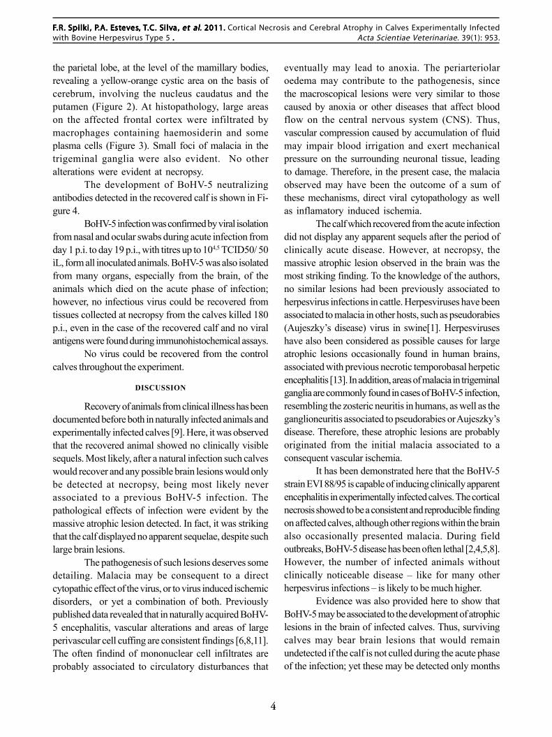
Three of the four BoHV-5 infected calvespresented two febrile peaks on days 2 (40.1oC) and 4(40.8oC) p.i. Two of them died presenting a set of severeclinical signs of nervous impairment (Table 1). On day14 p.i, one calf showed apathy, bruxism, dysphagia,pressing of the head against the walls of the bay, hypersalivation, tongue paralysis, hypermetria, and transientblindness but the signs become gradually milder and thehealth status of the animal improved until day 21 p.i.
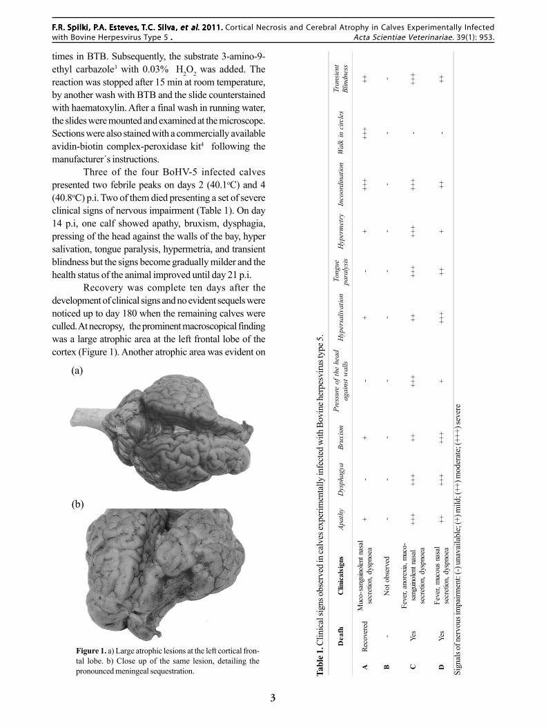
Recovery was complete ten days after thedevelopment of clinical signs and no evident sequels werenoticed up to day 180 when the remaining calves wereculled. At necropsy, the prominent macroscopical findingwas a large atrophic area at the left frontal lobe of thecortex (Figure 1). Another atrophic area was evident on
Deafh
Clinicalsigns
Apat
hyD
ysph
agya
Brux
ismPr
essu
re o
f the
hea
dag
ains
t wal
lsH
yper
saliv
atio
nTo
ngue
para
lysis
Hyp
erm
etry
Inco
ordi
natio
nW
alk
in c
ircle
sTr
ansie
ntBl
indn
ess
ARe
cove
red
Muc
o-sa
nguin
olen
t nas
alse
cret
ion,
dys
pnoe
a+
-+
-+
-+
+++
+++
++
B-
Not
obs
erve
d-
--
--
--
--
-
CYe
sFe
ver,
anor
exia,
muc
o-sa
nguin
olen
t nas
alse
cret
ion,
dys
pnoe
a++
+++
+++
+++
++++
+++
+++
+-
+++
DYe
sFe
ver,
muc
ous
nasa
lse
cret
ion,
dys
pnoe
a++
+++
+++
+++
+++
+++
-++
Figure 1. a) Large atrophic lesions at the left cortical fron-tal lobe. b) Close up of the same lesion, detailing thepronounced meningeal sequestration. Ta
ble 1
. Clin
ical
sign
s obs
erve
d in
calv
es ex
perim
enta
lly in
fect
ed w
ith B
ovin
e her
pesv
irus t
ype 5
.
Sign
als o
f ner
vous
impa
irmen
t: (-)
una
vaila
ble;
(+) m
ild; (
++) m
oder
ate;
(+++
) sev
ere
A
(a)
(a)
(b)
4
FFFFF.R..R..R..R..R. S S S S Spilkpilkpilkpilkpilki ,i ,i ,i ,i , P P P P P.A..A..A..A..A. E E E E Estststststeeeeevvvvveseseseses,,,,, TTTTT.C..C..C..C..C. S S S S Silvilvilvilvilva,a,a,a,a, et alet alet alet alet al..... 2011. 2011. 2011. 2011. 2011. Cortical Necrosis and Cerebral Atrophy in Calves Experimentally Infectedwith Bovine Herpesvirus Type 5 ..... Acta Scientiae Veterinariae. 39(1): 953.
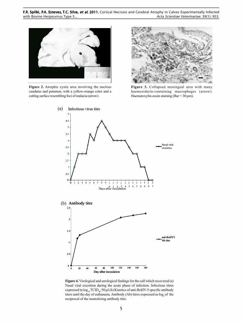
the parietal lobe, at the level of the mamillary bodies,revealing a yellow-orange cystic area on the basis ofcerebrum, involving the nucleus caudatus and theputamen (Figure 2). At histopathology, large areason the affected frontal cortex were infiltrated bymacrophages containing haemosiderin and someplasma cells (Figure 3). Small foci of malacia in thetrigeminal ganglia were also evident. No otheralterations were evident at necropsy.
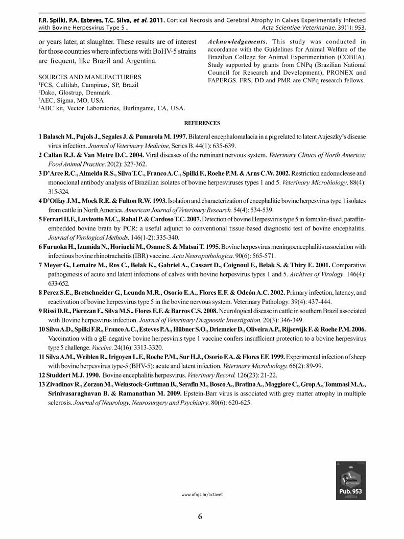
The development of BoHV-5 neutralizingantibodies detected in the recovered calf is shown in Fi-gure 4.
BoHV-5 infection was confirmed by viral isolationfrom nasal and ocular swabs during acute infection fromday 1 p.i. to day 19 p.i., with titres up to 104,5 TCID50/ 50ìL, form all inoculated animals. BoHV-5 was also isolatedfrom many organs, especially from the brain, of theanimals which died on the acute phase of infection;however, no infectious virus could be recovered fromtissues collected at necropsy from the calves killed 180p.i., even in the case of the recovered calf and no viralantigens were found during immunohistochemical assays.
No virus could be recovered from the controlcalves throughout the experiment.
DISCUSSION
Recovery of animals from clinical illness has beendocumented before both in naturally infected animals andexperimentally infected calves [9]. Here, it was observedthat the recovered animal showed no clinically visiblesequels. Most likely, after a natural infection such calveswould recover and any possible brain lesions would onlybe detected at necropsy, being most likely neverassociated to a previous BoHV-5 infection. Thepathological effects of infection were evident by themassive atrophic lesion detected. In fact, it was strikingthat the calf displayed no apparent sequelae, despite suchlarge brain lesions.
The pathogenesis of such lesions deserves somedetailing. Malacia may be consequent to a directcytopathic effect of the virus, or to virus induced ischemicdisorders, or yet a combination of both. Previouslypublished data revealed that in naturally acquired BoHV-5 encephalitis, vascular alterations and areas of largeperivascular cell cuffing are consistent findings [6,8,11].The often findind of mononuclear cell infiltrates areprobably associated to circulatory disturbances that
eventually may lead to anoxia. The periarteriolaroedema may contribute to the pathogenesis, sincethe macroscopical lesions were very similar to thosecaused by anoxia or other diseases that affect bloodflow on the central nervous system (CNS). Thus,vascular compression caused by accumulation of fluidmay impair blood irrigation and exert mechanicalpressure on the surrounding neuronal tissue, leadingto damage. Therefore, in the present case, the malaciaobserved may have been the outcome of a sum ofthese mechanisms, direct viral cytopathology as wellas inflamatory induced ischemia.
The calf which recovered from the acute infectiondid not display any apparent sequels after the period ofclinically acute disease. However, at necropsy, themassive atrophic lesion observed in the brain was themost striking finding. To the knowledge of the authors,no similar lesions had been previously associated toherpesvirus infections in cattle. Herpesviruses have beenassociated to malacia in other hosts, such as pseudorabies(Aujeszky’s disease) virus in swine[1]. Herpesviruseshave also been considered as possible causes for largeatrophic lesions occasionally found in human brains,associated with previous necrotic temporobasal herpeticencephalitis [13]. In addition, areas of malacia in trigeminalganglia are commonly found in cases of BoHV-5 infection,resembling the zosteric neuritis in humans, as well as theganglioneuritis associated to pseudorabies or Aujeszky’sdisease. Therefore, these atrophic lesions are probablyoriginated from the initial malacia associated to aconsequent vascular ischemia.
It has been demonstrated here that the BoHV-5strain EVI 88/95 is capable of inducing clinically apparentencephalitis in experimentally infected calves. The corticalnecrosis showed to be a consistent and reproducible findingon affected calves, although other regions within the brainalso occasionally presented malacia. During fieldoutbreaks, BoHV-5 disease has been often lethal [2,4,5,8].However, the number of infected animals withoutclinically noticeable disease – like for many otherherpesvirus infections – is likely to be much higher.
Evidence was also provided here to show thatBoHV-5 may be associated to the development of atrophiclesions in the brain of infected calves. Thus, survivingcalves may bear brain lesions that would remainundetected if the calf is not culled during the acute phaseof the infection; yet these may be detected only months
5
M
FFFFF.R..R..R..R..R. S S S S Spilkpilkpilkpilkpilki ,i ,i ,i ,i , P P P P P.A..A..A..A..A. E E E E Estststststeeeeevvvvveseseseses,,,,, TTTTT.C..C..C..C..C. S S S S Silvilvilvilvilva,a,a,a,a, et alet alet alet alet al..... 2011. 2011. 2011. 2011. 2011. Cortical Necrosis and Cerebral Atrophy in Calves Experimentally Infectedwith Bovine Herpesvirus Type 5 ..... Acta Scientiae Veterinariae. 39(1): 953.
Figure 2. Atrophic cystic area involving the nucleuscaudatus and putamen, with a yellow-orange color and acutting surface resembling foci of malacia (arrow).
Figure 3. Collapsed meningeal area with manyhaemosiderin-containing macrophages (arrow).Haematoxylin-eosin staining (Bar = 30 µm).
Figure 4. Virological and serological findings for the calf which recovered (a)Nasal viral excretion during the acute phase of infection. Infectious titresexpressed in log10 TCID50/50 µl.(b) Kinetics of anti-BoHV-5 specific antibodytiters until the day of euthanasia. Antibody (Ab) titres expressed as log2 of thereciprocal of the neutralizing antibody titre.
(a)
(b)
6
FFFFF.R..R..R..R..R. S S S S Spilkpilkpilkpilkpilki ,i ,i ,i ,i , P P P P P.A..A..A..A..A. E E E E Estststststeeeeevvvvveseseseses,,,,, TTTTT.C..C..C..C..C. S S S S Silvilvilvilvilva,a,a,a,a, et alet alet alet alet al..... 2011. 2011. 2011. 2011. 2011. Cortical Necrosis and Cerebral Atrophy in Calves Experimentally Infectedwith Bovine Herpesvirus Type 5 ..... Acta Scientiae Veterinariae. 39(1): 953.
www.ufrgs.br/actavet Pub. 953Pub. 953Pub. 953Pub. 953Pub. 953
or years later, at slaughter. These results are of interestfor those countries where infections with BoHV-5 strainsare frequent, like Brazil and Argentina.
SOURCES AND MANUFACTURERS1FCS, Cultilab, Campinas, SP, Brazil2Dako, Glostrup, Denmark.3AEC, Sigma, MO, USA4ABC kit, Vector Laboratories, Burlingame, CA, USA.
Acknowledgements . This study was conducted inaccordance with the Guidelines for Animal Welfare of theBrazilian College for Animal Experimentation (COBEA).Study supported by grants from CNPq (Brazilian NationalCouncil for Research and Development), PRONEX andFAPERGS. FRS, DD and PMR are CNPq research fellows.
REFERENCES
1 Balasch M., Pujols J., Segales J. & Pumarola M. 1997. Bilateral encephalomalacia in a pig related to latent Aujeszky’s diseasevirus infection. Journal of Veterinary Medicine, Series B. 44(1): 635-639.
2 Callan R.J. & Van Metre D.C. 2004. Viral diseases of the ruminant nervous system. Veterinary Clinics of North America:Food Animal Practice. 20(2): 327-362.
3 D’Arce R.C., Almeida R.S., Silva T.C., Franco A.C., Spilki F., Roehe P.M. & Arns C.W. 2002. Restriction endonuclease andmonoclonal antibody analysis of Brazilian isolates of bovine herpesviruses types 1 and 5. Veterinary Microbiology. 88(4):315-324.
4 D’Offay J.M., Mock R.E. & Fulton R.W. 1993. Isolation and characterization of encephalitic bovine herpesvirus type 1 isolatesfrom cattle in North America. American Journal of Veterinary Research. 54(4): 534-539.
5 Ferrari H.F., Luvizotto M.C., Rahal P. & Cardoso T.C. 2007. Detection of bovine Herpesvirus type 5 in formalin-fixed, paraffin-embedded bovine brain by PCR: a useful adjunct to conventional tissue-based diagnostic test of bovine encephalitis.Journal of Virological Methods. 146(1-2): 335-340.
6 Furuoka H., Izumida N., Horiuchi M., Osame S. & Matsui T. 1995. Bovine herpesvirus meningoencephalitis association withinfectious bovine rhinotracheitis (IBR) vaccine. Acta Neuropathologica. 90(6): 565-571.
7 Meyer G., Lemaire M., Ros C., Belak K., Gabriel A., Cassart D., Coignoul F., Belak S. & Thiry E. 2001. Comparativepathogenesis of acute and latent infections of calves with bovine herpesvirus types 1 and 5. Archives of Virology. 146(4):633-652.
8 Perez S.E., Bretschneider G., Leunda M.R., Osorio E.A., Flores E.F. & Odeón A.C. 2002. Primary infection, latency, andreactivation of bovine herpesvirus type 5 in the bovine nervous system. Veterinary Pathology. 39(4): 437-444.
9 Rissi D.R., Pierezan F., Silva M.S., Flores E.F. & Barros C.S. 2008. Neurological disease in cattle in southern Brazil associatedwith Bovine herpesvirus infection. Journal of Veterinary Diagnostic Investigation. 20(3): 346-349.
10 Silva A.D., Spilki F.R., Franco A.C., Esteves P.A., Hübner S.O., Driemeier D., Oliveira A.P., Rijsewijk F. & Roehe P.M. 2006.Vaccination with a gE-negative bovine herpesvirus type 1 vaccine confers insufficient protection to a bovine herpesvirustype 5 challenge. Vaccine. 24(16): 3313-3320.
11 Silva A.M., Weiblen R., Irigoyen L.F., Roehe P.M., Sur H.J., Osorio F.A. & Flores EF. 1999. Experimental infection of sheepwith bovine herpesvirus type-5 (BHV-5): acute and latent infection. Veterinary Microbiology. 66(2): 89-99.
12 Studdert M.J. 1990. Bovine encephalitis herpesvirus. Veterinary Record. 126(23): 21-22.13 Zivadinov R., Zorzon M., Weinstock-Guttman B., Serafin M., Bosco A., Bratina A., Maggiore C., Grop A., Tommasi M.A.,
Srinivasaraghavan B. & Ramanathan M. 2009. Epstein-Barr virus is associated with grey matter atrophy in multiplesclerosis. Journal of Neurology, Neurosurgery and Psychiatry. 80(6): 620-625.