Embed Size (px)
Citation preview

Correction
MEDICAL SCIENCESCorrection for “Regulator of G protein signaling 5 protectsagainst cardiac hypertrophy and fibrosis during biomechanicalstress of pressure overload,” by Hongliang Li, Chengwei He,Jinhua Feng, Yan Zhang, Qizhu Tang, Zhouyan Bian, Xue Bai,Heng Zhou, Hong Jiang, Scott P. Heximer, Mu Qin, He Huang,Peter. P. Liu, and Congxin Huang, which was first published July19, 2010; 10.1073/pnas.1008397107 (Proc Natl Acad Sci USA107:13818–13823).The authors wish to note the following: “The representative
images in Fig. 1A, Fig. 4B, and Fig. S1C appeared incorrectly.Original data for the entire study was available and used tocorrect the figures. These errors do not affect any conclusions inthe paper. We sincerely apologize for the mistake and in-convenience we may have caused.” The corrected Fig. 1, Fig. 4,and Fig. S1 appear below, with their respective legends. The SIhas been updated online.
E3858–E3861 | PNAS | April 17, 2018 | vol. 115 | no. 16 www.pnas.org
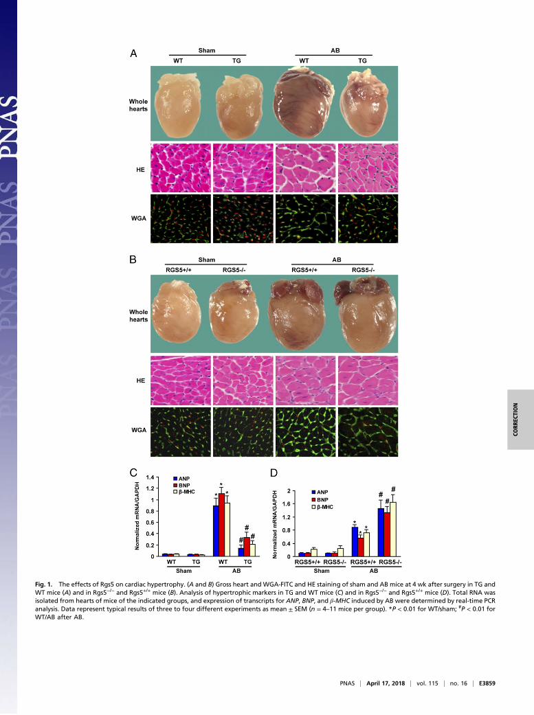
Fig. 1. The effects of Rgs5 on cardiac hypertrophy. (A and B) Gross heart and WGA-FITC and HE staining of sham and AB mice at 4 wk after surgery in TG andWT mice (A) and in Rgs5−/− and Rgs5+/+ mice (B). Analysis of hypertrophic markers in TG and WT mice (C) and in Rgs5−/− and Rgs5+/+ mice (D). Total RNA wasisolated from hearts of mice of the indicated groups, and expression of transcripts for ANP, BNP, and β-MHC induced by AB were determined by real-time PCRanalysis. Data represent typical results of three to four different experiments as mean ± SEM (n = 4–11 mice per group). *P < 0.01 for WT/sham; #P < 0.01 forWT/AB after AB.
PNAS | April 17, 2018 | vol. 115 | no. 16 | E3859
CORR
ECTION

Fig. 4. Inhibition of MEK-ERK1/2 signaling rescued abnormalities in Rgs5−/−mice. (A) U0126 blocked MEK1/2 and ERK1/2 phosphorylation mediated by AB in Rgs5−/−
mice. (B) Effects of U0126 on histological changes at 2 wk after surgery. (C) The effects of U0126 on fibrosis. Left, PSR staining; Right, statistical results of fibrotic areas.(D) The effects of U0126 on hypertrophic and fibrotic markers expression induced by AB in Rgs5−/− mice. mRNA was determined by real-time PCR analysis. Datarepresent typical results of three to five different experiments as mean ± SEM (n = 4–12 mice/per group). *P < 0.01 for PBS/sham values; #P < 0.01 for PBS/AB after AB.
E3860 | www.pnas.org

Published under the PNAS license.
Published online April 2, 2018.
www.pnas.org/cgi/doi/10.1073/pnas.1804573115
Fig. S1. Characterization of human Rgs5 transgenic mice. (A) Representative Western blots of human Rgs5 protein in the heart tissue from four lines of TGand WT mice. (B) Representative Western blot of human Rgs5 protein from different tissue of TG mice as indicated (A, lung; B, muscle; C, brain; D, heart; E,kidney; F, spleen; G, liver; H, testis). (C and D) Representative Western blots and quantitative results of endogenous Rgs5 protein levels in the heart from WTand TG mice after AB at time points indicated (n = 7). *P < 0.01 for 0 d group.
PNAS | April 17, 2018 | vol. 115 | no. 16 | E3861
CORR
ECTION

Regulator of G protein signaling 5 protects againstcardiac hypertrophy and fibrosis duringbiomechanical stress of pressure overloadHongliang Lia,b,1,2, Chengwei Hec,1, Jinhua Fenga,b, Yan Zhanga,b, Qizhu Tanga,b, Zhouyan Biana,b, Xue Baia,b,Heng Zhoua,b, Hong Jianga,b, Scott P. Heximerd, Mu Qina,b, He Huanga,b, Peter. P. Liue, and Congxin Huanga,b,2
aDepartment of Cardiology, Renmin Hospital of Wuhan University, Wuhan 430060, China; bCardiovascular Research Institute, Wuhan University, Wuhan430060, China; cDepartment of Medicine, Massachusetts General Hospital and Harvard Medical School, Boston, MA 02114; dDepartment of Physiology,University of Toronto, Toronto, ON, Canada M5S 3J9; and eDivision of Cardiology, Heart and Stroke/Richard Lewar Centre of Excellence, University HealthNetwork, University of Toronto, Toronto, ON, Canada M5S 3E2
Communicated by Alexander Leaf, Massachusetts General Hospital, Harvard Medical School, Charlestown, MA, June 15, 2010 (received for review March1, 2010)
The development of cardiac hypertrophy in response to increasedhemodynamic load and neurohormonal stress is initially a com-pensatory response that may eventually lead to ventricular di-lation and heart failure. Regulator of G protein signaling 5 (Rgs5) isa negative regulator of G protein-mediated signaling by inactivat-ing Gα(q) and Gα(i), which mediate actions of most known vaso-constrictors. Previous studies have demonstrated that Rgs5expresses among various cell types within mature heart andshowed high levels of Rgs5 mRNA in monkey and human hearttissue by Northern blot analysis. However, the critical role of Rgs5on cardiac remodeling remains unclear. To specifically determinethe role of Rgs5 in pathological cardiac remodeling, we used trans-genic mice with cardiac-specific overexpression of human Rgs5gene and Rgs5−/− mice. Our results demonstrated that the trans-genic mice were resistant to cardiac hypertrophy and fibrosisthrough inhibition of MEK-ERK1/2 signaling, whereas the Rgs5−/−
mice displayed the opposite phenotype in response to pressureoverload. These studies indicate that Rgs5 protein is a crucial com-ponent of the signaling pathway involved in cardiac remodelingand heart failure.
ERK1/2 | RGS5 | MEK1/2 | signal transduction
The heart undergoes adaptive hypertrophic growth to augmentcardiac output in response to a variety of pathological stimuli
including hypertension, ischemia, pressure overload, and inheri-ted gene mutations (1, 2). After a period of compensatory hyper-trophy, the myocardium undergoes functional and histologicaldeterioration in the setting of ongoing stress. Although much isknown about the pathways that promote hypertrophic responses,mechanisms that antagonize these pathways have not been asclearly defined. The discovery and functional clarification ofantihypertrophic targets are equally important for understandingthe molecular mechanisms underlying cardiac hypertrophy.Regulators of G-protein signaling (RGS) proteins are negative
regulators of G protein-mediated signaling that act as GTPaseaccelerating proteins for heterotrimeric G proteins. One memberof the RGS protein superfamily, RGS5, is expressed in vascular,cardiac, and skeletal muscle tissues (3). Indeed, it is a marker forangiogenic pericytes during neovascularization associated withskin wound healing and tumor angiogenesis (4, 5). RGS5 has beenreported to inhibit several Gαi- and Gαq-mediated signalingpathways in cardiovascular tissues, including those acting via thecardiovascular signaling molecules angiotensin II (Ang II) andendothelin-1 (3, 6). Notably, ribozyme-mediated knockdown ofRGS5 results in selective upreglation of Ang II-mediated activa-tion of ERK1/2 compared with other RGS proteins (3).Clinical work has shown that certain combinations of multiple
SNPs in Rgs5 may confer risk for hypertension (6). Consistentwith a role for RGS5 in blood pressure regulation, mean arterial
blood pressures were lower in Rgs5-deficient mice comparedwith WT controls (7, 8). Cho et al. (7) reported that the hypo-tensive phenotype may be related to increased NO susceptibilitycombined with increased and ERK1/2 activation in vascularsmooth muscle cells. In the heart, Rgs5 is known to be up-regulated in atria tissue of mice that overexpressed β2-adrenergicreceptor, as well as in atria of rats that were chronically ad-ministered the β-adrenergic agonist isoproterenol (9). However,the role of Rgs5 as a regulator of cardiac hypertrophy and fi-brosis has not previously been determined. In the present study,we show that the cardiac constitutive expression of human Rgs5protects against cardiac hypertrophy and fibrosis by blockingMEK-ERK1/2 signaling, whereas Rgs5−/− mice displayed theopposite phenotype in response to pressure overload. Ourstudies with cardiac-specific transgenic Rgs5 mice and Rgs5−/−
mice suggest that Rgs5 is a crucial modulator of cardiacremodeling and heart failure.
ResultsGeneration of Mice with Cardiac-Specific Overexpression of HumanRgs5. To examine the function of endogenous Rgs5 in the mouseheart in vivo, transgenic mice with cardiac-specific over-expression of human Rgs5 (i.e., TG mice) were generated byusing the α-myosin heavy chain promoter. We identified fivetransgenic founders by PCR analysis and established four in-dependent lines. These lines were born in a normal Mendeliandistribution. They also exhibited normal reproductive rate andsex distributions. The relative levels of Rgs5 protein expressionin the different lines were line no. 21 > 3 > 28 > 7. Among fourestablished lines of TG mice, line 3 was used for furtherexperiments (Fig. S1A). We analyzed Rgs5 protein levels invarious tissues by Western blot analysis using a human-specificanti-Rgs5 antibody. We found a robust expression of humanRgs5 protein in the heart, but did not detect it in other organs(Fig. S1B). To investigate whether Rgs5 expression is regulatedby pressure overload, WT mice were subjected to aortic banding(AB) for different durations. Rgs5 protein expression was in-creased by 3.7-fold over basal levels in the second week of AB.However, Rgs5 expression in the LV was markedly decreased
Author contributions: H.L., C. He, P.P.L., and C. Huang designed research; H.L., C. He, J.F.,Y.Z., Z.B., X.B., H.Z., and M.Q. performed research; S.P.H. contributed new reagents/ana-lytic tools; H.L., C. He, J.F., Y.Z., Q.T., Z.B., X.B., H.J., H.H., and C. Huang analyzed data; andH.L., C. He, and C. Huang wrote the paper.
The authors declare no conflict of interest.1H.L. and C. He contributed equally to this work.2To whom correspondence may be addressed. E-mail: [email protected] or [email protected].
This article contains supporting information online atwww.pnas.org/lookup/suppl/doi:10.1073/pnas.1008397107/-/DCSupplemental.
13818–13823 | PNAS | August 3, 2010 | vol. 107 | no. 31 www.pnas.org/cgi/doi/10.1073/pnas.1008397107

compared with basal levels after 8 wk of AB (Fig. S1 C and D).Thus, Rgs5 expression is regulated during LV remodeling in-duced by chronic pressure overload.
Effect of Rgs5 on Cardiac Hypertrophy. To determine whethercardiac overexpression of Rgs5 antagonized the hypertrophicresponse to pressure overload, WT littermates and TG micewere subjected to AB surgery or sham operation. TG miceshowed significant attenuation of hypertrophy after 4 wk of ABcompared with WT littermates, as measured by the ratios ofheart weight/body weight (HW/BW), lung weight/body weight(LW/BW), and cardiomyocyte cross-sectional area (Table S1).
No significant differences were observed in the sham-operatedTG and WT mice. TG also inhibited cardiac dilation, wallthickness, and dysfunction, as evidenced by improvements inechocardiographic measurements (Table S1). Gross heart andHE staining further confirmed the inhibitory effect of Rgs5 oncardiac remodeling after AB (Fig. 1A). The induction of hyper-trophic markers atrial natriuretic peptide (ANP), B-type natri-uretic peptide (BNP), and β-MHC was also severely blunted inTG mice in response to AB (Fig. 1C). To further test the role ofendogenous Rgs5 on cardiac hypertrophy, we applied AB toRgs5−/− and Rgs5+/+ mice. After 4 wk, AB caused significantincreases in HW/BW and CSA in both Rgs5−/− and Rgs5+/+ mice
Fig. 1. The effects of Rgs5 on cardiac hypertrophy. (A and B)Gross heart andWGA-FITC and HE staining of sham andABmiceat 4 wk after surgery in TG and WT mice (A) and in Rgs5−/− andRgs5+/+ mice (B). Analysis of hypertrophic markers in TG andWTmice (C) and in Rgs5−/− and Rgs5+/+ mice (D). Total RNA wasisolated from hearts of mice of the indicated groups, and ex-pression of transcripts for ANP, BNP, and β-MHC induced by ABwere determined by real-time PCR analysis. Data representtypical results of three to four different experiments as mean ±SEM (n = 4–11mice per group). *P < 0.01 for WT/sham; #P < 0.01for WT/AB after AB.
Li et al. PNAS | August 3, 2010 | vol. 107 | no. 31 | 13819
MED
ICALSC
IENCE
S

compared with sham operation. Interestingly, the percentageincreases inHW/BW,LW/BW, andCSAwere significantly greaterin Rgs5−/− than in WT mice (Table S2). The results of echocar-diographic measurements conducted at 4 weeks indicated thatcardiac function was decreased in both Rgs5−/− and Rgs5+/+ mice(Table S2). Rgs5−/− mice hearts dilated after pressure overload,with end-diastolic and end-systolic dimensions increasing andfractional shortening decreasing more than in Rgs5+/+ mousehearts (Table S2). These echocardiographic data were supportedby morphologic analysis and more detailed invasive pressure–volume analysis (Table S2 and Fig. 1B). AB-induced changes incardiac fetal genesANP,BNP, and β-MHCwere greater inRgs5−/−
than in Rgs5+/+ hearts (Fig. 1D). These results suggest that en-dogenous Rgs5 negatively regulates the extent of cardiac hyper-trophy in response to pressure overload.
Effect of Rgs5 on MEK-ERK1/2 Signaling Pathway. To explore themolecular mechanisms through which Rgs5 impairs the hyper-trophic response, we examined the state of activation of MAPKin TG and WT hearts in response to pressure overload. Wefound that the phosphorylated levels of MEK1/2, ERK1/2, p38,and JNK1/2 were significantly increased in AB-infused WThearts. However, the increased level of MEK1/2 and ERK1/2was almost completely blocked in TG hearts, whereas p38 andJNK1/2 was similarly activated in the two groups (Fig. 2). Al-though AKT signaling plays a crucial role in the regulation ofcardiac remodeling and apoptosis, we did not observe any dif-ferences in AKT activation between WT and TG mice (Fig. 2).Using Rgs5−/− and Rgs5+/+ mice, our further studies showedthat AB significantly increased MEK and ERK1/2 phosphory-lation in both Rgs5−/− and Rgs5+/+ mice. The increase of MEK-ERK1/2 signaling was more pronounced in Rgs5−/− than inRgs5+/+ mice (Fig. 2). Collectively, these data suggest thatconstitutive expression of Rgs5 blunts the activation of MEK-ERK1/2 signaling, although it has no effect on p38, JNK, or AKTactivation in hearts subjected to AB surgery. To further examinethe role of Rgs5 on MEK-ERK1/2 signaling in the heart, we usedAd-Rgs5 to overexpress Rgs5 and Ad-shRgs5 to knock downRgs5 protein expression (Fig. S2A) and exposed cultured neo-natal rat cardiomyocytes to 1 μM Ang II infected with Ad-Rgs5or Ad-shRgs5. Further studies showed that Ang II induceda significant phosphorylation of MEK and ERK1/2 that was al-
most completely blocked and sustained for all tested time pointsby overexpression of Rgs5 (Fig. S2B). More importantly, de-creased Rgs5 levels by infection of Ad-shRgs5 resulted in pro-nounced activation of MEK and ERK1/2 in cardiac myocytes(Fig. S2B). Our findings suggest that Rgs5 inhibits MEK-ERK1/2signaling in vitro and in vivo in response to hypertrophic stimuli.To examine whether MEK-ERK signaling has a causative role inRgs5-mediated inhibition of cardiac hypertrophy, further in vitroexperiments were performed. As expected, decreased Rgs5 lev-els led to pronounced hypertrophy induced by Ang II as assessedby ANP promoter activity and surface area measurements (Fig.S2 C and D). This response was strongly blunted by U0126,a MEK inhibitor that prevented ERK1/2 phosphorylation. Theseresults suggest that Rgs5 inhibits cardiac hypertrophy throughdirect inhibition of MEK-ERK1/2 signaling in cardiac myocytes.
Effect of Rgs5 on Fibrosis. To determine the extent of fibrosis inthe heart, paraffin-embedded slides were stained with picrosiriusred (PSR). Marked perivascular and interstitial fibrosis weredetected in the WT mice subjected to AB by PSR staining. Theextent of cardiac fibrosis was remarkably reduced in TG mice(Fig. 3A). Subsequent analysis of mRNA expression levels ofknown mediators of fibrosis including Tgfβ1, procollagen, type Iα1 (Col1α1), procollagen type III α1(Col3α1), and connectivetissue growth factor (Ctgf), also demonstrated a blunted responsein TG mice (Fig. 3C). Importantly, fibrosis was negligible inhearts from sham-operated Rgs5−/− and Rgs5+/+ mice. AfterAB, the fibrosis were present in Rgs5+/+ mice, but much moreprominent in the Rgs5−/− mice (Fig. 3B). The expression ofmarkers for fibrosis was also higher in Rgs5−/− than Rgs5+/+
mice (Fig. 3D). To confirm our in vivo data, we examined theeffect of Rgs5 on collagen synthesis in isolated cardiac fibroblastsby [3H]-proline incorporation assays. Cells were serum-starvedfor 24 h in 0.5% FCS after infection with Ad-Rgs5 and Ad-shRgs5, and then treated with 15 ng/mL TGF-β1 for the in-dicated time. TGF-β1–stimulated [3H]-proline incorporation wasattenuated by infection with Ad-Rgs5 and promoted by infectionwith Ad-shRgs5 (Fig. S3). To confirm the effects of Rgs5 oncollagen synthesis, luciferase assay demonstrated an increase ofpromoter activity of CTGF with Rgs5 inhibition and the con-verse with Rgs5 overexpression (Fig. S3). To further elucidatethe cellular mechanisms underlying the antifibrotic effects ofRgs5, we assessed the regulatory role of Rgs5 on Smad cascadeactivation. The increased level of Smad 2 phosphorylation andSmad2/3 nuclear translocation was attenuated in TG mice andpromoted in Rgs5−/− mice in response to AB (Fig. 3E).
Inhibition of MEK-ERK1/2 Signaling Rescued Abnormalities in Rgs5−/−
Mice. The aforementioned experimental results suggested thatRgs5 inhibits cardiac hypertrophy and fibrosis through blockingMEK-ERK1/2–dependent signaling pathways. To further con-firm these findings, we evaluated whether the abnormalities inRgs5−/− mice could be reversed through blocking MEK-ERK1/2signaling using U0126 in vivo. We therefore treated Rgs5−/−
mice with U0126 or PBS solution following AB. Western blotanalysis showed that MEK and ERK1/2 phosphorylation levelswere almost completely abrogated in our U0126-treated samplescompared with the PBS solution–treated control mice (Fig. 4A).The results showed that U0126 treatment significantly reversedthe deteriorative effects on cardiac dilation, wall thickness, andcardiac morphology, as well as fibrosis in response to 2 weeks ofAB compared with PBS solution treatment in Rgs5−/− mice(Table S3 and Fig. 4 B and C). U0126 treatment also significantlyreversed the increased mRNA levels of ANP, BNP, Ctgf, andCol1α1compared with those PBS-treated groups (Fig. 4D).These results suggest that inhibition of MEK-ERK1/2 signalingmarkedly reverses the deteriorative effects on cardiac hypertro-phy and fibrosis caused by absent Rgs5 expression in the heart.
Fig. 2. Effect of Rgs5 on MEK-ERK1/2 signaling pathway. Representativeblots of ERK1/2, p38, and JNK, and AKT phosphorylation and their totalprotein expression at 4 wk after AB surgery in Rgs5 transgenic mice and WTmice or in Rgs5−/− and Rgs5+/+ mice.
13820 | www.pnas.org/cgi/doi/10.1073/pnas.1008397107 Li et al.

These findings indicate that pharmacological inhibition of MEK-ERK1/2 signaling rescues abnormalities in Rgs5−/− mice in re-sponse to pressure overload.
DiscussionPrevious studies have demonstrated that Rgs proteins play im-portant role in the heart and vessels (3, 4). Rgs5 is a negative reg-ulator of G protein-mediated signaling by inactivating Gα(q) andGα(i) (3, 4). However, its function during cardiac hypertrophy andfibrosis was unclear. In the present study, we examined the role ofRgs5 in cardiac hypertrophy and fibrosis by using cardiac-specificRgs5 transgenic mice and KOmice. The results demonstrated thatelevated levels of Rgs5 protein expression in transgenic mice pro-foundly blunt hypertrophy, chamber dilation, and fibrosis via dis-ruption of MEK-ERK1/2 signaling following chronic pressureoverload. Conversely, loss of Rgs5 results in an exaggerated re-sponse of pathological cardiac remodeling and fibrosis. To our
knowledge, this is the first report to demonstrate an important roleof Rgs5 in the regulation of cardiac hypertrophy and fibrosis.The mechanism by which Rgs5 mediates its antihypertrophic
effects remains unclear. Cardiac hypertrophy is part of a com-pensatory response to mechanical loading and oxidative stresses
Fig. 3. The effects of Rgs5 on fibrosis. (A and B) PSR staining on histologicalsections of the LV was performed on indicated mice 4 weeks after AB. (Scalebar: 10 μm.) Fibrotic areas from histological sections were quantified usingan image analyzing system. (C and D) Real-time PCR analyses of Tgfβ1,Col1α1, Col3α1, and Ctgf were performed to determine mRNA expressionlevels in indicated mice. GAPDH was used as the sample loading control.Data represent typical results of three different experiments as mean± SEM(n = 4–6 mice/per group). *P < 0.01 for WT/sham. #P < 0.01 for WT/AB afterAB. (E) Representative blots of Smad-2 phosphorylation and Smad-2/3/4translocation from indicated groups 4 wk after AB in Rgs5 transgenic miceand WT mice or in Rgs5−/− and Rgs5+/+ mice (n = 4).
Fig. 4. Inhibition of MEK-ERK1/2 signaling rescued abnormalities in Rgs5−/−
mice. (A) U0126 blocked MEK1/2 and ERK1/2 phosphorylation mediated byAB in Rgs5−/− mice. (B) Effects of U0126 on histological changes at 2 wk aftersurgery. (C) The effects of U0126 on fibrosis. Left, PSR staining; Right, sta-tistical results of fibrotic areas. (D) The effects of U0126 on hypertrophic andfibrotic markers expression induced by AB in Rgs5−/− mice. mRNA was de-termined by real-time PCR analysis. Data represent typical results of three tofive different experiments as mean ± SEM (n = 4–12 mice/per group). *P <0.01 for PBS/sham values; #P < 0.01 for PBS/AB after AB.
Li et al. PNAS | August 3, 2010 | vol. 107 | no. 31 | 13821
MED
ICALSC
IENCE
S

that initiallymaintains cardiac output (1–3).With persistent stress,however, the compensatory hypertrophy can evolve into a decom-pensated state with profound changes in gene expression, con-tractile dysfunction, and extracellular remodeling. The signalingmechanism thatmediates the critical transition from compensatedhypertrophy to decompensated heart failure remains elusive.Recent studies demonstrated that MAPK signaling pathwaysplay a key role in the progress of cardiac hypertrophy (9–11). TheMAPK cascade consists of a sequence of successively acting ki-nases, including p38, JNKs, and ERKs, and is initiated in cardiacmyocytes by stress stimuli (9–11). After it has been activated, p38,JNKs, and ERKs phosphorylate a wide array of intracellular tar-gets, which include numerous transcription factors, resulting in thereprogramming of cardiac gene expression. The role that MEK1-ERK1/2 plays in the regulation of cardiac hypertrophy is an area ofongoing debate (9–11). It has been shown that ERK1/2 is activatedin cultured neonatal rat cardiomyocytes by agonist stimulation (12,13). Activation of MEK1 augmented cardiac hypertrophy in cul-tured cardiomyocytes whereas blocking MEK1 attenuated it.Similarly, use of the MEK1 inhibitor U0126 demonstrated thatERKs were required for the hypertrophic response induced byAng II andET-1 (12, 13).However, a number of additional studieshave disputed the importance ofMEK1-ERK1/2 in the regulationof cardiac hypertrophy, and one study has suggested that ERKactivation prevents cardiac hypertrophy (9–11). To examine themolecularmechanisms involved inRgs5’s ability to protect againstcardiac hypertrophy, we examined the status of MAPKs signalingin our hypertrophic models. An important finding of this study isthatMEKandERK1/2 activation were blocked almost completelyby cardiac expression of human Rgs5, whereas MEK and ERK1/2phosphorylation levels were enhanced further by loss of Rgs5 ex-pression in response to chronic pressure overload. However, thephosphorylationofp38, JNK1/2, andAKTwasnot affectedbyRgs5.Therefore MEK-ERK1/2 signaling was a critical pathway throughwhich Rgs5 influences cardiomyocyte growth. These findings areconsistent with two recent studies that showed that Rgs5 expressionblunts the activation of ERK1/2 in vascular smooth muscle cells (7,14). We believe that inhibition of MEK-ERK1/2 signaling in thecontext of the adult heart under stress, such as pressure overload,may therefore provide a therapeutic strategy to regress cardiac hy-pertrophy. Consistent with this notion, blocking MEK-ERK1/2signaling by U0126 rescued deteriorative cardiac dysfunction anddilation in Rgs5 mutant mice, indicating that the inhibitory ef-fects of Rgs5 on cardiac hypertrophy are mediated through MEK-ERK1/2 signaling.Pathological cardiac hypertrophy is accompanied by interstitial
and perivascular fibrosis and approaches to limit collagen de-position in the heart have been limited to date (15–18). Thepresentstudy revealed that Rgs5 blocks cardiac fibrosis in vivo and inhibitscollagen synthesis in vitro. To our knowledge, our study is the firstto report inhibition of fibrosis and TGF-β1–induced collagen syn-thesis in cardiac fibroblasts by Rgs5. In an attempt to elucidate themechanisms underlying the inhibitory effect of Rgs5 on fibrosis, weanalyzed key components of TGF-β1/Smad signal transduction.Blockade of this signaling pathway was predicted to blunt fibrosis(19). In line with these notions, our data suggest that Rgs5 abro-gates Smad 2 phosphorylation and Smad 2/3 translocation in bothcultured cardiac fibroblasts and hypertrophied hearts, thus inhib-iting collagen synthesis and fibrosis. Recent studies indicate thatTGF-β1/Smad signaling can be regulated by MEK-ERK1/2 sig-naling (9–11).We therefore examined the effects ofMEK-ERK1/2activation on fibrotic signaling and found that blocking MEK-ERK1/2 activation led to significant inhibition, whereas activationof MEK-ERK1/2 resulted in up-regulation of collagen synthesisand Smad 2/3 signaling. Furthermore, pharmacological inhibitionof MEK-ERK1/2 signaling by U0126 markedly reversed the exag-gerated fibrosis found in Rgs5−/−mice in vivo, indicating that Rgs5attenuates fibrosis by blocking MEK-ERK1/2 signaling. In con-
clusion, the present study defines the role of Rgs5 in maintainingcardiac contractility and in reducing fibrosis in response to hyper-trophic stimuli. The subcellular mechanism for the protective roleof Rgs5 on the development of cardiac hypertrophy appears to berelated to inhibition of the MEK-ERK1/2 signaling pathway. Ourstudy provides insights into themechanisms of cardiac hypertrophyand may have significant implications for the development ofstrategies for the treatment of cardiac hypertrophy through tar-geting of the Rgs5 signaling pathway.
Materials and MethodsMaterials, Animals, and Animal Models. Antibodies for the MAPK pathwayswere purchased from Cell Signaling Technology. The anti-Rgs5 antibody(reactive with mouse or human) was purchased from Abcam. The [3H]-leucineand [3H]-proline were purchased from Amersham. Other reagents were or-dered from different company as described in SI Materials and Methods. Allprotocols were approved by the Animal Care and Use Committee of RenminHospital of Wuhan University. Human Rgs5 cDNA construct containing full-length human Rgs5 cDNA was cloned downstream of the cardiac MHCpromoter. The detail for generation of cardiac specific transgenic mice ofRgs5 and animal models are described in SI Materials and Methods.
Blood Pressure, Echocardiography, and Histological Analysis. A microtip cath-eter transducer (SPR-839; Millar Instruments) was inserted into the rightcarotid artery and advanced into the left ventricle. After stabilization for 15min, the pressure signals and heart rate were recorded continuously with anARIA pressure–volume conductance system coupled with a Powerlab/4SP A/Dconverter, stored, and displayed on a personal computer as described pre-viously (18). Echocardiography was performed by Sonos 5500 ultrasound(Philips) with a 15-MHz linear array ultrasound transducer; details are pro-vided in SI Materials and Methods. For histological analysis, hearts wereexcised, washed with saline solution, and placed in 10% formalin. Heartswere cut transversely close to the apex to visualize the left and right ven-tricles. Several sections of heart (4–5 μm thick) were prepared and stainedwith H&E for histopathology or PSR for collagen deposition and then visu-alized by light microscopy. For myocyte cross-sectional area, sections werestained for membranes with FITC-conjugated wheat germ agglutinin (WGA;Invitrogen) and for nuclei with DAPI. A single myocyte was measured withan image quantitative digital analysis system (NIH Image, version 1.6). Theoutline of 100 myocytes was traced in each group.
Recombinant Adenoviral Vectors, Cultured Neonatal Rat Cardiac Myocytes, andFibroblasts. We used replication-defective adenoviral vectors encoding forthe entire coding region of Rgs5 gene (Open Biosystems) under the control ofthe cytomegalovirus promoter, and as a control, a similar adenoviral vectorencoding for the GFP gene (AdEasy XL adenoviral Vector system; Strategene).We ordered three rat shRgs5 constructs from SuperArray (cat. no. KR42418G)and then generated three Ad-shRgs5 adenoviruses and selected one that ledto a significant decrease in Rgs5 levels for further experiments. The details forgeneration of adenoviral, neonatal rat cardiac myocytes, and fibroblasts areprovided in SI Materials and Methods.
Protein and Collagen Synthesis Assays and Surface Area. Protein and collagensynthesis were assessed by [3H]-leucine and [3H]-proline incorporation asdescribed previously (19, 20). For the surface areas, the cells were fixed with3.7% formaldehyde in PBS solution, permeabilized in 0.1% Triton X-100 inPBS solution, and stained with α-actinin (Sigma) at a dilution of 1:100 bystandard immunocytochemical techniques. Details are provided in SI Mate-rials and Methods.
Reporter Assays, Real-Time RT-PCR, and Western Blotting. The luciferase ac-tivity was assessed as described in SI Materials and Methods. Real-time PCRwas used to detect the mRNA expression levels of hypertrophic and fibroticmarkers. Quantification of Western blots was performed by Odyssey in-frared imaging system (Li-Cor Biosciences) to detect protein expression. Thesecondary antibodies IRdye 800 antirabbit and IRdye 700 antimouse (Rock-land) were used at 1:2,500 and 1:5,000, respectively, in Odyssey blocking for1 h. The blots were scanned with the infrared Li-Cor scanner, allowing forsimultaneous detection of two targets (anti- phospho and –total protein) inthe same experiment. Details are provided in SI Materials and Methods.
Statistical Analysis. Data are expressed as means ± SEM. Differences amonggroups were tested by two-way ANOVA followed by post hoc Tukey test.
13822 | www.pnas.org/cgi/doi/10.1073/pnas.1008397107 Li et al.

Comparisons between two groups were performed by unpaired Student ttest. P < 0.05 was considered to be significantly different.
ACKNOWLEDGMENTS. This research was supported by National NaturalScience Foundation of China Grants 30900524, 30972954, and 30770733.
1. Berk BC, Fujiwara K, Lehoux S (2007) ECM remodeling in hypertensive heart disease. JClin Invest 117:568–575.
2. Wencker D, et al. (2003) A mechanistic role for cardiac myocyte apoptosis in heartfailure. J Clin Invest 111:1497–1504.
3. Manzur M, Ganss R (2009) Regulator of G protein signaling 5: A new player in vascularremodeling. Trends Cardiovasc Med 19:26–30.
4. Gu S, Cifelli C, Wang S, Heximer SP (2009) RGS proteins: Identifying new GAPs in theunderstanding of blood pressure regulation and cardiovascular function. Clin Sci(Lond) 116:391–399.
5. Hamzah J, et al. (2008) Vascular normalization in Rgs5-deficient tumours promotesimmune destruction. Nature 453:410–414.
6. Xiao B, Zhang Y, Niu WQ, Gao PJ, Zhu DL (2009) Haplotype-based association ofregulator of G-protein signaling 5 gene polymorphisms with essential hypertensionand metabolic parameters. Clin Chem Lab Med 47:1483–1488.
7. Cho H, et al. (2008) Rgs5 targeting leads to chronic low blood pressure and a leanbody habitus. Mol Cell Biol 28:2590–2597.
8. Nisancioglu MH, et al. (2008) Generation and characterization of rgs5 mutant mice.Mol Cell Biol 28:2324–2331.
9. Jean-Baptiste G, et al. (2005) Beta adrenergic receptor-mediated atrial specific up-regulation of RGS5. Life Sci 76:1533–1545.
10. Lorenz K, Schmitt JP, Vidal M, Lohse MJ (2009) Cardiac hypertrophy: targeting Raf/MEK/ERK1/2-signaling. Int J Biochem Cell Biol 41:2351–2355.
11. Molkentin JD (2004) Calcineurin-NFAT signaling regulates the cardiac hypertrophicresponse in coordination with the MAPKs. Cardiovasc Res 63:467–475.
12. Molkentin JD, Dorn GW, 2nd (2001) Cytoplasmic signaling pathways that regulate
cardiac hypertrophy. Annu Rev Physiol 63:391–426.13. Bueno OF, Molkentin JD (2002) Involvement of extracellular signal-regulated kinases
1/2 in cardiac hypertrophy and cell death. Circ Res 91:776–781.14. Nakamura T, et al. (2007) Mediating ERK 1/2 signaling rescues congenital heart
defects in a mouse model of Noonan syndrome. J Clin Invest 117:2123–2132.15. Cho H, Kozasa T, Bondjers C, Betsholtz C, Kehrl JH (2003) Pericyte-specific expression
of Rgs5: Implications for PDGF and EDG receptor signaling during vascular
maturation. FASEB J 17:440–442.16. Li HL, et al. (2007) Overexpression of myofibrillogenesis regulator-1 aggravates
cardiac hypertrophy induced by angiotensin II in mice. Hypertension 49:1399–1408.17. Li HL, et al. (2007) Targeted cardiac overexpression of A20 improves left ventricular
performance and reduces compensatory hypertrophy after myocardial infarction.
Circulation 115:1885–1894.18. Bian ZY, et al. (2010) LIM and cysteine-rich domains 1 regulates cardiac hypertrophy
by targeting calcineurin/nuclear factor of activated T cells signaling. Hypertension 55:
257–263.19. Cai J, et al. (2009) Targeted expression of receptor-associated late transducer inhibits
maladaptive hypertrophy via blocking epidermal growth factor receptor signaling.
Hypertension 53:539–548.20. Li HL, et al. (2006) Epigallocathechin-3 gallate inhibits cardiac hypertrophy through
blocking reactive oxidative species-dependent and –independent signal pathways.Free Radic Biol Med 40:1756–1775.
Li et al. PNAS | August 3, 2010 | vol. 107 | no. 31 | 13823
MED
ICALSC
IENCE
S