Embed Size (px)
Citation preview
Academic Journal of Animal Diseases 4(2): 87-103, 2015ISSN 2079-200X© IDOSI Publications, 2015DOI: 10.5829/idosi.ajad.2015.4.2.9512
Corresponding Author: Bemrew Admassu, Department of veterinary Pharmacy and Biomedical Sciences, Faculty of Veterinarymedicine, University of Gondar, P.O. Box: 196, Gondar, Ethiopia.
87
Contagious Bovine Pleuropneumonia in Ethiopia (Review Article)
Bemrew Admassu, Anmaw Shite and Wassie Molla
Department of Veterinary Pharmacy and Biomedical Sciences, Faculty of Veterinary Medicine, University of Gondar, Gondar, Ethiopia
Abstract: Contagious bovine pleuropneumonia is a disease of cattle caused by Mycoplasma mycoidessubsp.mycoidessmall colonies. The disease is characterized by a relatively long incubation period and a highlyvariable clinical course. Recovered animals may harbour the infection in lung sequestra: necrotic areas of lungtissue separated from the surrounding normal tissue by a fibrous capsule. Contagious bovine Pleuropneumoniais current disease of major concern throughout sub-Saharan Africa. The principal route of infection is by theinhalation of infective droplets from animals active or carrier cases of the disease. An essential part of thepathogenesis of the disease is thrombosis in the pulmonary vessels, probably prior to the development ofpneumonic lesions. It is manifested by anorexia, fever and respiratory signs such as dyspnoea, polypnoea,cough and nasal discharges. Diagnosis depends on the isolation of an etiological agent. The common methodsused for the diagnosis of the disease are complement fixation test and enzyme linked immune sorbent assays.It is considered to be a disease of economic importance. The disease is endemic in Ethiopia. The major controlmethod practiced in Ethiopia is vaccination. The main problems for control or eradication are the uncontrolledmovements of animals and the frequent occurrence of sub-acute or subclinical infections and the persistenceof chronic carriers after the clinical phase. Therefore, adequate control strategic measures should beimplemented for eradication of the disease such as test and slaughter, stamping out, quarantine andvaccination.
Key words: CBPP Control Strategies Diagnosis Epidemiology Ethiopia Mycoplasma Mycoides
INTRODUCTION has never experienced the disease [2]. There has been no
Contagious bovine pleuropneumonia (CBPP) is a conditions, MmmSC affects only the ruminants of thecontagious disease of cattle caused by Mycoplasma Bosgenus, i.e. mainly bovine and zebu cattle [1].mycoidessubspecies mycoidesSmall colonies (Mmmsc) The disease is an OIE-notifiable disease and was[1]. It has been known to occur in Europe since the 16 included among the former list “A” diseases [3, 4]. It is inth
century but it gained a world-wide distribution only a prominent cattle disease in Africa, where outbreaks ofduring the second half of the 19 century because of the disease reported from 20 countries 2006, with theth
increased international trade in live cattle. It was highest number of cases in Ethiopia, Angola anderadicated from many countries by the beginning of the Cameroon [5]. CBPP has been eradicated in Australia,20 century through stamping-out policies. However, the Europe, Asian and America through the application ofth
disease persists in many parts of Africa. The situation in restrictions to the movement of cattle, as well as test andAsia is unclear, but historically it was thought the disease slaughter policies combined with compensation forwas introduced into Europe from Asia in 19 century and livestock keepers. Such policies are difficult to apply inth
that wars of the 18 and 19 century resulted in its spread most African countries because of pastoralism, lack ofth
throughout the continent. From Europe it was taken to the economic resources and fragmented veterinary servicesrest of the world; South America is the only continent that [6, 7]. As a result, the disease remains endemic in Africa
reported outbreak in Europe since 1999. In natural
Acad. J. Anim. Diseases 4(2): 87-103, 2015
88
particularly in tropical and subtropical regions (West, cattle in a group [12]. An overview of the current state ofcentral, east and parts of southern Africa) of the continent techniques available for the diagnosis of CBPP clearly[6, 8]. The disease has serious implications for food demonstrates that recent advances in the study ofsecurity and peoples' livelihoods in affected countries [8]. immunology and molecular biology have and will
Contagious bovine pleuropneumonia is manifested continue to open avenues for improved CBPP diagnosis.by anorexia, fever and signs polypnoea, cough and nasal The tools currently available for CBPP diagnosis includedischarges. In the case of acute outbreaks under clinical signs, pathologic lesions, (Pleurisy, lungexperimental conditions, the mortality rate may be as high hepatization), identification and isolation of the agent,as 50% in the absence of antibiotic treatment. When an immunoblotting, serology and PCR techniques [13].outbreak first occurs in an area, the mortality will be high Ethiopia is a tropical African country in which mobilebut is often lower in the field following the primary pastoralism is dominant in the arid and semi-arid areas inoutbreak. Clinical signs are not always evident; sub-acute the eastern, northeastern and southeastern parts oftheor asymptomatic forms occur frequently as the clinical country [14]. Currently, CBPP is one of the most importantsigns in affected animals subside with partial recovery. In cattle diseases and impediments to livestock developmentthis case their lungs show typical encapsulated lesions in Ethiopia [8, 15]. Studies undertaken on CBPP so farcalled ‘Sequestra’. These animals may be responsible for revealed the existence of the disease in different parts ofunnoticed persistence of the infection in a herd or a the country with prevalence that varies from 4.3 % in Jijigaregion and play an important role in the epidemiology of [16] to 96 % in Western Gojjam[17]. The cattle populationthe disease [1, 9, 10]. at risk of CBPP and livestock production systems in CBPP
Its transmission occurs from direct and repeated endemic and epidemic zones of Ethiopia is estimated to becontacts between sick and healthy animals (Naïve one). a total of 13,325,700 heads of cattle [18]. Although theThere is no evidence of transmission through fomites as disease is endemic in the country and brings a highMmmSC does not persist in the environment. The economic loss in the livestock industry, there isnotprincipal route of infection is by the inhalation of infective enough information regarding its distribution and controldroplets from animals active or carrier cases of the in livestock industry as a priority disease in thedisease. Outbreaks tend to be more extensive in country[19].housedand in those in transit by train and on foot [9].Factors such as extremes of age, stress and concurrent Therefore the objectives of this review paper are:infections may predispose to tissue invasion [11]. In mostcontinents, control strategies are based on the early To review the epidemiology of CBPP in Ethiopiadetection of outbreaks, control of animal movements and To high lightsome diagnostic techniques of thea stamping-out policy. In Africa control of the disease is diseasebased on vaccination campaigns using attenuated To indicate control strategies of contagious bovineMmmSC strains such as T1/44 or T1sr [1, 9]. It is pleura pneumonia.considered to be a disease of economic importancebecause of its high mortality rate, production loss, Contagious Bovine Pleuropneumonia: The disease:increased production cost due to cost of, disease Contagious bovine pleuropneumonia (CBPP) is an acute,control, loss of weight and working ability, delayed sub-acute or chronic respiratory disease of cattle causedmarketing, reduced fertility, loss due to quarantine, loss of by a Mycoplasma called Mycoplasma mycoidescattle trade and reduced investment in livestock subspecies mycoides(Bovine biotype) SC (Small colony)production[3,9]. [20]. It is a serious threat and obstacle to livestock
Although the use of antibiotics is theoretically production and development in Sub-Saharan Africa, someprohibited, they are widely applied in the field. The Asian countriesand still occurring in some Europeanconsequences of these antibiotic treatments in terms of countries. Once introduced to a new area, initial losses areclinical efficacy, emergence of resistant strains and can be very high and its eradication is very difficultpersistence of chronic carriers have not been evaluated requiring major expenditure for control.CBPP is anyet. However, recent work has shown that antibiotic economically important and highly infectioustreatment of cattle may greatly reduce the transmission to septicaemia characterized by localization in the lungs andhealthy contacts but this requires treatment of all affected pleura [10].
Acad. J. Anim. Diseases 4(2): 87-103, 2015
89
It causes a respiratory disease that ranges from a Gram stain method, although they are classified as grampersistent, sub-clinical infection to an acute, sometimes negative. The preferred stains are; Giemsa, Castaneda,fatal disease. Anorexia, fever and respiratory signs, such Dienes and methylene blue [21].as dyspnoea, polypnoea, cough and nasal discharges, Members of the Mollicutesinfect a wide range ofare the main manifestation of CBPP. The main problems animal species and human being. Infections range fromfor control or eradication are the frequent occurrence of sub-clinical to severely debilitating and sometimes fatalsub-acute or asymptomatic infections and the persistence disease. Clinical manifestations include respiratory andof chronic carriers after the clinical phase [20]. uro-genital tract infections, arthritis, mastitis andHowever, clinical signs are not always evident and could septicaemia. Most pathogenic species exhibit a highbe confounded with other respiratory disease symptoms. degree of host specificity. Mycoplasmas are unique inSub-acute or asymptomatic forms occur frequently and microbiology because of their extremely small size andserve as a source for maintaining and spreading infection their growth on complex but cell-free media. Members ofin the herd. Most infections are limited to the respiratory the M. mycoidesgroup, M. capricolumgroup and Leach’stract, although arthritis occurs in calves. Sequestra and group 7 form the so-called M. mycoidescluster, whichchronic cases are possible but still remains debated the consists of six Mycoplasma species, subspecies orfact that these cases might be infectious in all cases and groups of strains, originating from bovines and goats [24].time (Lungersas a new source of infection) [21]. These six Mycoplasmas share serological and
Etiology: The Mycoplasmas (Mollicutes), formerly called diagnostic problems. In natural conditions, MycoplasmaPPLO (Pleuropneumonia-like organisms), are non- mycoides subspecies mycoides Small Colony typesporulating, Gram-negative, non-motile bacteria, which do (MmSC) affects only the ruminants of the Bos genusnot possess a determined shape of the cell. The (Mainly bovine). Two types of MycoplasmasareMollicutes are members of the order Mycoplasmatales recognized: large colony (LC) and small colony (SC). Theyand class Mollicutes (Softskin) and they are the smallest cannot be differentiated serologically but are differentof the free-living prokaryotes. Mollicutes is the correct morphologically, culturally and in their pathogenicity andterm to use when collectively referring to members in this can be distinguished through mouse protection tests.order; however, the trivial name mycoplasma(s) is also Large colony types occur almost exclusively in goats,used for this purpose [2]. rarely in sheep while SC types cause CBPP in cattle.
There are no internal membrane structures and no cell Mycoplasma mycoides subspecies mycoidesLC also causewall external to the plasma membrane; however, many mastitis, arthritis and, occasionally, Contagious Caprinestrains possess surface structures equivalent to a Pleuropneumonia and a fatal systemic disease in goats.capsule. With the exception of Acholeplasmas, They can be maintained readily in special culture mediaMycoplasmas depend on a supply of intact cholesterol, and in embryonated hens’ eggs [24].which they incorporate into the membrane, creatingsufficient osmotic stability for survival under normal Epidemiology: Host range-Contagious bovinephysiological conditions. The Acholeplasmasynthesize pleuropneumonia is predominantly the disease of thecarotenol as a substitute for cholesterol, but will genus Bos; both bovine and zebu cattle are naturallyincorporate cholesterol if it is provided. Their infected. There are many reported breed differences withpolymorphism is the consequence of the missing cell wall. respect to susceptibility. In general, European breeds areMycoplasmas are devoid of not only cell walls but also tends to be more susceptible than indigenous Africanlack the genetic capacity to produce one, which also breeds [25]. Only cattle and water buffalo have beenrenders the completely resistant to ß-lactam and other infected under experimental conditions [2]. There doescell-wall active drugs [22, 23]. Due to their small size seem to be some age resistance, animals less than three(0.1-0.3 mm) and their polymorphism, they are able to pass years of age are less resistant to experimental challengesthrough the usual bacteriological filters (0.1-0.3 mm). [26].Cell shapes include spherical, pear shaped, spiral shaped Geographical distribution-Contagious bovineand filamentous forms. Cell sometimes appear as chains pleuropneumonia is endemic in parts of Africa, Middleand beads, the result of a synchronized genomic East, Asia and sporadic outbreaks in some Europeanreplication and cell division. Mollicutesstain poorly with countries [27]. It is a problem of in parts of Asia,
genetic characteristics and this causes taxonomic and
Acad. J. Anim. Diseases 4(2): 87-103, 2015
90
especially India, China. Periodically, CBPP occurs in Risk Factors -Animal: Contagious BovineEurope and out breaks within the last decades have Pleuropneumonia occurs only in cattle; rare natural casesoccurred in Spain, Portugal and Italy. Contagious bovine have been observed in buffalo, yak, bison, reindeer andpleuropneumonia was eradicated from the USA in the antelopes and the disease has been producednineteenth century. It is of historical interest that the experimentally in captive Africa buffalo and white tailedBureau of Animal industries, which is the fore runner of deer [9]. A strong immunity develops after an attack of thethe USDA’S Animals and Plants Health inspection natural disease in cattle and vaccination plays anservice, was formed in 1884 specifically to eradicate CBPP. important part in control. The lack of a cell wall andThe USA was declared free of the disease only nine years endotoxins may enable mycoplasmas to colonize thelater in 1993. Currently, CBPP is not present in the western animal without inducing an immune response and thehemisphere [9]. Methods of transmission-Normally predilection for the mucosal membranes may also limit thetransmissions are by droplet infection from actively humoral response [1, 9].infected animals to susceptible animals in close proximity[2]. Outbreaks usually occur as the result of movement of Management: The occurrence and incidence of CBPP isinfected animals into a naïve herd. It is widely believed influenced by management system, disease controlthat the recovered animals harboring infectious policies and regulation of the country, knowledge of theorganisms, within a pulmonary sequestrum, may become disease by farmers, veterinarians and livestock fieldactive shedders when stressed. Cattle may be exposed to officers. The diagnosis capabilities of veterinaryinfections for a period of up to 8 months before the laboratory, disease surveillance and monitoring system,disease become established and this necessitates a long adequacy vaccination programs, government budgetperiod of quarantine before a herd can be declared to be allocated to control programs, desires of cattle ownersfree of the disease. Some inanimate objects such as and traders to control the disease are critically importantplacenta and urine can also remain infective for long manegment factors, which influence the effectiveness ofperiods; but this means of transmission is not general controlling disease in a country [9]. thought to be a problem [9, 28].
Incubation period-The time from natural exposure to Pathogen: Mycoplasma mycoides subspecies mycoidesovert signs of disease is variable but generally quite long. is sensitive to all environment influences, includingIt has been shown that healthy animals placed in CBPP disinfectants, heat and dry; do not ordinarily surviveinfected herd may begin showing signs of the disease 20 outside the animal body for more than a few hours.to 123 days. Experimentally subsequent to installation of Restriction enzyme analysis of strains of the organisma large quantity of infective materials at the trachea, found that European strains have different patterns thanbifurcation, the incubation period is 2 to 3 weeks [9]. African strains. The organism can be grouped into two
Mortality and Morbidity-The attack rate with CBPP major, epidemiologically distinct, clusters. One clusteris variable. With increased confinement of animals, contains strains isolated from different Europeanmorbidity rises. The mortality with CBPP is quite varied countries since 1980 and second cluster contains Africanand ranges from 10 to 70% in various outbreaks [2]. and Australian strains collected over the last 50 years.
Source of infection-The primary source of most of the The current European strain lack a substantial segment ofpathogenic mollicutes is the host that is infected with the genetic information which may have occurred by deletionagent [29].The focus of infection is often provided by events. A variety of potential virulence factors have beenrecovered carrier animals in which a pulmonary identified, including genes of encoding putative variables,sequestrum preserves a potential source of organisms for surface proteins, enzymes and transport proteinsperiods as long as 3 years. For many, it was thought that responsible for the production H O and the capsulecondition of stress due to starvation, exhaustions or which is thought to have toxic effect on the animal.intercurrent can cause the sequestrum to break down and Molecular epidemiology of CBPP by multilocus sequenceconvert the animal in to an active case. Experimental analysis of MmmSC strains found a clear distinctionevidence throws some doubt on this explanation, but between European and African strains. This indicates thatdroplet infection is usually associated with a donar lesion the CBPP outbreaks which occurred in European were notin the lung [9]. introduction from Africaand confirms true re-emergence.
2 2
Acad. J. Anim. Diseases 4(2): 87-103, 2015
91
The last strains isolated from an epidemic are usually of Hyper Acute Forms: The clinical signs observed in thelower virulence than the first strains. Generally, strains are hyper acute form are much accelerated. Affected animalsmost virulent when first isolated and lose their virulence may die within a week exhibiting classical respiratoryafter subculture [1,9]. signs. In fatal cases, death occurs after a variable course
Pathogenesis: Contagious bovine pleuropneumonia istypical example of multi-factorial diseases, where factors Acute Forms: The early stages of CBPP aresuch as intercurrent infections, crowding, inclement indistinguishable from any severe pneumonia withclimatic conditions, age, genetic constitution and stress pleurisy. Animals show dullness, anorexia and irregularfrom transportation, handling and experimentation are rumination with moderate fever and may show signs ofimportant determinants of the final outcome of infection. respiratory disease. Coughing is usually persistent and isAn essential part of the pathogenesis of the disease is slight or dry. Sometimes fever goes up to 40 – 42°C andthrombosis in the pulmonary vessels, probably prior to the animal prostrates with difficulty of movement. As thethe development of pneumonic lesions. The mechanism of typical lung lesions develop, the signs become moredevelopment of the thrombosis is not well understood, pronounced with increased frequency of coughing andbut is considered, at least in part, mediated through the animal becomes prostrate or stands with the backinduction of cytokines [30]. Contagious bovine arched, head extended and elbows abducted. Whilepleuropneumonia is lobar variety of pneumonia in which classical respiratory signs may be evident in calves,the inter-lobular septa are dilated and prominent due to a articularlocalization of the causative agent with attendantgreat out pouring of plasma and fibrin in to them and this arthritis usually predominates [2, 9].dilated septa that give the “Marbling” effect to the lungin these areas [10]. Subacute Forms: Signs may be limited to a slight cough
Bronchitis, bronchiolitis and alveolitis with only noticeable when the animal is exercised. Cattle thatpredominantly neutrophils and mononuclear cellular recover naturally are extremely weak and emaciated.response constitute the very early inflammation in Many infected animals develop chronic or milder forms ofMycoplasma pneumonia. Contagious bovine the disease, which may be either symptomless orpleuropneumonia is characterized by substantial unilateral associated with only a slight temporary rise in bodypulmonary necrosis, sometimes sequestration and marked temperature and some loss of condition. Recoveredserosanguinous fluid accumulation in interstitial and animals may be clinically normal but in some, an inactivepleura [31]. Vasculitis appears to be an important sequestrum forms in the lung, with a necrotic centre ofcomponent of the pathological changes in this disease, sufficient size to produce a toxaemia causing unthriftness,explaining the marked exudation and pleurisy. a chronic cough and mild respiratory distress on exercise.Thrombosis can explain ischemic necrosis and infarcts The length of the incubation period depends upon theof the lung. Death results from anoxia and presumably volume of the infective dose, the virulence of the strainfrom toxemia [21]. There are various substances produced and the immune state of the animal and it can last from aby the Mollicutes, which are potentially important in few days up to several months (In occasional instance updisease pathogenesis. Peroxide and super-oxide to 6 months) [31]. production may be important in disruption of host cell Depending on the résistance level of the animal andintegrity [27]. the intensity of exposure, the disease takes an hyper-
Mycoplasma phospholipases are potentially acute, acute to chronic, or the acute course is sometimesimportant in pneumonia for they may reduce surface followed by a chronic stage which may lastfor 2 to 3 yearstension of the alveolar surfactants, thus resulting in (Lunger) as a latent phase of the disease. The hyperatelectasis. A galactan polymer in M. mycoides ssp. acute form, involving up to10 percent of infectedmycoideshas been shown to modulate the immune animals, may be observed at the onset of an outbreak;response and promote dissemination [31]. death is sudden and is often not accompanied by any
Clinical signs-There is considerable variation in the other signs. The acute form is observed inseverity of clinical disease from hyper acute, acute, approximately20 per cent of the diseased animals. Thesub-acute to chronic form Radiostits et al. [9]. course is 5 to 7 days [21].
of from several days to 3 weeks [29].
Acad. J. Anim. Diseases 4(2): 87-103, 2015
92
The earliest signs are a sudden onset of fever to 40°C cells around bronchioles. There is also lymphatic edema,or more and, in milking cows, a drop in milk yield, anorexia with distension of sub pleural lymphatics. Necrosis canand cessation of rumination. There is severe depression occur early and tends to have a lobular distribution. It isand the animals stand apart or lag behind a traveling often demarcated from living tissue by a zone ofgroup and stop eating. The clinical symptoms start with leucocytes and nuclear debris [1, 24, 33].the characteristics short, dry cough, which becomes more A connective tissue capsule develops rapidly, butand more painful. Later, the cough usually becomes more the necrotic material may persist for many months.severe; the animals shows signs of pain, standing with Resolution of the pneumonia is by slow connective tissuearched back and extension of the head and neck forwards replacement of damaged tissue. This starts around bloodand downwards, increased grunting respiration, salivation vessels. A layer of mononuclear cells borders theand nasal discharge. At this stage one could try to get connective tissue on the necrotic side and connectivesample of thoracic fluid from the chest by tapping before tissue gradually moves in to replace the dead tissue [33].any fibrin is formed that would hamper the sampling (And Diagnosis and Diagnostic Techniques- Theauscultation of the lung is possible at this stage to diagnosis of CBPP is based on a history of contact withidentify formation of liquid [31]. infected animals, clinical findings, immuno-diagnosis
Pathology-Gross Pathology: In acute CBPP, there is asevere fibrinous pneumonia with copious pleural Identification of the Agent: The causal organism can beexudates. The latter is a striking feature and there may be isolated from samples taken eitherfrom live animals or atup to 30 liters’ of yellow exudates, containing clots, in the necropsy. Samples taken from live animals are nasalchest cavity. One or both lungs may be partially or swabs or nasal discharges, broncho-alveolar lavage orcompletely consolidated, giving a characteristic marbled transtracheal washing and pleural fluid collectedappearance. Affected areas are swollen, vary from pink to aseptically by puncture made in the lower part of thedark red, have a moderately firm consistency and exude thoracic cavity between the seventh and eighth ribs.clear fluid and sometimes blood from cut surfaces. Blood may also be cultured [1, 31].The interlobular septa are grossly thickened. Pleural Samples taken at necropsy are lungs with lesions,surfaces over affected areas are thickened, grey. Nature of pleural fluid (‘Lymph’), lymph nodes of the broncho-the disease is too red and is often covered by friable, pulmonary tract and synovial fluid from those animalsyellow fibrin. Local lymph nodes are enlarged, edematous with arthritis. The samples should be collected fromand may contain areas of necrosis [24, 32].In chronic lesions at the interface between diseased and normalcases, necrotic lung tissue becomes encapsulated to form tissue. The agent can be detected by culture, nucleic acida sequestrum of 1 to 20 cm diameter. The tissue within the methods and immunological tests described below.sequestrum [plural = sequestra] tends to retain much of Bacteriological identification of the agent is more complexthe architecture of the acute lesion, but may eventually and can be done by biochemical tests, nucleic acidbecome calcified or liquefied. The lesion may either break recognition methods and immunological methods. Theseopen to release viable mycoplasmas or be resorbed. methods are described here in general terms; however, itPleural adhesions are commonly found in chronic cases is recommended that the definitive identification be done[2,9]. by an OIE Reference Laboratory. The presence of
Histopathology: Microscopically, the earliest pulmonary of the lesions and a negative result is not conclusive,lesions consist of foci of catarrhal bronchiolitis, with particularly after treatment with an antibiotic. Whendistension of the lymphatic in the interlobular septa and dispatching samples to the laboratory, it is advisable tothickened alveolar walls. At the same time, or soon after, use a transport medium that will protect the mycoplasmasblood vessels and lymphatic become thrombosis and and prevent proliferation of other bacteria (heart-infusionalveoli are filled with fluid and cells (Alveolar broth without peptone and glucose, 10%yeast extract,macrophages and sometimes polymorphonuclear 20% serum, 0.3% agar, 500 International Units [IU]/mlleucocytes). There is proliferation of the cells in lymphatic penicillin, thallium acetate 0.2 g/litre). The samples mustfollicles and an increase in the population of mononuclear be kept cool at 4°C if stored for a few days or frozen at or
tests, necropsy findings and cultural examination [9, 24].
pathogens varies greatly with the stage of development
Acad. J. Anim. Diseases 4(2): 87-103, 2015
93
below –20°C for a longer period. For laboratory-to- Biochemical Tests: For routine field use, thelaboratory transfer, lung fragments or pleural fluid can immunological tests and PCR are sufficient, but wherealso be freeze-dried [1]. these give dubious results, biochemical tests may be
Culture: Mycoplasma mycoidessubspeciesmycoidesSmall reference laboratory [31]. For this purpose, after two orColoniesneeds appropriate media to grow[1]. But it is not three subcultures, antibiotics should be omitted fromintrinsically difficult to grow, unlike other fastidious the medium to check if the isolate is a mycoplasma or anMycoplasmas such as one causing CCPP, but requires a L-form of a bacterium that will regain its original form infully functioning bacteriological laboratory with access the medium without inhibitors. Once this test is done andto special Mycoplasms media [5]. In attempting isolation, after cloning (At least three colonies should be selected),2-3 blind passages may be required. Many attempts to the organism can be identified using biochemical testsisolate fail because the organism is labile, is often present [34].in small quantities and is demanding in its growth Mycoplasma mycoides subspecies mycoides Smallrequirements. The media should contain a basic medium Coloniesis sensitive to digitonin (Like all members of the(Such as heart-infusion or peptone), yeast extract order Mycoplasmatales), does not produce ‘film and(Preferably fresh) and horse serum (10%). Several other spots’, ferments glucose, reduces tetrazolium saltscomponents can be added, such as glucose, glycerol, (Aerobically or anaerobically), does not hydrolyseDNA and fatty acids, but the effects vary with the strains arginine, has no phosphatase activity and has no or weak[1]. To avoid growth of other bacteria, inhibitors, such as proteolytic properties. For these tests, special media havepenicillin, colistin or thallium acetate, are necessary. The been developed that include the same basic ingredientsmedia can be used as broth or solid medium with 1.0–1.2% (Heart-infusion broth or Bacto PPLO [Pleuropneumonia-agar [2]. like organisms] broth, horse serum, 25% yeast extract
All culture media prepared should be subjected to solution, 0.2% DNA solution), to which is added 1% of aquality and must support growth of Mycoplasma spp. 50% glucose solution for glucose hydrolysis, 4% of a 38%from small inocula. The reference strain should be arginine HCl solution for arginine hydrolysis and 1% of acultured in parallel with the suspicious samples to ensure 2% triphenyltetrazolium chloride solution for tetrazoliumthat the tests are working correctly. After grinding in reduction, plus a pH indicator (e.g. phenol red). (Note: abroth containing antibiotics, the lung samples are diluted pH indicator should not be added to a medium containingtenfold to minimize contaminating bacteria and are triphenyltetrazolium chloride). For demonstration ofinoculated into five tubes of broth and on to solid proteolysis, growth is carried out on casein agar and/ormedium. The pleural fluid can be inoculated directly coagulated serum agar [1, 34].without previous dilution. Hermetic sealing of the Petri Once the biochemical characteristics have beendishes or the uses of incubators with controlled humidity checked, one of the following immunological tests can beare recommended in order to avoid desiccation. To ensure performed to confirm the identification: disk growththe best conditions for mycoplasma growth, a CO inhibition test (DGIT), fluorescent antibody test (FAT)2
incubator or candle jar should be used. The tubes and the dot immunobinding on a membrane filter (MF-dot)and petri dishes are inspected at day 5 and at day 10. test. The isolation and identification of the CBPP agentIn fluid medium, a homogeneous cloudiness usually can be difficult and time consuming and depends onappears within 2–4 days, frequently with a silky, careful of the appropriate procedures and media. Whenfragile filament called a ‘Comet’, which is characteristic of possible, classical bacteriology laboratories should set up(Or M. capricolumsubspcapripneumoniae, the cause of a special section for work only with mycoplasmas [31].contagious caprine pleuropneumonia). During thefollowing days a uniform opacity develops with whirls Serological Tests: Serological tests for CBPP are valid atwhen shaken. On agar media, the colonies are small the herd level only. Tests on single animals can be(1mm in diameter) and have the classical appearance of misleading, either because the animal is in the early stage‘fried eggs’ with a dense centre. At this stage, the indirect of disease, before specific antibodies are produced, or itfluorescent antibody (IFA) test or PCR can be performed may be in the chronic stage of the disease when very few[1]. animals are seropositive [33].
used. These biochemical tests should be carried out by a
Acad. J. Anim. Diseases 4(2): 87-103, 2015
94
Complement fixation (A test suitable for determining of the c-ELISA and the CF test are similar and(3)freedom from disease and a prescribed test for antibodies are detected by the c-ELISA in an infectedinternational trade):The Campbell & Turner complement herd very soon after they can be detected by the CFT andfixation (CF) test remains the recommended procedure c-ELISA antibody persists for a longer period of time [36].(Although the current method is slightly different from the The c-ELISA is now provided as a readymade kit thatoriginal one) and it widely used in all countries where contains all the necessary reagents including precoatedinfection occurs [25]. plates kept in sealed aluminum foil. The kit has been
It is recommended that any fixation of complement, especially designed to be robust and offer a goodeven partial (25, 50 or 75%), at a serum dilution of 1/10 repeatability. As a consequence, sera are analyzed inshould be followed by additional investigations. The single wells. The substrate has been modified and is nowlimitations of the CF test are well known. With a TMB (Tetra methyl Benzedrine) in a liquid buffer and thesensitivity of 70% and a specificity of 98% (7), the CF test reading is at 450 nm. The substrate color turns from palecan detect nearly all sick animals with acute lesions, but green to blue in the first place and becomes yellow oncea rather smaller proportion of animals in the early stages the stopping solution has been added. Monoclonalof the disease or of animals with chronic lesions. In antibody (MAb) controls exhibit a darker color whileaddition, therapeutic interventions and improperly strong positive serum controls are very pale. The cut-offconducted prophylactic operations (Partial slaughter of point has been set at 50% and should be valid in everythe herd) may increase the number of false-negative country [1].reactions. However, for groups of animals (Herd orepidemiological unit) the CF test is capable of detecting Immunoblotting Test: An immunoenzymatic testpractically 100% of infected groups. The nature of the designated the immunoblotting test (IB test) has beenpathogenesis of the disease is such that the incubation developed and is of diagnostic value. A field evaluationperiod, during which antibodies are undetectable by the indicated a higher sensitivity and specificity than the CFCF test, may last for several months. Despite the high test. A core profile of antigenic bands, present both inspecificity of the CF test, false-positive results can occur, experimentally and naturally infected cattle areof which an important cause is serological cross-reactions immunodominant. The more accurate picture of thewith other mycoplasmas, particularly other members of the immune status of animals given by this test is due to theM. mycoidescluster. The validity of the results has to be possibility of a more precise analysis of the host’sconfirmed by post-mortem and bacteriological examination immune response in relation to the electrophoretic profileand serological tests on blood taken at the time of of MmmSC antigens; thus the test overcomes problemsslaughter [35]. related to nonspecific binding. It should be used primarily
Competitive enzyme-linked immunosorbent assay as a confirmatory test, after other tests and should be(A prescribed test for international trade): A competitive used in all cases in which the CF test has given aenzyme-linked immunosorbent assay (C-ELISA) suspected false result [1, 32].developed by the OIE Collaborating Centre for thediagnosis and control of animal diseases in tropical Nucleic Acid Recognition Methods: Radio labeled orcountries has undergone evaluation [1, 13]. enzyme probes have been developed, but have been
An indirect ELISA based on the use of a lipoprotein superseded by the more convenient and safe PCRantigen is currently being validated by the IAEA. In May technology. The PCR is sensitive, highly specific, rapid2004, the c-ELISA was designated as an OIE prescribed and relatively easy to perform. Primers specific for thetest for international trade by the OIE International M. mycoidescluster and MmmSC have been reported andCommittee. Compared with the CF test, the c-ELISA has PCR assays have been developed, including a newequal sensitivity and greater specificity. Advice on the technique that permits the identification of the T1 vaccinalavailability of reagents can be obtained from the OIE strains [37]Reference Laboratories for CBPP or the OIE Collaborating Using samples such as lung exudates allows theCentre for ELISA and Molecular Techniques in Animal PCR to be performed directly after differentialDisease Diagnosis Validation tests that have been carried centrifugations to remove inflammatory cells and pelletout in several African and European countries would mycoplasmas. For fragments, the PCR is applied afterindicate [13] that(1)the true specificity of the c-ELISA has DNA extraction. The PCR can also be performed on urinebeen reported to be at least 99.9%; (2) that the sensitivity or blood. The main advantage of the PCR technique is
Acad. J. Anim. Diseases 4(2): 87-103, 2015
95
that it can be applied to poorly preserved samples animals that survive the longest can appear very similar to(Contaminated or without any viable mycoplasmas as may the marbling lesion of CBPP, there may be yellow fluid inoccur following antibiotic treatment). If direct detection of the chest and the affected lung may adhere to the insideDNA from the organ under test fails, specimens should be of the rib cage. Thus, in the individual case distinguishingenriched by culturing them in an appropriate medium for between HS and CBPP can be difficult[31].24–48 hours, followed by attempted detection of DNAfrom culture [1, 37]. Bacterial or Viral Bronchopneumonia: Clinical signs may
The PCR has become the primary tool for resemble closely those of acute CBPP. Post-mortemidentification of MmmSC. If a sample is PCR positive in a examination shows usually both lungs to be affected,CBPP-free zone, the test confirmed by a second and fibrinous exudates may be present but not to the samedifferent PCR; infection can be confirmed by the use of extent as in CBPP. While dark, solid areas of lung may beonly one immunological test. One of the problems with seen, these are usually restricted to the anterior lobes (notPCR is the possible occurrence of contamination if the the diaphragmatic lobe as in CBPP) and marbled lungs arenecessary precautions and quality management system not often seen [9].are not implemented correctly in the diagnostic laboratory.Great care must be taken to respect the strict separation Theileriosis (East Coast Fever): Coughing, nasal andbetween those parts of the laboratory that may ocular discharge and diarrhea are observed. Affectedcontaminated with PCR products (Such as the cattle show general enlargement of superficial lymphelectrophoresis room) and those parts of the laboratory nodes and especially those of the head. The lungsdevoted to preparing the reagents [31]. contain much clear liquid which is also present in the
Differential diagnosis: In carrying out a CBPP chest cavity; the airways in the lung may be filled withdiagnosis, it is necessary to differentiate this disease from white froth. Cigarette urn-like ulcers are seen in theother diseases which may present similar clinical signs or abomasal folds. Neither pneumonia nor inflammations oflesions. The way the disease behaves in the herd is as the pleura are present [20].important as the findings in a single animal when carryingout an investigation. The following diseases should be Ephemeral Fever: In most cases this is a self-limitingconsidered in differential diagnosis of CBPP [2, 9, 10, 31]. disease of short duration; most affected cattle recover
Rinderpest: The confusion with rinderpest results from fluctuates with two or more peaks. Pneumonia is not athe fever and discharges observed from the eyes, nose main feature of the disease but a secondary pneumoniaand mouth. However, the characteristic lesions of can occur with lung oedema and emphysema in a smallrinderpest those are essentially erosions in the mouth and proportion of cases. Confusion with CBPP arises from thethroughout the digestive tract, together with the profuse, presence of fever, discharges from the eyes and drippingoften bloody, diarrhoea in advanced cases, should enable of saliva from the mouth, lameness and swollen jointseasy differentiation from CBPP in which these are not (But in animals of all ages unlike CBPP) [32].seen. Lung lesions are seen in more chronic cases ofrinderpest and these consist of red areas of collapse Abscesses: They can be mistaken for sequestra. Whentogether with emphysema of lung lobules and the septa cut open the contents of abscesses are seen to beseparating them. At this stage the erosive lesions of offensive smelling, liquid purulent material, absent inrinderpest may have healed [31]. sequestra. In abscesses a total destruction of the lung
Foot-and-mouth Disease: Salivation, lameness and fever also cause some confusion [2].are the cause of confusion [21].
Haemorrhagicsepticaemia: This is a very acute disease resemble sequestra but they are degenerative cheese-likeand most affected animals die within 6 to 72 hours after lesions, sometimes calcified. The lung tissue is destroyedthe onset of clinical signs. Buffaloes are particularly and the same lesions are also seen in lymph nodes in thesusceptible. Oedema of the throat and neck to the brisket chest. The capsule of the tubercular nodules is not wellis often very pronounced. The lung lesions seen in defined when compared to that of sequestra [9].
quickly, even those which are severely affected. The fever
tissue occurs. Old thickly encapsulated hydatid cysts can
Tuberculosis: Tubercular nodules can superficially
Acad. J. Anim. Diseases 4(2): 87-103, 2015
96
Farcy: The lung lesions of farcy differ from sequestra as transboundary animal disease, like CBPP [38]. Control ofthey are filled with foul smelling purulent material animal movement (Quarantine and isolation): testing of(Same as abscesses). Similar lymph node lesions are suspected animals, slaughtering those infected andalways present [31]. disposing of carcass by burial or burning (Stumping out),
Actinobacillosis: The pulmonary lesions, when found,could be mistaken for sequestra. Lesions are generalized Stamping- Out: The ideal method to control a trans-and seldom present in lungs [24]. boundary disease like CBPP is the application of the
Echinococcal (Hydatid) Cysts: These cysts having a and exposed animals along with attendant zoo-sanitarydouble wall and contain a clear liquid, often calcified when measures. This strategy is generally design to forold [2]. slaughtering of animals during the epidemicity of the
Foreign Body Reticulum Pericarditis: Mostly one animal This policy will probably be most important foris affected. The two diseases could be clinically countries with highly developed livestock industries.misunderstood, but not epidemiologically and It involves the irradiation of disease by distraction of allpathologically [31]. infected animals [32]. It should not be contemplated
Treatment- Under practical field conditions, when the unless there are adequate provisions for compensation.disease out breaks in a new area, treatment is not If there is no any compensation for stumping out, thenapplicable and not recommended because of reasons of producers, particularly small scale producers are reluctantdisease prevention (Seifert, 1996). Treatment is usually to participate and if they participate it may mean that noundertaken and indicated only in areas where the disease longer can afford to produce. In order to avoidis endemic [10], but in practice farmers are treating their decapitalization, small scale producers who rely on solelyanimals when they have no other alternative. Although on their animals for income may move their animals acrossthe Mycoplasmas are susceptible to a number of the border rather than killing them, farther spreadingantibiotics invitro, treatment failures are common [21]. infection [32].Commonly used antibiotics include tetracyclines, tylosin,erythromycin, lincomycin, spectinomycin and tilmicosin Test and Slaughter Infected Animal: In eradication[21]. Tyrosine and spiramycin are effective in the control campaign, infected animals may be slaughtered to removeof excessive vaccination reactions and should be of value source of infection. Eradication of a disease from herdsin the treatment of clinical cases. Resistance to some of after involves a test and removal strategies, in each allthese antimicrobials has been noted. Animals that do animals are tested and only those positive are removednot respond to treatment often become carriers. Penicillin and slaughtered [39].is of little value, streptomycin has some curative effect[9, 10]. Quarantine: Uncontrolled animal movements during
Control and prevention- To make the most efficient transhumance, trade and cattle theft have facilitated theuse of the increasingly scarce resources, disease control spread of the disease throughout the world. Although theprograms must be tailored to the needs of particular quarantine and checkpoints have been in place, weakcommunities and to high-priority cattle populations to legislation and a lack of means and resources to enforceensure their efficacy, acceptance and sustainability and control of livestock movements are making the situationtherefore economic evaluation should be generalized [20]. worse [40]. Then this is strategy for isolation of animalsThe major obstacles to the control and eradication of the that are either infected or suspecting of being so, or ofdisease are: difficulty in controlling of animal movements, non infected animals that are at risk. It is also important toespecially in sub-Saharan Africa, complications of isolate animals suspected of being infected, untilapplying quarantine and slaughter policies, lack of rapid infections is either confirmed or discounted by clinicalpen side diagnostic test, in effective vaccine and in examination or laboratory testing. Within each quarantinesufficient funds to implement control policies [9]. A areas, clinical cases are separated and confirmed to avariety of management options exists when local, national hospitalized zone and such animals are slaughtered underor international authorities face decisions on strict veterinary supervision [39].
vaccination of susceptible animals [27].
stamping out policy of complete elimination of infected
disease to reduce the risk of transmission [20, 39].
Acad. J. Anim. Diseases 4(2): 87-103, 2015
97
Vaccination: Where the application of the stamping out great plagues which continue to devastate cattle herds onpolicy of eradication is not feasible the control of CBPP which so many people are dependent in the lowlands.has relied on preventive immunoprophylaxis using live In the highlands, the consecutive yearly blanketattenuated cultures of the causative agent along with vaccinations with combined Rinderpest and CBPP haverestriction of cattle movement if possible [25]. CBPP certainly contained the disease to a relatively low levelvaccination is the method that is currently in use in most during the past years. But with the adoption of a strategyAfrican countries employing the vaccine strains T /44 or towards Rinderpest eradication, the vaccinations in the1
its streptomycin resistant derivative T -SR for a long time, highlands have ceased since 1992/93 [15].1
liquid culture vaccine were successfully used to control Generally, the irregularity and low rate ofCBPP especially in East Africa and Australia[41]. vaccinations since 1993 seem to contribute to the
Economic Importance of CBPP: CBPP is considered to be The usual blanket coverage was around 50% and nevera disease of economic importance because of its high reached the desired 80-100% level [15].mortality rate, production loss, increased production cost According to eleven years (1992 – 2002 G. C.) diseasedue to cost of disease control, loss of weight and working outbreak reports by Federal Ministry of Agriculture,ability, delaying marketing, reduced fertility, loss due to several CBPP epidemics have been recorded from thequarantine, loss of cattle trade, reduced investment in south, south-west, west, north-west and north-eastlivestock production[3, 9]. In addition to these, it leads to regions of the country. The passive disease outbreakin imposition of rigorous limitations to international trades reports from 1992-2002 shows 587 outbreaks, 16,806 caseson CBPP-affected countries in accordance with world and 3,262 deaths. The highest record was in 1998 whenorganization of Animal Health (OIE) regulations [7, 42]. 187 outbreaks with 5,652 cases and 1071 deaths were
The financial and economic loss caused by the reported [45]. However, this data cannot be used todisease in Africa is significant.Otte et al. [38] reported determine, the level and geographic feature of the disease,that the continent has lost approximately 2 billion US determine the importance of the disease, set priorities fordollar per year due to death of livestock from the disease. the use of resources for disease control activities, plan,Contagious bovine pleuropneumonia has been causing implement and monitor diseases control program, orsignificant economic loss on the agriculture sectors and demonstrate disease status to trading activities. Due tothe national economy. It accounts for a loss of over 206.5 the insidious nature of the disease, such official data domillion Ethiopian birr per year [43]. Thus, over the last not necessarily convey the extent of the problem causeddecades, the country has lost a substantial market share by CBPP in Ethiopia [18].and foreign exchange earnings due to frequent bans by Studies under taken on CBPP so far revealed thethe Middle East countries [19]. existence of the disease in different parts of Ethiopia with
The Epidemiology of Cbpp in Ethiopia: The origin of CBPP in western Gojjam [17]. Studies conducted in Westernin Central, West and East Africa is obscure and it has Ethiopia [46, 47], Northwest Ethiopia [48], Southernbeen suggested that the infection was introduced by zebu Ethiopia [49] and different regions of the country [50]cattle when they first migrated to the African continent. revealed that CBPP is posing a major threat to cattle inThere is a suggestion that CBPP was introduced into East many parts of the country thereby causing considerableAfrica from India by the army of field Marshal Napier economic losses through morbidity and mortality andwhen he invaded Ethiopia in 1867-1868 [26], warranting for serious attention [18]. The cattlewhileTulasneet al. [44] have reported that the traditional population at risk of CBPP and livestock productionpractice of provoking "Willems reaction" was systems in CBPP endemic and epidemic zones of Ethiopiarediscovered by willemms in 1854. This indicates that is estimated to be a total of 13,325,700 heads of cattle.CBPP had existed in Africa before 1854 [44]. All of them are considered to be at risk of CBPP, of which
After Rinderpest has been brought under control, 5,510,700 are in endemic zones and 7,815,000 are inCBPP is considered to be among the most important cattle epidemic zones. Generally, based on the availablediseases and impediments to livestock development in information, the epidemiological situation of CBPP isEthiopia, particularly in the lowlands. CBPP is one of the found in various parts of Ethiopia (Fig. 1) [18]:
increased incidence of the disease and its further spread.
the prevalence of that vary from 43%in Jijiga [16] to 96%
Acad. J. Anim. Diseases 4(2): 87-103, 2015
98
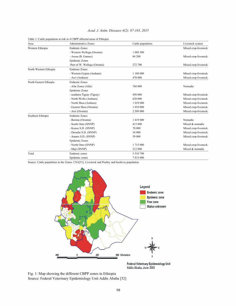
Table 1: Cattle population at risk in 4 CBPP affected areas of EthiopiaArea Administrative Zones Cattle population Livestock systemWestern Ethiopia Endemic Zones Mixed crop-livestock
- Western Wellega (Oromia) 1 005 500- Asosa (B. Gumuz) 84 200 Mixed crop-livestockEpidemic Zones-Part of W. Wellega (Oromia) 272 700 Mixed crop-livestock
North Western Ethiopia Endemic Zones- Western Gojam (Amhara) 1 188 000 Mixed crop-livestock- Awi (Amhara) 470 000 Mixed crop-livestock
North Eastern Ethiopia Endemic Zones- Afar Zones (Afar) 768 000 NomadicEpidemic Zones- southern Tigray (Tigray) 450 000 Mixed crop-livestock- North Wello (Amhara) 620 000 Mixed crop-livestock- North Shoa (Amhara) 1 018 000 Mixed crop-livestock- Eastern Shoa (Oromia) 1 019 000 Mixed crop-livestock- Arsi (Oromia) 2 509 000 Mixed crop-livestock
Southern Ethiopia Endemic Zones- Borena (Oromia) 1 419 000 Nomadic- South Omo (SNNP) 413 000 Mixed & nomadic- Konso S.D. (SNNP) 70 000 Mixed crop-livestock- Derashe S.D. (SNNP) 34 000 Mixed crop-livestock- Amaro S.D. (SNNP) 59 000 Mixed crop-livestockEpidemic Zones- North Omo (SNNP) 1 715 000 Mixed crop-livestock- Maji (SNNP) 212 000 Mixed & nomadic
Total Endemic zones 5 510 700Epidemic zones 7 815 000
Source: Cattle population in the Zones: CSA[51], Livestock and Poultry and beehives population
Fig. 1: Map showing the different CBPP zones in EthiopiaSource: Federal Veterinary Epidemiology Unit Addis Ababa [52]
Acad. J. Anim. Diseases 4(2): 87-103, 2015
99
Western Ethiopia including Western Wollega and causative Mycoplasma species. Sick animals for autopsyAssosa Zones (And possible a part of Gambella Region) and bacteriological specimen collection were out looked.are considered endemic and epidemic; Southern Ethiopia: Postmortem examination and sample collection was(Southern Nation, Nationalities and People region, performed on 7 recently dead animals. The clinical andSNNPR). Borena Zone as a whole (OromiaRegion), pathological findings encountered and the bacteriologicalinfected since long time, is an endemic area and as well as the biochemical tests performed, established thecharacterized by pastoralism; South Omo, KonsoDerashe outbreak to be CBPP [53].and Amaro Zones (SNNP Region) are considered asendemic, with recent outbreaks in the neighboring Zones Clinical and Necropsy Findings: Clinical examinations ofsuch as Bench Maji and North Omo Zones; Gondar and infected animals revealed nasal discharge, coughing,Gojam areas have declared numerous outbreaks since labored breathing, disinclination to move and postures1993 and South Gondar and West Gojam are categorized that showed the animal was fighting to get enoughas epidemic areas. West Gojam zone comprises of seven oxygen. The profound lesions observed on postmortemdistricts, namely Burie-Wonberma, Denbecha, Jabitenan, showed adhesion of the pleura with the chest wall and theDega-Damot, Quarit, Sekela and Achefer, of which the lung and consolidated lung tissues with characteristicfirst two districts are considered CBPP endemic and the marbling. The pleural cavity was full of copious,last four districts are considered CBPP free; the highlands yellowish-colored clear fluid. Heavy deposits of fibrinof North Shewa were considered as CBPP free, however, flocculates were encountered [53].Wondimu [49] reported sero-prevalence rate of 54% usingCFT; Southern Tigray seems to be recently infected with Bacteriological Findings: Evidence of the growth ofsero-prevealence rate of 50% reported in 1996 [49] and Mycoplasma organisms was based on a change in colorthis can be categorized as epidemic; AgewAwi zone ofthe growth medium from pink to yellow.Moderatecomprises of four districts, nameyDangela, Ankesha- turbidity with a whitish deposit atthe bottom of theguagusa, Gungua and Banja- shikudad, of which the first culture vessels were additional parameters used tothree are considered as CBPP endemic and the last is with determineMycoplasma growth. Both the tissuesporadic occurrence (Table 1). Here mixed crop-livestock sampleprocessed and the pleural fluid culturedproduction system is practiced and the dominant werepositive for Mycoplasma growth after incubation forlivestock species are cattle; North Eastern Ethiopia: Afar 72–120 hours in broth culture media.Gram-stained smearsRegion as a whole and Northern Somali Region may be from these culturesshowed the presence of gram-considered as endemic, with recent outbreaks negative, pleomorphic organisms composed ofencroaching on the edge of endemic are in Southern coccoid,cocco-bacillary and filamentousTigray, North Wello, North Shewa, East Shewa, (Amhara organisms.Giemsa-stained preparations from theRegion) and Arsi Zone (Oromia Region); Eastern Ethiopia: culturesuspensions revealed coccoid, pear-shaped,andIn Somali Region except one zone, Shinille, which is filamentous microorganisms. Growth onsolid medium wasconsidered to be CBPP epidemic zone, the status of the characterized by the presence of microcolonies with adisease in all the other zones is unknown. Once typical nipple-shaped appearance after 7 days ofintroduced to a new area, initial losses in pastoral incubation.The colonies were observed undercommunities can be very high and its eradication is very invertedmicroscope (32X) with transmitted light.difficult requiring major expenditure for control [49]. Biochemical and Biological Properties: Positive
The Cbpp Diagnosis in Ethiopia: Epidemiological fermentation, digitonin sensitivity,ability to pass throughinvestigation to obtain a general picture of the way the a 0.45- m membrane, growth inhibition and sensitivitydisease has behaved in the herd; clinical examination: how tochloramphenicol and tetracycline. Negativeresults werethe animals of a herd are affected by the disease; Post- seen for arginine hydrolysis,urea breakdown, growth onmortem examination to observe the characteristic lesions serum-freemedia and sensitivity to pencillin [16, 53].in organs of dead and\or slaughtered animals; laboratoryexamination to confirm the presence of infection [16]. Serological Tests: The compliment fixation test on serum
An outbreak of contagious bovine pleuropneumonia is still the most useful methods of detecting infection.(CBPP) was investigated in the Somali National Regional It is a rapid, simple and easy to perform andState, Eastern Ethiopia, to isolate and identify the interpret the results. It is more specific than ELISA tests.
resultswere seen for growth in aerobic conditions,glucose
Acad. J. Anim. Diseases 4(2): 87-103, 2015
100
It lacks sensitivity for serum samples having a very low Veterinarian should aware of the pastoralist about theantibody level. ELISA tests detect late and persistentinfections while CFT detects early infections [54].
CBPP Control Methods (Strategies) in Ethiopia: Themajor control method practiced in Ethiopia is vaccination.The control of CBPP by vaccination has been carried outfor the last 30 years. Previously consecutive yearlyblanket vaccination with combined Rinderpest and CBPPvaccine was adopted as a strategy to control CBPP. It wasthis strategy that is believed to have contained thedisease to a relatively low level until 1992/93. And thismethod was considered as a successful achievement inthe control of CBPP. However with the adoption of astrategy towards Rinderpest eradication, the vaccinationsin the highlands and most parts of the Somali region haveceased since 1992/93. Besides, the vaccination coveragewas around 50% and did not reach the desired 80 – 100%level. Currently, CBPP control in Ethiopia is based ontargeted and ring vaccination in the face of outbreaks [55].
CONCLUSIONS
Contagious bovine pleuropneumonia is highlycontagious disease of cattle caused by Mycoplasmamyciodes subspecies mycoides SC type. The disease isfound in different parts of the world; especially it is theproblem of developing countries, Ethiopia due to lack ofenough diagnostic tools, well trained personnel,economy, strategic epidemiological surveillance for theeradication of the diseases. The epidemiological situationof the disease is found in various parts of Ethiopia. It is anendemic disease in most parts of the country. Contagiousbovine pleuropneumonia is possing a major threat tocattle in many parts of the country thereby causingconsiderable economic losses through morbidity andmortality. Diagnosis of the disease in Ethiopia isperformed through clinical finding, necropsy finding,culturing and serological technique. The main controlstrategy in Ethiopia is done through vaccination; andsometimes control is done by restricting the movement ofcattle. However, vaccination cover is usually not veryhigh, due to financial and government policy constraints.Therefore, based on the above conclusions, the followingrecommendations are forwarded.
Adequate funding should be available to controlCBPP in Ethiopia, as well as other countries.Research in the improvements of vaccine shouldcontinue and include the possibility of differentiationbetween vaccinated and non-vaccinated animals.
problem of the disease. Strategic control of CBPP should be progressive andbased on impact assessment and cost benefitanalysis done with appropriate methods includingparticipatory techniques to cover regional, national,zone level.A veterinarian should well trained to performdiagnosis of the disease through the availablediagnostic tools.Research should be done on the epidemiologicalsituation of the disease
REFERENCES
1. Office International Des Epizooties (OIE), 2008.Manual of Diagnostic Tests and Vaccines forTerrestrial Animals (Mammals, birds and bees),6 ed., (Office International Des Epizooties, Paris),th
pp: 712-724.2. Andrews, A.H., R.W. Blowey, H. Boyd and R.G.
Eddy, 2004. Bovine Medicine Diseases andHusbandry of Cattle, 2 ed., (Blackwell publishing, And
Gay, ustralia), pp: 868-874.3. Tambi, N.E., W.O. Maina and C. Ndi, 2006. An
estimation of the economic impact of contagiousbovine pleuropneumonia in Africa, Revuescientifiqueet technique - Office International DesÉpizooties, 25: 999-1012.
4. Schubert, E., K. Sachse, J. Jores and M. Heller, 2011.Serological testing of cattle experimentally infectedwith Mycoplasma mycoides subsp. mycoides smallcolony using four different tests reveals a variety ofseroconversion patterns, BMC veterinary Research,7: 72.
5. Nicholas, R, R. Ayling and L. McAuliffe, 2008.Mycoplasma diseases of ruminants, (CABinternational, Biddles Ltd, Kings Lynn Norfolk, UK.),pp: 69-97.
6. Neiman, M., C. Hamsten, J.M. Schwenk, G. Bo¨lskeand A. Persson, 2009. Multiplex Screening of SurfaceProteins from Mycoplasma mycoides subsp.mycoides Small Colony for an Antigen CocktailEnzyme-Linked Immunosorbent Assay, Clinical andvaccine Immunology, 16: 1665-1674.
7. Sacchini, F., M. Luciani, R. Salini, M. Scacchia, A.Pini, R. Lelli, J. Naessens, J. Poole and J. Jores, 2012.Plasma levels of TNF- TNF- IFN- , IL-4 and IL-10during a course of experimental contagious bovinepleuropneumonia, BMC Veterinary Research, 8: 44.
Acad. J. Anim. Diseases 4(2): 87-103, 2015
101
8. Amanfu, W., 2009. Contagious bovine 19. Belachew, H. and E. Jemberu, 2003. Challenges andpleuropneumonia (lung sickness in Africa, opportunities of livestock marketing in Ethiopia. In:Onderstepoort Journal of Veterinary Research, Yilma, J. and Gatachew, G. (eds), Proceedings of the76: 13-17. 10 annual conference of the Ethiopian Society of
9. Radiostits, O.M., C.C Gay, K.W. Hinchcliff and Animal Production (ESAP) held in Addis Ababa,P.D. Constable, 2007. Veterinary Medicine, a Ethiopia, August 24-26, 2002.Textbook of the Diseases of Cattle, Sheep, Pigs, 20. Office International Des Épizooties (OIE), 2002.Goats and Horses, 10 ed., (Sounders Elsevier, Manual of Standards for Diagnostic Tests andth
Spain), pp: 1131-1135. Vaccines.10. Radiostits, O.M., D.C. Blood and C.C. Gay, 1994. 21. Walker, L.R., 1999. Mollicutes: In Hirsh, D. C. and
Veterinary Medicine: A textbook of the diseases of Zee, Y. C. Veterinary microbiology. Blackwellcattle, sheep, pigs, goats and horses. 8 ed. Baillière Science, Inc., pp: 165-172.th
Tindall, pp: 910-913. 22. Kasper, D.L., A.S. Fauci, D.C. Longo, E. Brownwald,11. Thomson, G.R., 2005. Contagious bovine S.L. Houser and J.L. Jameson, 2005. Horison’s
pleuropneumonia and poverty. A strategy for principles of internal medicine. 16 ed.USA: Mcrrowaddressing the effects of the disease in sub- Saharan Hill., pp: 1008-1009.Africa, Research report, (DFID animal health 23. Quinn, P.J., M.E. Carter, B. Markey and G.R. Carter,programme, centre for Tropical Veterinary Medicine, 1994. Clinical Veterinary Microbiology, 1 . Mosby,University of Edinburgh, UK). London, pp: 320-325.
12. Huschle, O., K. Godinho and R.A.J. Nicholas, 2004. 24. Office International Des Épizooties (OIE), 2000.Denofloxacin treatment of cattle affected by CBPP. Consultative group on Contagious BovineVet. Rec., 155: 404. Pleuropneumonia (CBPP). Report of second meeting.
13. Goffe, C. and F. Thiaucourt, 1998. A competitive Reviving progressive control of CBPP in Africa,ELISA for the specific diagnosis of contagious Rome, Italy.bovine pleuropneumonia (CBPP). Vet. Microbial, 25. Provost, A., P. Perreau, A. Breard, C. Le Goff,60: 179-191. J.L. Martel and G.S. Cottew, 1987. Péripneumonie
14. Tegegne, A., T. Mengistie, T. Desalew, W. Teka and contagious bovine. Rev. Sci. Tech. Off. Int. Epiz.,E. Dejen, 2009. Transhumance cattle production 6: 565-624.system in North Gondar, Amhara Region, Ethiopia: Is 26. Masiga, W.W. and R.S. Windsor, 1978. Someit sustainable? Improving Productivity and Market evidence of age susceptibility to CBPP. Research inSuccess (IPMS) of Ethiopian Farmers Project, Veterinary Science, 2: 333.(International Livestock Research Institute (ILRI), 27. Quinn, P.J., B.K. Markey, M.E. Carter, W.J. DonnellyAddis Ababa, Ethiopia). and F.C. Leonard, 2002. Veterinary Microbiology and
15. Ministry of Agriculture (MOA), 2003. Monthly Microbial Disease, 2 ed. Blackwell science, USA,animal health status report; Ministry of Agriculture pp: 189-195.Veterinary Services, Epidemiology Unit, Addis 28. Windsor, R.S. and W.N. Masiga, 1977. InvestigationsAbaba, Ethiopia. in to the roles of carrier animals in the spreads of
16. Gedlu, M., 2004. Serological, clinical and participatory CBPP. Res. Vet. Sci., 23: 224-229.epidemiological survey of CBPP in Somali Region, 29. Hirsh, D.C., N.J. Maclachlan and R.L. Walker, 2004.Ethiopia. MSc thesis, Addis Ababa University, Veterinary microbiology. 2 ed. Blackwellscience,Faculty of veterinary medicine, Debreziet, Ethiopia. pp: 240-243.
17. Yigezu, L.M. and F. Roger, 1997. CBPP European 30. Rosendal, S., 1993. Mycoplasma. In: Gyles, C. L andUnion Project component 2: Improvement of Charles, O. T. Pathogenesis of bacterial infections indiagnostic methods competitive ELISA Kit animals. 2 ed. Ames, IA: Iowa State University,assessment Report of the second semester year 2. pp: 297-311.National Veterinary Institute, Ethiopia. 31. FAO EMPRES Unit, 1997. Recognizing CBPP, A Field
18. Afework, Y., 2000. Analysis of CBPP situation in Manual for Recognition. EMPRES FAO AnimalEthiopia, Past and Present. Ministry of Agriculture, Health Service Animal Production and HealthAddis Ababa, Ethiopia. Division Rome, Italy.
th
th
st
nd
nd
nd
Acad. J. Anim. Diseases 4(2): 87-103, 2015
102
32. FAO, 2004. Animal diseases control issues and Competitive ELISA and LppQ ELISA with Post-impacts. FAO Corporate Document Repository, mortem Findings in the Diagnosis of Contagiousproject on livestock industrialization, Trade and Bovine Pleuropneumonia (CBPP), Tropical Animalsocial health. Health and Production, 43: 1057-106.
33. Masiga, W. N., J. Domenech and R. S. Windsor, 43. Laval, G., 1999. Cost analysis of contagious bovine1996. Manifestation and epidemiology of Contagious pleuropneumonia in Ethiopia, (Unpublished MScBovine Pleuropneumonia in Africa. Rev. Sci. Tech. thesis, Claude Bernard University).off. Int. Epiz., 15: 1241-1262. 44. Tulasne, J.J., J.K. Litamoi, B. Moreine, L. Dedieu,
34. Freundt, E.A., H. Erno and R.M. Lemcke, 1999. V.J. Palypa, M. Yami, A. Izzeldin, D. Sylla andIdentification of mycoplasmas. In: Methods in A. Bensaid, 1996. CBPP vaccines, Actual SituationMicrobiology, Vol. 13, Bergen T. & Norris J.R., eds. and need for improvements. Scientific review of theAcademic Press, London, UK, pp: 377-396. OIE 1996 on animal mycoplasmoses 1373- 1396.
35. Martel, J.L., R. Nicholas, J. Noorduizen, A. Pini and Vet. Microbial., 47: 305-315.J. Regalla, 2004. Diagnostic tests for contagions 45. MOA, 2002. Monthly Animal Health Statusbovine pleuropneumonia. Report of the Scientific Report; Ministry of Agriculture VeterinaryCommittee on Animal Health and Animal Welfare. Services, Epidemiology Unit. Addis Ababa,Commission of the European Community. Ethiopia.
36. Niang, M., M. Diallo, O. Cisse, M. Kone, M. 46. Regassa, F., 2001. Herd prevalence of ContagiousDoucoure, J.A. Roth, V. Balcer-Rodrigues and Bovine Pleuropneumonia (CBPP), BovineDedieu, 2006. Pulmonary and serum antibody Tuberculosis and Dictyocaulosis in Bodjiworeda,responses elicited in zebu cattle experimentally west wellega. Addis Ababa University, Faculty ofinfected with Mycoplasma mycoides subsp. Veterinary Medicine, Debrezeit, Ethiopia, DVMmycoides SC by contact exposure. Vet. Res., thesis.37: 733-38. 47. Beyene, D., 1997. Sero-epidemiological investigation
37. Taylor, T.K., J.B. Bashiruddin and A.R. Gould, 1992. of CBPP in Illubabor and Wellega. Addis AbabaRelationships between members of the Mycoplasma University, Faculty of Veterinary Medicine,mycoides cluster as shown by DNA probes and DebreZeit, Ethiopia, DVM thesis.sequence analysis. Int. J. Syst. Bact., 42: 593-601. 48. Takele, G., 1998. Epidemiological Survey of CBPP in
38. Otte, M.J., R. Nugent and A. McLeod, 2004. Awi and Western Gojam zone of Amhara Region andTransboundery animal diseases: Assessment of Comparison of CFT and C-ELISA for the Diagnosissocio-economic impacts and institutional responses, of CBPP. Addis Ababa University and FreeLivestock policy discussion paper No.9, (Food and University of Berlin, MSc thesis.Agriculture Organization Livestock Information and 49. Wondimu, D., 1996. Contagious BovinePolicy Branch, AGAL). Pleuropneumonia (CBPP): Prevalence and Evaluation
39. Thrusfield, M., 2005. Veterinary epidemiology. 3 ed. of Post-Vaccination immune response (North Omo,rd
Black well., pp: 384-386. Konso&Dirashe Regions/Ethiopia). Addis Ababa40. Msami, H.M., T. Ponela-Mlelwa, B.J. Mtei and University, Faculty of Veterinary Medicine,
A.M. Kapaga, 2001. Contagious Bovine Debrezeit, Ethiopia, DVM thesis.Pleuropneumonia in Tanzania: Current status. 50. NAHRC, 2000. Report on CBPP Study in Ethiopia.Tropical Animal Health and Production, 33: 21-28. Serosurveillance results National Animal Health
41. Litamoi, J.K., 2000. Overview of CBPP vaccine Research Centre Ethiopian Agricultural Researchproduction and quality in Africa. In: Report of Organization (EARO) Sebeta, Ethiopia.second meeting of the FAO/OIE/OAU/IAEA 51. CSA, 1998. Agricultural Sample Survey 1997/98,consultative group on Contagious Bovine volume II. Statistical bulletin Number 193, CSA,Pleuropneumonia (CBPP). Rome, Italy. Addis Ababa, Ethiopia.
42. Muuka, G., B.M. Hang'ombe, K.S. Nalubamba, 52. Federal Epidemiology Unit, 2003. National policy andS. Kabilika, L. Mwambazi and J.B. Muma, 2011. strategy for the control of CBPP. Addis Ababa,Comparison of Complement Fixation Test, Ethiopia.
Acad. J. Anim. Diseases 4(2): 87-103, 2015
103
53. Regassa, F., E. Gelaye, A. Zeleke and T. Sori, 2005. 55. MOA, 1997. Livestock Development project. MinistryIsolation and identification of MmmSC Bovine of Agriculture, the Federal Democratic Republic ofbiotype in Eastern Ethiopia. Intern. J. Appl. Res. Vet. Ethiopia. Addis Ababa, Ethiopia.Med., 3(1): 32-38.
54. Kassaye, D. and W. Molla, 2012. Seroprevalence ofCBPP at export quarantine centers in and aroundAdama, Ethiopia. Tropical animal production, 41: 1-7.