Embed Size (px)
Citation preview

Connective Tissue
Mamoun KremliAl-Maarefa College

Objectives• What is connective tissue• Types of connective tissues• Functions of connective tissues– Relation of structure and function

Tissues • Four fundamental tissues are recognized:– Epithelial tissue– Connective tissue– Muscular tissue– Nervous tissue

Connective Tissue• Consists of two basic
elements:– Cells, and– Extra-cellular matrix (abundant)
(dominant part)• Fibers, and• Ground substance
– liquid, gel, or solid
• Function– Binds and/or supports other
tissue

Connective Tissue• Connective tissue is clearly different from
neighboring tissues

Connective Tissue• Connective tissue is clearly different from
neighboring tissues

Connective Tissue• Connective tissue is clearly different from
neighboring tissues

Connective Tissue Cells• Fibroblasts:– Secrete both fibers and ground substance of the
matrix (wandering)
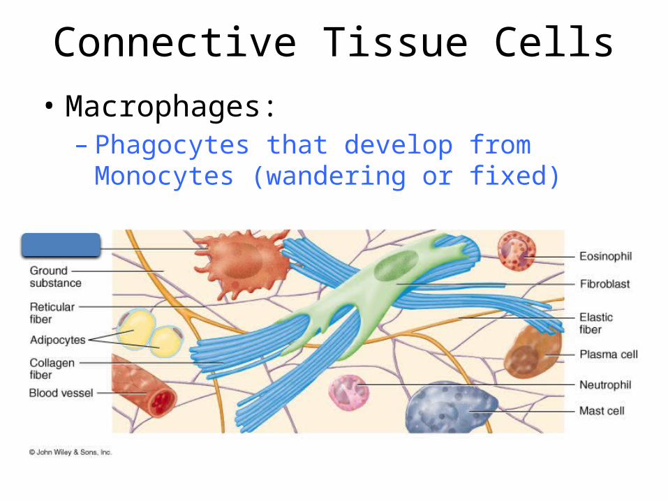
Connective Tissue Cells• Macrophages:– Phagocytes that develop from Monocytes
(wandering or fixed)

Connective Tissue Cells• Plasma Cells:– Antibody secreting cells that develop from B-
Lymphocytes (wandering)

Connective Tissue Cells• Mast Cells– Produce histamine that help dilate small blood
vessels in reaction to injury (wandering)

Connective Tissue Cells• Adipocytes:– Fat cells that store triglycerides, support, protect
and insulate (fixed)

Connective Tissue Cells

Fibroblasts• Active fibroblasts have extensions
Extensions of fibroblasts (arrow-heads) are seen with the cell or alone, depending on section plane

Fibroblasts• Active fibroblasts have extensions
Electrom micrograph of fibrocyte with cytoplasmic extensions interdigitating among collagen fibers, X 26,000

Matrix Fibers• Collagen Fibers• Elastic Fibers• Reticular Fibers

Matrix Fibers• Collagen Fibers:– Large fibers made of the protein collagen– The most abundant fibers– Promote tissue flexibility

Matrix Fibers• Elastic Fibers:– Intermediate fibers made of the protein Elastin– Branching fibers that allow for stretch and recoil
Polarizing microscopypicrosirius-stained collagen,elastic fibers are stained by Orcein

Matrix Fibers• Reticular Fibers:– Small delicate, branched fibers– Have same chemical composition of Collagen– Forms structural framework for organs such as
spleen and lymph nodes.

Matrix Fibers
Collagen
Elastin

Elastic and Collagen Fibers

Matrix Ground Substance • Hyaluronic Acid:– Complex combination of polysaccharides and
proteins found in “true” or proper connective tissue
• Chondroitin sulfate:– Jellylike ground substance of cartilage, bone, skin
and blood vessels• Other ground Substances:– Dermatin sulfate, keratin sulfate, and
adhesion proteins

Types of Connective Tissue1. True (Proper) Connective Tissue– Loose Connective Tissue• Aereolar, Adipose, Reticular
– Dense Connective Tissue
2. Supportive Connective Tissue– Cartilage– Bone
3. Liquid Connective Tissue– Blood

Loose Connective Tissue• Areolar tissue–Widely distributed under epithelia
• Adipose tissue–Hypodermis, within abdomen, breasts
• Reticular connective tissue– Lymphoid organs such as lymph nodes

Areolar Connective Tissue• Structure:
– all 3 types of fibers– several types of cells– semi-fluid ground substance
• Present in:– subcutaneous layer– mucous membranes– around blood vessels, nerves
and organs• Function:
– strength, support and elasticity

Adipose Connective Tissue:• Structure:
– adipocytes; "signet ring" appearing fat cells. They store energy in the form of triglycerides (lipids)
• Present in:– subcutaneous layer– around organs– yellow marrow of long bones
• Function:– supports, protects and
insulates– serves as an energy reserve

Adipose Connective Tissue

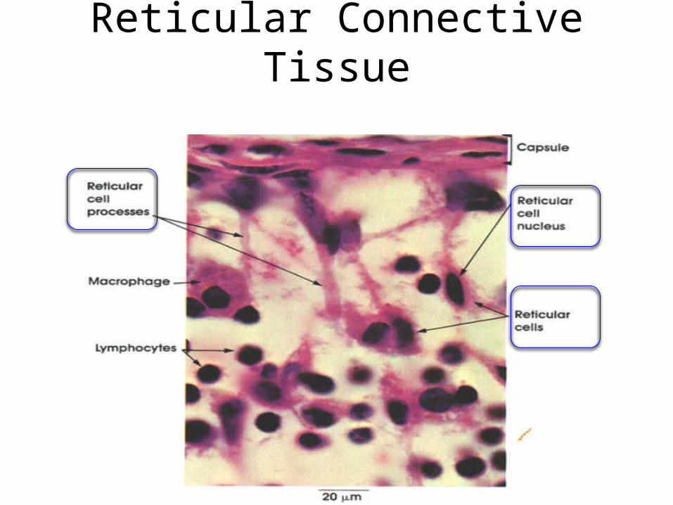
Reticular Connective Tissue• Structure:
– fine interlacing reticular fibers
– reticular cells• Present in:
– liver, spleen and lymph nodes
• Function:– forms the framework
(stroma) of organs– binds together smooth
muscle tissue cells

Reticular Connective Tissue• Structure:
– fine interlacing reticular fibers
– reticular cells• Present in:
– liver, spleen and lymph nodes
• Function:– forms the framework
(stroma) of organs– binds together smooth
muscle tissue cells
Reticular Connective Tissue

Reticular Connective Tissue
Reticular Fibers
Collagen Fibers
Thyroid gland, Scanning electron microscopy, X 2500Kuehnel, Color Atlas of Cytology, Histology, and Microscopic Anatomy

Types of Connective Tissue1. True (Proper) Connective Tissue– Loose Connective Tissue• Aereolar, Adipose, Reticular
– Dense Connective Tissue
2. Supportive Connective Tissue– Cartilage– Bone
3. Liquid Connective Tissue– Blood

Dense Connective Tissue• Contains more numerous and thicker fibers
and far fewer cells than loose CT
• Types:– Dense regular connective tissue• Tendons and ligaments
– Dense irregular connective tissue• Dermis of skin, submucosa of digestive tract

Dense Regular Connective Tissue• Structure:– bundles of collagen fibers
and fibroblasts• Present in:– Tendons,– Ligaments– aponeuroses
• Function:– provides strong
attachment between various structures Tendon

Dense Regular Connective Tissue

Dense Irregular Connective Tissue• Structure:– randomly-arranged collagen
fibers and– few fibroblasts
• Present in:– fasciae,– dermis of skin– joint capsules– heart valves
• Function:– provides strength

Dense Irregular Connective Tissue• Structure:– randomly-arranged collagen
fibers and– few fibroblasts
• Present in:– fasciae,– dermis of skin– joint capsules– heart valves
• Function:– provides strength Eyelid, Azan stain
Kuehnel, Color Atlas of Cytology, Histology, and Microscopic Anatomy

Dense Irregular Connective Tissue• Structure:– randomly-arranged collagen
fibers and– few fibroblasts
• Present in:– fasciae,– dermis of skin– joint capsules– heart valves
• Function:– provides strength
Renal capsule, Scanning electron microscopy, X 5000
Kuehnel, Color Atlas of Cytology, Histology, and Microscopic Anatomy

Types of Connective Tissue1. True (Proper) Connective Tissue– Loose Connective Tissue• Aereolar, Adipose, Reticular
– Dense Connective Tissue
2. Supportive Connective Tissue– Cartilage– Bone
3. Liquid Connective Tissue– Blood

Cartilage• Structure:
– Jelly-like matrix (chondroitin sulfate)
– collagen and elastic fibers– Chondrocytes (within spaces in
the matrix called lacunae)– surrounded by a membrane
(perichondrium)– has NO blood vessels or nerves
except in the perichondrium• Function:
– Collagen fibers provide strength– chondroitin sulfate provides
resilience
Perichondrium Perichondrium
Hayaline Cartilage

Cartilage• Types:–Hyaline cartilage– Fibro-cartilage– Elastic cartilage

Hyaline Cartilage• Most abundant type• Structure:
– Fine collagen fibers embedded in a gel-type matrix
– Occasional chondrocytes inside lacunae
• Present in:– embryonic skeleton– at the ends of long bones (joints)– in the nose and in respiratory
structures• Function:
– flexible, provides support– allows movement at joints

Hyaline Cartilage

Hyaline Cartilage• Covers articular surfaces

Fibrocartilage• Structure
– bundles of collagen in the matrix that are usually more visible under microscopy
• Present in:– Intervertebral discs,– Menisci of the knee,– Pubic Symphysis,– Tendon insertion on apophyseal
hayaline cartilage• Function:
– Support and fusion– shock absorption

Fibrocartilage

Fibrocartilage
Picrosirius-Hematoxilin stain of fibrocartilage, with abundant collagen fibers

Elastic Cartilage• Structure– Threadlike network of
elastic fibers within the matrix
• Present in:– external ear– auditory tubes– epiglottis
• Function:– gives support,– maintains shape– allows flexibility

Elastic Cartilage
Resorcin stain selectively staining the elastic fibers of elastic cartilage tissueCells are not stained

Elastic Cartilage
1 Elastic fibers, 2 Cartilage Cells, 3 perichondrium
Kuehnel, Color Atlas of Cytology, Histology, and Microscopic Anatomy

Types of Connective Tissue1. True (Proper) Connective Tissue– Loose Connective Tissue• Aereolar, Adipose, Reticular
– Dense Connective Tissue
2. Supportive Connective Tissue– Cartilage– Bone
3. Liquid Connective Tissue– Blood

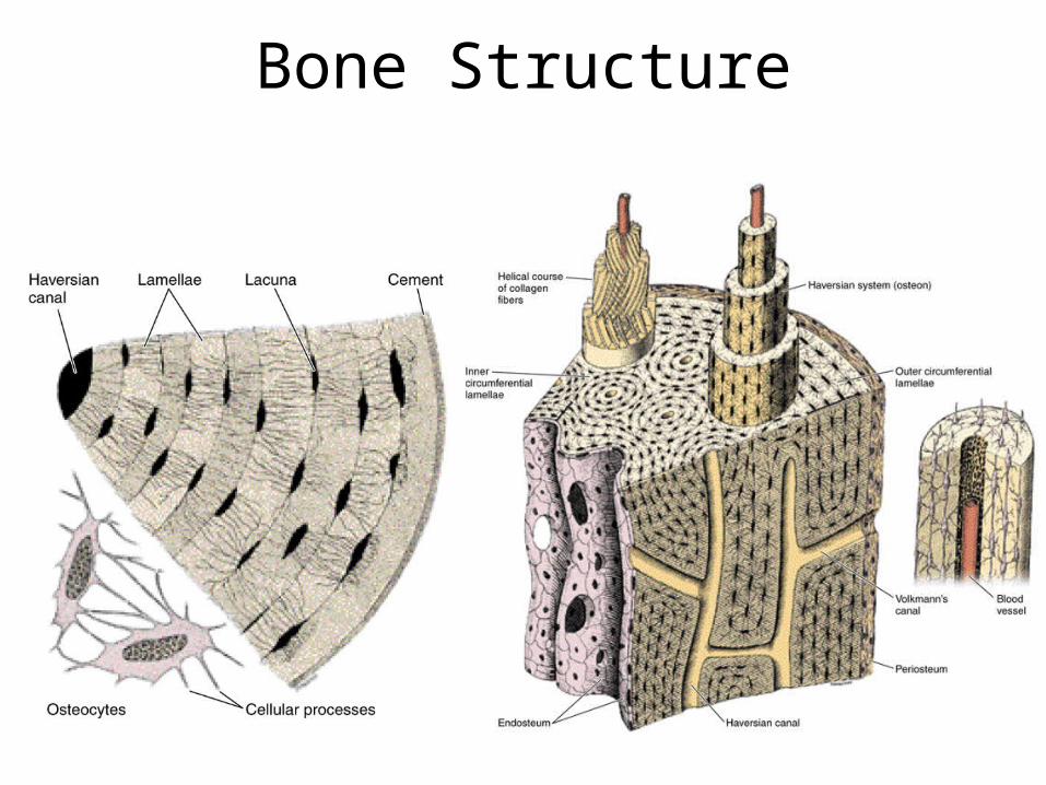
Bone• Structure– The hardest CT– Osteocytes in small cavities- lacunae– Impregnated with calcium salts
• Types:– Spongy (cancellous)– Compact (cortical)

Bone Types• Spongy (cancellous)– Loose rods of bones– Found inside body
of bones, and ends of arms and legs
• Compact (cortical)– Tightly organized– Found in shafts of
long bones

Bone Structure
Cancellous Bone
Cortical Bone

Bone Structure
Bone Structure

Bone Structure

Bone Structure
Section of a Haversian system (Osteone)

Bone Cells• Osteoblasts:– build bone – Bone deposition
• Osteocytes:– Osteoblasts: surrounded
by the matrix they formed
• Osteoclasts:– resorb (eat) bone– Bone resorption

Bone Cells• Osteoblasts:– build bone
• Osteocytes:– osteoblasts surrounded by matrix they formed

Bone Cells• Osteoclasts:– Resorb (eat) bone

Bone Cells• Osteoclasts:– Resorb (eat) bone

Types of Connective Tissue1. True (Proper) Connective Tissue– Loose Connective Tissue• Aereolar, Adipose, Reticular
– Dense Connective Tissue
2. Supportive Connective Tissue– Cartilage– Bone
3. Liquid Connective Tissue– Blood– Lymph

Blood• RBC• Neutrophils• Lymphocytes• Monocytes• Platelets

Blood• RBC• Neutrophils• Lymphocytes• Monocytes• Platelets
www.lab.anhb.uwa.edu.au

Blood• RBC• Neutrophils• Lymphocytes• Monocytes• Platelets
www.lab.anhb.uwa.edu.au

Blood• RBC• Neutrophils• Lymphocytes• Monocytes• Platelets
www.lab.anhb.uwa.edu.au

Blood• RBC• Neutrophils• Lymphocytes• Monocytes• Platelets
www.lab.anhb.uwa.edu.au

LymphContains lymphatic fluid and WBC

Summary• What is connective tissue• Structure: Consists of two basic elements:– Cells, and– Extra-cellular matrix (abundant) (dominant part)• Fibers, and• Ground substance (liquid, gel, or solid)
• Function– Binds and/or supports other tissue

Summary
1. True (Proper) Connective Tissue– Loose CT (areolar, adipose, reticular)– Dense CT (regular, irregular)
2. Supportive Connective Tissue– Cartilage– Bone
3. Liquid Connective Tissue– Blood– Lymph
Types of Connective Tissue: