Embed Size (px)
Citation preview

FluorescentHydrophobicpolymer core
Conjugated Targeting moieties bound to surface: e.g. Antibody / oligonucleotide / protein / fab fragments / azide / streptavidin
Iron Oxide or Gadolinium (Magnetic manipulation / MRI)
Conjugated Polymer Nanoparticles (CPNs™) Highly Fluorescent Imaging Agents for Multiple Cellular Applications
Alderley ParkNether Alderley, Cheshire, SK10 4TG, [email protected]
Conjugated Polymer Nanoparticles:CPNs™ are exceptionally fluorescent, nanoparticulate,labelling probes that can be utilised in cellular imagingand diagnostics applications. They are highly stable in awide range of pHs and temperatures, and produce veryintense light emissions that are immensely brighter thanconventional technologies. CPNs™ do not photo-bleach,(with over 12 months of stability), allowing significantlymore sensitive detection of analytes than otherfluorophores in techniques such as immunocytochemistry,ELISA and flow cytometry. The greater stability alsomakes the results highly reproducible. CPNs™ can betaken up by cells through endocytosis or targeted tospecific regions by linking them to antibodies, proteins,nucleotides or other targeting moieties. CPNs™ matchstandard fluorescent filters and laser lines, and areavailable in a range of wavelengths covering the visiblespectrum – including near-IR.
Why are they so much Brighter, Sensitive and Stable?Derived from TV display OLED technology, the polymer core was originally designed to cope with years ofelectrical excitation, making it extremely stable. Quantum Yield (QY) is frequently and mistakenly assumedas a measure for brightness while the Extinction Coefficient’s (ExCoeff) are often ignored. However, it isthe extremely large ExCoeffs that give CPNs™ their incredible brightness and sensitivity.
Brightness = QY x ExCoeff
Structural Properties:CPNs™ are water-soluble micelles with a light-emitting polymer core and are manufactured at anapproximate size of 70-80 nm, although particles can be as small as 15-50 nm. Encapsulation of the corewithin a biocompatible surfactant also increases its hydrophilicity and this ‘core-shell’ structure provides aready base for the covalent bonding of functionalising molecules for targeting proteins or nucleic acids.CPNs™ also incorporate iron oxide, allowing CPNs™ and the molecules or cells to which they areattached to be manipulated using magnets to direct movement and facilitate purification. The iron oxidecan also be visualised by utilising Magnetic Resonance Imaging (MRI) as a contrast reagent.

Biological Properties:The intense brightness of CPNs™dramatically increases the sensitivity ofapplications such as flow cytometry,ELISA, IHC and microscopy, with singlenanoparticles being detectable in flowcytometry and immunocytochemistry. Thisenables the study of individual proteins insamples and cells. Streptavidin andantibodies can be covalently conjugated toCPNs™ via the surfactant’s carboxylicacid groups using N-ethyl-N’-dimethylaminopropyl-carbodiimide (EDC)chemistry. These targeted CPNs™ can be
0
10000
20000
30000
40000
50000
60000
70000
80000
0 20 40 60 80 100 120
FI
Sig
na
l
[CRP] µg / ml
FI intensity vs CRP concentration for 4 concentrations of detection Ab at 1µg / ml capture Ab
10 ng / ml 20 ng / ml 50 ng / ml 100 ng / ml
0
10000
20000
30000
40000
50000
60000
70000
0 20 40 60 80 100 120
FI
Sig
na
l
[CRP] µg / ml
FI intensity vs CRP concentration for 4 concentrations of detection Ab at 20µg / ml capture Ab
10 ng / ml 20 ng / ml 50 ng / ml 100 ng / ml
EliSA:
Cells 293T adherent: 24well plate – 1ml volumewith added 1µl 10µl or100µl 475 CPN. Culturetime 48 hr. Analysis byflow cytometry & confocalmicroscopy.
Flow Cytometry
readily used in existing assays, with the increased brightness improving performance and increasingsensitivity. When conjugated to an oligonucleotide, the combination is thermally stable, requiring nocold storage. Other surface chemistries are also available, such as Thiol and Azide for click chemistry.
CPNs™ can be linked to a wide range of antibodies such as CD-4, CD-8, CD-34, INF-g, MOC-31, andmouse/rabbit IgG. Utilisation of the magnetic properties of CPNs™ during conjugation, by simple, carefulplacing of a magnetic, significantly increases binding. When applied to a range of applications, such asELISA, this can further increase sensitivity.
Dot blot for 1ul dots of IgG at indicated quantities probed with CPN550+anti-Mouse IgG.
0
0.1
0.2
0.3
0.4
0.5
0.6
0.7
0.8
-Strep +Strep +Strep + Mag
Increase in binding due to magnet
CPN concentration and incubation time for live uptake experiments
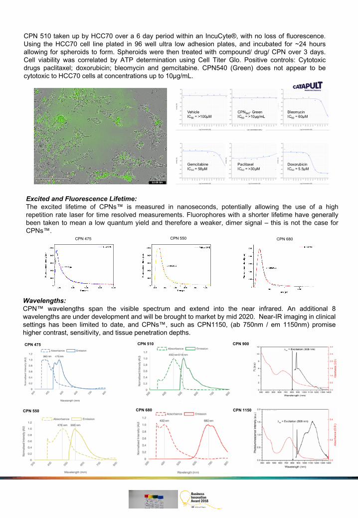
CPN 510 taken up by HCC70 over a 6 day period within an IncuCyte®, with no loss of fluorescence.Using the HCC70 cell line plated in 96 well ultra low adhesion plates, and incubated for ~24 hoursallowing for spheroids to form. Spheroids were then treated with compound/ drug/ CPN over 3 days.Cell viability was correlated by ATP determination using Cell Titer Glo. Positive controls: Cytotoxicdrugs paclitaxel; doxorubicin; bleomycin and gemcitabine. CPN540 (Green) does not appear to becytotoxic to HCC70 cells at concentrations up to 10μg/mL.
Wavelengths:CPN™ wavelengths span the visible spectrum and extend into the near infrared. An additional 8wavelengths are under development and will be brought to market by mid 2020. Near-IR imaging in clinicalsettings has been limited to date, and CPNs™, such as CPN1150, (ab 750nm / em 1150nm) promisehigher contrast, sensitivity, and tissue penetration depths.
CPN 550
CPN 475 CPN 510
CPN 1150CPN 680
CPN 900
Excited and Fluorescence Lifetime:The excited lifetime of CPNs™ is measured in nanoseconds, potentially allowing the use of a highrepetition rate laser for time resolved measurements. Fluorophores with a shorter lifetime have generallybeen taken to mean a low quantum yield and therefore a weaker, dimer signal – this is not the case forCPNs™.
CPN 680CPN 475 CPN 550

Protocol for Conjugation of CPN™ to Targeting Molecules, e.g. Streptavidin
CPNs™ are readily conjugated to proteins using EDC (N-ethyl-N’-dimethylaminopropyl-carbodiimide) tolink amine groups (-NH2) on the protein to the carboxyl groups on the surface of the CPN™ (-COOH).Attachment of proteins such as antibodies or streptavidin will generate highly selective CPNs™ for thedetection of target molecules. The brightness of the CPNs™ ensures the detection is highly sensitive, withsingle molecules being detected in flow cytometry and immunocyto / histochemistry. The affinity oftargeting molecules varies greatly and an initial titration of the CPN™: targeting molecule ratio will need tobe undertaken. Similarly, final usage dilution of the CPN™ - targeting molecule conjugate will need to bedetermined empirically.
Protocol
Reagents
1. Add 50µl CPN™ (0.1mg/ml) to 1.5ml tube
2. Add 1µl of HEPES 1M
3. Add 1µl PEG 8000 5% w/v
4. Add 3µl streptavidin (1mg/ml) and vortex (several ratios of CPN™: targeting molecule should be tested to identify the optimum conditions, e.g. 10-100μl targeting molecule (1mg/ml))
5. Add 1µl freshly prepared EDC solution 5mg/ml
6. Shake mixture for 4 hours at room temperature
7. The CPN™:Streptavidin conjugates can then be purified from the reaction components by precipitating them using a magnet.
8. Re-suspend in 20μl sterile Resuspension Buffer (HEPES (20mM), BSA 0.1%, Tween-20 0.02%)
HEPES (1M), pH 7.4
PEG 8000 (5% w/v)
Streptavidin (1mg/ml)
EDC [N-(3-Dimethylaminopropyl)-N’-ethylcarbodiimide hydrochloride] 5mg/ml (Freshly made and used immediately. Discard any unused)
Bovine Serum Albumin (10% w/v)
Tween-20
Neodymium magnet