Embed Size (px)
Citation preview
1356
CORRESPONDENCE
Is Human T-Cell Lymphotropic Virus Type I MoreClever than Human Immunodeficiency Virus Type 1?
SIR—Casseb [1] hypothesizes that human T-cell lymphotropicvirus type I (HTLV-I) is “more clever” than human immuno-deficiency virus type 1 (HIV-1). One of his reasons is thatHTLV-I, unlike HIV-1, has “an exceptionally low capability forcausing death in the host.” “Cleverness” is not a biologicalconcept but rather an anthropomorphism. Casseb probably isreferring to the biological success of a virus.
The only measure of the success of an organism is Dar-winian—that is, successful viruses transmit more copies of theirgenes to new hosts, and unsuccessful ones transmit fewer (ornone, thus dying out completely). As has been pointed out, itis not necessarily in the evolutionary interest of a virus to havelow pathogenicity [2]. For example, some arthropod-borne vi-ruses are transmitted more efficiently when their host is debil-itated and unable to fend off insect vectors. HIV-1 seems to beparticularly successful in transmitting itself to new hosts, al-though most transmissions probably take place when the hostis feeling well. It is probable that host death caused by HIV-1is merely an unfortunate (from the host’s point of view) epi-phenomenon, although in fact we do not know if this is thecase. HTLV-I may seem “clever” because it does not cause hostmortality, but we cannot say that HIV-1 is less “clever” becauseit often kills its host. HIV-1, however deadly, seems to be re-markably evolutionarily successful at this point in time, andthere seems to be no shortage of hosts for the virus to infectin the future.
Daniel B. HrdyDivision of Infectious and Immunologic Diseases,
University of California Davis Medical Center,Sacramento, California
References
1. Casseb J. Is human T-cell lymphotropic virus type I more clever than humanimmunodeficiency virus type 1? Clin Infect Dis 1998;27:1309–10.
2. Ewald P. Evolution of infectious diseases. New York: Oxford University Press,1994.
Reprints or correspondence: Dr. Daniel B. Hrdy, 21440 Road 87, Winters, CA95694.
Clinical Infectious Diseases 1999;29:1356q 1999 by the Infectious Diseases Society of America. All rights reserved.1058-4838/1999/2905-0059$03.00
Reply
SIR—Hrdy points out that HIV type 1 (HIV-1) seems to beremarkably successful evolutionarily. This observation is not indisagreement with my published hypothesis [1]. I agree that
viral adaptation in the host is critical for biological success,and it is usually driven by natural selection. However, my pointwas that human T cell lymphotropic virus type I (HTLV-I) isbetter adapted to humans than is HIV-1 at this time. The reasonis that it is a less pathogenic virus, and so has more time fortransmission. Natural viral adaptation is slow and gradual, andin the case of HTLV-I in humans, it has probably takenthousands of years. In contrast, HIV-1 has not had time toadapt; it is thought to have begun infecting humans much morerecently, only a few decades ago [2].
During adaptation, the transmission of some genes may con-tribute to the stability of the host cell, such as happens withthe genes tax/rex of HTLV-I. Although similar genes are presentin HIV-1, this adaptation, which is quite clever, has not beenreported in HIV infection [3]. HIV-1 without the nef gene isless pathogenic, and the progression of HIV-1 disease seems tobe only attenuated [4]. HIV-1 infection has the capacity to de-velop into the deadly immunosuppression state after 7–14 years,and this length of time seems to be enough to explain themillions of infections worldwide. However, during this time, thevirulence of HIV-1 does not decrease, although we may be ableto reduce it in the future with the use of new antiretroviralagents.
Ebola virus is a good example of virulence-defeating viraladaptation [5]. Even though Ebola virus infection is the mostpathogenic and transmissible viral disease in humans, few caseshave been reported. These cases usually appear in short out-breaks that disappear when all the hosts have died. It seemsthat Ebola virus is a very new pathogen in humans and is notwell adapted. If one agrees that evolutionary success is depen-dent upon adaptation in the host, reliable transmission, andlow pathogenicity, then HTLV-I has been more successful thanHIV-1.
Jorge CassebEmılio Ribas Institute of Infectious Diseases and Laboratory of
Immunogenetics and Experimental Transplantation, Medical School atSao Paulo University, Sao Paulo, Brazil
References
1. Casseb J. Is human T-cell lymphotropic virus type I more clever than humanimmunodeficiency virus type 1? Clin Infect Dis 1998;27:1309–10.
2. Zhu T, Korber BT, Nahmias AJ, Hooper E, Sharp PM, Ho DD. An AfricanHIV-1 sequence from 1959 and implications for the origin of the epidemic.Nature 1998;391:594–5.
3. Kirchhoff F, Greenough TC, Brettler DB, Sullivan JL, Desrosiers RC. Absenceof intact nef sequences in a long-term survivor with non-progressive HIV-1 infection. N Engl J Med 1995;332:228–32.
4. Learmont JC, Geczy AF, Mills J, et al. Immunologica and virologic statusafter 14 to 18 years of infection with an attenuated strain of HIV-1. Areport from the Sydney Blood Bank Cohort. N Engl J Med 1999;340:1715–22.
5. Peters CJ, LeDuc JW. An introduction to Ebola: the virus and the disease.Clin Infect Dis 1999;29(Suppl 1):S9–16.
at Maastricht U
niversity on October 27, 2014
http://cid.oxfordjournals.org/D
ownloaded from
CID 1999;29 (November) Correspondence 1357
Reprints or correspondence: Dr. Jorge Casseb, Avenue Doctor Arnaldo455, 2d Floor, Room 2345, Sao Paulo, Brazil 01246-903 ([email protected]).
Clinical Infectious Diseases 1999;29:1356–7q 1999 by the Infectious Diseases Society of America. All rights reserved.1058-4838/1999/2905-0060$03.00
Risk of Candida Infection from ContaminatedAortic Valve Allografts
SIR—We note the recent case report by Kuehnert et al. [1] ofCandida albicans endocarditis following the use of an aorticvalve allograft. Culture of a tissue specimen from the allograftwas positive for C. albicans during tissue bank processing, butculture of a sample of the allograft was negative after disin-fection in a solution that included amphotericin B and flucon-azole. The concentration of these antifungal agents and thetemperature and duration of exposure were not reported, sincesuch data are the proprietary information of Cryolife (Marietta,GA). Kuehnert et al. remind us that decontamination processesfor allografts may not always be effective. They suggest thatprocessing protocols may need to be standardized in accordancewith newer recommendations such as those of Gall and col-leagues [2], which allude to discarding valves with fungal con-tamination at trimming. The editorial response [3] to the reportby Kuehnert et al. states the following: “The severity of fungalendocarditis and the lack of efficacy of a disinfection processthat involves soaking the valve in an antimicrobial solutioncontaining amphotericin B and fluconazole show the necessityto discard the allograft in case of fungal contamination.” Boththe case report and the accompanying editorial response tendto favor the practice of the American Association of TissueBanks, which is to routinely discard valves with positive fungalcultures; however, it must be pointed out that disinfecting so-lutions used by both the American Association of Tissue Banksand the Prince Charles Hospital [2] do not contain any anti-fungal agents.
Allograft valves have been used in our hospital since 1962[4]. Our antimicrobial solution contains one antifungal agent,amphotericin B (at the relatively high concentration of 25 mg/L) [5]. Tissue is exposed to 2 changes of this solution at 47Cfor a total of 48 h. Tissue specimens for fungal culture areobtained three times after disinfection: immediately after 48 hof treatment with antimicrobial solution [5]; immediately beforecryopreservation, after being kept in antibiotic-free medium for<7 days; and after being thawed at the time of grafting. From1985 to December 1998, we processed 1621 allograft tissues:1348 aortic valves, 224 pulmonary valves, and 49 sections ofaorta. We reviewed our records and found that during this time,51 allografts with positive predisinfection and negative post-disinfection fungal cultures were grafted into patients. Fromthe 51 fungal culture–positive allografts, 52 fungal isolates wererecovered: 29 filamentous fungi (e.g., Aspergillus and Penicil-
lium species) and 23 yeasts (of which 14 were C. albicans and9 were other Candida species). There have been 2 cases of fungalendocarditis in Auckland, New Zealand, during this period,but neither case was associated with allograft tissue.
Although our own experience is small (23 patients with pre-viously Candida-positive allografts over 13 years), there maybe other investigators (e.g., Cryolife) who could provide moreextensive data that would allow estimation of the incidence ofsubsequent candidal endocarditis. It is unlikely that such a com-plication of allografting would go unreported; therefore, a rea-sonably accurate estimate should be obtainable.
Our data suggest that amphotericin B is an effective agentfor disinfecting allografts contaminated with Candida speciesso that they are suitable for grafting into patients. Althoughthe use of antifungal agents in disinfecting solutions may neverguarantee total freedom from risk of subsequent Candida en-docarditis, it is not our experience that they are “ineffective”in removing fungi from potential allografts. In our situation,unless there is a substantial increase in tissue donation, nonuseof allografts with positive predisinfection fungal cultures willresult in the greater use of mechanical and bioprosthetic valves.Before any protocol that involves mandatory discarding of allvalves with a positive predisinfection fungal culture is adopted,or indeed recommended, more information on the possible riskof subsequent endocarditis is needed. Only then can this riskbe balanced against the use of another valve type.
Marianne G. Strickett, Lois C. Armiger,Arthur J. Morris, and David A. Haydock
Departments of Microbiology and Cardiovascular Surgery, Green LaneHospital, Auckland, New Zealand
References
1. Kuehnert MJ, Clark E, Lockhart SR, Soll DR, Chia J, Jarvis WR. Candidaalbicans endocarditis associated with a contaminated aortic valve allograft:implications for regulation of allograft processing. Clin Infect Dis 1998;27:688–91.
2. Gall K, Smith S, Willmette C, Wong M, O’Brien M. Allograft heart valvesterilisation: a six-year in-depth analysis of a twenty-five-year experiencewith low dose antibiotics. J Thorac Cardiovasc Surg 1995;110:680–7.
3. Joly V, Carbon C. Editorial response: unfortunate in vitro selection of resistantCandida albicans with severe clinical consequences. Clin Infect Dis 1998;27:692–4.
4. Barratt-Boyes BG, Roche AHG. A review of aortic valve homografts over asix and one-half year period. Ann Surg 1969;170:483–92.
5. Strickett MG, Barratt-Boyes BG, MacCulloch D. Disinfection of human heartvalve allografts with antibiotics in low concentration. Pathology 1983;15:457–62.
Reprints or correspondence: Dr. Arthur Morris, Microbiology Laboratory,Green Lane Hospital, Green Lane West, Auckland, New Zealand ([email protected]).
Clinical Infectious Diseases 1999;29:1357q 1999 by the Infectious Diseases Society of America. All rights reserved.1058-4838/1999/2905-0061$03.00
at Maastricht U
niversity on October 27, 2014
http://cid.oxfordjournals.org/D
ownloaded from
1358 Correspondence CID 1999;29 (November)
Reply
SIR—We thank Strickett et al. for sharing their data concerningdisinfection of allograft tissues contaminated with fungal or-ganisms. They have extensive experience with tissue bank pro-cessing and report that 51 of 1621 allograft tissues collectedand processed from 1985–1998 were found to be positive forfungal organisms (23 for Candida species), but after beingproven culture-negative on three occasions after disinfection ina solution containing amphotericin B, these allografts were sub-sequently implanted into patients. Strickett et al. imply thatsince no episodes of allograft-associated fungal endocarditiswere reported from Auckland, New Zealand (the city in whichtheir institution is located) during the 13 years that the datawere collected, their disinfection process for these allografts wassuccessful.
We remain concerned about the use of allografts known tohave been contaminated with fungal organisms. Disinfectionsometimes can be ineffective in removing fungal organisms fromallograft tissue and, as our case report illustrates, may resultin postimplantation fungal endocarditis caused by a highly re-sistant organism [1]. The data of Strickett et al. are interestingbut raise a number of questions. What is their overall disin-fection failure rate? Of the allografts found to be contaminatedwith fungi after disinfection, how many were culture-positiveat each of the occasions when a tissue sample was routinelycultured? What is their procedure when culture of an allograftspecimen obtained at the time of grafting turns positive afterthe time of implantation?
We understand that allograft tissues are a limited and val-uable resource and agree that provision of data by tissue bankprocessing facilities would be helpful to evaluate differences inprocessing of allograft tissue found to be contaminated withfungal organisms. However, we question the benefit of a policythat advocates the use of disinfected allografts known to havebeen contaminated with fungal organisms if there is a risk offungal endocarditis in the recipient when the disinfection pro-cess fails. Until sufficient data are available to show that thebenefits of such a policy outweigh this risk, we agree with therecommendation of the American Association of Tissue Banksthat allograft valves with positive fungal cultures be discarded.
Matthew J. Kuehnert and William R. JarvisHospital Infections Program, Centers for Disease Control
and Prevention, Atlanta, Georgia
Reference
1. Kuehnert MJ, Clark E, Lockhart SR, Soll DR, Chia J, Jarvis WR. Candidaalbicans endocarditis associated with a contaminated aortic valve allograft:implications for regulation of allograft processing. Clin Infect Dis 1998;27:688–91.
Reprints or correspondence: Dr. Matthew J. Kuehnert, Hospital InfectionsProgram, Centers for Disease Control and Prevention, 1600 Clifton Road, Mail-stop E-69, Atlanta, GA 30333 ([email protected]).
Clinical Infectious Diseases 1999;29:1358q 1999 by the Infectious Diseases Society of America. All rights reserved.1058-4838/1999/2905-0062$03.00
Computer Keyboards as Reservoirs for Acinetobacterbaumannii in a Burn Hospital
SIR—In the January 1999 issue of Clinical Infectious Diseases,there were 2 articles plus an editorial about Acinetobacter [1–3].One of the articles was from Hong Kong [1], and the other wasfrom Germany [3], thereby indicating the widespread distri-bution of this increasingly emerging nosocomial pathogen. Inone of these articles, Wisplinghoff et al. [3] found that priornosocomial colonization with Acinetobacter baumannii at a dis-tant site was strongly associated with the acquisition of A. bau-mannii bloodstream infections in burn patients; they aptly con-cluded that their data underscore the need for effective controlmeasures for this emerging nosocomial problem.
In the past few years at our pediatric burn hospital here inthe United States, we too have become increasingly aware ofthe presence of A. baumannii and have developed improvedcontrol measures because of this microbe. In contrast to thestudies from Germany and France, which showed A. baumanniiwas highly endemic in hospitals there, we found the microor-ganism is not highly endemic in our hospital. Of the 587 acuteburn patients admitted to our hospital between January 1996and December 1998, only 8.3% had cultures of specimens ob-tained at admission that yielded Acinetobacter. Further, ourrate of transfer of A. baumannii to patients tends to be quitelow; specifically, in 1996 and 1998, only 4.2% of our acute burnpatients acquired Acinetobacter colonization (less than 1 trans-fer per month).
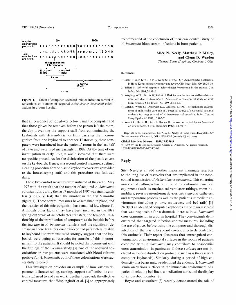
In the spring of 1997, however, we noticed an increase in thenumber of acquired A. baumannii colonizations in our pediatricburn patients (figure 1). Cultures of environmental specimensshowed that Acinetobacter was on various surfaces in the pa-tients’ rooms, especially on the plastic covers that fit over thebedside computer keyboards. Observation revealed that glovedpatient care staff moved back and forth between the patientand the keyboard and that ungloved support staff, who nevertouched the patient, entered and retrieved data from the samecomputer. We hypothesized that the ungloved personnel werecontaminating the keyboard with A. baumannii that was thenpicked up by the gloved patient care staff who transferred itto the patient. In essence, the keyboard was acting as an A.baumannii reservoir, a hypothesis that is consistent with studiesshowing that Acinetobacter species can survive for long periodson dry surfaces [4, 5].
Two control measures were instituted. First, it was required
at Maastricht U
niversity on October 27, 2014
http://cid.oxfordjournals.org/D
ownloaded from
CID 1999;29 (November) Correspondence 1359
Figure 1. Effect of computer keyboard–related infection control in-terventions on number of acquired Acinetobacter baumannii coloni-zations in a burn hospital.
that all personnel put on gloves before using the computer andthat those gloves be removed before the person left the room,thereby preventing the support staff from contaminating thekeyboards with Acinetobacter or from carrying the microor-ganism from one keyboard to another. Historically, these com-puters were introduced into the patients’ rooms in the last halfof 1996 and were used increasingly in 1997. At the time of ourinvestigation in early 1997, it was discovered that there wereno specific procedures for the disinfection of the plastic coverson the keyboards. Hence, as a second control measure, a definedcleaning procedure for the plastic keyboard covers was providedto the housekeeping staff, and this procedure was followeddaily.
These two control measures were initiated at the end of May1997 with the result that the number of acquired A. baumanniicolonizations during the last 7 months of 1997 was significantlyless ( , x2 test) than the number in the first 5 monthsP ! .05(figure 1). These control measures have remained in place, andthe transfer of this microorganism has remained low (figure 1).Although other factors may have been involved in the 1997spring outbreak of acinetobacter transfers, the temporal rela-tionship of the introduction of computers at the bedside beforethe increase in A. baumannii transfers and the significant de-crease in these transfers once two control parameters relativeto keyboard use were instituted strongly suggest that the key-boards were acting as reservoirs for transfer of this microor-ganism to the patients. It should be noted that, consistent withthe findings of the German study [3], two of the acquired col-onizations in our patients were associated with blood culturespositive for A. baumannii; both of these colonizations were suc-cessfully resolved.
This investigation provides an example of how various de-partments (housekeeping, nursing, support staff, infection con-trol, etc.) need to and can work together to provide the effectivecontrol measures that Wisplinghoff et al. [3] so appropriately
recommended at the conclusion of their case-control study ofA. baumannii bloodstream infections in burn patients.
Alice N. Neely, Matthew P. Maley,and Glenn D. Warden
Shriners Burns Hospitals, Cincinnati, Ohio
References
1. Siau H, Yuen K-Y, Ho P-L, Wong SSY, Woo PCY. Acinetobacter bacteremiain Hong Kong: prospective study and review. Clin Infect Dis 1999;28:26–30.
2. Seifert H. Editorial response: acinetobacter bacteremia in the tropics. ClinInfect Dis 1999;28:31–2.
3. Wisplinghoff H, Perbix W, Seifert H. Risk factors for nosocomial bloodstreaminfections due to Acinetobacter baumannii: a case-control study of adultburn patients. Clin Infect Dis 1999;28:59–66.
4. Getchell-White SI, Donowitz LG, Groschel DHM. The inanimate environ-ment of an intensive care unit as a potential source of nosocomial bacteria:evidence for long survival of Acinetobacter calcoaceticus. Infect ControlHosp Epidemiol 1989;10:402–7.
5. Wendt C, Dietze B, Dietz E, Ruden H. Survival of Acinetobacter baumanniion dry surfaces. J Clin Microbiol 1997;35:1394–7.
Reprints or correspondence: Dr. Alice N. Neely, Shriners Burns Hospital, 3229Burnet Avenue, Cincinnati, OH 45229-3095 ([email protected]).
Clinical Infectious Diseases 1999;29:1358–9q 1999 by the Infectious Diseases Society of America. All rights reserved.1058-4838/1999/2905-0063$03.00
Reply
SIR—Neely et al. add another important inanimate reservoirto the long list of reservoirs that are implicated in the noso-comial transmission of Acinetobacter baumannii. This emergingnosocomial pathogen has been found to contaminate medicalequipment (such as mechanical ventilator tubings, room hu-midifiers, pressure monitoring devices, intravascular catheters,and temperature probes) as well as the patient’s immediate en-vironment (including pillows, mattresses, and bed rails) [1].Neely et al. identified computer keyboards as the main reservoirthat was responsible for a dramatic increase in A. baumanniicross-transmission in a burns hospital. They convincingly dem-onstrated that targeted infection control measures, includingthe use of gloves before using the computer and thorough dis-infection of the plastic keyboard covers, effectively controlledthis outbreak. Their report illustrates that unrecognized con-tamination of environmental surfaces in the rooms of patientscolonized with A. baumannii may contribute to nosocomialcross-transmission, in particular, if these surfaces are not in-cluded in routine disinfection protocols (such as is the case withcomputer keyboards). Similarly, during a period of high en-demicity in a burns unit, we identified the endemic A. baumanniistrain on various surfaces in the immediate environment of apatient, including bed linen, a medication table, and the displayof an overbed monitor [2].
Boyce and coworkers [3] recently demonstrated the role of
at Maastricht U
niversity on October 27, 2014
http://cid.oxfordjournals.org/D
ownloaded from

1360 Correspondence CID 1999;29 (November)
environmental contamination with methicillin-resistant Staph-ylococcus aureus in the epidemiology of this pathogen. Theyalso found that nurses who had no direct contact with affectedpatients but who touched contaminated inanimate surfaces con-taminated their gloves with the endemic methicillin-resistant S.aureus strain. Unlike these researchers, Neely and colleaguesdid not confirm their observations by molecular strain typingto demonstrate the clonal identity of A. baumannii recoveredfrom patients and environmental specimens. However, their ep-idemiological data strongly support their conclusion. Furtherstudies are necessary to establish the levels of contaminationwith A. baumannii as well as the implications of persistence ofthese organisms on environmental surfaces that are importantfor patient-to-patient transmission.
Harald Seifert and Hilmar WisplinghoffInstitute of Medical Microbiology and Hygiene,
University of Cologne, Cologne, Germany
References
1. Bergogne-Berezin E, Towner KJ. Acinetobacter spp. as nosocomial pathogens:microbiological, clinical and epidemiological features. Clin Microbiol Rev1996;9:148–65.
2. Seifert H, Boullion B, Schulze A, Pulverer G. Plasmid DNA profiles of Aci-netobacter baumannii: clinical application in a complex endemic setting.Infect Control Hosp Epidemiol 1994;15:520–8.
3. Boyce JM, Potter-Bynoe G, Chenevert C, King T. Environmental contami-nation due to methicillin-resistant Staphylococcus aureus: possible infectioncontrol implications. Infect Control Hosp Epidemiol 1997;18:622–7.
Reprints or correspondence: Dr. Harald Seifert, Institute of Medical Micro-biology and Hygiene, University of Cologne, Goldenfelsstrasse 19-21, 50935 Co-logne, Germany ([email protected]).
Clinical Infectious Diseases 1999;29:1359–60q 1999 by the Infectious Diseases Society of America. All rights reserved.1058-4838/1999/2905-0064$03.00
Necrotizing Fasciitis Associated with Klebsiellapneumoniae Liver Abscess
SIR—We read with interest the article by Dylewski and Dy-lewski [1] that described an Indian patient with necrotizing fas-ciitis associated with Klebsiella pneumoniae liver abscess. Met-astatic soft-tissue infections in conjunction with klebsiella liverabscess are rare [2–4]. However, the case reported by Dylewskiand Dylewski is not the first reported case of necrotizing fasciitisin association with klebsiella liver abscess. In 1996, Chou andKou [4] described a Taiwanese patient with K. pneumoniae liverabscess in conjunction with endophthalmitis and necrotizingfasciitis. Here we describe two other patients with similarmanifestations.
A 71-year-old man with diabetes was admitted to the hospitalfor evaluation of a 2-week history of fever, chills, and right
upper abdominal pain. He had progressively painful swellingon the left eye 4 days after the onset of fever and a 10-dayhistory of left leg swelling with erythema. Physical examinationshowed an acutely ill man with a temperature of 37.57C. Hisleft eye was blind with extensive hypopyon, severe proptosis,and moderate edema and erythema in the eyelid. His left lowerleg had marked swelling with induration and severe tenderness.Laboratory studies disclosed the following: initial WBC count,
/L; serum glucose level, 203 mg/dL; serum albumin913.1 3 10level, 1.8 g/dL; and alkaline phosphatase level, 651 U/L. A CTscan of the abdomen showed an 8-cm intrahepatic abscess,bilateral renal abscesses, and a pancreatic abscess. A presump-tive diagnosis of liver abscess with endogenous endophthalmitiswas made, and therapy with ceftriaxone was started.
Sonography-guided percutaneous drainage and fasciotomywere performed, and necrotizing fasciitis was observed. Cul-tures of pus specimens from the liver abscess and left leg bothyielded K. pneumoniae. The two isolates belonged to capsularserotype K1. On hospital day 11, the left eye was evisceratedand the skin grafted was normalized. The patient was dis-charged 53 days after admission. He remained free of liverabscess for 1 year of follow-up.
A 40-year-old man with diabetes presented with a 1-weekhistory of fever, chills, nausea, and vomiting and a 3-day historyof painful swelling on the left leg. At the time of physical ex-amination, his temperature was 37.97C. The left leg had markedswelling and severe tenderness. Laboratory studies disclosedthe following pertinent values: WBC count, ; he-928.6 3 10 /Lmoglobin level, 85 g/L; serum glucose level, 313 mg/dL; serumalbumin level, 1.7 g/dL; alkaline phosphatase level, 485 U/L;and total bilirubin level, 6.8 mg/dL. A presumptive diagnosisof necrotizing fasciitis was made, and fasciotomy was per-formed immediately.
Cultures of blood and a left leg wound specimen both yieldedK. pneumoniae. On the next day, a CT scan of the abdomenshowed multiple low-density poorly enhanced nodules in bothlobes of the liver and a large subcapsular hematoma in the leftkidney. Therapy with parenteral cefazolin and gentamicin wasadministered for 37 days. He remained asymptomatic during2 years of follow-up.
Diabetes is an apparent risk factor for klebsiella liver abscessin Taiwan [2–5]. All 4 patients described in the literature whohad liver abscess and fasciitis also had diabetes, either for sev-eral years or at the time of presentation [1, 4]. It is worthinvestigating further to determine the relationship between di-abetes and klebsiella liver abscess with fasciitis. To our knowl-edge, this is the first time that K. pneumoniae capsular serotypeK1 has been reported in conjunction with liver abscess andendophthalmitis.
There are few data on the role of K. pneumoniae capsularserotypes in liver abscess and endophthalmitis. Noble et al. [6]had described 1 endogenous endophthalmitis patient due toKlebsiella aerogenes serotype K2. In northern Taiwan, Kleb-
at Maastricht U
niversity on October 27, 2014
http://cid.oxfordjournals.org/D
ownloaded from

CID 1999;29 (November) Correspondence 1361
siella serotype K1 is the most common serotype of Klebsiellaisolates causing bacteremia [7]. We speculate that serotype K1of K. pneumoniae might be the predominant strain causing liverabscess in Taiwan.
Bor-Shen Hu, Yeu-Jun Lau, Zhi-Yuan Shi,and Yu-Hui Lin
Section of Infectious Diseases, Department of Internal Medicine,Taichung Veterans General Hospital, Taichung, Taiwan,
Republic of China
References
1. Dylewski JS, Dylewski I. Necrotizing fasciitis with klebsiella liver abscess. ClinInfect Dis 1998;27:1561–2.
2. Cheng DL, Liu YC, Yen MY, et al. Pyogenic liver abscess: clinical manifes-tations and value of percutaneous catheter drainage treatment. J FormosMed Assoc 1990;89:571–6.
3. Chang FY, Chou MY, Fan RL, et al. A clinical study of Klebsiella liver abscess.J Formos Med Assoc 1988;87:282–7.
4. Chou FF, Kou HK. Endogenous endophthalmitis associated with pyogenichepatic abscess. J Am Coll Surg 1996;182:33–6.
5. Chou FF, Sheen-Chen SM, Chen YS, et al. The comparison of clinical courseand results of treatment between gas-forming and non-gas-forming pyo-genic liver abscess. Arch Surg 1995;130:401–5.
6. Noble CJ, Winch J, Munton CG. Metastatic endophthalmitis [letter]. J Infect1983;6:100–1.
7. Peng HL, Wang PY, Wu JL, et al. Molecular epidemiology of Klebsiella pneu-moniae. Chin J Microbiol Immunol 1991;24:264–71.
Reprints or correspondence: Dr. Bor-Shen Hu, 160 Taichung Harbor RoadSec. 3, Section of Infectious Diseases, Department of Internal Medicine, Tai-chung Veterans General Hospital, Taichung, Taiwan, Republic of China ([email protected]).
Clinical Infectious Diseases 1999;29:1360–1q 1999 by the Infectious Diseases Society of America. All rights reserved.1058-4838/1999/2905-0065$03.00
Reply
SIR—We are pleased to see that additional cases of necrotizingfasciitis in association with klebsiella liver abscess are beingreported from Taiwan. For the record, our patient acquired hisillness in June 1994, and we had tried to have our report pub-lished in this and other journals over a period of 3 years. Itwas only after the review article by Wang et al. [1] that ourcase report was allowed to be published as a letter. Of note,the article by Chou and Kou [2] was not found in the referenceslisted in the bibliographies done by Dr. Wang and ourselves.A MEDLINE search of the literature using the key words“klebsiella liver abscess” and “necrotizing fasciitis” did not turnup the article by Chou and Kou, and even their abstract doesnot mention necrotizing fasciitis. There are no details given inthe report on how the diagnosis was made.
We appreciate Hu et al. pointing out the case reported byChou and Kou and two new cases of necrotizing fasciitis with
klebsiella liver abscess. Fortunately, this association remains arare occurrence in North America.
Joe Dylewski and Irving DylewskiSt. Mary’s Hospital, Montreal, Quebec, Canada
References
1. Wang J-H, Liu Y-C, Lee SS-J, et al. Primary liver abscess due to Klebsiellapneumoniae in Taiwan. Clin Infect Dis 1998;26:1434–8.
2. Chou FF, Kou HK. Endogenous endophthalmitis associated with pyogenichepatic abscess. J Am Coll Surg 1996;182:33–6.
Reprints or correspondence: Dr. Joe Dylewski, St. Mary’s Hospital, 3830 La-combe Avenue, Montreal, Quebec H3T 1M5, Canada ([email protected]).
Clinical Infectious Diseases 1999;29:1361q 1999 by the Infectious Diseases Society of America. All rights reserved.1058-4838/1999/1905-0066$03.00
Orbital Cellulitis Due to Streptococcus pneumoniaein a Previously Healthy Adult
SIR—We read with great interest the brief report of adult pneu-mococcal cellulitis by Parada and Maslow [1]. The authorsstated that “pneumococcal cellulitis was universally associatedwith bacteremia” and summarized that “pneumococcal skininfections represent 2 distinctive clinical syndromes: facial cel-lulitis in persons with systemic lupus erythematosus and he-matologic disorders and limb cellulitis in persons with diabetesmellitus and substance abuse.” We describe a case of Strepto-coccus pneumoniae orbital cellulitis without bacteremia in a pre-viously healthy adult.
A 77-year-old woman with a medical history of coronaryartery disease and hypertension was in her usual state of goodhealth when she started to have a dull aching pain in her lefteye while she was playing bridge. She looked in the mirror andnoticed mild redness in her left eye. The next day, her left eyewas more red with intense pain and more swollen, and the skinbelow the eye was hard as a rock. The swelling became so severethat the patient had to pull down her lower eyelid to see. Thepatient denied fever, chills, headache, impairment of vision,nausea, vomiting, or diarrhea. She denied any history oftrauma, sinus disease, or insect bite. She presented to the emer-gency department.
Her temperature was 37.17C; pulse rate, 80; respiratory rate,18; and blood pressure, 106/72 mm Hg. There was markedlycongested conjunctiva of the left eye with marked induratedswelling of the lower eyelid that extended to the maxillary area.The patient also had difficulty looking outward (lateral gaze)with her left eye. There was purulent discharge from the lefteye. The rest of her physical examination was unremarkable.The white blood cell count was /L (95% neutrophils).916.5 3 10A CT scan of the orbits showed marked soft-tissue swelling
at Maastricht U
niversity on October 27, 2014
http://cid.oxfordjournals.org/D
ownloaded from
1362 Correspondence CID 1999;29 (November)
involving the left orbit with no evidence of orbital extensionof cellulitis or bony erosive changes. A CT scan of the paranasalsinuses was unremarkable.
The patient was treated with 2 g of intravenous ceftriaxonein the emergency department after blood specimens for cultureswere obtained; culture of the purulent eye discharge was alsoperformed. The patient was seen by an ophthalmologist whosediagnosis was left orbital cellulitis with sixth cranial nerve palsy;her vision was 20/30 in both eyes. She was treated with cefazo-lin (1 g intravenously every 8 h) on the second day of hospi-talization; at this time, the patient started to feel better with amild decrease in her pain and swelling below the eye. The pa-tient’s condition continued to improve with a marked decreasein pain and swelling of the left eye, and she had no problemwith her lateral gaze. She was discharged on the third day; hermedication at discharge was cephalexin, and she recovereduneventfully.
Cultures of blood obtained at admission had no growth.Culture of the left eye drainage yielded moderate growth S.pneumoniae resistant to penicillin (MIC, 1.5 mg/mL) but sus-ceptible to ceftriaxone (MIC, 0.5 mg/mL) and cefotaxime (MIC,1.0 mg/mL). Disk diffusion tests revealed that the S. pneumoniaeisolate was susceptible to cefazolin, clindamycin, and vanco-mycin but resistant to erythromycin and tetracycline.
Periorbital cellulitis due to S. pneumoniae is known to occurin children [2–4], but S. pneumoniae orbital cellulitis with in-fections of orbital contents marked by proptosis and ophthal-moplegia is rare. DiNubile et al. [5] described a case of S.pneumoniae orbital cellulitis of the right eye in an adult withinsulin-dependent diabetes mellitus, retinopathy, blindness, andan enucleated right eye. Our patient had no compromising un-derlying disease and was in a state of good health except for
a history of coronary artery disease and hypertension. Herblood cultures had no growth. Similar to the case reported byDiNubile et al, S. pneumoniae was cultured from eye drainagefrom our patient, and she recovered uneventfully.
The pathogenesis of orbital cellulitis in our patient is notclear. The CT scan of her paranasal sinuses was unremarkable,and she had no documented bacteremia. Furthermore, therewas no history of trauma or surgery. Nevertheless, S. pneu-moniae should be added to the list of organisms capable ofcausing orbital cellulitis in previously healthy adults.
Sameh Naseib1 and Chatrchai Watanakunakorn1,2
1Department of Internal Medicine, St. Elizabeth Health Center,Youngstown, and 2Northeastern Ohio Universities College of Medicine,
Rootstown, Ohio
References
1. Parada JP, Maslow JN. Adult pneumococcal cellulitis: case report and review.Clin Infect Dis 1999;28:918.
2. Thirumoorthi MC, Asmar BI, Dajani AS. Violaceous discoloration in pneu-mococcal cellulitis. Pediatrics 1978;62:492–3.
3. Sankrithi LM, Lipuma JJ. Clinically inapparent meningitis complicating per-iorbital cellulitis. Pediatr Emerg Care 1991;7:28–9.
4. Shapiro ED, Wald ER, Brozanski BA. Periorbital cellulitis and paranasalsinusitis: a reappraisal. Pediatr Infect Dis 1982;1:91–4.
5. DiNubile MJ, Albornoz A, Stumacher RJ, et al. Pneumococcal soft-tissueinfections: possible association with connective tissue diseases. J Infect Dis1991;163:897–900.
Reprints or correspondence: Dr. C. Watanakunakorn, St. Elizabeth HealthCenter, 1044 Belmont Avenue, P.O. Box 1790, Youngstown, OH 44501-1790.
Clinical Infectious Diseases 1999;29:1361–2q 1999 by the Infectious Diseases Society of America. All rights reserved.1058-4838/1999/2905-0067$03.00
at Maastricht U
niversity on October 27, 2014
http://cid.oxfordjournals.org/D
ownloaded from