Embed Size (px)
Citation preview
LUND UNIVERSITY
PO Box 117221 00 Lund+46 46-222 00 00
Complications to peptic ulcer and peptic ulcer surgery
Åhsberg, Kristina
2010
Link to publication
Citation for published version (APA):Åhsberg, K. (2010). Complications to peptic ulcer and peptic ulcer surgery. Department of Surgery, ClinicalSciences Lund, Lund University.
General rightsCopyright and moral rights for the publications made accessible in the public portal are retained by the authorsand/or other copyright owners and it is a condition of accessing publications that users recognise and abide by thelegal requirements associated with these rights.
• Users may download and print one copy of any publication from the public portal for the purpose of private studyor research. • You may not further distribute the material or use it for any profit-making activity or commercial gain • You may freely distribute the URL identifying the publication in the public portalTake down policyIf you believe that this document breaches copyright please contact us providing details, and we will removeaccess to the work immediately and investigate your claim.
1
Bulletin No. 137 from the Department of Surgery, Lund University, Sweden
COMPLICATIONS TO PEPTIC ULCER
AND PEPTIC ULCER SURGERY
Kristina Åhsberg
Akademisk avhandling
i ämnet klinisk medicin med inriktning kirurgi som med vederbörligt tillstånd av
Medicinska fakulteten vid Lunds Universitet för avläggande av doktorsexamen, kommer att offentligen försvaras i
Föreläsningssal 3, Centralblocket, Universitetssjukhuset i Lund, fredagen den 4 juni 2010, kl 09.00
Handledare: Fakultetsopponent: Docent Christer Staël von Holstein Professor Kurt Borch Kirurgiska kliniken Universitetssjukhuset i Linköping
Lund University, Faculty of Medicine Doctoral Dissertation Series 2010:65
3
Bulletin No. 137 from the Department of Surgery, Lund University, Sweden
COMPLICATIONS TO PEPTIC ULCER AND PEPTIC ULCER SURGERY
Kristina Åhsberg, MD Department of Surgery
Lund University Lund University Hospital
Sweden
Lund University, Faculty of Medicine Doctoral Dissertation Series 2010:65
4
© Kristina Åhsberg, MD, 2010 e-mail: [email protected] Supervisor: Associate Professor Christer Staël von Holstein, MD, PhD Department of Surgery, Clinical Sciences Lund Lund University and Lund University Hospital 221 85 Lund, Sweden Bulletin No. 137 from the Department of Surgery, Lund University, Sweden ISSN 1652-8220 ISBN 978-91-86443-81-8 Lund University, Faculty of Medicine Doctoral Dissertation Series 2010:65 Printed by Media-Tryck, Lund, Sweden

5
CONTENTS CONTENTS 5
ORIGINAL PAPERS 7
INTRODUCTION 9
Peptic ulcer surgery 10
Sequelae after partial gastrectomy 14
Long term morbidity and mortality after peptic ulcer surgery 20
Epidemiology of peptic ulcer 21
Ethiology of peptic ulcer disease and peptic ulcer bleeding 24
Peptic ulcer bleeding and other causes of gastrointestinal bleeding 29
AIMS OF THIS THESIS 31
MATERIAL AND METHODS 33
Study I. 33
Study II 34
Study III 35
Study IV and V 38
RESULTS 43
Study I 43
Study II 45
Study III 47
Study IV 53
Study V 65
DISCUSSION 79
Complications to peptic ulcer surgery (Study I and II) 79
Complications to peptic ulcer (Study III-V) 84
CONCLUSIONS 93

6
CLINICAL IMPLICATIONS AND FUTURE ASPECTS 95
POPULÄRVETENSKAPLIG SAMMANFATTNING 97
ACKNOWLEDGEMENTS 101
REFERENCES 103
7
ORIGINAL PAPERS This thesis is based on the following papers, which will be referred to in the text by their Roman numerals (I-V):
I. Åhsberg K, Hammar E, Staël von Holstein C. Mucosal changes in the gastric remnant: long-term effects of bile reflux diversion and Helicobacter pylori infection. Eur J Gastroenterol Hepatol 2003;15:35-40
II. Åhsberg K, Olsson H, Staël von Holstein C.
Increased mortality in prostate carcinoma and smoking related disease after PCV: a long-term follow-up study. Scand J Gastroenterol 2009;44(8):947-51
III. Åhsberg K, Ye W, Lu Y, Zheng Z, Staël von Holstein C. Hospitalization of and mortality from bleeding peptic ulcer in Sweden. A nation-wide time-trend analysis. Submitted
IV. Åhsberg K, Höglund P, Staël von Holstein C. Aspirin may decrease the risk of in-hospital mortality after peptic ulcer bleeding. Submitted
V. Åhsberg K, Höglund P, Kim W, Staël von Holstein C.
Impact of bleeding promoting drugs on localization and outcome of gastrointestinal bleeding. Submitted

8
9
INTRODUCTION Few other gastrointestinal disorders have gone through such a revolutionary change during the last decades as peptic ulcer disease regarding both causative factors and management.
Before the introduction of modern antisecretory drugs in form of histamine-2-receptor antagonists (H2RA) in 1976, surgery was the only method available to cure peptic ulcer. Up to 1970 partial gastrectomy was the most common procedure, with low ulcer recurrence rate and low per-operative mortality. However, some patients got symptoms of epigastric pain and bile vomiting post-operatively due to the inevitable reflux of enterogastric contents, so called reflux gastritis (Ritchie 1984). Even worse, patients were shown to carry an increased risk of malignant disease in the resected stomach as well as in other organs after partial gastrectomy (Domellof 1977; Caygill 1987; Lundegårdh 1991; Staël von Holstein 1995; Bahmanyar 2007; Luo 2007). The enterogastric reflux is believed to be the major ethiological factor also in gastric stump carcinoma, although other ethiological factors have not been ruled out. In 1970 the more physiological procedure parietal cell vagotomy was introduced, but long-term mortality and morbidity after this kind of operation is not well known. The first part of this thesis (study I and II) is devoted to evaluate complications to peptic ulcer surgery in form of long-term sequelae.
In the mid 1980’s the Helicobacter pylori was discovered by Warren and Marshall and its ethiological association with peptic ulcer disease was established (Marshall 1984). Eradication therapies against H. pylori infection together with effective antisecretory proton pump inhibitors (PPI), introduced in the late 1980’s, have made most peptic ulcers possible to treat merely with pharmacological therapy. The need for elective ulcer surgery has therefore diminished to almost zero and hospitalization rates for uncomplicated ulcer disease has declined (Lewis 2002; Kang 2006; Post 2006; Wang 2010).
There are however diverting reports of the incidence of complicated ulcer during the last decades (Higham 2002; Paimela 2002; van Leerdam 2003; Ohmann 2005; Kang 2006; Lassen 2006; Post 2006; Hermansson 2009) and mortality after bleeding ulcer is shown to vary between 5 to 15% in different studies (Rockall 1995; van Leerdam 2003; Kang 2006). The second part of this thesis (study III-V) is devoted to investigate the incidence of and mortality in peptic ulcer bleeding in Sweden and to assess the impact of increased use of drugs with ulcerogenic and/or bleeding promoting side-effects, as low dose aspirin, non-steroidal anti-inflammatory drugs (NSAIDs), steroids, warfarin and selective serotonin re-uptake inhibitors (SSRIs) on outcome after peptic ulcer bleeding and on localization of gastrointestinal bleed.
10
Peptic ulcer surgery
Gastric resections
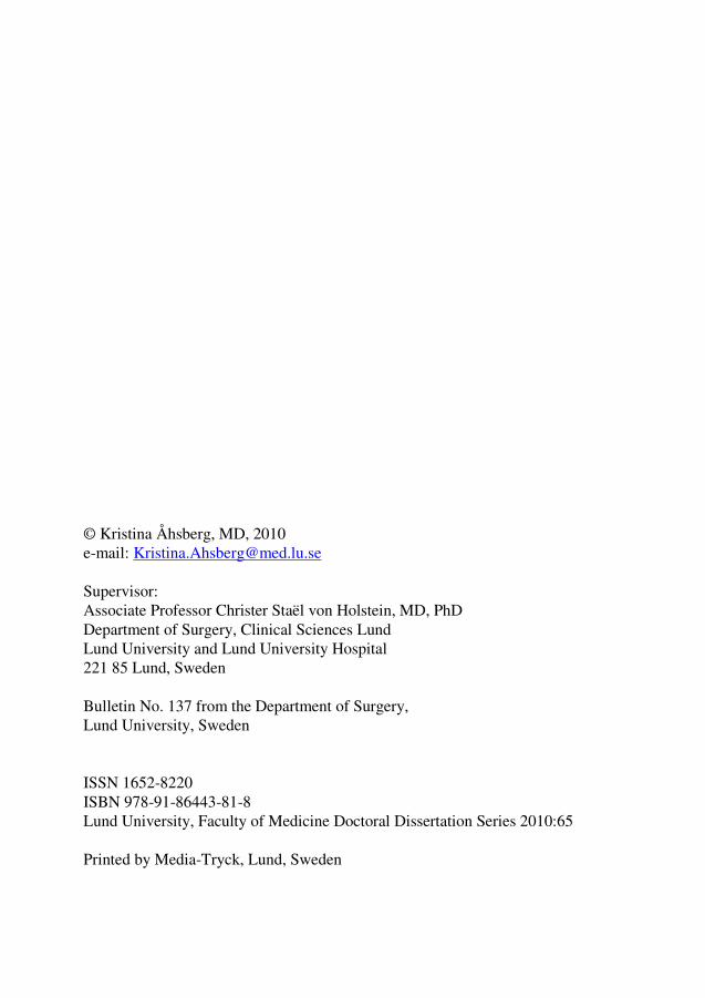
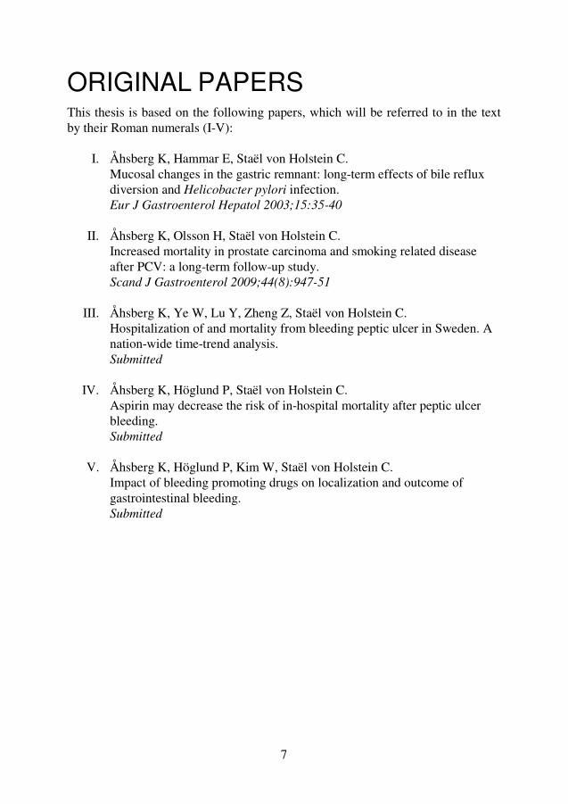
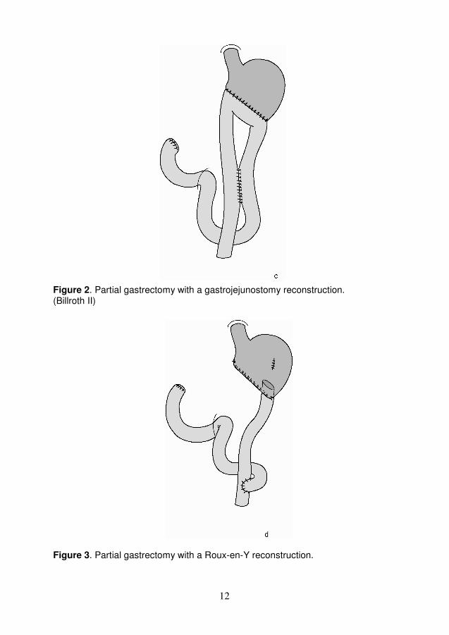
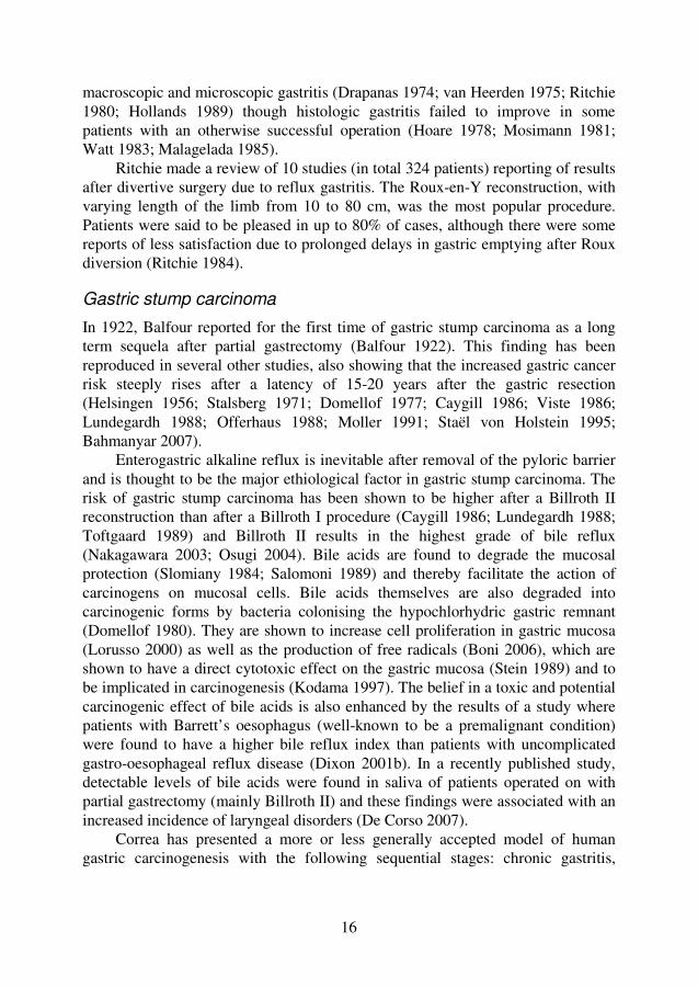
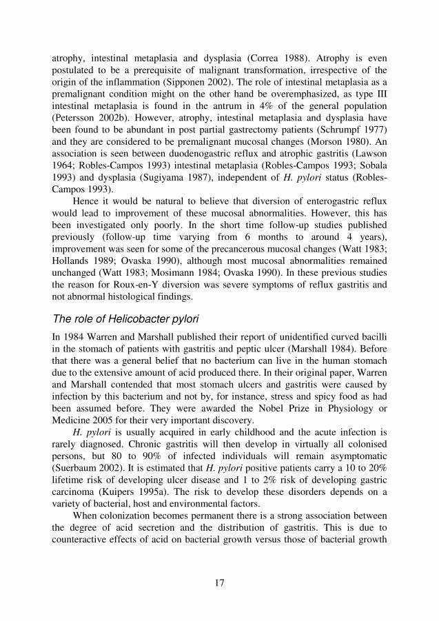
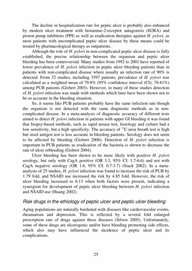
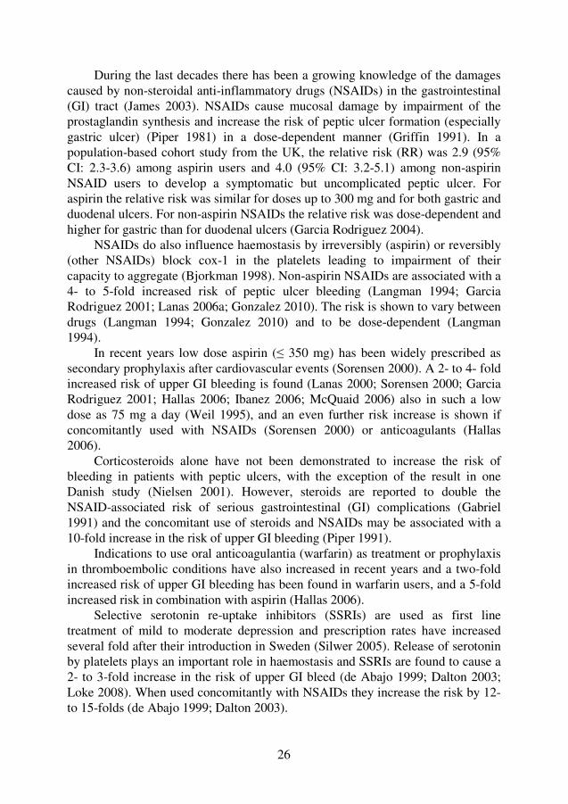
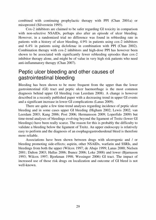
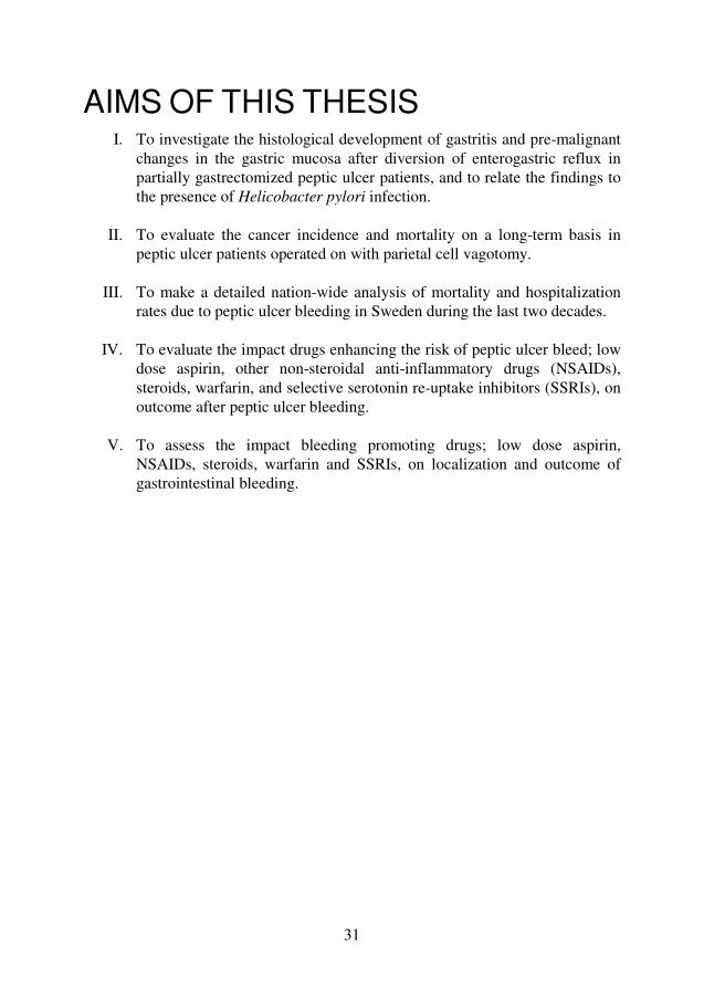
Peptic ulcer disease has been treated by surgery for more than one hundred years. In 1881, Billroth performed the first successful partial gastrectomy with gastro-duodenostomy (Billroth I) (Fig. 1) in a patient with distal gastric carcinoma (Wiese 1929). One year later, Rydygier was the first to surgically treat ulcer disease, by performing a similar resection in a patient with gastric outlet obstruction (Rydygier 1882). With improved anaesthesia this kind of operation together with partial gastrectomy with gastrojejunostomy (Billroth II - introduced in 1885) (Fig. 2), soon became common procedures against benign peptic ulcer disease (Wiese 1929).
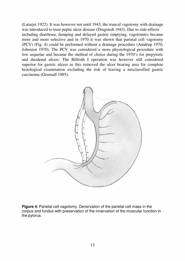
Unfortunately, some patients got troublesome symptoms of bile vomiting and epigastric pain after these operations (Wolfler 1881). The ethiology of these symptoms was not clearly addressed until much later – see chapter of sequelae after partial gastrectomy. To prevent duodenal contents to reach the remaining stomach a side-to-side enteroanastomosis a bit below the Billroth II gastroenteroanastomosis was suggested by Braun (Braun 1892), and Roux invented a Y shaped reconstruction. Bile and pancreatic juice was diverted from the stomach by a separate duodenojejunal limb reaching the jejunum by an end-to-side enteroanastomosis several decimeters below the gastroenteroanastomis (Roux 1897) (Fig. 3)
Finsterer recommended in 1918 that the gastric resection should include two thirds of the stomach, to inhibit acid secretion permanently by markedly reducing the parietal cell mass. (Finsterer 1918).
Vagotomy and drainage procedures
Around 1900 the gastroenteroanastomosis without resection (GE) (Mayo 1906) and different variations of pyloroplasties (PP) were introduced to treat stenosis due to peptic ulcer disease (Fronmüller 1886; Mikulicz 1888; Jaboulay 1892; Finney 1902). During the first decades of the twentieth century GE was considered an easy and relatively safe procedure to cure peptic ulcer also without a stenosis. After some years it was however abandoned as a sole therapeutic procedure due to a 50 per cent recurrence rate of stomal ulcers (Finney 1929). The Billroth II operation took over and became the standard procedure, reaching its peak in the 1950’s.
Already in 1814 Brodie described animal experiments of inhibited gastric secretion after vagotomy (Brodie 1814). Latarjet made an exact description of the vagal innervation of the stomach in 1922 and pointed out the importance of a drainage procedure (GE or PP) to prevent gastric retention after vagotomy

11
Figure 1. Partial gastrectomy with a gastroduodenostomy reconstruction (Billroth I)
12
Figure 2. Partial gastrectomy with a gastrojejunostomy reconstruction. (Billroth II) Figure 3. Partial gastrectomy with a Roux-en-Y reconstruction.
13
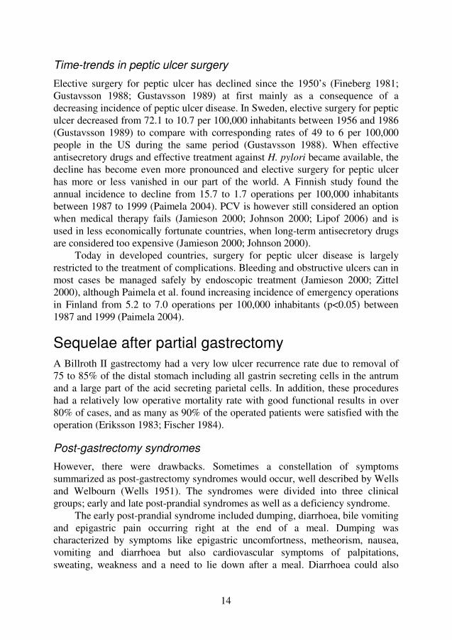
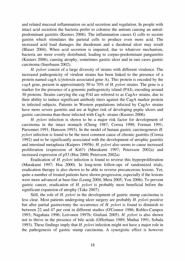
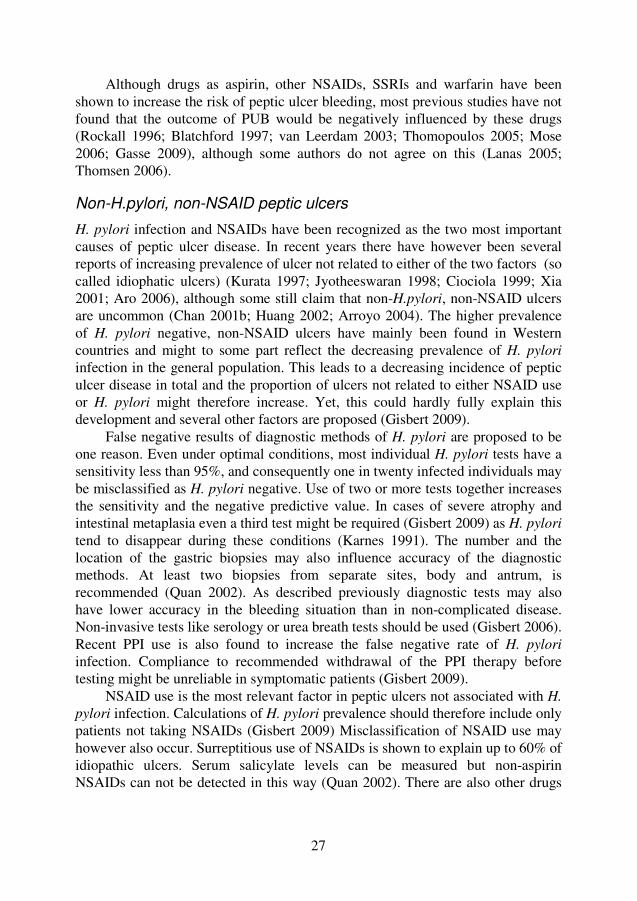
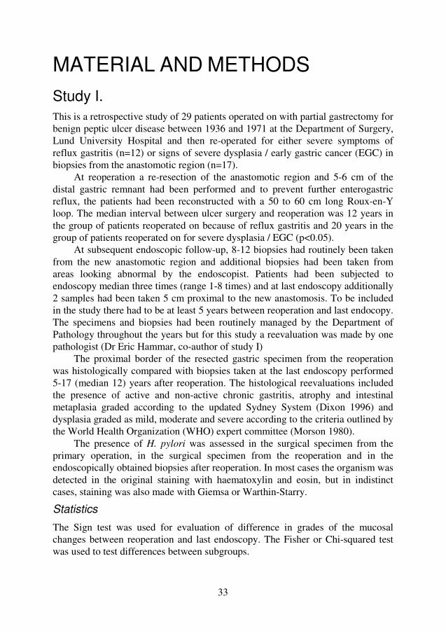
(Latarjet 1922). It was however not until 1943, the truncal vagotomy with drainage was introduced to treat peptic ulcer disease (Dragstedt 1943). Due to side-effects including diarrhoea, dumping and delayed gastric emptying, vagotomies became more and more selective and in 1970 it was shown that parietal cell vagotomy (PCV) (Fig. 4) could be performed without a drainage procedure (Amdrup 1970; Johnston 1970). The PCV was considered a more physiological procedure with few sequelae and became the method of choice during the 1970’s for prepyloric and duodenal ulcers. The Billroth I operation was however still considered superior for gastric ulcers as this removed the ulcer bearing area for complete histological examination excluding the risk of leaving a misclassified gastric carcinoma (Greenall 1985).
Figure 4. Parietal cell vagotomy. Denervation of the parietal cell mass in the corpus and fundus with preservation of the innervation of the muscular function in the pylorus.
14
Time-trends in peptic ulcer surgery
Elective surgery for peptic ulcer has declined since the 1950’s (Fineberg 1981; Gustavsson 1988; Gustavsson 1989) at first mainly as a consequence of a decreasing incidence of peptic ulcer disease. In Sweden, elective surgery for peptic ulcer decreased from 72.1 to 10.7 per 100,000 inhabitants between 1956 and 1986 (Gustavsson 1989) to compare with corresponding rates of 49 to 6 per 100,000 people in the US during the same period (Gustavsson 1988). When effective antisecretory drugs and effective treatment against H. pylori became available, the decline has become even more pronounced and elective surgery for peptic ulcer has more or less vanished in our part of the world. A Finnish study found the annual incidence to decline from 15.7 to 1.7 operations per 100,000 inhabitants between 1987 to 1999 (Paimela 2004). PCV is however still considered an option when medical therapy fails (Jamieson 2000; Johnson 2000; Lipof 2006) and is used in less economically fortunate countries, when long-term antisecretory drugs are considered too expensive (Jamieson 2000; Johnson 2000).
Today in developed countries, surgery for peptic ulcer disease is largely restricted to the treatment of complications. Bleeding and obstructive ulcers can in most cases be managed safely by endoscopic treatment (Jamieson 2000; Zittel 2000), although Paimela et al. found increasing incidence of emergency operations in Finland from 5.2 to 7.0 operations per 100,000 inhabitants (p<0.05) between 1987 and 1999 (Paimela 2004).
Sequelae after partial gastrectomy A Billroth II gastrectomy had a very low ulcer recurrence rate due to removal of 75 to 85% of the distal stomach including all gastrin secreting cells in the antrum and a large part of the acid secreting parietal cells. In addition, these procedures had a relatively low operative mortality rate with good functional results in over 80% of cases, and as many as 90% of the operated patients were satisfied with the operation (Eriksson 1983; Fischer 1984).
Post-gastrectomy syndromes
However, there were drawbacks. Sometimes a constellation of symptoms summarized as post-gastrectomy syndromes would occur, well described by Wells and Welbourn (Wells 1951). The syndromes were divided into three clinical groups; early and late post-prandial syndromes as well as a deficiency syndrome.
The early post-prandial syndrome included dumping, diarrhoea, bile vomiting and epigastric pain occurring right at the end of a meal. Dumping was characterized by symptoms like epigastric uncomfortness, metheorism, nausea, vomiting and diarrhoea but also cardiovascular symptoms of palpitations, sweating, weakness and a need to lie down after a meal. Diarrhoea could also
15
occur solely without other dumping symptoms. The symptoms were due to increased intestinal motility caused by the rapid emptying of the gastric remnant into the jejunum. Improvement could often be achieved by smaller and more frequent meals, intake of liquids first after the meal and by excluding dairy products and sweets. An adaptation of the jejunum to the new post-operative situation also often occurred.
Bile vomiting was suggested by Wells and Welbourn to be due to chronic intermittent obstruction of the afferent loop. Accumulated bile and pancreatic juice were rapidly emptied into the stomach, giving rise to vomiting. The pain was thought to emerge from gastric distention and slow emptying of the remnant stomach.
The late post-prandial or the hypoglycaemic syndrome was also caused by the rapid emptying of the gastric remnant. Glucose was absorbed in the jejunum more rapidly than normal resulting in asymptomatic hyperglycaemia, but after two or three hours, symptoms of hypoglycaemia might follow due to overproduction of insulin.
The deficiency syndrome included weight loss quite often seen after gastrectomies due to reduced calorie intake, impaired digestion and fat malabsorption (Lawrence 1960). Iron deficiency anaemia (Holt 1970), vitamin B12 deficiency due to removal of intrinsic factor and calcium deficiency (Mellstrom 1982) might also occur.
Alkaline reflux gastritis
In 1965, Toye and Williams (Toye 1965) reported of an intriguing single subject human experiment and suggested that reflux of upper intestinal content into the gastric remnant after partial gastrectomy was responsible for a bile vomiting syndrome which was distinct from and unassociated with the afferent loop syndrome.
One year earlier Lawson had shown in experimental studies that reflux of duodenal contents into the stomach of dogs caused superficial gastritis, atrophic gastritis and epithelial proliferation with increased mitotic activity. The greatest reaction was caused by a mixture of bile and pancreatic juice and the least by pancreatic juice alone. When a Roux-en-Y reconstruction primarily was performed, deviating the duodenal content from the stomach, instead of a Billroth I gastrectomy, the gastric mucosa remained normal (Lawson 1964).
Further on, it was postulated that excessive enterogastric reflux was the principle cause of a syndrome with symptoms of epigastric pain, nausea, vomiting, weight-loss, hypochlorohydria and anaemia associated with severe signs of endoscopic and histologic gastritis. This syndrome was referred to as alkaline reflux gastritis, and it was shown that diversion of the enterogastric reflux in selected patients resulted in marked improvement of symptoms as well as both
16
macroscopic and microscopic gastritis (Drapanas 1974; van Heerden 1975; Ritchie 1980; Hollands 1989) though histologic gastritis failed to improve in some patients with an otherwise successful operation (Hoare 1978; Mosimann 1981; Watt 1983; Malagelada 1985).
Ritchie made a review of 10 studies (in total 324 patients) reporting of results after divertive surgery due to reflux gastritis. The Roux-en-Y reconstruction, with varying length of the limb from 10 to 80 cm, was the most popular procedure. Patients were said to be pleased in up to 80% of cases, although there were some reports of less satisfaction due to prolonged delays in gastric emptying after Roux diversion (Ritchie 1984).
Gastric stump carcinoma
In 1922, Balfour reported for the first time of gastric stump carcinoma as a long term sequela after partial gastrectomy (Balfour 1922). This finding has been reproduced in several other studies, also showing that the increased gastric cancer risk steeply rises after a latency of 15-20 years after the gastric resection (Helsingen 1956; Stalsberg 1971; Domellof 1977; Caygill 1986; Viste 1986; Lundegardh 1988; Offerhaus 1988; Moller 1991; Staël von Holstein 1995; Bahmanyar 2007).
Enterogastric alkaline reflux is inevitable after removal of the pyloric barrier and is thought to be the major ethiological factor in gastric stump carcinoma. The risk of gastric stump carcinoma has been shown to be higher after a Billroth II reconstruction than after a Billroth I procedure (Caygill 1986; Lundegardh 1988; Toftgaard 1989) and Billroth II results in the highest grade of bile reflux (Nakagawara 2003; Osugi 2004). Bile acids are found to degrade the mucosal protection (Slomiany 1984; Salomoni 1989) and thereby facilitate the action of carcinogens on mucosal cells. Bile acids themselves are also degraded into carcinogenic forms by bacteria colonising the hypochlorhydric gastric remnant (Domellof 1980). They are shown to increase cell proliferation in gastric mucosa (Lorusso 2000) as well as the production of free radicals (Boni 2006), which are shown to have a direct cytotoxic effect on the gastric mucosa (Stein 1989) and to be implicated in carcinogenesis (Kodama 1997). The belief in a toxic and potential carcinogenic effect of bile acids is also enhanced by the results of a study where patients with Barrett’s oesophagus (well-known to be a premalignant condition) were found to have a higher bile reflux index than patients with uncomplicated gastro-oesophageal reflux disease (Dixon 2001b). In a recently published study, detectable levels of bile acids were found in saliva of patients operated on with partial gastrectomy (mainly Billroth II) and these findings were associated with an increased incidence of laryngeal disorders (De Corso 2007).
Correa has presented a more or less generally accepted model of human gastric carcinogenesis with the following sequential stages: chronic gastritis,
17
atrophy, intestinal metaplasia and dysplasia (Correa 1988). Atrophy is even postulated to be a prerequisite of malignant transformation, irrespective of the origin of the inflammation (Sipponen 2002). The role of intestinal metaplasia as a premalignant condition might on the other hand be overemphasized, as type III intestinal metaplasia is found in the antrum in 4% of the general population (Petersson 2002b). However, atrophy, intestinal metaplasia and dysplasia have been found to be abundant in post partial gastrectomy patients (Schrumpf 1977) and they are considered to be premalignant mucosal changes (Morson 1980). An association is seen between duodenogastric reflux and atrophic gastritis (Lawson 1964; Robles-Campos 1993) intestinal metaplasia (Robles-Campos 1993; Sobala 1993) and dysplasia (Sugiyama 1987), independent of H. pylori status (Robles-Campos 1993).
Hence it would be natural to believe that diversion of enterogastric reflux would lead to improvement of these mucosal abnormalities. However, this has been investigated only poorly. In the short time follow-up studies published previously (follow-up time varying from 6 months to around 4 years), improvement was seen for some of the precancerous mucosal changes (Watt 1983; Hollands 1989; Ovaska 1990), although most mucosal abnormalities remained unchanged (Watt 1983; Mosimann 1984; Ovaska 1990). In these previous studies the reason for Roux-en-Y diversion was severe symptoms of reflux gastritis and not abnormal histological findings.
The role of Helicobacter pylori
In 1984 Warren and Marshall published their report of unidentified curved bacilli in the stomach of patients with gastritis and peptic ulcer (Marshall 1984). Before that there was a general belief that no bacterium can live in the human stomach due to the extensive amount of acid produced there. In their original paper, Warren and Marshall contended that most stomach ulcers and gastritis were caused by infection by this bacterium and not by, for instance, stress and spicy food as had been assumed before. They were awarded the Nobel Prize in Physiology or Medicine 2005 for their very important discovery.
H. pylori is usually acquired in early childhood and the acute infection is rarely diagnosed. Chronic gastritis will then develop in virtually all colonised persons, but 80 to 90% of infected individuals will remain asymptomatic (Suerbaum 2002). It is estimated that H. pylori positive patients carry a 10 to 20% lifetime risk of developing ulcer disease and 1 to 2% risk of developing gastric carcinoma (Kuipers 1995a). The risk to develop these disorders depends on a variety of bacterial, host and environmental factors.
When colonization becomes permanent there is a strong association between the degree of acid secretion and the distribution of gastritis. This is due to counteractive effects of acid on bacterial growth versus those of bacterial growth
18
and related mucosal inflammation on acid secretion and regulation. In people with intact acid secretion the bacteria prefer to colonise the antrum causing an antral-predominant gastritis (Kusters 2006). The inflammation causes G cells to secrete gastrin which stimulates the parietal cells to produce even more acid. The increased acid load damages the duodenum and a duodenal ulcer may result (Blaser 2004). When acid secretion is impaired, due to whatever mechanism, bacteria are more evenly distributed, leading to corpus-predominant pangastritis (Kusters 2006), causing atrophy, sometimes gastric ulcer and in rare cases gastric carcinoma (Suerbaum 2002).
H. pylori consist of a large diversity of strains with different virulence. The increased pathogenicity of virulent strains has been linked to the presence of a protein named cagA (cytotoxin associated gene A). This protein is encoded by the cagA gene, present in approximately 50 to 70% of H. pylori strains. The gene is a marker for the presence of a genomic pathogenicity island (PAI), encoding around 30 proteins. Strains carrying the cag PAI are referred to as CagA+ strains, due to their ability to induce significant antibody titers against the CagA marker protein in infected subjects. Patients in Western populations infected by CagA+ strains have more severe gastritis and are at higher risk of developing peptic ulcers and gastric carcinoma than those infected with CagA- strains (Kusters 2006).
H. pylori infection is shown to be a major risk factor for development of carcinoma in the intact stomach (Cheng 1987; Correa 1990; Forman 1991; Parsonnet 1991; Hansson 1993). In the model of human gastric carcinogenesis H.
pylori infection is found to be the most common cause of chronic gastritis (Correa 1992) and to be significantly associated with the development of atrophic gastritis and intestinal metaplasia (Kuipers 1995b). H. pylori also seems to cause increased proliferation (expression of Ki67) (Murakami 1997; Petersson 2002a) and increased expression of p53 (Hsu 2000; Petersson 2002a)
Eradication of H. pylori infection is found to reverse this hyperproliferation (Murakami 1997; Hsu 2000). In long-term follow-ups of randomized trials, eradication therapy is also shown to be able to reverse precancerous lesions. Yet, quite a number of treated patients have shown progression, especially if the lesions were more advanced at base-line (Leung 2004; Mera 2005; You 2006). To prevent gastric cancer, eradication of H. pylori is probably most beneficial before the significant expansion of atrophy (Take 2007).
Still, the role of H. pylori in the development of gastric stump carcinoma is less clear. Most patients undergoing ulcer surgery are probably H. pylori positive but after partial gastrectomy the occurrence of H. pylori is found to diminish to between 22 and 47 per cent in different studies (O'Connor 1986; Robles-Campos 1993; Nagahata 1996; Leivonen 1997b; Giuliani 2005). H. pylori is also shown not to thrive in the presence of bile acids (Offerhaus 1989; Mathai 1991; Sobala 1993). These findings imply that H. pylori infection might not have a major role in the pathogenesis of gastric stump carcinoma. A synergistic effect is however
19
suggested as the highest levels of intestinal metaplasia (Sobala 1993) and the highest levels of cell proliferation (Lynch 1995; Leivonen 1997a) are found in patients with both H. pylori infection and bile reflux. In multivariate analyses independent positive associations between H. pylori infection and atrophic-metaplastic lesions (p=0.02) and the grade of the lesions (p=0.005) are found in the gastric remnant after benign ulcer surgery (Giuliani 2005).
Histologically, chronic gastritis caused by H. pylori is characterized by surface epithelial degeneration, glandular atrophy, intestinal metaplasia and an inflammatory cell response that involve neutrophil polymorphs (activity), monocytes, lymphocytes and plasma cells (Dixon 1994). On the other hand, the gastritis caused by enterogastric reflux (chemical or reactive gastritis) has been suggested to be another entity (Dixon 1986; O'Connor 1986; Offerhaus 1989); rich in foveolar hyperplasia (elongation, tortuosity and hypercellularity of the gastric pits), vasodilatation and congestion of capillaries in the superficial lamina propria, oedema and a paucity of both chronic inflammatory cells and of neutrophil polymorphs (Dixon 1986). This probably explains why histologic gastritis shown in the gastric remnant sometimes did not improve after Roux-en-Y reconstruction although there were objective criteria of a successful diversion (Hoare 1978; Mosimann 1981; Watt 1983; Malagelada 1985) Patients probably had chronic gastritis related to H. pylori infection and not only gastritis caused by enterogastric reflux.
Other possible risk factors of gastric stump carcinoma
The antrectomy itself in the partial gastrectomy procedure might enhance gastric mucosal atrophy through the loss of the trophic effect of gastrin (Tatsuta 1982; Freston 1995). The bacteria colonising in the hypochlorhydric environment of the resected stomach (Muscroft 1981; Enander 1982) are also shown to transform ingested nitrate to nitrite (Tannenbaum 1983) and catalyse the formation of N-nitroso compounds known to be highly carcinogenic (Ruddell 1976; Tannenbaum 1983).
Excessively salty food and low intake of ascorbic acid and carotenoids (Correa 1992) as well as smoking (Hansson 1994) are associated with an increased risk of developing carcinoma in the non-operated stomach. The role of these factors in the pathogenesis of gastric stump carcinoma is not clear. Gastric carcinogenesis is thought to be a multifactorial process (Correa 1992; Sipponen 2002) and the cancer development in the resected stomach is probably also due to both environmental and host factors.
20
Long term morbidity and mortality after peptic ulcer surgery
Partial gastrectomy
Patients operated on with a partial gastrectomy for benign peptic ulcer disease have in some studies been found to have an overall increased mortality (Fischer 1984; Staël von Holstein 1995) although not agreed on by all (Eriksson 1983; Lundegårdh 1991). As discussed in the previous section, an increased risk of gastric stump carcinoma after partial gastric resection, though with a delay of 15 to 20 years after ulcer surgery, has been found in several studies (Stalsberg 1971; Domellof 1977; Caygill 1986; Viste 1986; Lundegardh 1988; Offerhaus 1988; Moller 1991; Staël von Holstein 1995; Molloy 1997; Bahmanyar 2007).
Discouraging, an increased risk of malignancy also in other gastrointestinal organs has been found; oesophagus (Caygill 1987; Lundegårdh 1991), biliary tract (Caygill 1987; Lundegårdh 1991), pancreas (Caygill 1987; Staël von Holstein 1995; Tascilar 2002; Luo 2007) and colon and rectum (Caygill 1987; Staël von Holstein 1995) although there are some studies with contrary results (Fisher 1994; Hedberg 1997; Munnangi 1997). Altered bacterial flora leading to an increased rate of carcinogenic N-nitroso compounds, secondary bile acids as well as H.
pylori infection have been suggested as potential ethiological factors (Fisher 1994; Luo 2007) whereas others believe in confounding by smoking (Hedberg 1997).
The smoking habits of ulcer patients may also explain the increased risk of cancer found in lungs (Eriksson 1983; Lundegårdh 1991; Moller 1991; Staël von Holstein 1995; Staël von Holstein 1997; Tascilar 2002) and bladder (Caygill 1987; Moller 1991). The explanation of the increased risk of cancer in breast (Caygill 1988) and male genital organs (Staël von Holstein 1995; Staël von Holstein 1997) also found after partial gastrectomy for peptic ulcer disease is not so clear.
The patients were also shown to have increased mortality in non-malignant, especially smoking related, disease (Eriksson 1983; Lundegårdh 1991; Staël von Holstein 1995) as well as in suicide (Eriksson 1983; Fischer 1984; Staël von Holstein 1995).
Vagotomy and drainage
An excessive overall mortality rate has been found after vagotomy with drainage (Watt 1984) and after selective gastric vagotomy with antrectomy (Ditlevsen 1989). An increased risk of gastric carcinoma is shown (Watt 1984; Ditlevsen 1989) though in contrast with a Swedish register study (Lundegårdh 1994). Increased incidence of (Mullan 1990) and mortality in (Watt 1984) colorectal cancer have also been found as well as an increased incidence of (Ekbom 1998)
21
and mortality in (Watt 1984) pulmonary carcinoma. Among non-malignant disease, excessive mortality has been found in cerebrovascular events, bronchopneumonia (Watt 1984) and suicide (Ditlevsen 1989).
In a recently published study increased risks of gastric, bronchial and laryngeal cancers, but not of colorectal cancer, were found in a cohort of patients previously operated on with vagotomy and drainage for peptic ulcer disease (Jenkins 2007). Vagotomy with drainage roughly results in the same post-operative situation as partial gastrectomy regarding presence of enterogastric reflux and hypochlorhydria. Together with the smoking habits of ulcer patients this probably explains the long-term morbidity and mortality seen after vagotomy with drainage.
Parietal cell vagotomy (PCV)
PCV was considered to be a more physiological procedure with very low post-operative morbidity, although ulcer recurrence rates have ranged between 4.3 and 26% (Goligher 1978; Madsen 1980; Staël von Holstein 1987).
Long-term cancer incidence and mortality after PCV are poorly investigated. A Danish study found no increased mortality up to 13 years after PCV in a cohort of 307 patients (Ditlevsen 1989) and a Swedish population based cohort study found no increased risk of stomach cancer up to 18 years after PCV (Lundegårdh 1994).
Register studies have a problem separating PCV from less selective vagotomies as they have the same operation code. In the absence of a code for antrectomy or drainage procedure, there is however a good assumption that a PCV was the procedure performed at this occasion. However, there might be patients operated on with a PCV who previously had been subjected to a gastric resection or drainage procedure. Therefore, to assess the effects of PCV, a thorough evaluation of medical records is essential, to be certain that the cohort consists of patients operated on with no other gastric operation than PCV. Side-effects of surgery might not appear until after many years so long follow-up periods are also important.
Epidemiology of peptic ulcer
Time trends in incidence and prevalence of peptic ulcer disease
There has been a decline in hospitalization rates for peptic ulcer disease (PUD) in most Western countries from the 1950’s. In England, Scotland and Wales, 386 per 100,000 inhabitants were admitted for PUD in 1958-1960, decreasing to 285 per 100,000 inhabitants in 1970-1972 (Brown 1976). From Scotland is reported declining hospitalization rates in peptic ulcer from 1982 to 2002; 251 to 120 per 100,000 inhabitants (Kang 2006). In a database from the Department of Veterans

22
Affairs in the US, hospitalization rates were found to decline from 236 per 100,000 inhabitants 1970-1974 to 102 per 100,000 inhabitants 1990-1995 (El-Serag 1998). Also from the US, with data from the National Hospital Discharge Survey, hospitalization rates for PUD were reported to decrease significantly from 205 to 165 per 100,000 inhabitants between 1992 and 1999 (Lewis 2002) though the rates were higher compared with the results in the other studies evaluating almost the same time period. In a nation-wide study from the Netherlands, admission rates for peptic ulcer more than halved between 1980 and 2003; 126 to 54 per 100,000 inhabitants (Post 2006). In a very recently published study reporting data from a 20% stratified sample of all hospitalizations in the US, the number of hospitalizations for PUD decreased from 222,601 in 1993 to 156,108 in 2006 (rates per 100,000 inhabitants not given) (Wang 2010). However, in another study from the US no significant trend was found in number of duodenal and gastric ulcers as discharge diagnoses at five large hospitals between 1996 and 2005 (Manuel 2007).
Still, many patients with PUD (especially uncomplicated disease) do not require hospitalization. Incidence and prevalence of PUD might therefore be more appropriately investigated including also general practice and hospital based outpatient clinics. From Belgium is reported of a decreasing annual incidence of physician-diagnosed PUD from 397 to 186 per 100,000 inhabitants between 1994 and 2003 (Bartholomeeusen 2007). A study from Denmark using both in- and outpatients registers showed decreasing annual incidence from 180 to 142 per 100,000 inhabitants of physician-diagnosed PUD between 1993 and 2002 (Lassen 2006). The incidence of endoscopically diagnosed PUD was found to decrease also in a Spanish study from 217 to 142 per 100,000 inhabitants between 1985 and 2000(Perez-Aisa 2005). From a General Practice Research Database in England and Wales the annual age-standardized prevalence of physician-diagnosed PUD decreased from 210 to 120 per 100,000 inhabitants between 1994 and 1998 (Kang 2002). In a population-based cohort study from the UK uncomplicated PUD was found to decrease from 110 to 52 per 100,000 inhabitants between 1997 and 2005 (Cai 2009).
The true incidence and prevalence of peptic ulcer disease (PUD) is nevertheless almost impossible to determine. The most reliable study of physician-diagnosed prevalence is from Sweden. In the Kalixandra study both symptomatic and asymptomatic patients were included. A randomly selected representative sample of adults (n=3000) was sent a questionnaire on gastrointestinal symptoms. A subsample of respondents (n=1001) were then randomly offered to undergo an upper endoscopy, irrespective of whether they had reported symptoms or not (73% response rate). Overall, 4.1% had PUD (corresponding to 4100 per 100,000 inhabitants after extrapolation), of which 19.5% were asymptomatic (Aro 2006). Comparing this prevalence with the lower rates in the other studies suggests that quite a big proportion of individuals with PUD may remain undiagnosed. In
23
asymptomatic disease the first sign of PUD might be a severe complication like peptic ulcer bleeding (Sung 2009).
Time trends in hospitalization rates of peptic ulcer bleeding
As described there are decreasing incidence and prevalence of PUD during the last decades. Temporal trends of hospitalization rates for complicated ulcer are more divergent (Sung 2009). In this thesis focus is set on the most common of complications to peptic ulcer disease - bleeding.
There are some nation-wide population based time-trend analyses from Western Europe, reporting of hospitalization rates for peptic ulcer bleeding during the last decades. In Finland (1972-1992) (Paimela 2002) hospitalization rates increased for ulcer haemorrhage (numeric rates not given), especially among elderly women with gastric ulcer. In England (1989-1999) (Higham 2002) hospitalization rates for ulcer haemorrhage also increased overall 41 to 47 per 100,000 inhabitants, but especially among the elderly with duodenal ulcer bleeding.
In Scotland (1982-2002) (Kang 2006) a general declining trend was found, 59.5 to 44.6 per 100,000, but among the elderly the admission rates increased. For gastric ulcer (GU) haemorrhage the increase was found only among men but for duodenal ulcer (DU) haemorrhage there was an increase in both sexes.
In the Netherlands (1980-2003) (Post 2006) a gender difference was also found. Among women both GU (4.8 to 6.5 per 100,000) and DU (4.0 to 4.3 per 100,000) bleeding slightly increased, whereas among men bleeding from DU decreased (11.6 to 8.6 per 100,000) and GU bleeding initially increased (7.6 to 10.5 per 100,000) but returned to its original level in the late 1990’s.
In not nation-wide but regional population based studies the overall incidence of ulcer haemorrhage was found unchanged, in Denmark, 55 to 57 per 100,000 inhabitants between 1993 and 2002 (Lassen 2006), the Netherlands 24.2 to 21.7 per 100,000 between 1993 and 2000 (van Leerdam 2003) and Germany 51.4 to 48.7 per 100,000 between 1990 and 2000 (Ohmann 2005), though in the German study DU bleeding increased in those 70 years or older.
In a Swedish national survey an increased hospitalization rate was found in bleeding GU and DU from 1974 to 1988 followed by a decrease up to 2002 (annual rates not given) (Hermansson 2009). A decrease in hospitalization rate for peptic ulcer bleeding among women 36.1 to 23.2 and men 71.1 to 28.7 per 100,000 inhabitants is shown in a local hospital study from Southern Sweden between 1987 and 2004 (Sadic 2009).
Mortality from peptic ulcer bleeding
Annual age standardized mortality rates per 100,000 inhabitants for both DU and GU haemorrhage has been shown to decline between 1982 and 2002 in Scotland in
24
all age groups (Kang 2006). Case fatality rate in form of in-hospital mortality was however in the same study found to increase after DU bleeding (9.5 to 11.4% in women and 4.9 to 6.2% in men) but to decrease after GU bleeding (12.3 to 6.5% in women and 8.0 to 4.5% in men) (Kang 2006).
From Holland is reported of unchanged in hospital mortality of around 15% between 1993 and 2000 (van Leerdam 2003). In one hospital in the UK 30-day mortality rate did not significantly change between 1995 and 2003 (10.5 and 14.6%, respectively (Lim 2006). From a local hospital study in Southern Sweden is also reported of low 30-day mortality rates of 1.2 to 3.4% between 1987 and 2004 (Sadic 2009).
Mortality has been shown to be higher in older age groups (Blatchford 1997; Kang 2006) and in patients with severe co-morbidity (Rockall 1995; van Leerdam 2003). Consequently, higher mortality rates are found in patients who start to bleed while already hospitalized for another reason than in patients who start to bleed at home (33 and 11%, respectively) (Rockall 1995) (42 and 6.7% respectively) (Blatchford 1997).
Ethiology of peptic ulcer disease and peptic ulcer bleeding
Helicobacter pylori
Peptic ulcer disease develops when the protective mechanisms of the gastric mucosa are overwhelmed by the damaging effects of gastric acid and pepsin. H.
pylori infection is generally accepted as a major cause of peptic ulcer (Marshall 1984). For pathogenesis see also the previous section of the role of H. pylori in gastric carcinogenesis.
The decline in overall peptic ulcer disease is probably due to decreased prevalence of H. pylori in the population (Sung 2009). Infection is normally acquired in early childhood and only rarely thereafter. The annual incidence is found to be less than 0.5% (Kuipers 1993; Parsonnet 1995). It causes a life long inflammation of the gastric mucosa (Kuipers 1993) if not eradicated, although the infection might disappear spontaneously as the gastric mucosa becomes increasingly atrophic and inhospitable to colonisation (Karnes 1991). In industrialized countries it is nowadays uncommon to find infected children, but the prevalence increases with age to around 10% in ages 18 to 30 years and about 50% for those over 60 years (Pounder 1995). Due to improvement in socioeconomic standards there has been a decrease in the rate of H. pylori infection in subsequent birth cohorts since almost a century in our part of the world (Roosendaal 1997), but the prevalence in immigrants is usually higher in corresponding age-groups (Loffeld 2003).
25
The decline in hospitalization rate for peptic ulcer is probably also enhanced by modern ulcer treatment with histamine-2-receptor antagonists (H2RA) and proton pump inhibitors (PPI) as well as eradication therapies against H. pylori, as most patients with uncomplicated peptic ulcer disease by these means could be treated by pharmacological therapy as outpatients.
Although the role of H. pylori in non-complicated peptic ulcer disease is fully established, the precise relationship between the organism and peptic ulcer bleeding has been controversial. Many studies from 1992 to 2001 have reported of lower prevalence of H. pylori infection in peptic ulcer bleeding patients than in patients with non-complicated disease where usually an infection rate of 90% is detected. From 32 studies, including 3597 patients, prevalence of H. pylori was calculated as a weighted mean of 79.8% (95% confidence interval (CI); 78-81%) among PUB patients (Gisbert 2003). However, in many of these studies detection of H. pylori infection was made with methods which later have been shown not to be so accurate in the bleeding situation.
So, it seems like PUB patients probably have the same infection rate though the organism is not detected with the same diagnostic methods as in non-complicated disease. In a meta-analysis of diagnostic accuracy of different tests aimed to detect H. pylori infection in patients with upper GI bleeding it was found that biopsy-based methods, such as rapid urease test, histology and culture had a low sensitivity, but a high specificity. The accuracy of 13C-urea breath test is high but stool antigen test is less accurate in bleeding patients. Serology does not seem to be affected by bleeding (Gisbert 2006). Detection of H. pylori infection is important in PUB patients as eradication of the bacteria is shown to decrease the rate of ulcer rebleeding (Gisbert 2004).
Ulcer bleeding has been shown to be more likely with positive H. pylori serology, but only with CagA positive (OR 3.3, 95% CI: 1.7-6.6) and not with CagA negative serology (OR 1.6, 95% CI: 0.7-3.7) (Stack 2002). In a meta-analysis of 25 studies, H. pylori infection was found to increase the risk of PUB by 1.79 fold, and NSAID use increased the risk by 4.85 fold. However, the risk of ulcer bleeding increased to 6.13 when both factors were present, indicating a synergism for development of peptic ulcer bleeding between H. pylori infection and NSAID use (Huang 2002).
Risk drugs in the ethiology of peptic ulcer and peptic ulcer bleeding
Aging populations are naturally burdened with diseases like cardiovascular events, rheumatism and depression. This is reflected by a several fold enlarged prescription rate of drugs against these diseases (Silwer 2005). Unfortunately, some of these drugs are ulcerogenic and/or have bleeding promoting side effects, which also may have influenced the incidence of peptic ulcer and its complications.
26
During the last decades there has been a growing knowledge of the damages caused by non-steroidal anti-inflammatory drugs (NSAIDs) in the gastrointestinal (GI) tract (James 2003). NSAIDs cause mucosal damage by impairment of the prostaglandin synthesis and increase the risk of peptic ulcer formation (especially gastric ulcer) (Piper 1981) in a dose-dependent manner (Griffin 1991). In a population-based cohort study from the UK, the relative risk (RR) was 2.9 (95% CI: 2.3-3.6) among aspirin users and 4.0 (95% CI: 3.2-5.1) among non-aspirin NSAID users to develop a symptomatic but uncomplicated peptic ulcer. For aspirin the relative risk was similar for doses up to 300 mg and for both gastric and duodenal ulcers. For non-aspirin NSAIDs the relative risk was dose-dependent and higher for gastric than for duodenal ulcers (Garcia Rodriguez 2004).
NSAIDs do also influence haemostasis by irreversibly (aspirin) or reversibly (other NSAIDs) block cox-1 in the platelets leading to impairment of their capacity to aggregate (Bjorkman 1998). Non-aspirin NSAIDs are associated with a 4- to 5-fold increased risk of peptic ulcer bleeding (Langman 1994; Garcia Rodriguez 2001; Lanas 2006a; Gonzalez 2010). The risk is shown to vary between drugs (Langman 1994; Gonzalez 2010) and to be dose-dependent (Langman 1994).
In recent years low dose aspirin (≤ 350 mg) has been widely prescribed as secondary prophylaxis after cardiovascular events (Sorensen 2000). A 2- to 4- fold increased risk of upper GI bleeding is found (Lanas 2000; Sorensen 2000; Garcia Rodriguez 2001; Hallas 2006; Ibanez 2006; McQuaid 2006) also in such a low dose as 75 mg a day (Weil 1995), and an even further risk increase is shown if concomitantly used with NSAIDs (Sorensen 2000) or anticoagulants (Hallas 2006).
Corticosteroids alone have not been demonstrated to increase the risk of bleeding in patients with peptic ulcers, with the exception of the result in one Danish study (Nielsen 2001). However, steroids are reported to double the NSAID-associated risk of serious gastrointestinal (GI) complications (Gabriel 1991) and the concomitant use of steroids and NSAIDs may be associated with a 10-fold increase in the risk of upper GI bleeding (Piper 1991).
Indications to use oral anticoagulantia (warfarin) as treatment or prophylaxis in thromboembolic conditions have also increased in recent years and a two-fold increased risk of upper GI bleeding has been found in warfarin users, and a 5-fold increased risk in combination with aspirin (Hallas 2006).
Selective serotonin re-uptake inhibitors (SSRIs) are used as first line treatment of mild to moderate depression and prescription rates have increased several fold after their introduction in Sweden (Silwer 2005). Release of serotonin by platelets plays an important role in haemostasis and SSRIs are found to cause a 2- to 3-fold increase in the risk of upper GI bleed (de Abajo 1999; Dalton 2003; Loke 2008). When used concomitantly with NSAIDs they increase the risk by 12- to 15-folds (de Abajo 1999; Dalton 2003).
27
Although drugs as aspirin, other NSAIDs, SSRIs and warfarin have been shown to increase the risk of peptic ulcer bleeding, most previous studies have not found that the outcome of PUB would be negatively influenced by these drugs (Rockall 1996; Blatchford 1997; van Leerdam 2003; Thomopoulos 2005; Mose 2006; Gasse 2009), although some authors do not agree on this (Lanas 2005; Thomsen 2006).
Non-H.pylori, non-NSAID peptic ulcers
H. pylori infection and NSAIDs have been recognized as the two most important causes of peptic ulcer disease. In recent years there have however been several reports of increasing prevalence of ulcer not related to either of the two factors (so called idiophatic ulcers) (Kurata 1997; Jyotheeswaran 1998; Ciociola 1999; Xia 2001; Aro 2006), although some still claim that non-H.pylori, non-NSAID ulcers are uncommon (Chan 2001b; Huang 2002; Arroyo 2004). The higher prevalence of H. pylori negative, non-NSAID ulcers have mainly been found in Western countries and might to some part reflect the decreasing prevalence of H. pylori infection in the general population. This leads to a decreasing incidence of peptic ulcer disease in total and the proportion of ulcers not related to either NSAID use or H. pylori might therefore increase. Yet, this could hardly fully explain this development and several other factors are proposed (Gisbert 2009).
False negative results of diagnostic methods of H. pylori are proposed to be one reason. Even under optimal conditions, most individual H. pylori tests have a sensitivity less than 95%, and consequently one in twenty infected individuals may be misclassified as H. pylori negative. Use of two or more tests together increases the sensitivity and the negative predictive value. In cases of severe atrophy and intestinal metaplasia even a third test might be required (Gisbert 2009) as H. pylori tend to disappear during these conditions (Karnes 1991). The number and the location of the gastric biopsies may also influence accuracy of the diagnostic methods. At least two biopsies from separate sites, body and antrum, is recommended (Quan 2002). As described previously diagnostic tests may also have lower accuracy in the bleeding situation than in non-complicated disease. Non-invasive tests like serology or urea breath tests should be used (Gisbert 2006). Recent PPI use is also found to increase the false negative rate of H. pylori infection. Compliance to recommended withdrawal of the PPI therapy before testing might be unreliable in symptomatic patients (Gisbert 2009).
NSAID use is the most relevant factor in peptic ulcers not associated with H.
pylori infection. Calculations of H. pylori prevalence should therefore include only patients not taking NSAIDs (Gisbert 2009) Misclassification of NSAID use may however also occur. Surreptitious use of NSAIDs is shown to explain up to 60% of idiopathic ulcers. Serum salicylate levels can be measured but non-aspirin NSAIDs can not be detected in this way (Quan 2002). There are also other drugs
28
and herbal medications that might be harmful to the gastric mucosa and all medicines recently taken by the patient should be scrutinized.
Smoking has been suggested to explain a major part of idiopathic ulcers (Kurata 1997). Smoking and nicotine have been shown to increase the risk of peptic ulcer by a lot of different mechanisms (Maity 2003). When adjustment was made for age, NSAID use and H. pylori infection, smoking has even been shown to be an independent risk factor for both gastric and duodenal ulcer formation (Konturek 2003). Smoking and nicotine may also potentiate the effects of H.
pylori and NSAIDs in the peptic ulcer pathogenesis (Maity 2003) and the rate of H. pylori infection has been found to be higher in smokers than non-smokers (Konturek 2003). However, in some studies no difference was found in smoking history between patients with non-H. pylori, non-NSAID ulcers and H. pylori associated ulcers (Xia 2000) and ulcer relapse after eradication is also shown to be independent of smoking (Chan 1997b; Quan 2002).
Some studies have reported that H. pylori negative ulcer patients are likely to be older (Kemppainen 1998). Age might be related to idiopathic ulcer disease due to weakening of the gastric mucosal defence mechanisms. It is speculated that in patients with underlying vascular disease the mucosal blood flow may be reduced, resulting in decreased ability of delivering nutrients to epithelial cells, transporting neutralizing bicarbonate and disposing of back-diffused acid (Kemppainen 1997). An alternative explanation is that early H. pylori infection in old patients has led to atrophy and intestinal metaplasia, with subsequent elimination of H. pylori (Kemppainen 1998).
Other diseases affecting the duodenal mucosa might be misinterpreted as an ulcer. Zollinger-Ellison syndrome has to be excluded for instance (Quan 2002). However, when all these factors are taken into account it is suggested that the proportion of true idiopathic ulcers is very small (Gisbert 2009).
Preventive strategies against peptic ulcer bleeding
NSAID use and H.pylori infection are by far the most important risk factors of peptic ulcer disease and peptic ulcer bleeding (PUB). As previously described a synergistic effect between these factors in the development of PUB is found (Huang 2002) and if H. pylori infection is detected, eradication is recommended to prevent recurrence of ulcer bleed (Gisbert 2004).
Compared with maintenance PPI therapy, eradication of H. pylori has been found to be more effective, and much cheaper, in preventing recurrence of peptic ulcer bleeding (Sung 1997). In patients with no previous history of ulcer disease, who are to start NSAID therapy (for instance arthritis patients) eradication of H.pylori is also shown to reduce the risk of symptomatic or complicated ulcer disease (Chan 1997a). High-risk patients for PUB (previous history of bleeding ulcer) on NSAIDS are however probably better off if eradication of H. pylori is
29
combined with continuing prophylactic therapy with PPI (Chan 2001a) or misoprostol (Silverstein 1995).
Cox-2 inhibitors are claimed to be safer regarding GI toxicity in comparison with non-selective NSAIDs, perhaps also after an episode of ulcer bleeding. However, in a randomized trial no difference was found in rebleeding rate in patients with a history of ulcer bleeding, 4.9% in patients using cox-2 inhibitors and 6.4% in patients using diclofenac in combination with PPI (Chan 2002). Combination therapy with cox-2 inhibitors and high-dose PPI has however been shown to be associated with significantly fewer rebleeding episodes than cox-2 inhibitor therapy alone, and might be of value in very high risk patients who need anti-inflammatory therapy (Chan 2007).
Peptic ulcer bleeding and other causes of gastrointestinal bleeding Bleeding has been shown to be more frequent from the upper than the lower gastrointestinal (GI) tract and peptic ulcer haemorrhage is the most common diagnosis behind upper GI bleeding (van Leerdam 2008). A change is however described in a recently published paper with a decreasing trend in upper GI events and a significant increase in lower GI complications (Lanas 2009).
There are quite a few time-trend analyses regarding incidence of peptic ulcer bleeding and in some cases upper GI bleeding (Higham 2002; Lewis 2002; van Leerdam 2003; Kang 2006; Post 2006; Hermansson 2009; Loperfido 2009) but time-trend analyses of bleedings evolving beyond the ligament of Treitz (lower GI bleedings) have been really scarce. The reason for this is probably the difficulty to validate a bleeding below the ligament of Treitz. An upper endoscopy is relatively easy to perform and the diagnosis of an esophagogastroduodenal bleed is therefore more reliable.
Associations have been shown between drugs with ulcerogenic and / or bleeding promoting side-effects; aspirin, other NSAIDs, warfarin and SSRIs, and bleedings from both the upper (Wilcox 1997; de Abajo 1999; Lanas 2000; Nielsen 2001; Dalton 2003; Hallas 2006; Ibanez 2006; Loke 2008) and lower (Bjarnason 1993; Wilcox 1997; Bjorkman 1998; Wessinger 2006) GI tract. The impact of increased use of those risk drugs on localization and outcome of GI bleed is not well-known.

30
31
AIMS OF THIS THESIS I. To investigate the histological development of gastritis and pre-malignant
changes in the gastric mucosa after diversion of enterogastric reflux in partially gastrectomized peptic ulcer patients, and to relate the findings to the presence of Helicobacter pylori infection.
II. To evaluate the cancer incidence and mortality on a long-term basis in
peptic ulcer patients operated on with parietal cell vagotomy.
III. To make a detailed nation-wide analysis of mortality and hospitalization rates due to peptic ulcer bleeding in Sweden during the last two decades.
IV. To evaluate the impact drugs enhancing the risk of peptic ulcer bleed; low
dose aspirin, other non-steroidal anti-inflammatory drugs (NSAIDs), steroids, warfarin, and selective serotonin re-uptake inhibitors (SSRIs), on outcome after peptic ulcer bleeding.
V. To assess the impact bleeding promoting drugs; low dose aspirin,
NSAIDs, steroids, warfarin and SSRIs, on localization and outcome of gastrointestinal bleeding.

32
33
MATERIAL AND METHODS
Study I. This is a retrospective study of 29 patients operated on with partial gastrectomy for benign peptic ulcer disease between 1936 and 1971 at the Department of Surgery, Lund University Hospital and then re-operated for either severe symptoms of reflux gastritis (n=12) or signs of severe dysplasia / early gastric cancer (EGC) in biopsies from the anastomotic region (n=17).
At reoperation a re-resection of the anastomotic region and 5-6 cm of the distal gastric remnant had been performed and to prevent further enterogastric reflux, the patients had been reconstructed with a 50 to 60 cm long Roux-en-Y loop. The median interval between ulcer surgery and reoperation was 12 years in the group of patients reoperated on because of reflux gastritis and 20 years in the group of patients reoperated on for severe dysplasia / EGC (p<0.05).
At subsequent endoscopic follow-up, 8-12 biopsies had routinely been taken from the new anastomotic region and additional biopsies had been taken from areas looking abnormal by the endoscopist. Patients had been subjected to endoscopy median three times (range 1-8 times) and at last endoscopy additionally 2 samples had been taken 5 cm proximal to the new anastomosis. To be included in the study there had to be at least 5 years between reoperation and last endocopy. The specimens and biopsies had been routinely managed by the Department of Pathology throughout the years but for this study a reevaluation was made by one pathologist (Dr Eric Hammar, co-author of study I)
The proximal border of the resected gastric specimen from the reoperation was histologically compared with biopsies taken at the last endoscopy performed 5-17 (median 12) years after reoperation. The histological reevaluations included the presence of active and non-active chronic gastritis, atrophy and intestinal metaplasia graded according to the updated Sydney System (Dixon 1996) and dysplasia graded as mild, moderate and severe according to the criteria outlined by the World Health Organization (WHO) expert committee (Morson 1980).
The presence of H. pylori was assessed in the surgical specimen from the primary operation, in the surgical specimen from the reoperation and in the endoscopically obtained biopsies after reoperation. In most cases the organism was detected in the original staining with haematoxylin and eosin, but in indistinct cases, staining was also made with Giemsa or Warthin-Starry.
Statistics
The Sign test was used for evaluation of difference in grades of the mucosal changes between reoperation and last endoscopy. The Fisher or Chi-squared test was used to test differences between subgroups.
34
Study II An evaluation was made of 405 patients registered as operated on with a parietal cell vagotomy (PCV) for peptic ulcer disease at Lund University Hospital between 1971 and 1980. After exclusion of post-operative deaths, re-operations, misclassified patients and patients with a cancer diagnosis before the PCV operation, 383 patients were included in the study population.
Median age at operation was 46 years and 309 (81%) were men. Ninety-three per cent of patients were operated on because of duodenal or prepyloric peptic ulcer. Other diagnoses were reflux oesophagitis and gastritis. A minority of patients had mild cardiovascular or respiratory disease. None had severe co-morbidity. Out of available information of smoking habits, 75% of the men and 69% of the women were smokers.
A questionnaire was sent to the patients who had moved to another health-care region to gather information of their medical history after the PCV operation and a 100% response rate was reached. In case of diseased patients the medical records from their local hospitals were evaluated.
Thirty-seven patients had been reoperated during follow-up, 17 with truncal vagotomy and pyloroplasty (PP), 11 patients with gastric resection and 9 patients with PP or gastroenterostomy.
Based on previous studies regarding long-term results after ulcer surgery, the following diagnoses were investigated: carcinoma of the stomach, pancreas, colon, respiratory organs and male genital organs, as well as benign respiratory diseases, cardiovascular diseases and suicide. Patient data on cancer incidence of these selected malignancies and mortality in these malignant diseases as well as the benign conditions were compiled and compared with the population in the Southern Swedish Health-Care Region through the South Swedish Tumour Registry. The diseases were classified according to the International Classification of Diseases, ICD-8, ICD-9 and ICD-10.
Statistics
Standardized incidence ratios (SIR) and standardized mortality ratios (SMR) were calculated with 95% confidence intervals (CI). For cancer incidence, person-years at risk were calculated from date of the PCV operation until death, emigration or until 31 December 2005. Members of the cohort were censored after first detection of cancer at any location. Median follow-up was 31 years (range 25-34 years) with a total of 9288.6 person-years at risk.
For mortality, the follow-up ended on 31 December 2002 as there were no reference data available for calculation of SMR after this date. During follow-up, 143 patients (37%) died. Median follow-up of mortality was 28 years (range 22-31 years) with 9076.3 person-years at risk.
35
Study III All hospitalizations in Sweden from 1987 to 2005 at departments with primary responsibility for patients with peptic ulcer haemorrhage (main- or co-diagnosis at discharge) were retrieved from the Swedish Hospital Discharge Register. In order to identify the departments that had primary responsibility for peptic ulcer bleeding a questionnaire was sent to all emergency hospitals in Sweden in December 2005. In 60 out of totally 63 emergency hospitals, patients with bleeding ulcer were handled at Departments of Surgery. In only three hospitals bleeding ulcer patients were hospitalized at another department. (Departments of Medicine, Gastroenterology and Emergency conditions, respectively). The study period started in 1987 as this was the first year the registry had a 100 per cent national coverage. The cohort thus consisted of totally 129 687 hospitalisations for peptic ulcer of which 58 445 hospitalizations were coded as bleeding ulcer.
The 9th and 10th revision of International Classification of Diseases were used to identify the ulcer cohort in the Swedish Hospital Discharge Registry and the following diagnoses were included; bleeding gastric ulcer (GU); 531A, 531E, K25.0, K25.4, bleeding duodenal ulcer (DU); 532A, 532E K26.0, K26.4, bleeding gastroduodenal ulcer (without specified location); 533A, 533E, K27.0, K27.4, perforated gastric ulcer 531B, 531C, 531F, 531G, K25.1, K25.2, K25.5, K25.6, perforated duodenal ulcer 532B, 532C, 532F, 532G, K26.1, K26.2, K26.5, K26.6, perforated gastroduodenal ulcer 533B, 533C, 533F, 533G, K27.1, K27.2, K27.5, K27.6, unspecified (not bleeding, not perforated) gastric ulcer (UNS) 531D, 531H, 531X, K25.3, K25.7, K25.9, duodenal ulcer UNS 532D, 532H, 532X, K26.3, K26.7, K26.9 and gastrodeuodenal ulcer UNS 533D, 533H, 533X, K27.3, K27.7, K27.9. Ulcers classified as both bleeding and perforated were assigned to the perforated ulcers. Gastrojejunal ulcers (recurrences after gastric surgery) 534 and K28. were not included in this study.
Statistics
Annual hospitalization rates per 100,000 inhabitants of total ulcer, bleeding ulcer, perforated ulcer and ulcer UNS were calculated. Further calculations of bleeding ulcer were performed with reference to ulcer location, gender and age groups. Bleeding ulcer fatality rate within 30 days after admission date was calculated by Life table method and standardised to the gender and age distribution of total bleeding hospitalization episodes in Sweden from 1987 to 2005. Linear regression was used to test secular trends of hospitalization and mortality rates. Negative binomial regression was used to compare hospitalization and mortality rates among sub-groups. Logarithm of the number at risk (population in hospitalization rate and effective sample size in mortality) was used as offset. Validity of negative binomial regression models were justified by Deviance value divided by degree of freedom..
36
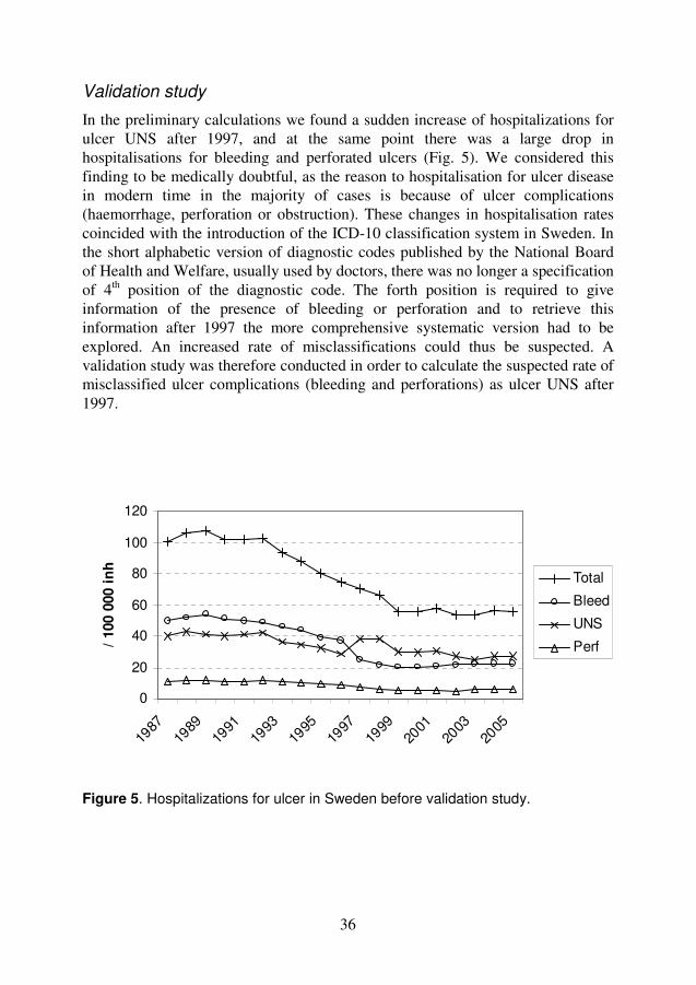
Validation study
In the preliminary calculations we found a sudden increase of hospitalizations for ulcer UNS after 1997, and at the same point there was a large drop in hospitalisations for bleeding and perforated ulcers (Fig. 5). We considered this finding to be medically doubtful, as the reason to hospitalisation for ulcer disease in modern time in the majority of cases is because of ulcer complications (haemorrhage, perforation or obstruction). These changes in hospitalisation rates coincided with the introduction of the ICD-10 classification system in Sweden. In the short alphabetic version of diagnostic codes published by the National Board of Health and Welfare, usually used by doctors, there was no longer a specification of 4th position of the diagnostic code. The forth position is required to give information of the presence of bleeding or perforation and to retrieve this information after 1997 the more comprehensive systematic version had to be explored. An increased rate of misclassifications could thus be suspected. A validation study was therefore conducted in order to calculate the suspected rate of misclassified ulcer complications (bleeding and perforations) as ulcer UNS after 1997. Figure 5. Hospitalizations for ulcer in Sweden before validation study.
0
20
40
60
80
100
120
1987
1989
1991
1993
1995
1997
1999
2001
2003
2005
/ 100 0
00 i
nh
Total
Bleed
UNS
Perf
37
A random sampling of totally 400 emergency hospitalizations at Departments of Surgery, with a discharge diagnosis of ulcer UNS were drawn from the Swedish Hospital Discharge Register, 200 from the period 1993-1994 and 200 from 1999-2000. The sampling number was chosen after the following calculations: With an assumption of a base-line misclassification of diagnoses in the Swedish Hospital Discharge Register of around 10%, 195 individuals in each group were required to detect an increase to around 20 per cent of misclassified ulcer complications as ulcer UNS after the introduction of ICD-10 (80% power, two-sided test, α= 0.05).
In cooperation with the National Board of Health and Welfare, questionnaires were sent to the departments where the patients had been hospitalised, for evaluation of the medical records to ascertain a potential misclassification of a perforated or bleeding ulcer. The response rate was 96.8% (387/400). Data were unidentified before further processing and analysing.
The criteria of a misclassified bleeding ulcer was either a) operation for bleeding ulcer or b) symptoms/history of bleeding at admission together with an endoscopically verified ulcer with no other bleeding source or c) an endoscopically verified bleeding ulcer. The criteria of a misclassified perforated ulcer were either operation for perforated ulcer or in another way verified perforated ulcer.
In the validation study the median age increased from 69 to 74 years (p=0.0132). Symptoms or patient history of bleeding (hematemesis or melena) at time for admission increased by 53% and was much more frequent in the latter period (52.3% compared to 34.2%, p=0.000047, Fishers´s exact test). Diagnostic endoscopy was frequently performed during both periods (84.0 and 89.2%, ns). The diagnosis of ulcer disease was verified (through surgery, endoscopy, radiology or autopsy) to a higher extent in the latter period (81.0 and 88.8% respectively, p=0.033). The verification of bleeding was made in about 20% of patients (blood in stomach, ongoing bleeding, visible vessel, haematin coloured spot or blood clot covering ulcer base) and did not change between periods.
In summary, 65/190 patients met the criteria of a misclassified bleeding ulcer before the introduction of ICD-10 in comparison with 98/197 patients after the introduction of ICD-10. An increase from 34.2 to 49.7% (p=0.002). The mis-classification of perforated ulcer increased from 1/190 (0.5%) to 12/197 (6.1%) (p=0.003).
Extrapolation of data
The percentage of misclassified bleeding or perforated ulcers found in the validation study was subtracted from the crude rate of hospitalisations for ulcer UNS in the cohort. The result from the period 1993-1994 was used for 1987-1996 (before the introduction of ICD-10) and the result from 1999-2000 was used for 1997-2005 (after the introduction of ICD-10. (In one Swedish region ICD-10 was
38
introduced in 1998)). The number of hospitalizations subtracted from the ulcer UNS cases was then added to the bleeding or perforated ulcer cases in the cohort. Chi-square tests showed no difference regarding the distribution in age, gender and ulcer location between the misclassified bleeding cases in the validation study and the UNS cases in the cohort. The same extrapolation percentage was therefore applied across all subgroups of age, gender and ulcer location.
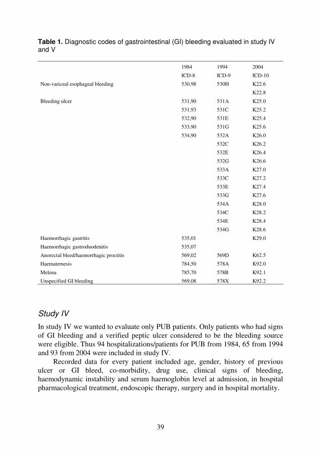
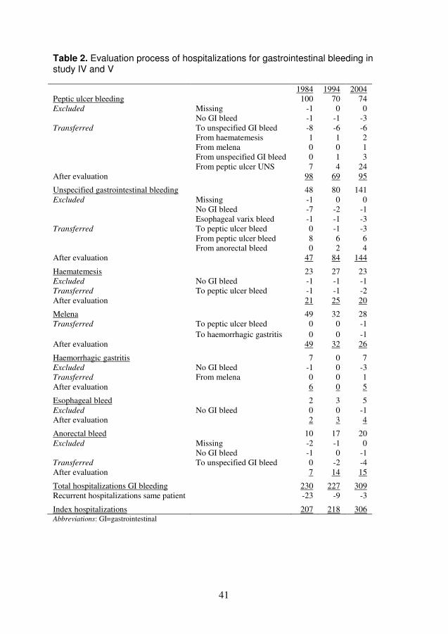
Study IV and V A retrospective review of totally 766 hospitalizations with a discharge diagnosis of gastrointestinal bleeding during three distinct time periods: 1984, 1994 and 2004 at the Department of Surgery (and for 2004 also the Department of Emergency conditions due to re-organisation) at Lund University Hospital in Sweden was performed. The diagnostic codes included in these studies are summarized in Table 1. Bleeding from oesophageal varices were not included in these studies.
Additionally 193 hospitalizations for unspecified peptic ulcer were evaluated according to the results of the validation study performed in study III where a diagnostic misclassification rate of bleeding ulcer in 34-50% of unspecified ulcer cases was found. Seven of 63 (11.1%) in 1984, four of 41 (9.8%) in 1994 and 24 of 89 (27%) in 2004 were found to be misclassified bleeding ulcers and consequently added to the bleeding ulcer group.
Every medical record was carefully studied and patients found to have an incorrect diagnosis were excluded (non bleeders) or transferred to the correct diagnostic group. If a patient had more than one hospitalization with the same localization of GI bleed during the same year, only the index hospitalization was included. Twenty-three patients were hospitalized twice, three patients three times and one patient seven times. After exclusion of these 35 recurrent hospitalizations, 731 hospitalizations/patients were included in the study. The evaluation process is shown in Table 2 below.
Lund University Hospital provides highly specialised health care for rare conditions in the population in the Southern part of Sweden but for common emergency conditions like GI bleeding it serves the population in a defined catchment area in the mid-west of Skane County. The population in the catchment area grew from 151,711 in 1984, to 170,727 in 1994 to 289,560 in 2004.
39
Table 1. Diagnostic codes of gastrointestinal (GI) bleeding evaluated in study IV and V 1984 1994 2004
ICD-8 ICD-9 ICD-10
Non-variceal esophageal bleeding 530,98 530H K22.6
K22.8
Bleeding ulcer 531,90 531A K25.0
531,93 531C K25.2
532,90 531E K25.4
533,90 531G K25.6
534,90 532A K26.0
532C K26.2
532E K26.4
532G K26.6
533A K27.0
533C K27.2
533E K27.4
533G K27.6
534A K28.0
534C K28.2
534E K28.4
534G K28.6
Haemorrhagic gastritis 535,01 K29.0
Haemorrhagic gastroduodenitis 535,07
Anorectal bleed/haemorrhagic proctitis 569,02 569D K62.5
Haematemesis 784,50 578A K92.0
Melena 785,70 578B K92.1
Unspecified GI bleeding 569,08 578X K92.2
Study IV
In study IV we wanted to evaluate only PUB patients. Only patients who had signs of GI bleeding and a verified peptic ulcer considered to be the bleeding source were eligible. Thus 94 hospitalizations/patients for PUB from 1984, 65 from 1994 and 93 from 2004 were included in study IV.
Recorded data for every patient included age, gender, history of previous ulcer or GI bleed, co-morbidity, drug use, clinical signs of bleeding, haemodynamic instability and serum haemoglobin level at admission, in hospital pharmacological treatment, endoscopic therapy, surgery and in hospital mortality.
40
Study V
In study V, patients were grouped into upper GI bleed (UPGIB) and lower GI bleed (LGIB) according to statements in the records of bleeding symptoms and bleeding sources found in investigations of the GI tract. Upper GI bleed was further divided into peptic ulcer bleed (PUB) and non-ulcer upper GI bleed (NUUPGIB). PUB was defined as a peptic ulcer, considered to be the bleeding source, found at endoscopy or surgery. NUUPGIB was defined as haematemesis or blood in nasogastric tube at presentation and/or blood or a bleeding source in the upper GI tract, other than ulcer, found at endoscopy or surgery. LGIB consisted of the other GI bleedings and were considered to evolve from a level below the Ligamentum of Treitz, i.e. no haematemesis or blood in nasogastric tube and/or no bleeding source found at upper endoscopy.
The following patient data was recorded: age, gender, co-morbidity, drug use, serum haemoglobin level and haemodynamic instability at admission, diagnostic modalities (gastroscopy, colonoscopy) and in-hospital mortality.
Statistics
Incidence and mortality rates per 100,000 inhabitants were calculated by Poisson regression with 95 per cent confidence intervals (CI) and Tukey corrected p-values for multiple comparisons. In univariate analyses Kruskal-Wallis or Wilcoxon tests were used for continuous variables and Fisher’s exact test for categorical variables. Logistic regression models were used in the multivariable analyses. The level of significance was set at p<0.05. Data were analysed using the Hmisc and Design packages of the R software (R Foundation for Statistical Computing, Vienna, Austria), version 2.6.2.
41
Table 2. Evaluation process of hospitalizations for gastrointestinal bleeding in study IV and V 1984 1994 2004 Peptic ulcer bleeding 100 70 74 Excluded Missing -1 0 0 No GI bleed -1 -1 -3 Transferred To unspecified GI bleed -8 -6 -6 From haematemesis 1 1 2 From melena 0 0 1 From unspecified GI bleed 0 1 3 From peptic ulcer UNS 7 4 24 After evaluation 98 69 95 Unspecified gastrointestinal bleeding 48 80 141 Excluded Missing -1 0 0 No GI bleed -7 -2 -1 Esophageal varix bleed -1 -1 -3 Transferred To peptic ulcer bleed 0 -1 -3 From peptic ulcer bleed 8 6 6 From anorectal bleed 0 2 4 After evaluation 47 84 144 Haematemesis 23 27 23 Excluded No GI bleed -1 -1 -1 Transferred To peptic ulcer bleed -1 -1 -2 After evaluation 21 25 20 Melena 49 32 28 Transferred To peptic ulcer bleed 0 0 -1 To haemorrhagic gastritis 0 0 -1 After evaluation 49 32 26 Haemorrhagic gastritis 7 0 7 Excluded No GI bleed -1 0 -3 Transferred From melena 0 0 1 After evaluation 6 0 5 Esophageal bleed 2 3 5 Excluded No GI bleed 0 0 -1 After evaluation 2 3 4 Anorectal bleed 10 17 20 Excluded Missing -2 -1 0 No GI bleed -1 0 -1 Transferred To unspecified GI bleed 0 -2 -4 After evaluation 7 14 15 Total hospitalizations GI bleeding 230 227 309 Recurrent hospitalizations same patient -23 -9 -3 Index hospitalizations 207 218 306 Abbreviations: GI=gastrointestinal

42
43
RESULTS
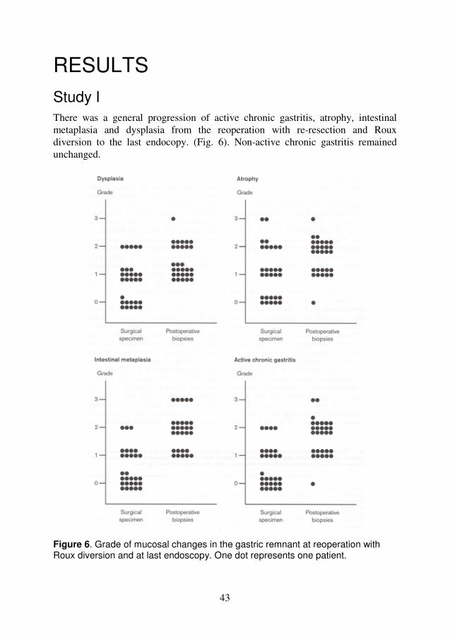
Study I There was a general progression of active chronic gastritis, atrophy, intestinal metaplasia and dysplasia from the reoperation with re-resection and Roux diversion to the last endocopy. (Fig. 6). Non-active chronic gastritis remained unchanged.
Figure 6. Grade of mucosal changes in the gastric remnant at reoperation with Roux diversion and at last endoscopy. One dot represents one patient.
44
The same development was seen when patients were subgrouped according to primary ulcer location (gastric or duodenal ulcer) or according to cause of reoperation (reflux gastritis or severe dysplasia / EGC). However, the subgroups were too small in some cases for adequate statistical evaluation.
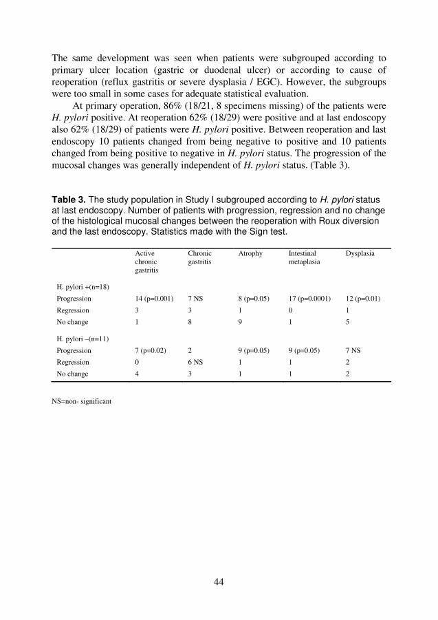
At primary operation, 86% (18/21, 8 specimens missing) of the patients were H. pylori positive. At reoperation 62% (18/29) were positive and at last endoscopy also 62% (18/29) of patients were H. pylori positive. Between reoperation and last endoscopy 10 patients changed from being negative to positive and 10 patients changed from being positive to negative in H. pylori status. The progression of the mucosal changes was generally independent of H. pylori status. (Table 3). Table 3. The study population in Study I subgrouped according to H. pylori status at last endoscopy. Number of patients with progression, regression and no change of the histological mucosal changes between the reoperation with Roux diversion and the last endoscopy. Statistics made with the Sign test.
Active chronic gastritis
Chronic gastritis
Atrophy Intestinal metaplasia
Dysplasia
H. pylori +(n=18)
Progression 14 (p=0.001) 7 NS 8 (p=0.05) 17 (p=0.0001) 12 (p=0.01)
Regression 3 3 1 0 1
No change
1 8 9 1 5
H. pylori –(n=11)
Progression 7 (p=0.02) 2 9 (p=0.05) 9 (p=0.05) 7 NS
Regression 0 6 NS 1 1 2
No change 4 3 1 1 2
NS=non- significant
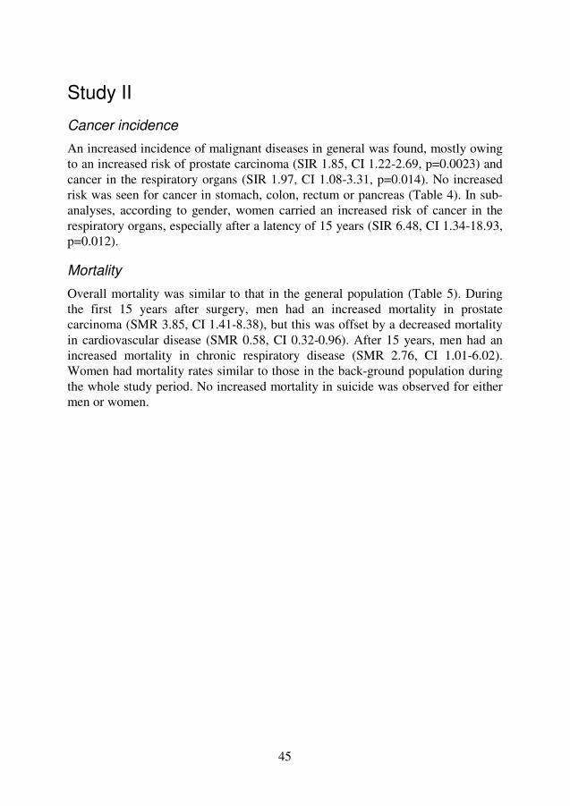
45
Study II
Cancer incidence
An increased incidence of malignant diseases in general was found, mostly owing to an increased risk of prostate carcinoma (SIR 1.85, CI 1.22-2.69, p=0.0023) and cancer in the respiratory organs (SIR 1.97, CI 1.08-3.31, p=0.014). No increased risk was seen for cancer in stomach, colon, rectum or pancreas (Table 4). In sub-analyses, according to gender, women carried an increased risk of cancer in the respiratory organs, especially after a latency of 15 years (SIR 6.48, CI 1.34-18.93, p=0.012).
Mortality
Overall mortality was similar to that in the general population (Table 5). During the first 15 years after surgery, men had an increased mortality in prostate carcinoma (SMR 3.85, CI 1.41-8.38), but this was offset by a decreased mortality in cardiovascular disease (SMR 0.58, CI 0.32-0.96). After 15 years, men had an increased mortality in chronic respiratory disease (SMR 2.76, CI 1.01-6.02). Women had mortality rates similar to those in the back-ground population during the whole study period. No increased mortality in suicide was observed for either men or women.
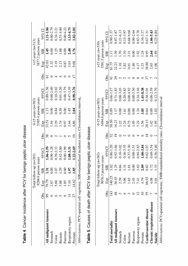
46
Tab
le 4
. Can
cer
inci
denc
e a
fter
PC
V f
or b
enig
n pe
ptic
ulc
er d
isea
se
T
otal
fol
low
-up
(n=3
83)
9288
.6 p
erso
n-ye
ars
0-
15 y
ears
(n=
383)
53
85.4
per
son-
year
s >1
5 ye
ars
(n=3
13)
3877
.2 p
erso
n ye
ars
O
bs
Exp
SI
R
95%
CI
Obs
E
xp
SIR
95
%C
I O
bs
Exp
SI
R
95%
CI
All
mal
igna
nt t
umou
rs
99
79.7
9 1.
31
1.06
-1.5
9 38
33
.51
1.13
0.
80-1
.56
61
42.0
3 1.
45
1.11
-1.8
6 St
omac
h 0
2.87
0.
00
0.00
-1.2
8 0
1.54
0.
00
0.00
-2.4
0 0
1.32
0.
00
0.00
-2.7
9 C
olon
6
5.60
1.
07
0.39
-2.3
3 2
2.39
0.
84
0.10
-3.0
2 4
3.19
1.
25
0.34
-3.2
1 R
ectu
m
5 3.
88
1.29
0.
42-3
.00
1 1.
74
0.58
0.
01-3
.20
4 2.
13
1.88
0.
51-4
.80
Pan
crea
s 0
1.85
0.
00
0.00
-1.9
9 0
0.97
0.
00
0.00
-3.7
9 0
0.87
0.
00
0.00
-4.2
2 R
espi
rato
ry o
rgan
s 14
7.
10
1.97
1.
08-3
.31
5 3.
44
1.45
0.
47-3
.39
9 3.
63
2.48
1.
13-4
.71
Pro
stat
e 27
14
.62
1.85
1.
22-2
.69
10
4.86
2.
04
0.98
-3.7
6 17
9.
68
1.76
1.
02-2
.81
Ab
bre
via
tio
ns:
PC
V=p
arie
tal c
ell v
agot
omy,
SIR
=sta
ndar
dize
d in
cide
nce
ratio
, CI=
conf
iden
ce in
terv
al,
Tab
le 5
. Cau
ses
of d
eath
aft
er P
CV
for
be
nig
n p
eptic
ulc
er d
isea
se.
T
otal
fol
low
-up
(n=3
83)
9076
.3 p
erso
n-ye
ars
0-15
yea
rs (
n=38
3)
5494
.8 p
erso
n-ye
ars
>15
year
s (n
=330
) 35
81.5
per
son
year
s
Obs
E
xp
SIR
95
%C
I O
bs
Exp
SI
R
95%
CI
Obs
E
xp
SIR
95
% C
I T
otal
mor
talit
y 14
3 14
0.25
1.
02
0.86
-1.2
0 52
58
.50
0.89
0.
66-1
.17
91
81.7
5 1.
11
0.90
-1.3
7 A
ll m
alig
nant
tum
ours
48
38
.45
1.25
0.
92-1
.66
19
16.2
1 1.
17
0.71
-1.8
3 29
22
.23
1.30
0.
87-1
.87
Stom
ach
2 2.
39
0.84
0.
10-3
.02
0 1.
22
0.00
0.
00-3
.03
2 1.
18
1.70
0.
21-6
.13
Col
on
4 3.
06
1.31
0.
36-3
.35
2 1.
24
1.61
0.
20-5
.83
2 1.
82
1.10
0.
13-3
.97
Rec
tum
3
1.65
1.
81
0.37
-5.3
0 0
0.75
0.
00
0.00
-4.9
2 3
0.91
3.
31
0.68
-9.6
8 P
ancr
eas
0 2.
45
0.00
0.
00-1
.51
0 1.
05
0.00
0.
00-3
.50
0 1.
40
0.00
0.
00-2
.64
Res
pira
tory
org
ans
12
7.41
1.
62
0.84
-2.8
3 4
3.24
1.
24
0.34
-3.1
6 8
4.17
1.
92
0.83
-3.7
8 P
rost
ate
10
4.79
2.
09
1.00
-3.8
4 6
1.56
3.
85
1.41
-8.3
8 4
3.23
1.
24
0.34
-3.1
7 C
ardi
ovas
cula
r di
seas
e 55
66
.93
0.82
0.
62-1
.07
18
28.1
3 0.
64
0.38
-1.0
1 37
38
.80
0.95
0.
67-1
.31
Chr
onic
res
pira
tory
dis
ease
7
4.08
1.
72
0.69
-3.5
4 0
1.42
0.
00
0.00
-2.5
9 7
2.65
2.
64
1.06
-5.4
3 Su
icid
e 4
3.01
1.
33
0.36
-3.4
0 2
1.95
1.
02
0.12
-3.7
0 2
1.06
1.
89
0.23
-6.8
4
Ab
bre
via
tio
ns:
PC
V=p
arie
tal c
ell v
agot
omy,
SM
R=s
tand
ardi
zed
mor
talit
y ra
tio, C
I=co
nfid
ence
inte
rval
47
Study III
Hospitalizations for bleeding ulcer
Admission rates are expressed as hospitalizations per 100 000 inhabitants per year. The hospitalization rate for peptic ulcer in total decreased from 102.1 to 54.8 during the study period 1987 to 2005. Hospitalizations for bleeding ulcer decreased from 63.9 to 35.3, for perforated ulcer from 11.9 to 7.7 and for ulcer UNS from 26.3 to 11.8 (All rates p<0.0001) (Fig. 7). Figure 7. Annual hospitalization rates for ulcer in Sweden 1987 to 2005. The declining trend of bleeding ulcer from 1987 to 2005 was significant for both sexes (p<0.0001) (Fig. 8). The male to female ratio decreased from 1.69 to 1.37 during the study period. Median age increased for both men (67 to 73 years) (p<0.0001) and women (74 to 78 years) (p<0.0001). The hospitalization rates were much higher among the elderly and the decrease in hospitalization rates was lower among the elderly compared to that of the younger age groups though the decrease was significant in all age groups (p<0.0001) (Table 6).
Admission year
/100
00
0 in
ha
bita
nts
0
20
40
60
80
100
120
1987 1989 1991 1993 1995 1997 1999 2001 2003 2005
TotalBleedingUnspecifiedPerforation
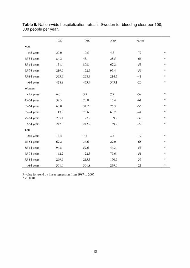
48
Table 6. Nation-wide hospitalization rates in Sweden for bleeding ulcer per 100, 000 people per year.
1987 1996 2005 %diff
Men
<45 years 20.0 10.5 4.7 -77 *
45-54 years 84.2 45.1 28.5 -66 *
55-64 years 131.4 80.8 62.2 -53 *
65-74 years 219.0 172.9 97.4 -56 *
75-84 years 363.6 268.9 214.5 -41 *
>84 years 428.8 433.4 343.1 -20 *
Women
<45 years 6.6 3.9 2.7 -59 *
45-54 years 39.5 23.8 15.4 -61 *
55-64 years 60.0 34.7 26.3 -56 *
65-74 years 113.0 78.6 63.2 -44 *
75-84 years 205.4 177.9 139.2 -32 *
>84 years 242.3 242.2 189.2 -22 *
Total
<45 years 13.4 7.3 3.7 -72 *
45-54 years 62.2 34.6 22.0 -65 *
55-64 years 94.8 57.6 44.3 -53 *
65-74 years 162.2 122.3 79.6 -51 *
75-84 years 269.6 215.3 170.9 -37 *
>84 years 301.0 301.8 239.0 -21 *
P-value for trend by linear regression from 1987 to 2005 * <0.0001
49
Figure 8. Annual hospitalization rates for bleeding ulcer by gender in Sweden 1987 to 2005.
Figure 9. Annual hospitalization rates for bleeding gastric and duodenal ulcer in Sweden 1987 to 2005.
Admission year
/10
0 0
00
inh
ab
itan
ts
0
20
40
60
80
100
1987 1989 1991 1993 1995 1997 1999 2001 2003 2005
MenWomen95% CI
Admission year
/100
00
0 in
ha
bita
nts
0
10
20
30
40
1987 1989 1991 1993 1995 1997 1999 2001 2003 2005
GU bleedingDU bleeding95% CI

50
Both gastric and duodenal ulcer bleeding decreased from 1987 to 2005 (Fig. 9). Admissions for gastric ulcer bleeding decreased among men from 35.6 to 20.2 and among women from 26.3 to 17.1 and the decrease was more pronounced among younger people although hospitalization rates declined in all age groups (p<0.0001) (men over 85 years, p=0.0002) (Table 7). Hospitalizations for duodenal ulcer hemorrhage decreased in males from 40.5 to 20.1 and in females from 18.9 to 12.3. The hospitalization rates for duodenal ulcer hemorrhage decreased in all age groups (p<0.0001) except among women over 85 years of age where the rate was stable (p=0.0942) (Table 8). Table 7. Nation-wide hospitalization rates in Sweden for bleeding gastric ulcer per 100,000 people per year.
1987 1996 2005 %diff
Men
<45 years 7.6 4.8 2.2 -72 *
45-54 years 35.2 22.1 14.9 -58 *
55-64 years 55.7 43.6 30.0 -46 *
65-74 years 98.5 83.7 47.2 -52 *
75-84 years 176.1 145.7 105.2 -40 *
>84 years 213.2 232.9 181.0 -15 **
Women
<45 years 3.1 2.0 1.5 -53 *
45-54 years 19.1 13.4 8.4 -56 *
55-64 years 30.2 19.6 15.1 -50 *
65-74 years 68.4 44.5 35.6 -48 *
75-84 years 116.3 110.4 82.1 -29 *
>84 years 132.4 140.9 110.3 -17 *
Total
<45 years 5.4 3.4 1.8 -67 *
45-54 years 27.3 17.8 11.7 -57 *
55-64 years 42.6 31.5 22.6 -47 *
65-74 years 82.4 62.7 41.1 -50 *
75-84 years 140.6 124.9 91.9 -35 *
>84 years 157.9 169.6 133.2 -16 * P-value for trend by linear regression from 1987 to 2005 * <0.0001, ** =0.0002
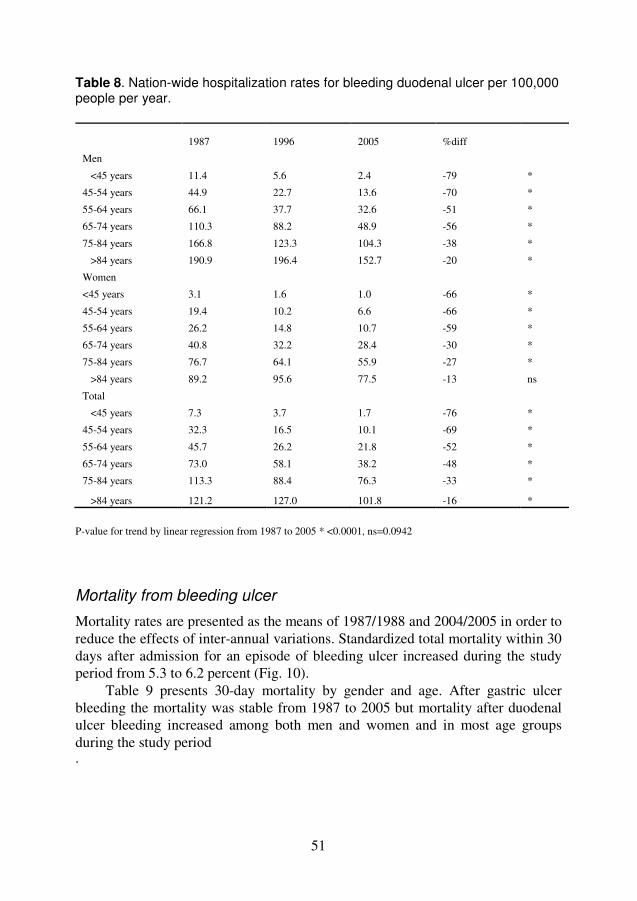
51
Table 8. Nation-wide hospitalization rates for bleeding duodenal ulcer per 100,000 people per year.
1987 1996 2005 %diff
Men
<45 years 11.4 5.6 2.4 -79 *
45-54 years 44.9 22.7 13.6 -70 *
55-64 years 66.1 37.7 32.6 -51 *
65-74 years 110.3 88.2 48.9 -56 *
75-84 years 166.8 123.3 104.3 -38 *
>84 years 190.9 196.4 152.7 -20 *
Women
<45 years 3.1 1.6 1.0 -66 *
45-54 years 19.4 10.2 6.6 -66 *
55-64 years 26.2 14.8 10.7 -59 *
65-74 years 40.8 32.2 28.4 -30 *
75-84 years 76.7 64.1 55.9 -27 *
>84 years 89.2 95.6 77.5 -13 ns
Total
<45 years 7.3 3.7 1.7 -76 *
45-54 years 32.3 16.5 10.1 -69 *
55-64 years 45.7 26.2 21.8 -52 *
65-74 years 73.0 58.1 38.2 -48 *
75-84 years 113.3 88.4 76.3 -33 *
>84 years 121.2 127.0 101.8 -16 *
P-value for trend by linear regression from 1987 to 2005 * <0.0001, ns=0.0942
Mortality from bleeding ulcer
Mortality rates are presented as the means of 1987/1988 and 2004/2005 in order to reduce the effects of inter-annual variations. Standardized total mortality within 30 days after admission for an episode of bleeding ulcer increased during the study period from 5.3 to 6.2 percent (Fig. 10).
Table 9 presents 30-day mortality by gender and age. After gastric ulcer bleeding the mortality was stable from 1987 to 2005 but mortality after duodenal ulcer bleeding increased among both men and women and in most age groups during the study period .
52
Table 9. Standardized 30 day mortality (%) after ulcer bleeding in Sweden. Standardized by age and gender wherever applicable.
1987/1988 2004/2005 p-value
for trend
Total ¤# 5.3% 6.2% 0.0014
<65 years # 1.0% 2.8% 0.054
65-74 years # 3.7% 5.9% 0.020
>74 years # 9.4% 9.6% 0.0092
Men total ¤ 5.9% 6.1% 0.019
<65 years 0.8% 2.2% 0.24
65-74 years 3.8% 5.7% 0.046
>74 years 10.2% 9.7% 0.065
Women total ¤ 4.9% 6.6% 0.015
<65 years 1.4% 3.7% 0.069
65-74 years 3.6% 6.3% 0.048
>74 years 8.1% 9.4% 0.030
Men GU Total ¤ 5.1% 5.2% 0.57
<65 years 0.8% 2.3% 0.72
65-74 years 3.5% 3.3% 0.54
>74 years 8.6% 9.1% 0.26
Women GU Total ¤ 4.2% 5.5% 0.22
<65 years 0.5% 3.8% 0.27
65-74 years 2.6% 4.9% 0.16
>74 years 7.8% 6.9% 0.62
Men DU Total ¤ 6.0% 6.1% 0.023
<65 years 0.6% 2.6% 0.042
65-74 years 2.8% 6.3% 0.030
>74 years 11.2% 9.1% 0.14
Women DU Total ¤ 5.9% 7.6% 0.013
<65 years 1.9% 4.7% 0.010
65-74 years 4.8% 8.2% 0.16
>74 years 9.3% 10.3% 0.019
Linear regression was used to test for secular trend. Abbreviations: GU=gastric ulcer, DU=duodenal ulcer. Standardized according to the age (¤) and/or gender (#) distribution of bleeding ulcer hospi-talizations in Sweden 1987-2005.
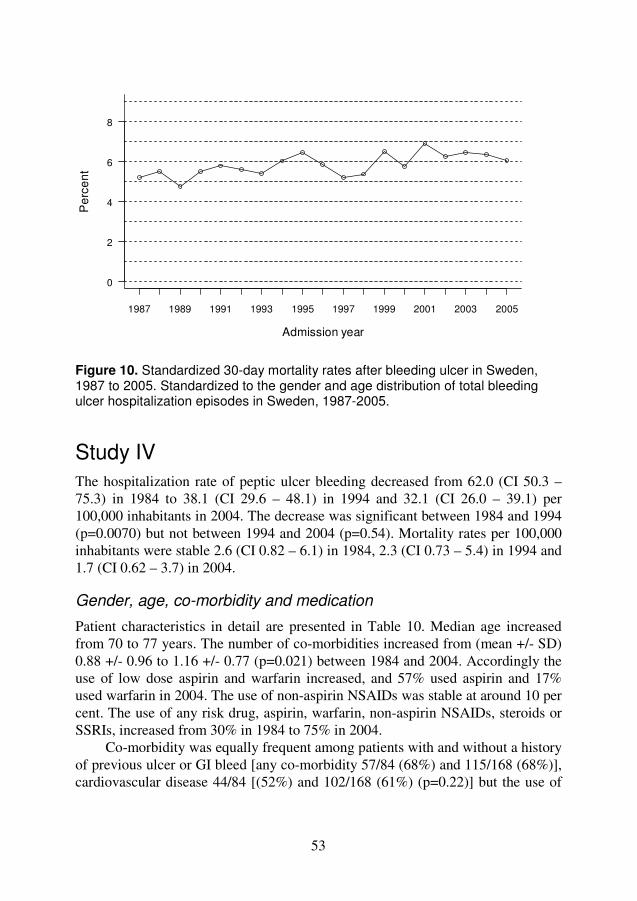
53
Figure 10. Standardized 30-day mortality rates after bleeding ulcer in Sweden, 1987 to 2005. Standardized to the gender and age distribution of total bleeding ulcer hospitalization episodes in Sweden, 1987-2005.
Study IV The hospitalization rate of peptic ulcer bleeding decreased from 62.0 (CI 50.3 – 75.3) in 1984 to 38.1 (CI 29.6 – 48.1) in 1994 and 32.1 (CI 26.0 – 39.1) per 100,000 inhabitants in 2004. The decrease was significant between 1984 and 1994 (p=0.0070) but not between 1994 and 2004 (p=0.54). Mortality rates per 100,000 inhabitants were stable 2.6 (CI 0.82 – 6.1) in 1984, 2.3 (CI 0.73 – 5.4) in 1994 and 1.7 (CI 0.62 – 3.7) in 2004.
Gender, age, co-morbidity and medication
Patient characteristics in detail are presented in Table 10. Median age increased from 70 to 77 years. The number of co-morbidities increased from (mean +/- SD) 0.88 +/- 0.96 to 1.16 +/- 0.77 (p=0.021) between 1984 and 2004. Accordingly the use of low dose aspirin and warfarin increased, and 57% used aspirin and 17% used warfarin in 2004. The use of non-aspirin NSAIDs was stable at around 10 per cent. The use of any risk drug, aspirin, warfarin, non-aspirin NSAIDs, steroids or SSRIs, increased from 30% in 1984 to 75% in 2004.
Co-morbidity was equally frequent among patients with and without a history of previous ulcer or GI bleed [any co-morbidity 57/84 (68%) and 115/168 (68%)], cardiovascular disease 44/84 [(52%) and 102/168 (61%) (p=0.22)] but the use of
Admission year
Pe
rce
nt
0
2
4
6
8
1987 1989 1991 1993 1995 1997 1999 2001 2003 2005

54
aspirin was less frequent [20/84 (24 %) and 66/168 (40%) (p=0.016)] and the use of H2RA or PPI was more frequent [19/84 (23%) and 8/168 (5%) (p<0.001)] among patients with, compared with those without, a history of previous ulcer or GI bleed.
Therapy and outcome
Detailed data of in hospital treatment, discharge status and mortality rates are found in Table 11. Intravenous pharmacological treatment and endoscopic therapy became more frequent and surgery became less frequent from 1984 to 2004 Prescriptions at discharge of acid reducing therapy and eradication therapy against H. pylori increased. Case fatality rate ranged between 4 and 8% (not significant). Patients who started to bleed while they were hospitalized for another reason had a higher mortality (13, 17, 14%) compared with those who started to bleed at home (2, 4, 4%) (p=0.004).
Data by ulcer location in 2004 is shown in Table 12. History of previous ulcer or GI bleed and endoscopic interventions were more common among duodenal ulcer patients whereas use of any “risk drug” was related to gastric ulcer disease.
Table 13 shows data for PUB patients by mortality. Patients who died during hospitalization were older, had more co-morbidities (mean number of co-morbidities (+/-SD) was 2.15 (+/-0.90) compared with 0.94 (+/-0.83)) and had lower serum haemoglobin levels at presentation in comparison with those who were alive at discharge. Use of “risk drugs” at presentation did not negatively influence the fatality rate. In the multivariable logistic regression model shown in Table 14, age above 65 years, number of co-morbidities and haemodynamic instability were independent risk factors for in hospital mortality whereas the use of aspirin at start of bleed decreased the risk of mortality significantly.

55
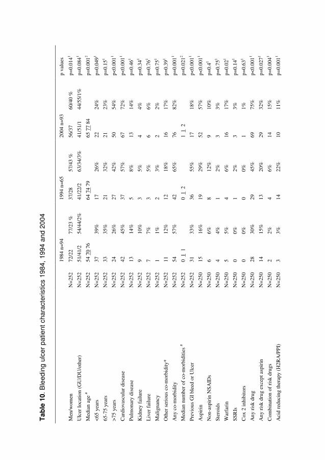
56
Tab
le 1
0. B
leed
ing
ulce
r p
atie
nt c
hara
cter
istic
s 19
84,
1994
an
d 20
04
19
84 n
=94
1994
n=6
5 20
04 n
=93
p va
lues
Men
/wom
en
N=
252
72/2
2 77
/23
%
37/2
8 57
/43
%
56/3
7 60
/40
%
p=0.
0141
Ulc
er lo
cati
on (
GU
/DU
/oth
er)
N=
252
51/4
1/2
54/4
4/2%
41
/22/
2 63
/34/
3%
41/5
1/1
44/5
5/1%
p=
0.08
41
Med
ian
age
# N
=25
2 54
70
76
64 7
4 79
65
77
84
p=0.
0012
<65
yea
rs
N=
252
37
39%
17
26
%
22
24%
p=
0.04
91
65-7
5 ye
ars
N=
252
33
35%
21
32
%
21
23%
p=
0.15
1
>75
yea
rs
N=
252
24
26%
27
42
%
50
54%
p<
0.00
11
Car
diov
ascu
lar
dise
ase
N=
252
42
45%
37
57
%
67
72%
p<
0.00
11
Pulm
onar
y di
seas
e
N=
252
13
14%
5
8%
13
14%
p=
0.46
1
Kid
ney
failu
re
N=
252
9 10
%
3 5%
4
4%
p=0.
341
Liv
er f
ailu
re
N=
252
7 7%
3
5%
6 6%
p=
0.76
1
Mal
igna
ncy
N=
252
1 1%
2
3%
2 2%
p=
0.75
1
Oth
er s
erio
us c
o-m
orbi
dity
* N
=25
2 11
12
%
12
18%
16
17
%
p=0.
391
Any
co-
mor
bidi
ty
N=
252
54
57%
42
65
%
76
82%
p=
0.00
11
Med
ian
num
ber
of c
o-m
orbi
ditie
s #
N=
252
0 1
1
0 1
2
1 1
2
p=0.
0212
Prev
ious
GI
blee
d or
Ulc
er
N=
252
31
33%
36
55
%
17
18%
p<
0.00
11
Asp
irin
N
=25
0 15
16
%
19
29%
52
57
%
p<0.
0011
Non
-asp
irin
NSA
IDs
N=
250
6 6%
8
12%
9
10%
p=
0.41
Ster
oids
N
=25
0 4
4%
1 2%
3
3%
p=0.
751
War
fari
n N
=25
0 5
5%
4 6%
16
17
%
p=0.
021
SSR
Is
N=
250
0 0%
1
2%
3 3%
p=
0.14
1
Cox
2 in
hibi
tors
N
=25
0 0
0%
0 0%
1
1%
p=0.
631
Any
ris
k dr
ug
N=
250
28
30%
29
45
%
69
75%
p<
0.00
11
Any
ris
k dr
ug e
xcep
t asp
irin
N
=25
0 14
15
%
13
20%
29
32
%
p=0.
0271
Com
bina
tion
of r
isk
drug
s N
=25
0 2
2%
4 6%
14
15
%
p=0.
0041
Aci
d re
duci
ng th
erap
y (H
2RA
/PP
I)
N=
250
3 3%
14
22
%
10
11%
p=
0.00
11
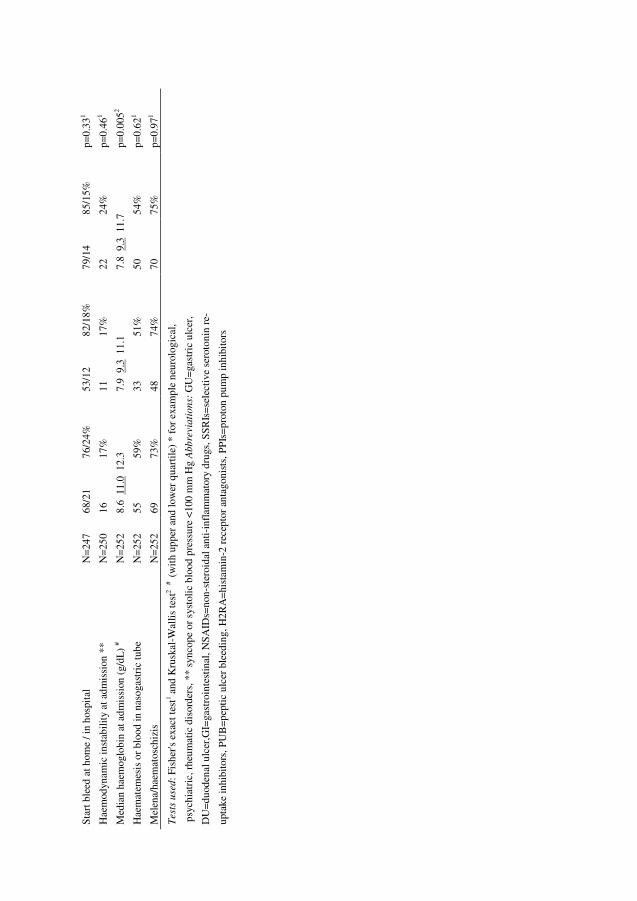
57
Star
t ble
ed a
t hom
e / i
n ho
spit
al
N=
247
68/2
1 76
/24%
53
/12
82/1
8%
79/1
4 85
/15%
p=
0.33
1
Hae
mod
ynam
ic in
stab
ility
at a
dmis
sion
**
N=
250
16
17%
11
17
%
22
24%
p=
0.46
1
Med
ian
haem
oglo
bin
at a
dmis
sion
(g/
dL)
# N
=25
2 8.
6 1
1.0
12.
3 7.
9 9
.3 1
1.1
7.8
9.3
11.
7 p=
0.00
52
Hae
mat
emes
is o
r bl
ood
in n
asog
astr
ic tu
be
N=
252
55
59%
33
51
%
50
54%
p=
0.62
1
Mel
ena/
haem
atos
chiz
is
N=
252
69
73%
48
74
%
70
75%
p=
0.97
1
Test
s u
sed
: Fis
her's
exa
ct te
st1 a
nd K
rusk
al-W
allis
test
2 #
(w
ith
uppe
r an
d lo
wer
qua
rtile
) *
for
exam
ple
neur
olog
ical
,
psy
chia
tric
, rhe
umat
ic d
isor
ders
, **
sync
ope
or s
ysto
lic b
lood
pre
ssur
e <1
00 m
m H
g A
bb
revia
tio
ns:
GU
=ga
stri
c ul
cer,
DU
=du
oden
al u
lcer
,GI=
gast
roin
test
inal
, NSA
IDs=
non-
ster
oida
l ant
i-in
flam
mat
ory
drug
s, S
SRIs
=se
lect
ive
sero
toni
n re
-
upta
ke in
hibi
tors
, PU
B=p
epti
c ul
cer
blee
ding
, H2R
A=
hist
amin
-2 r
ecep
tor
anta
goni
sts,
PPI
s=pr
oton
pum
p in
hibi
tors
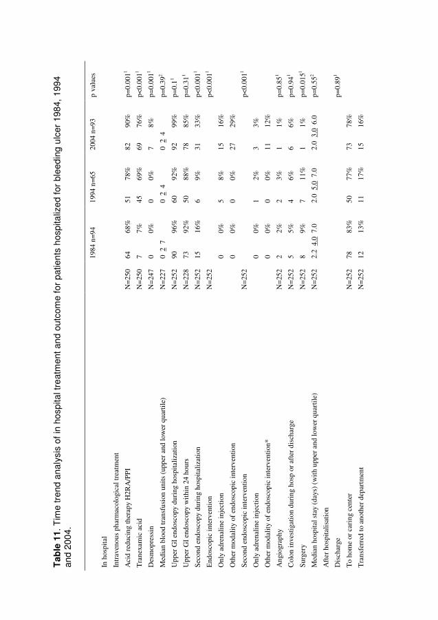
58
Tab
le 1
1. T
ime
tren
d an
aly
sis
of in
hos
pita
l tre
atm
ent a
nd o
utco
me
for
patie
nts
hos
pita
lize
d fo
r bl
eed
ing
ulc
er 1
984,
199
4
and
200
4.
1984
n=9
4 19
94 n
=65
2004
n=9
3 p
valu
es
In h
ospi
tal
Intr
aven
ous
phar
mac
olog
ical
trea
tmen
t
Aci
d re
duci
ng th
erap
y H
2RA
/PP
I N
=25
0 64
68
%
51
78%
82
90
%
p=0.
0011
Tra
nexa
mic
aci
d N
=25
0 7
7%
45
69%
69
76
%
p<0.
0011
Des
mop
ress
in
N=
247
0 0%
0
0%
7 8%
p=
0.00
11
Med
ian
bloo
d tr
ansf
usio
n un
its (
uppe
r an
d lo
wer
qua
rtile
) N
=22
7 0
2 7
0
2 4
0
2 4
p=
0.39
2
Upp
er G
I en
dosc
opy
duri
ng h
ospi
taliz
atio
n N
=25
2 90
96
%
60
92%
92
99
%
p=0.
11
Upp
er G
I en
dosc
opy
wit
hin
24 h
ours
N
=22
8 73
92
%
50
88%
78
85
%
p=0.
311
Seco
nd e
ndos
copy
dur
ing
hosp
italiz
atio
n N
=25
2 15
16
%
6 9%
31
33
%
p<0.
0011
End
osco
pic
inte
rven
tion
N
=25
2
p<
0.00
11
Onl
y ad
rena
line
inje
ctio
n
0 0%
5
8%
15
16%
Oth
er m
odal
ity o
f en
dosc
opic
inte
rven
tion
0
0%
0 0%
27
29
%
Seco
nd e
ndos
copi
c in
terv
enti
on
N=
252
p<0.
0011
Onl
y ad
rena
line
inje
ctio
n
0 0%
1
2%
3 3%
Oth
er m
odal
ity o
f en
dosc
opic
inte
rven
tion*
0 0%
0
0%
11
12%
Ang
iogr
aphy
N
=25
2 2
2%
2 3%
1
1%
p=0.
851
Col
on in
vest
igat
ion
duri
ng h
osp
or a
fter
dis
char
ge
N=
252
5 5%
4
6%
6 6%
p=
0.94
1
Surg
ery
N=
252
8 9%
7
11%
1
1%
p=0.
0151
Med
ian
hosp
ital s
tay
(day
s) (
wit
h up
per
and
low
er q
uart
ile)
N=
252
2.2
4.0
7.0
2.
0 5
.0 7
.0
2.0
3.0
6.0
p=
0.55
2
Aft
er h
ospi
talis
atio
n
Dis
char
ge
p=
0.89
1
To
hom
e or
car
ing
cent
er
N=
252
78
83%
50
77
%
73
78%
Tra
nsfe
rred
to a
noth
er d
epar
tmen
t N
=25
2 12
13
%
11
17%
15
16
%
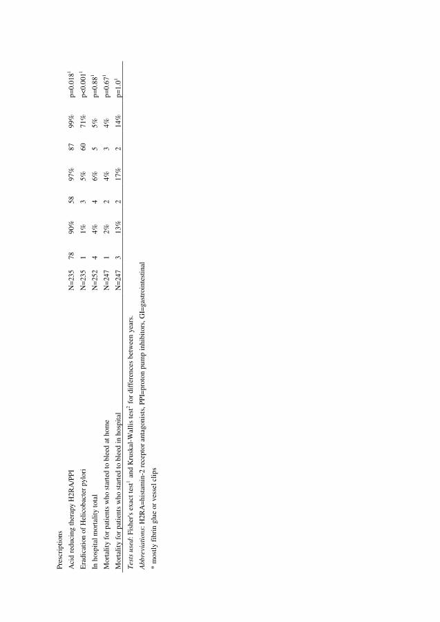
59
Pres
crip
tion
s
Aci
d re
duci
ng th
erap
y H
2RA
/PP
I N
=23
5 78
90
%
58
97%
87
99
%
p=0.
0181
Era
dica
tion
of
Hel
icob
acte
r py
lori
N
=23
5 1
1%
3 5%
60
71
%
p<0.
0011
In h
ospi
tal m
orta
lity
tota
l N
=25
2 4
4%
4 6%
5
5%
p=0.
881
Mor
talit
y fo
r pa
tien
ts w
ho s
tart
ed to
ble
ed a
t hom
e N
=24
7 1
2%
2 4%
3
4%
p=0.
671
Mor
talit
y fo
r pa
tien
ts w
ho s
tart
ed to
ble
ed in
hos
pita
l N
=24
7 3
13%
2
17%
2
14%
p=
1.01
Test
s u
sed
: Fis
her's
exa
ct te
st1 a
nd K
rusk
al-W
allis
test
2 fo
r di
ffer
ence
s be
twee
n ye
ars.
Abb
revi
ati
on
s: H
2RA
=hi
stam
in-2
rec
epto
r an
tago
nist
s, P
PI=
prot
on p
ump
inhi
bito
rs, G
I=ga
stro
inte
stin
al
* m
ostl
y fi
brin
glu
e or
ves
sel c
lips
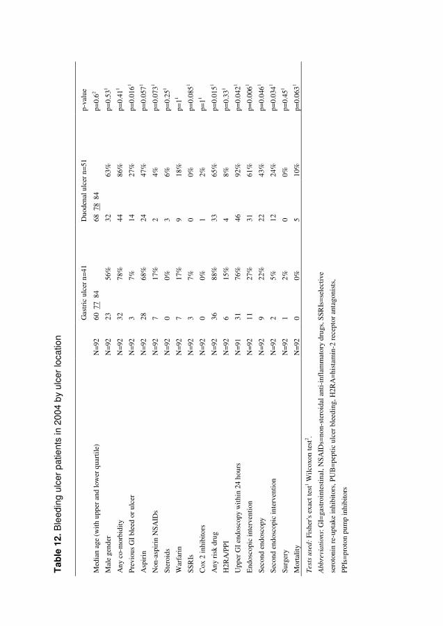
60
Tab
le 1
2. B
leed
ing
ulce
r p
atie
nts
in 2
004
by
ulce
r lo
catio
n
G
astr
ic u
lcer
n=4
1
Duo
dena
l ulc
er n
=51
p-va
lue
Med
ian
age
(wit
h up
per
and
low
er q
uart
ile)
N=
92
60 7
7 8
4 68
78
84
p=0.
62
Mal
e ge
nder
N
=92
23
56
%
32
63%
p=
0.53
1
Any
co-
mor
bidi
ty
N=
92
32
78%
44
86
%
p=0.
411
Prev
ious
GI
blee
d or
ulc
er
N=
92
3 7%
14
27
%
p=0.
0161
Asp
irin
N
=92
28
68
%
24
47%
p=
0.05
71
Non
-asp
irin
NSA
IDs
N=
92
7 17
%
2 4%
p=
0.07
31
Ster
oids
N
=92
0
0%
3 6%
p=
0.25
1
War
fari
n N
=92
7
17%
9
18%
p=
11
SSR
Is
N=
92
3 7%
0
0%
p=0.
0851
Cox
2 in
hibi
tors
N
=92
0
0%
1 2%
p=
11
Any
ris
k dr
ug
N=
92
36
88%
33
65
%
p=0.
0151
H2R
A/P
PI
N=
92
6 15
%
4 8%
p=
0.33
1
Upp
er G
I en
dosc
opy
wit
hin
24 h
ours
N
=91
31
76
%
46
92%
p=
0.04
21
End
osco
pic
inte
rven
tion
N
=92
11
27
%
31
61%
p=
0.00
61
Seco
nd e
ndos
copy
N
=92
9
22%
22
43
%
p=0.
0461
Seco
nd e
ndos
copi
c in
terv
enti
on
N=
92
2 5%
12
24
%
p=0.
0341
Surg
ery
N=
92
1 2%
0
0%
p=0.
451
Mor
talit
y N
=92
0
0%
5 10
%
p=0.
0631
Test
s u
sed
: Fi
sher
's e
xact
test
1 Wilc
oxon
test
2 .
Abb
revi
ati
on
s: G
I=ga
stro
inte
stin
al, N
SAID
s=no
n-st
eroi
dal a
nti-
infl
amm
ator
y dr
ugs,
SSR
Is=
sele
ctiv
e
sero
toni
n re
-upt
ake
inhi
bito
rs, P
UB
=pe
ptic
ulc
er b
leed
ing,
H2R
A=
hist
amin
-2 r
ecep
tor
anta
goni
sts,
PPIs
=pr
oton
pum
p in
hibi
tors

61
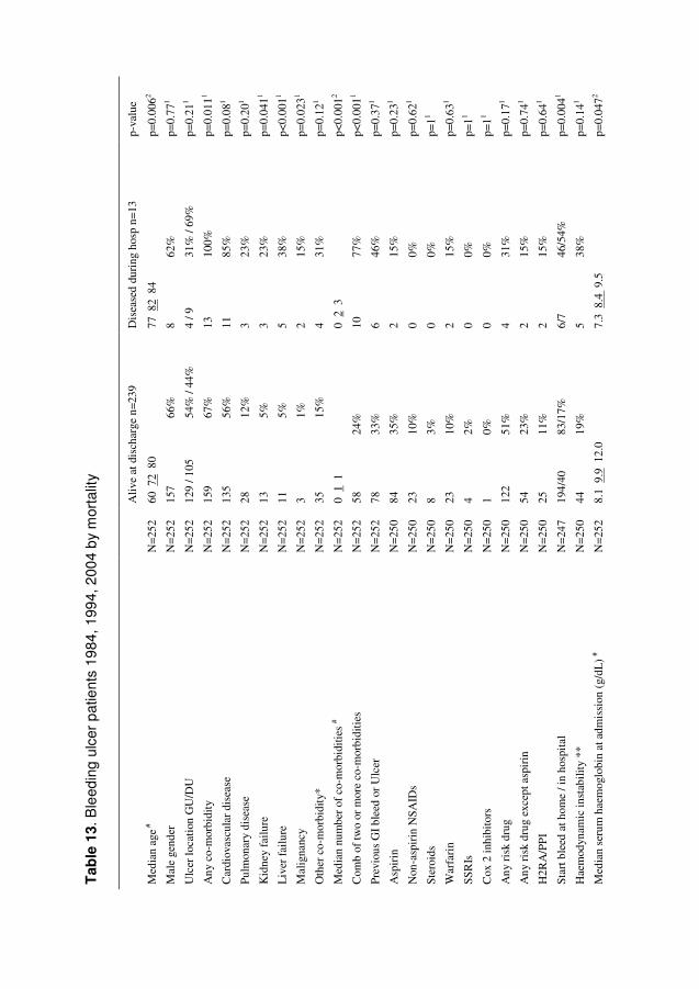
62
Tab
le 1
3. B
leed
ing
ulce
r p
atie
nts
1984
, 19
94,
200
4 b
y m
orta
lity
A
live
at d
isch
arge
n=2
39
Dis
ease
d du
ring
hos
p n=
13
p-va
lue
Med
ian
age #
N
=25
2 60
72
80
77
82
84
p=0.
0062
Mal
e ge
nder
N
=25
2 15
7 66
%
8 62
%
p=0.
771
Ulc
er lo
cati
on G
U/D
U
N=
252
129
/ 105
54
% /
44%
4
/ 9
31%
/ 69
%
p=0.
211
Any
co-
mor
bidi
ty
N=
252
159
67%
13
10
0%
p=0.
0111
Car
diov
ascu
lar
dise
ase
N=
252
135
56%
11
85
%
p=0.
081
Pulm
onar
y di
seas
e N
=25
2 28
12
%
3 23
%
p=0.
201
Kid
ney
failu
re
N=
252
13
5%
3 23
%
p=0.
0411
Liv
er f
ailu
re
N=
252
11
5%
5 38
%
p<0.
0011
Mal
igna
ncy
N=
252
3 1%
2
15%
p=
0.02
31
Oth
er c
o-m
orbi
dity
* N
=25
2 35
15
%
4 31
%
p=0.
121
Med
ian
num
ber
of c
o-m
orbi
ditie
s #
N=
252
0 1
1
0 2
3
p<0.
0012
Com
b of
two
or m
ore
co-m
orbi
diti
es
N=
252
58
24%
10
77
%
p<0.
0011
Prev
ious
GI
blee
d or
Ulc
er
N=
252
78
33%
6
46%
p=
0.37
1
Asp
irin
N
=25
0 84
35
%
2 15
%
p=0.
231
Non
-asp
irin
NSA
IDs
N=
250
23
10%
0
0%
p=0.
621
Ster
oids
N
=25
0 8
3%
0 0%
p=
11
War
fari
n N
=25
0 23
10
%
2 15
%
p=0.
631
SSR
Is
N=
250
4 2%
0
0%
p=11
Cox
2 in
hibi
tors
N
=25
0 1
0%
0 0%
p=
11
Any
ris
k dr
ug
N=
250
122
51%
4
31%
p=
0.17
1
Any
ris
k dr
ug e
xcep
t asp
irin
N
=25
0 54
23
%
2 15
%
p=0.
741
H2R
A/P
PI
N=
250
25
11%
2
15%
p=
0.64
1
Star
t ble
ed a
t hom
e / i
n ho
spit
al
N=
247
194/
40
83/1
7%
6/7
46/5
4%
p=0.
0041
Hae
mod
ynam
ic in
stab
ility
**
N=
250
44
19%
5
38%
p=
0.14
1
Med
ian
seru
m h
aem
oglo
bin
at a
dmis
sion
(g/
dL)
# N
=25
2 8.
1 9
.9 1
2.0
7.3
8.4
9.5
p=
0.04
72
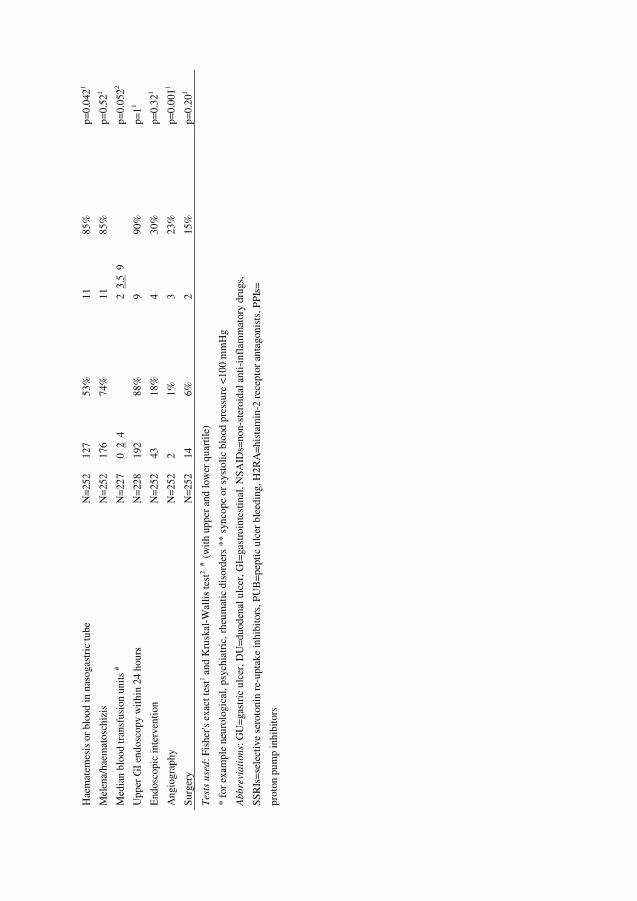
63
Hae
mat
emes
is o
r bl
ood
in n
asog
astr
ic tu
be
N=
252
127
53%
11
85
%
p=0.
0421
Mel
ena/
haem
atos
chiz
is
N=
252
176
74%
11
85
%
p=0.
521
Med
ian
bloo
d tr
ansf
usio
n un
its #
N=
227
0 2
4
2 3
.5 9
p=
0.05
22
Upp
er G
I en
dosc
opy
wit
hin
24 h
ours
N
=22
8 19
2 88
%
9 90
%
p=11
End
osco
pic
inte
rven
tion
N
=25
2 43
18
%
4 30
%
p=0.
321
Ang
iogr
aphy
N
=25
2 2
1%
3 23
%
p=0.
0011
Surg
ery
N=
252
14
6%
2 15
%
p=0.
201
Test
s u
sed
: Fis
her's
exa
ct te
st1 a
nd K
rusk
al-W
allis
test
2 #
(w
ith
uppe
r an
d lo
wer
qua
rtile
)
* fo
r ex
ampl
e ne
urol
ogic
al, p
sych
iatr
ic, r
heum
atic
dis
orde
rs *
* sy
ncop
e or
sys
tolic
blo
od p
ress
ure
<10
0 m
mH
g
Abb
revi
ati
on
s: G
U=
gast
ric
ulce
r, D
U=
duod
enal
ulc
er, G
I=ga
stro
inte
stin
al, N
SAID
s=no
n-st
eroi
dal a
nti-
infl
amm
ator
y dr
ugs,
SSR
Is=
sele
ctiv
e se
roto
nin
re-u
ptak
e in
hibi
tors
, PU
B=p
epti
c ul
cer
blee
ding
, H2R
A=
hist
amin
-2 r
ecep
tor
anta
goni
sts,
PPI
s=
prot
on p
ump
inhi
bito
rs
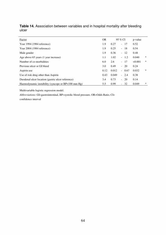
64
Table 14. Association between variables and in hospital mortality after bleeding ulcer Factor OR 95 % CI p-value
Year 1994 (1984 reference) 1.9 0.27 - 17 0.52
Year 2004 (1984 reference) 1.9 0.25 - 18 0.54
Male gender 1.9 0.36 - 12 0.48
Age above 65 years (1 year increase) 1.1 1.02 - 1.2 0.040 *
Number of co-morbidities 6.0 2.6 - 17 <0.001 *
Previous ulcer or GI bleed 3.0 0.49 - 20 0.24
Aspirin use 0.12 0.012 - 0.67 0.032 *
Use of risk drug other than Aspirin 0.43 0.049 - 2.4 0.38
Duodenal ulcer location (gastric ulcer reference) 3.4 0.73 - 20 0.14
Haemodynamic instability (syncope or BP<100 mm Hg) 5.5 0.99 - 32 0.049 * Multivariable logistic regression model.
Abbreviations: GI=gastrointestinal, BP=systolic blood pressure, OR=Odds Ratio, CI=
confidence interval
65
Study V
Time-trend analyses of incidence and mortality rates
The incidence of non-variceal GI bleeding decreased from 136.4 in 1984, to 127.7 in 1994 and 105.7 per 100,000 in 2004 and was due to a decline in the incidence of PUB. The incidence of NUUPGIB and LGIB did not change. Mortality rates after GI bleed from any localization per 100,000 inhabitants did not change between years (Table 15). Table 15. Standardized hospitalizations and mortality rates in gastrointestinal (GI) bleeding 1984, 1994, 2004. Number of patients per 100,000 inhabitants with 95% confidence intervals (CI). 1984 1994 2004
Pat / 100000 95 % CI
Pat / 100000 95 % CI
Pat / 100000 95 % CI
Hospitalizations
Total GI bleed 136.4 118.7 - 155.9 127.7 111.5 - 145.4 105.7 94.3 - 118.0
Total upper GI bleed 91.0 76.6 - 107.0 72.0 60.1 - 85.5 62.5 53.8 - 72.1
Peptic ulcer bleed 62.0 50.3 - 75.3 38.1 29.6 - 48.1 32.1 26.0 - 39.1
Other upper GI bleed 29.0 21.3 - 38.4 34.0 26.0 - 43.5 30.4 24.5 - 37.2
Lower GI bleed 45.5 35.6 - 57.1 55.6 45.2 - 67.6 43.2 36.0 - 51.2
Mortality
Total GI bleed 4.61 1.98 - 8.92 4.10 1.76 - 7.93 7.25 4.57 - 10.8
Total upper GI bleed 3.30 1.18 - 7.08 3.51 1.40 - 7.12 3.80 1.97 - 6.51
Peptic ulcer bleed 2.64 0.82 - 6.12 2.34 0.73 - 5.44 1.73 0.62 - 3.71
Other upper GI bleed 0.66 0.38 - 2.90 1.17 0.19 - 3.62 2.07 0.82 - 4.20
Lower GI bleed 1.32 0.22 - 4.07 0.59 0.03 - 2.58 3.45 1.73 - 6.06 Poisson regression with Tukey correction for multiple comparisons.
Diagnostics of UPGIB and LGIB
All PUB patients were subjected to upper endoscopy except a few who were either diagnosed by surgery or were very recently hospitalized and diagnosed with an ulcer. Bleeding sources found among NUUPGIB and LGIB patients are presented in Table 16. Among NUUPGIB patients an upper endoscopy was done in 60, 61 and 66% of cases in 1984, 1994 and 2004. Among LGIB patients, upper GI endoscopy was carried out during hospitalization in 29, 31 and 43% of patients. Colon investigation was performed in 65, 53 and 55% of patients.
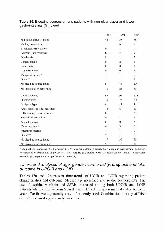
66
Table 16. Bleeding sources among patients with non-ulcer upper and lower gastrointestinal (GI) bleed
1984 1994 2004
Non-ulcer upper GI bleed 44 58 88
Mallory-Weiss tear 1 6 7
Esophagitis (incl ulcers) 0 1 9
Gastritis (incl erosions) 6 7 9
Duodenitis 9 1 2
Benign polyps 0 2 1
Ex ulceratio 0 0 1
Angiodysplasia 0 0 2
Malignant tumors * 1 1 5
Other ** 1 1 1
No bleeding source found 8 16 20
No investigation performed 18 23 31
Lower GI bleed 69 95 125
Diverticulosis 15 23 20
Benign polyps 6 11 4
Anorectal bleed (incl proctitis) 14 6 11
Inflammatory bowel disease 0 1 2
Meckel's diverticulum 0 1 1
Angiodysplasia 0 0 3
Cancer coli/recti 4 3 6
Infectious enteritis 1 1 0
Other*** 3 1 6
No bleeding source found 17 35 51
No investigation performed 9 13 21
* stomach (2), pancreas (2), duodenum (3), ** iatrogenic damage caused by biopsy and gastrostomal catheters, ***bleed after extirpation of polyps (4), after purging (1), stomal bleed (2), aorto enteric fistula (1), intestinal ischemia (1), hepatic cancer perforated to colon (1)
Time-trend analyses of age, gender, co-morbidity, drug use and fatal outcome in UPGIB and LGIB
Tables 17a and 17b present time-trends of UGIB and LGIB regarding patient characteristics and outcome. Median age increased and so did co-morbidity. The use of aspirin, warfarin and SSRIs increased among both UPGIB and LGIB patients whereas non-aspirin NSAIDs and steroid therapy remained stable between years. Coxibs were generally very infrequently used. Combination therapy of “risk drugs” increased significantly over time.

67
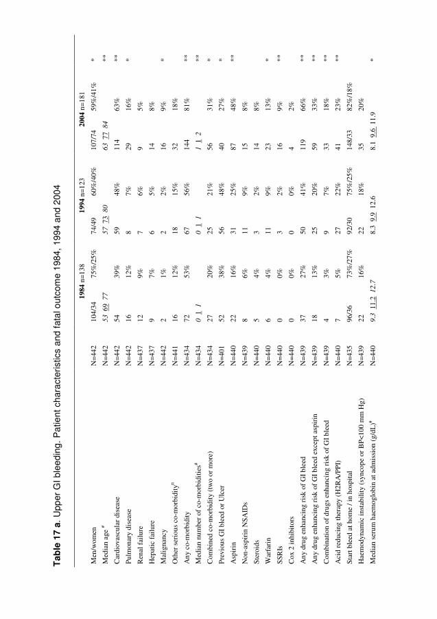
68
Tab
le 1
7 a
. U
pper
GI b
lee
din
g. P
atie
nt c
har
acte
ristic
s an
d fa
tal o
utco
me
1984
, 19
94 a
nd 2
004
19
84 n
=138
19
94 n
=123
20
04 n
=181
Men
/wom
en
N=
442
104/
34
75%
/25%
74
/49
60%
/40%
10
7/74
59
%/4
1%
*
Med
ian
age
# N
=44
2 5
3 6
9 7
7
57 7
3 8
0
63 7
7 8
4
**
Car
diov
ascu
lar
dise
ase
N=
442
54
39%
59
48
%
114
63%
**
Pulm
onar
y di
seas
e
N=
442
16
12%
8
7%
29
16%
*
Ren
al f
ailu
re
N=
437
12
9%
7 6%
9
5%
Hep
atic
fai
lure
N
=43
7 9
7%
6 5%
14
8%
Mal
igna
ncy
N=
442
2 1%
2
2%
16
9%
*
Oth
er s
erio
us c
o-m
orbi
dity
¤ N
=44
1 16
12
%
18
15%
32
18
%
Any
co-
mor
bidi
ty
N=
434
72
53%
67
56
%
144
81%
**
Med
ian
num
ber
of c
o-m
orbi
ditie
s# N
=43
4 0
1 1
0
1 1
1
1 2
**
Com
bine
d co
-mor
bidi
ty (
two
or m
ore)
N
=43
4 27
20
%
25
21%
56
31
%
*
Prev
ious
GI
blee
d or
Ulc
er
N=
401
52
38%
56
48
%
40
27%
*
Asp
irin
N
=44
0 22
16
%
31
25%
87
48
%
**
Non
-asp
irin
NSA
IDs
N=
439
8 6%
11
9%
15
8%
Ster
oids
N
=44
0 5
4%
3 2%
14
8%
War
fari
n N
=44
0 6
4%
11
9%
23
13%
*
SSR
Is
N=
440
0 0%
3
2%
16
9%
**
Cox
2 in
hibi
tors
N
=44
0 0
0%
0 0%
4
2%
Any
dru
g en
hanc
ing
risk
of
GI b
leed
N
=43
9 37
27
%
50
41%
11
9 66
%
**
Any
dru
g en
hanc
ing
risk
of
GI b
leed
exc
ept a
spir
in
N=
439
18
13%
25
20
%
59
33%
**
Com
bina
tion
of d
rugs
enh
anci
ng r
isk
of G
I ble
ed
N=
439
4 3%
9
7%
33
18%
**
Aci
d re
duci
ng th
erap
y (H
2RA
/PP
I)
N=
440
7 5%
27
22
%
41
23%
**
Star
t ble
ed a
t hom
e / i
n ho
spit
al
N=
435
96/3
6 73
%/2
7%
92/3
0 75
%/2
5%
148/
33
82%
/18%
Hae
mod
ynam
ic in
stab
ility
(sy
ncop
e or
BP<
100
mm
Hg)
N
=43
9 22
16
%
22
18%
35
20
%
Med
ian
seru
m h
aem
oglo
bin
at a
dmis
sion
(g/
dL)#
N=
440
9.3
11.
2 1
2.7
8.
3 9
.9 1
2.6
8.1
9.6
11.
9 *
69
Med
ian
bloo
d tr
ansf
usio
n un
its#
N=
359
0 2
6
0 2
4
0 2
4
Med
ian
hosp
ital s
tay
(day
s) #
N=
442
2.0
3.5
5.8
1.
0 4
.0 6
.0
2.0
3.0
6.0
In h
ospi
tal m
orta
lity
tota
l N
=44
2 5
4%
6 5%
11
6%
Mor
talit
y fo
r pa
tien
ts w
ho s
tart
to b
leed
at h
ome
N=
440
1 1%
4
4%
5 3%
Mor
talit
y fo
r pa
tien
ts w
ho s
tart
to b
leed
in h
ospi
tal
N=
440
4 11
%
2 7%
6
18%
Tes
ts u
sed
: Fi
sher
's ex
act
test
and
W
ilcox
on t
est,
*p<0
.05,
**p
<0.0
01.
# (with
upp
er a
nd l
ower
qua
rtile
), ¤ f
or e
xam
ple
neur
olog
ical
, ps
ychi
atri
c,
rheu
mat
ic d
isor
ders
, ±(g
/dL
) A
bbre
via
tio
ns:
GI=
gast
roin
test
inal
, N
SAID
s=no
n-st
eroi
dal a
nti-
infl
amm
ator
y dr
ugs,
SSR
Is=s
elec
tive
sero
toni
n re
-upt
ake
inhi
bito
rs,H
2RA
= hi
stam
in-2
rec
epto
r an
tago
nist
s, P
PIs=
prot
on p
ump
inhi
bito
rs
70
Tab
le 1
7 b
. Lo
wer
GI
ble
edin
g. P
atie
nt c
har
acte
ristic
s an
d fa
tal o
utco
me
1984
, 19
94 a
nd 2
004
19
84 n
=69
1994
n=9
5 20
04 n
=125
Men
/wom
en
N=
289
32/3
7 46
%/5
4%
43/5
2 45
%/5
5%
68/5
7 54
%/4
6%
Med
ian
age
# N
=28
9 51
69
79
58 7
6 8
5
63 7
5 8
3
*
Car
diov
ascu
lar
dise
ase
N=
289
23
33%
43
45
%
84
67%
**
Pulm
onar
y di
seas
e
N=
289
9 13
%
4 4%
17
14
%
*
Ren
al f
ailu
re
N=
275
0 0%
8
9%
14
11%
*
Hep
atic
fai
lure
N
=27
0 1
2%
2 2%
5
4%
Mal
igna
ncy
N=
289
3 4%
6
6%
12
10%
Oth
er s
erio
us c
o-m
orbi
dity
¤ N
=28
7 8
12%
16
17
%
23
18%
Any
co-
mor
bidi
ty
N=
263
33
52%
52
63
%
95
81%
**
Med
ian
num
ber
of c
o-m
orbi
ditie
s# N
=26
3 0
1 1
0
1 1
1
1 2
**
Com
bine
d co
-mor
bidi
ty (
two
or m
ore)
N
=26
3 8
13%
19
23
%
38
32%
*
Prev
ious
GI
blee
d or
Ulc
er
N=
218
27
45%
33
35
%
27
42%
Asp
irin
N
=28
6 5
7%
18
19%
49
40
%
**
Non
-asp
irin
NSA
IDs
N=
286
7 10
%
16
17%
11
9%
Ster
oids
N
=28
5 4
6%
7 8%
10
8%
War
fari
n N
=28
6 2
3%
11
12%
23
19
%
*
SSR
Is
N=
286
0 0%
2
2%
13
10%
*
Cox
2 in
hibi
tors
N
=28
6 0
0%
0 0%
3
2%
Any
dru
g en
hanc
ing
risk
of
GI b
leed
N
=28
5 16
24
%
44
47%
79
64
%
**
Any
dru
g en
hanc
ing
risk
of
GI b
leed
exc
ept a
spir
in
N=
285
12
18%
30
32
%
48
39%
*
Com
bina
tion
of d
rugs
enh
anci
ng r
isk
of G
I ble
ed
N=
285
2 3%
8
9%
24
19%
*
Aci
d re
duci
ng th
erap
y (H
2RA
/PP
I)
N=
288
4 6%
12
13
%
31
25%
*
Star
t ble
ed a
t hom
e / i
n ho
spit
al
N=
288
60/9
87
%/1
3%
78/1
7 82
%/1
8%
90/3
4 73
%/2
7%
*
Hae
mod
ynam
ic in
stab
ility
(sy
ncop
e or
BP<
100
mm
Hg)
N
=28
4 3
4%
8 8%
14
12
%
Med
ian
seru
m h
aem
oglo
bin
at a
dmis
sion
(g/
dL)
# N
=28
2 10
.0 1
1.9
14.
2 8.
8 1
0.7
12.
6 8.
3 9
.8 1
1.8
**
71
Med
ian
bloo
d tr
ansf
usio
n un
its#
N=
153
0 2
3
0.25
2 4
0
2 2
*
Med
ian
hosp
ital s
tay
(day
s) #
N=
289
1 3
7
2 3
5
1 3
8
In h
ospi
tal m
orta
lity
tota
l N
=28
9 2
3%
1 1%
10
8%
*
Mor
talit
y fo
r pa
tien
ts w
ho s
tart
to b
leed
at h
ome
N=
288
0 0%
0
0%
3 3%
Mor
talit
y fo
r pa
tien
ts w
ho s
tart
to b
leed
in h
ospi
tal
N=
288
2 22
%
1 6%
7
21%
Test
s u
sed
: Fis
her's
exa
ct te
st a
nd W
ilcox
on te
st, *
p<0.
05, *
*p<0
.001
# with
upp
er a
nd lo
wer
qua
rtile
, ¤ for
exa
mpl
e ne
urol
ogic
al, p
sych
iatr
ic, r
heum
atic
dis
orde
rs.
Abb
revi
ati
on
s: G
I=ga
stro
inte
stin
al,
NSA
IDs=
non-
ster
oida
l ant
i-in
flam
mat
ory
drug
s, S
SRIs
=se
lect
ive
sero
toni
n re
-upt
ake
inhi
bito
rs,
H2R
A=
hist
amin
-2 r
ecep
tor
anta
goni
sts,
PP
Is=
prot
on p
ump
inhi
bito
rs
72
Tab
le 1
8 a
. P
atie
nts
with
upp
er G
I ble
ed 1
984,
199
4, 2
004
by
mor
talit
y
A
live
at d
isch
arge
n=4
20
Dis
ease
d du
ring
hos
p n=
22
Med
ian
age
# N
=44
2 5
8 7
2 8
1
64 8
0 8
4
*
Mal
e ge
nder
N
=44
2 27
2 65
%
13
59%
Any
co-
mor
bidi
ty
N=
434
262
63%
21
10
0%
**
Car
diov
ascu
lar
dise
ase
N=
442
213
51%
14
64
%
Pulm
onar
y di
seas
e N
=44
2 47
11
%
6 27
%
*
Ren
al f
ailu
re
N=
437
25
6%
3 14
%
Hep
atic
fai
lure
N
=43
7 23
6%
6
27%
*
Mal
igna
ncy
N=
442
14
3%
6 27
%
**
Oth
er c
o-m
orbi
dity
¤ N
=44
1 61
15
%
5 24
%
Med
ian
num
ber
of c
o-m
orbi
ditie
s# N
=43
9 0
1 1
1
2 2
**
Com
bina
tion
of tw
o or
mor
e co
-mor
bidi
ties
N=
434
95
23%
13
62
%
**
Asp
irin
N
=44
0 13
6 33
%
4 18
%
Non
-asp
irin
NSA
IDs
N=
439
33
8%
1 5%
Ster
oids
N
=44
0 20
5%
2
9%
War
fari
n N
=44
0 36
9%
4
18%
SSR
I N
=44
0 19
5%
0
0%
C
ox 2
inhi
bito
rs
N=
440
4 1%
0
0%
Any
dru
g en
hanc
ing
risk
of
GI b
leed
N
=43
9 19
8 47
%
8 36
%
Any
dru
g en
hanc
ing
risk
of
GI b
leed
exc
ept a
spir
in
N=
439
97
23%
5
23%
Com
bina
tion
of d
rugs
enh
anci
ng r
isk
of G
I ble
ed
N=
439
43
10%
3
14%
H2R
A/P
PI
N=
440
70
17%
5
23%
Star
t ble
ed a
t hom
e / i
n ho
spit
al
N=
435
326/
88
79%
/21%
10
/11
48%
/52%
*
Hae
mod
ynam
ic in
stab
ility
(sy
ncop
e or
BP<
100
mm
Hg)
N
=43
9 68
16
%
11
50%
**
Med
ian
seru
m h
aem
oglo
bin
at a
dmis
sion
(g/
dL) #
N
=44
0 8.
5 1
0.2
12.
5 7.
4 9
.3 1
0.4
*
Med
ian
bloo
d tr
ansf
usio
n un
its#
N=
359
0 2
4
3 7
14
**
Test
s u
sed
: Fis
her's
exa
ct te
st a
nd W
ilcox
on te
st *
p<0.
05, *
*p<
0.00
1 # w
ith
uppe
r an
d lo
wer
qua
rtile
, ¤fo
r ex
ampl
e ne
urol
ogic
al, p
sych
iatr
ic, r
heum
atic
dis
orde
rs
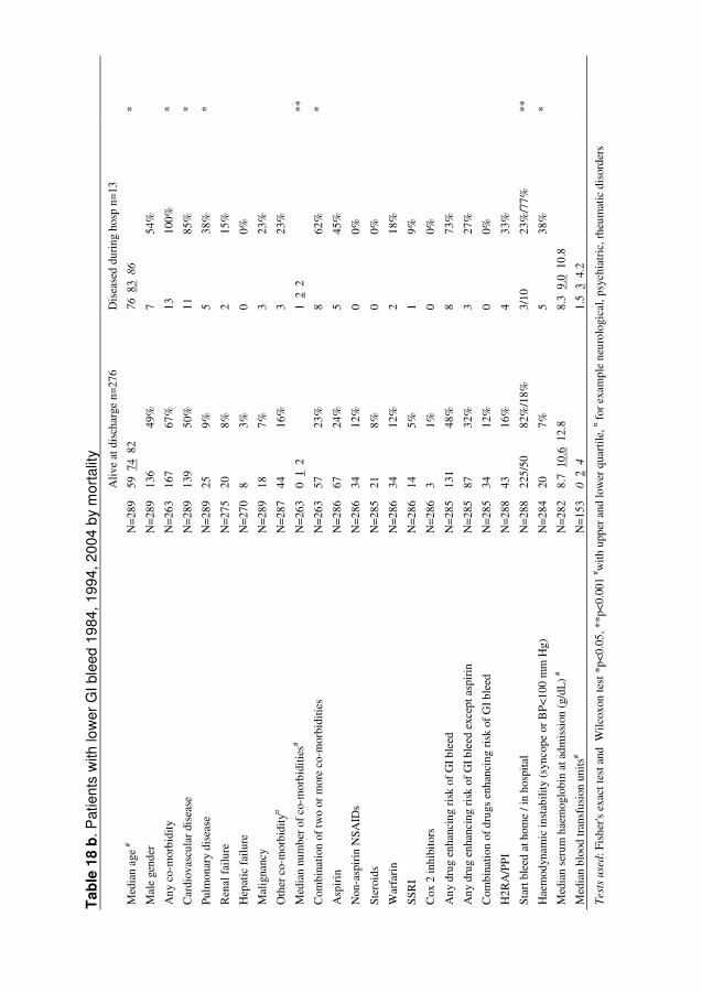
73
Tab
le 1
8 b
. Pat
ient
s w
ith lo
wer
GI b
leed
19
84,
199
4, 2
004
by
mor
talit
y
A
live
at d
isch
arge
n=2
76
Dis
ease
d du
ring
hos
p n=
13
Med
ian
age
# N
=28
9 59
74
82
76 8
3 8
6
*
Mal
e ge
nder
N
=28
9 13
6 49
%
7 54
%
Any
co-
mor
bidi
ty
N=
263
167
67%
13
10
0%
*
Car
diov
ascu
lar
dise
ase
N=
289
139
50%
11
85
%
*
Pulm
onar
y di
seas
e N
=28
9 25
9%
5
38%
*
Ren
al f
ailu
re
N=
275
20
8%
2 15
%
Hep
atic
fai
lure
N
=27
0 8
3%
0 0%
Mal
igna
ncy
N=
289
18
7%
3 23
%
Oth
er c
o-m
orbi
dity
¤ N
=28
7 44
16
%
3 23
%
Med
ian
num
ber
of c
o-m
orbi
ditie
s# N
=26
3 0
1 2
1
2 2
**
Com
bina
tion
of tw
o or
mor
e co
-mor
bidi
ties
N
=26
3 57
23
%
8 62
%
*
Asp
irin
N
=28
6 67
24
%
5 45
%
Non
-asp
irin
NSA
IDs
N=
286
34
12%
0
0%
Ster
oids
N
=28
5 21
8%
0
0%
War
fari
n N
=28
6 34
12
%
2 18
%
SSR
I N
=28
6 14
5%
1
9%
Cox
2 in
hibi
tors
N
=28
6 3
1%
0 0%
Any
dru
g en
hanc
ing
risk
of
GI b
leed
N
=28
5 13
1 48
%
8 73
%
Any
dru
g en
hanc
ing
risk
of
GI b
leed
exc
ept a
spir
in
N=
285
87
32%
3
27%
Com
bina
tion
of d
rugs
enh
anci
ng r
isk
of G
I ble
ed
N=
285
34
12%
0
0%
H2R
A/P
PI
N=
288
43
16%
4
33%
Star
t ble
ed a
t hom
e / i
n ho
spit
al
N=
288
225/
50
82%
/18%
3/
10
23%
/77%
**
Hae
mod
ynam
ic in
stab
ility
(sy
ncop
e or
BP<
100
mm
Hg)
N
=28
4 20
7%
5
38%
*
Med
ian
seru
m h
aem
oglo
bin
at a
dmis
sion
(g/
dL)
# N
=28
2 8.
7 1
0.6
12.
8 8.
3 9
.0 1
0.8
Med
ian
bloo
d tr
ansf
usio
n un
its#
N=
153
0 2
4
1.5
3 4
.2
Test
s u
sed
: Fis
her's
exa
ct te
st a
nd W
ilcox
on te
st *
p<0.
05, *
*p<
0.00
1 # w
ith u
pper
and
low
er q
uart
ile, ¤ f
or e
xam
ple
neur
olog
ical
, psy
chia
tric
, rhe
umat
ic d
isor
ders
74
Haemodynamic instability did not differ between years but was more common among UPGIB patients, 18 versus 9% (p<0.001). Median serum haemoglobin levels at presentation decreased in both groups. Case fatality rate ranged between 4 and 6% (p=0.65) in UPGIB patients and between 1 to 8% (p=0.033) in LGIB patients
Fatal outcome during hospitalization for UPGIB and LGIB
Univariate analyses of UPGIB and LGIB by mortality are shown in tables 18a and 18b. The diseased patients were older. There were no gender differences. All patients who died during hospitalization for GI bleed had some co-morbidity. Use of drugs enhancing the risk of GI bleed had no influence on outcome. In-hospital mortality was higher among patients who started to bleed while already hospitalized than among patients who started to bleed at home.
Table 19 shows a multivariable logistic regression model for in-hospital mortality in GI bleed in total. Haemodynamic instability at presentation, age above 65 years and number of co-morbidities were independent risk factors of fatal outcome when adjustment was made for localization of GI bleed (upper versus lower), gender and use of drugs.
Table 19. Association between variables and in hospital mortality in gastro-intestinal bleeding in total Factor OR 95 % CI
Year 1994 (1984 reference) 0.93 0.25 - 3.4
Year 2004 (1984 reference) 1.8 0.63 - 5.8
Lower GI bleed (Upper GI bleed reference) 1.1 0.46 - 2.5
Male gender 1.2 0.51 - 2.7
Age above 65 years (1 year increase) 1.1 1.02 - 1.1 *
Number of co-morbidities 2.6 1.7 - 4.0 **
Aspirin 0.50 0.20 - 1.2
Non-aspirin NSAIDs 0.30 0.016 - 1.6
Steroids 0.42 0.059 - 1.7
Warfarin 0.76 0.24 - 2.1
SSRIs 0.23 0.011 - 1.7
Haemodynamic instability 6.7 2.8 - 16 **
Multivariable logistic regression model. *p<0.05, **p<0.001
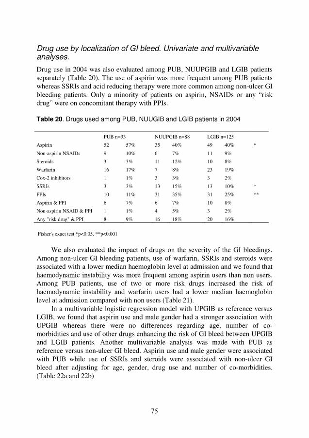
75
Drug use by localization of GI bleed. Univariate and multivariable analyses.
Drug use in 2004 was also evaluated among PUB, NUUPGIB and LGIB patients separately (Table 20). The use of aspirin was more frequent among PUB patients whereas SSRIs and acid reducing therapy were more common among non-ulcer GI bleeding patients. Only a minority of patients on aspirin, NSAIDs or any “risk drug” were on concomitant therapy with PPIs.
Table 20. Drugs used among PUB, NUUGIB and LGIB patients in 2004 PUB n=93 NUUPGIB n=88 LGIB n=125
Aspirin 52 57% 35 40% 49 40% *
Non-aspirin NSAIDs 9 10% 6 7% 11 9%
Steroids 3 3% 11 12% 10 8%
Warfarin 16 17% 7 8% 23 19%
Cox-2 inhibitors 1 1% 3 3% 3 2%
SSRIs 3 3% 13 15% 13 10% *
PPIs 10 11% 31 35% 31 25% **
Aspirin & PPI 6 7% 6 7% 10 8%
Non-aspirin NSAID & PPI 1 1% 4 5% 3 2%
Any "risk drug" & PPI 8 9% 16 18% 20 16%
Fisher's exact test *p<0.05, **p<0.001
We also evaluated the impact of drugs on the severity of the GI bleedings.
Among non-ulcer GI bleeding patients, use of warfarin, SSRIs and steroids were associated with a lower median haemoglobin level at admission and we found that haemodynamic instability was more frequent among aspirin users than non users. Among PUB patients, use of two or more risk drugs increased the risk of haemodynamic instability and warfarin users had a lower median haemoglobin level at admission compared with non users (Table 21).
In a multivariable logistic regression model with UPGIB as reference versus LGIB, we found that aspirin use and male gender had a stronger association with UPGIB whereas there were no differences regarding age, number of co-morbidities and use of other drugs enhancing the risk of GI bleed between UPGIB and LGIB patients. Another multivariable analysis was made with PUB as reference versus non-ulcer GI bleed. Aspirin use and male gender were associated with PUB while use of SSRIs and steroids were associated with non-ulcer GI bleed after adjusting for age, gender, drug use and number of co-morbidities. (Table 22a and 22b)

76
Table 21. The impact of drugs on haemodynamic instability and serum haemoglobin (Hb) levels in peptic ulcer bleedings and non-ulcer GI bleedings
Peptic ulcer bleeding (n=252)
Haemodynamic instability1 Median Hb level2#
N OR 95 % CI Drug Yes Drug No
Aspirin 86 1.4 0.7 - 2.8 8.3 9.8 11.9 7.9 9.8 11.8
NSAIDs 23 0.85 0.2 - 2.7 7.4 8.5 10.2 8.1 9.9 11.9
Steroids 8 2.5 0.4 - 14 7.7 9.2 10.0 8.0 9.8 11.9
Warfarin 25 2.1 0.7 - 5.6 6.7 8.2 10.0 8.1 9.9 12.0 *
SSRIs 4 0.0 0.0 - 6.2 11.6 14.0 16.1 7.9 9.8 11.8 *
Cox-2 inhibitors 1 0.0 0.0 - 159 5.7 5.7 5.7 8.0 9.8 11.9
Any risk drug 126 1.3 0.6 - 2.5 7.8 9.6 11.6 8.1 10.3 12.0 Any risk drug except aspirin 56 1.5 0.7 - 3.2 7.2 9.0 10.8 8.2 10.2 12.0 * Combination of risk drugs 20 3.0 1.0 - 8.7 * 7.6 9.0 10.4 8.0 9.8 11.9 Combinations except aspirin 4 1.4 0.03 - 17 5.7 6.4 7.5 8.0 9.8 11.9 *
H2RA/PPIs 27 0.48 0.09 - 1.7 7.8 8.9 10.9 8.0 9.9 11.9 Non-ulcer gastrointestinal bleeding (n=479)
Haemodynamic instability1 Median Hb level2#
N OR 95 % CI Drug Yes Drug No
Aspirin 126 2.0 1.0 - 3.7 * 8.8 10.0 12.2 8.9 10.6 13.3
NSAIDs 45 0.75 0.19 - 2.2 8.7 10.3 12.7 8.8 10.6 13.0
Steroids 35 1.7 0.54 - 4.4 8.1 9.9 10.8 8.9 10.6 13.0 *
Warfarin 51 1.5 0.59 - 3.6 7.7 9.3 10.5 8.9 10.7 13.0 **
SSRIs 30 0.85 0.16 - 2.9 8.1 9.1 10.4 8.9 10.6 13.0 *
Cox-2 inhibitors 6 0.0 0.0 - 8.5 6.7 8.8 10.0 8.8 10.6 13.0
Any risk drug 219 1.7 0.91 - 3.1 8.5 9.9 12.0 9.0 11.6 13.7 ** Any risk drug except aspirin 136 1.3 0.66 - 2.4 8.1 9.5 11.3 9.0 11.3 13.4 ** Combination of risk drugs 60 1.7 0.71 - 3.7 8.1 9.4 11.8 9.0 10.7 13.0 ** Combinations except aspirin 25 1.0 0.19 - 3.7 8.0 9.9 11.7 8.9 10.6 13.0
H2RA/PPIs 95 0.89 0.38 - 1.9 8.6 9.9 11.7 8.9 10.8 13.0 *
Fisher’s exact test1 and Wilcoxon test2 *p<0.05, **p<0.001 Abbreviations: OR=odds ratio, CI=confidence interval, GI=gastrointestinal. # with upper and lower quartile.
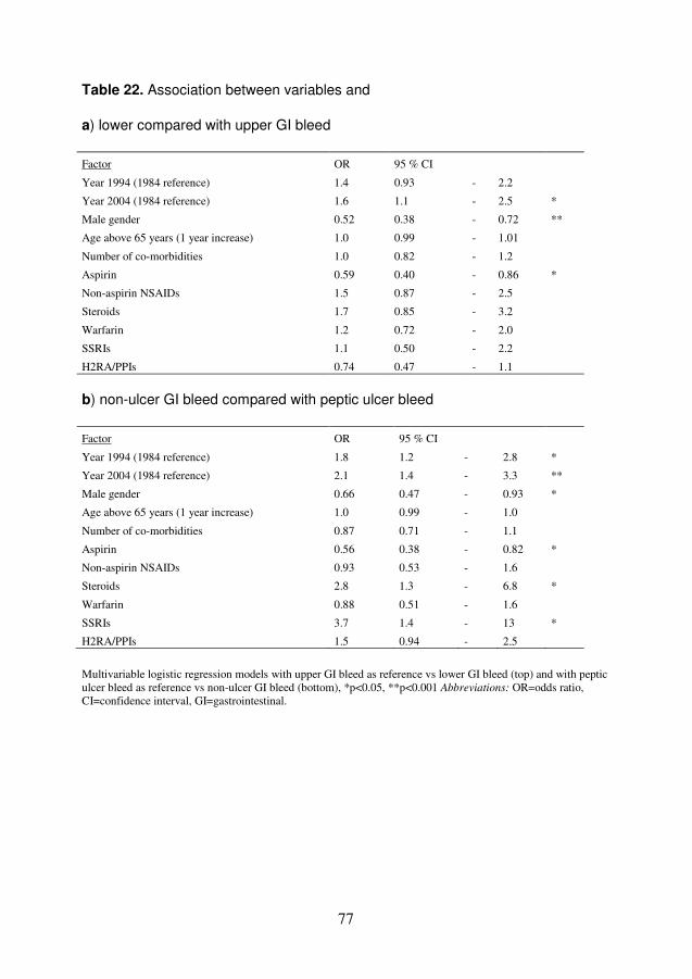
77
Table 22. Association between variables and a) lower compared with upper GI bleed
Factor OR 95 % CI
Year 1994 (1984 reference) 1.4 0.93 - 2.2
Year 2004 (1984 reference) 1.6 1.1 - 2.5 *
Male gender 0.52 0.38 - 0.72 **
Age above 65 years (1 year increase) 1.0 0.99 - 1.01
Number of co-morbidities 1.0 0.82 - 1.2
Aspirin 0.59 0.40 - 0.86 *
Non-aspirin NSAIDs 1.5 0.87 - 2.5
Steroids 1.7 0.85 - 3.2
Warfarin 1.2 0.72 - 2.0
SSRIs 1.1 0.50 - 2.2
H2RA/PPIs 0.74 0.47 - 1.1
b) non-ulcer GI bleed compared with peptic ulcer bleed
Factor OR 95 % CI
Year 1994 (1984 reference) 1.8 1.2 - 2.8 *
Year 2004 (1984 reference) 2.1 1.4 - 3.3 **
Male gender 0.66 0.47 - 0.93 *
Age above 65 years (1 year increase) 1.0 0.99 - 1.0
Number of co-morbidities 0.87 0.71 - 1.1
Aspirin 0.56 0.38 - 0.82 *
Non-aspirin NSAIDs 0.93 0.53 - 1.6
Steroids 2.8 1.3 - 6.8 *
Warfarin 0.88 0.51 - 1.6
SSRIs 3.7 1.4 - 13 *
H2RA/PPIs 1.5 0.94 - 2.5
Multivariable logistic regression models with upper GI bleed as reference vs lower GI bleed (top) and with peptic ulcer bleed as reference vs non-ulcer GI bleed (bottom), *p<0.05, **p<0.001 Abbreviations: OR=odds ratio, CI=confidence interval, GI=gastrointestinal.

78
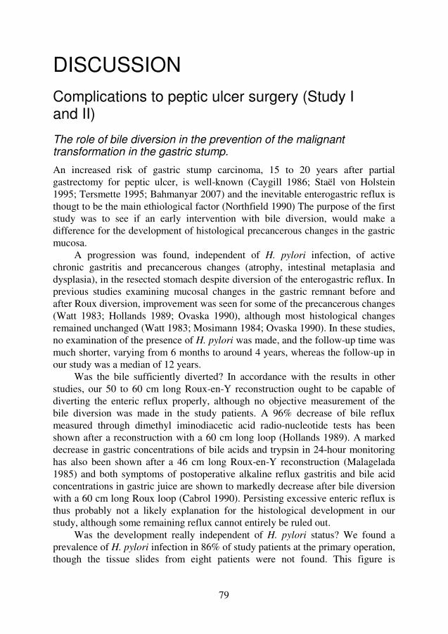
79
DISCUSSION
Complications to peptic ulcer surgery (Study I and II)
The role of bile diversion in the prevention of the malignant transformation in the gastric stump.
An increased risk of gastric stump carcinoma, 15 to 20 years after partial gastrectomy for peptic ulcer, is well-known (Caygill 1986; Staël von Holstein 1995; Tersmette 1995; Bahmanyar 2007) and the inevitable enterogastric reflux is thougt to be the main ethiological factor (Northfield 1990) The purpose of the first study was to see if an early intervention with bile diversion, would make a difference for the development of histological precancerous changes in the gastric mucosa.
A progression was found, independent of H. pylori infection, of active chronic gastritis and precancerous changes (atrophy, intestinal metaplasia and dysplasia), in the resected stomach despite diversion of the enterogastric reflux. In previous studies examining mucosal changes in the gastric remnant before and after Roux diversion, improvement was seen for some of the precancerous changes (Watt 1983; Hollands 1989; Ovaska 1990), although most histological changes remained unchanged (Watt 1983; Mosimann 1984; Ovaska 1990). In these studies, no examination of the presence of H. pylori was made, and the follow-up time was much shorter, varying from 6 months to around 4 years, whereas the follow-up in our study was a median of 12 years.
Was the bile sufficiently diverted? In accordance with the results in other studies, our 50 to 60 cm long Roux-en-Y reconstruction ought to be capable of diverting the enteric reflux properly, although no objective measurement of the bile diversion was made in the study patients. A 96% decrease of bile reflux measured through dimethyl iminodiacetic acid radio-nucleotide tests has been shown after a reconstruction with a 60 cm long loop (Hollands 1989). A marked decrease in gastric concentrations of bile acids and trypsin in 24-hour monitoring has also been shown after a 46 cm long Roux-en-Y reconstruction (Malagelada 1985) and both symptoms of postoperative alkaline reflux gastritis and bile acid concentrations in gastric juice are shown to markedly decrease after bile diversion with a 60 cm long Roux loop (Cabrol 1990). Persisting excessive enteric reflux is thus probably not a likely explanation for the histological development in our study, although some remaining reflux cannot entirely be ruled out.
Was the development really independent of H. pylori status? We found a prevalence of H. pylori infection in 86% of study patients at the primary operation, though the tissue slides from eight patients were not found. This figure is
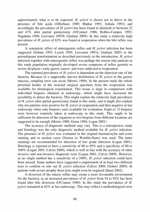
80
approximately what is to be expected. H. pylori is shown not to thrive in the presence of bile acids (Offerhaus 1989; Mathai 1991; Sobala 1993) and accordingly the prevalence of H. pylori has been found to diminish to between 22 and 47% after partial gastrectomy (O'Connor 1986; Robles-Campos 1993; Nagahata 1996; Leivonen 1997b; Giuliani 2005). In this study a relatively high prevalence of H. pylori of 62% was found at reoperation when the bile reflux was present.
A synergistic effect of enterogastric reflux and H. pylori infection has been suggested (Sobala 1993; Lynch 1995; Leivonen 1997a; Giuliani 2005) in the premalignant transformation as described previously in the introduction. H. pylori infection together with enterogastric reflux was perhaps the reason why patients in this study population originally developed severe symptoms of reflux gastritis or severe dysplasia / early gastric cancer and were subjected to reoperation.
The reported prevalence of H. pylori is dependent on the detection rate of the bacteria. Because of a supposedly uneven distribution of H. pylori in the gastric mucosa, sampling error can occur (Morris 1989). In the present study the whole proximal border of the resected surgical specimen from the reoperation was available for histological examination. This tissue is large in comparison with individual biopsies obtained at endoscopy, which might have increased the possibility to detect the bacteria. This might explain the relatively high prevalence of H. pylori after partial gastrectomy found in this study, and it might also explain why ten patients were positive for H. pylori at reoperation and then negative at last endoscopy when only biopsies were available for evaluation. Eight to 12 biopsies were however routinely taken at endoscopy in this study. This ought to be sufficient for detection of the organism as two biopsies from different locations are suggested to be enough (Morris 1989; Genta 1994; Logan 2001)
The accuracy of diagnostic methods may vary. This is a retrospective study and histology was the only diagnostic method available for H. pylori infection. The presence of H. pylori was evaluated in the original haematoxylin and eosin staining and in unclear cases Giemsa or Warthin-Starry was used. The latter stainings are recommended for detection of low grade infection (Logan 2001). Histology is reported to have a sensitivity of 88 to 95% and a specificity of 90 to 100% (Logan 2001; Calvet 2009), which is well in line with the accuracy of other invasive and non-invasive diagnostic tests (Logan 2001; Calvet 2009). However, as no single method has a sensitivity of a 100%, H. pylori infection could have been missed. Some authors have suggested a requirement of at least two different tests to confirm or rule out H. pylori infection (Calvet 2009; Gisbert 2009). In patients with severe atrophy three tests might even be required (Quan 2002).
A diversion of the enteric reflux may create a more favourable environment for the bacteria, as an increased prevalence of H. pylori from 54 to 92% has been found after bile diversion (O'Connor 1989). In this study the prevalence of H.
pylori remained at 62% at last endoscopy. This may reflect a methodological error

81
especially in those who were positive at reoperation and then negative at last endoscopy. Another explanation could be that patients have been subjected to eradication therapy against H. pylori.
The bacteria is known to be able to transform into a coccoid form when exposed to an unfavourable environment (Azevedo 2007), for instance in the presence of bile acids (Nilius 1993). The virulence of this coccoid shape has been under debate in the scientific community with one part maintaining that the coccoid form of the bacteria represents a degraded, nonviable form of the cell and others arguing that the coccoid cells constitute a survival strategy of the bacteria forming a viable but nonculturable state (Azevedo 2007). The detection of the coccoid form might however be difficult and it is often overlooked. The resting coccoid state is a speculative but reasonable explanation to our finding of 10 patients being negative in H. pylori status at reoperation, when bile reflux was present, and then positive at last endoscopy, when the living conditions for the bacteria again had improved. The annual incidence of H. pylori infection in adulthood is less than 0.5% (Kuipers 1993; Parsonnet 1995) and reinfection is therefore not likely, at least not in all 10 cases.
A progression of precancerous mucosal changes despite diversion of the enterogastric reflux and independent of H pylori infection was found. This may indicate that other factors (for instance dietary factors and smoking habits) could be of greater importance in the gastric stump carcinogenesis than previously thought. The carcinogenesis in the non-operated stomach is postulated to be multifactorial with both host and environmental ethiological factors (Correa 1992), and this is probably true also in the gastric remnant after partial gastrectomy.
The progression of precancerous changes might also have passed a point of no return when the bile diversion occurred. Dixon has postulated that reversal might become impossible in certain forms of atrophy and intestinal metaplasia. Regeneration of normal oxyntic mucosa following true glandular atrophy with replacement fibrosis seems likely to be limited and restoration of normal differentiation in intestinal metaplasia is improbable in the presence of stable mutations in stem cells (Dixon 2001a). This is also in line with a study from Japan where eradication of H. pylori before the significant expansion of atrophy was found to be most beneficial to prevent gastric cancer development (Take 2007).
Long-term morbidity and mortality after parietal cell vagotomy
Data on long-term cancer incidence and mortality after PCV is scarce. In a cohort of ulcer patients operated on with PCV 22-31 years previously, the overall mortality was found to be unchanged in comparison with the general population. This strengthens the belief of PCV as a more physiological procedure in comparison with partial gastrectomy or vagotomy with drainage procedures. Patients were quite young at the time for PCV (median 46 years) and only 37%
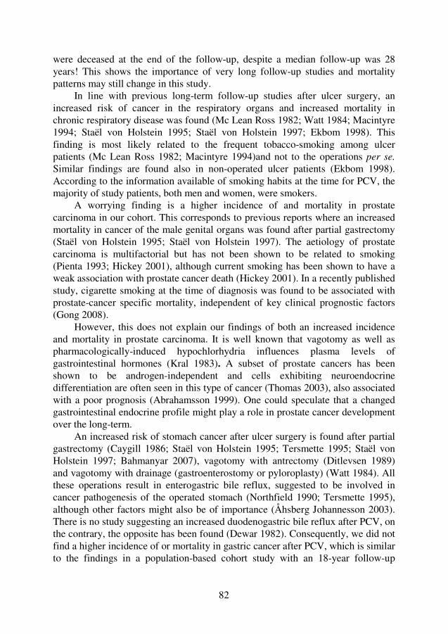
82
were deceased at the end of the follow-up, despite a median follow-up was 28 years! This shows the importance of very long follow-up studies and mortality patterns may still change in this study.
In line with previous long-term follow-up studies after ulcer surgery, an increased risk of cancer in the respiratory organs and increased mortality in chronic respiratory disease was found (Mc Lean Ross 1982; Watt 1984; Macintyre 1994; Staël von Holstein 1995; Staël von Holstein 1997; Ekbom 1998). This finding is most likely related to the frequent tobacco-smoking among ulcer patients (Mc Lean Ross 1982; Macintyre 1994)and not to the operations per se.
Similar findings are found also in non-operated ulcer patients (Ekbom 1998). According to the information available of smoking habits at the time for PCV, the majority of study patients, both men and women, were smokers.
A worrying finding is a higher incidence of and mortality in prostate carcinoma in our cohort. This corresponds to previous reports where an increased mortality in cancer of the male genital organs was found after partial gastrectomy (Staël von Holstein 1995; Staël von Holstein 1997). The aetiology of prostate carcinoma is multifactorial but has not been shown to be related to smoking (Pienta 1993; Hickey 2001), although current smoking has been shown to have a weak association with prostate cancer death (Hickey 2001). In a recently published study, cigarette smoking at the time of diagnosis was found to be associated with prostate-cancer specific mortality, independent of key clinical prognostic factors (Gong 2008).
However, this does not explain our findings of both an increased incidence and mortality in prostate carcinoma. It is well known that vagotomy as well as pharmacologically-induced hypochlorhydria influences plasma levels of gastrointestinal hormones (Kral 1983). A subset of prostate cancers has been shown to be androgen-independent and cells exhibiting neuroendocrine differentiation are often seen in this type of cancer (Thomas 2003), also associated with a poor prognosis (Abrahamsson 1999). One could speculate that a changed gastrointestinal endocrine profile might play a role in prostate cancer development over the long-term.
An increased risk of stomach cancer after ulcer surgery is found after partial gastrectomy (Caygill 1986; Staël von Holstein 1995; Tersmette 1995; Staël von Holstein 1997; Bahmanyar 2007), vagotomy with antrectomy (Ditlevsen 1989) and vagotomy with drainage (gastroenterostomy or pyloroplasty) (Watt 1984). All these operations result in enterogastric bile reflux, suggested to be involved in cancer pathogenesis of the operated stomach (Northfield 1990; Tersmette 1995), although other factors might also be of importance (Åhsberg Johannesson 2003). There is no study suggesting an increased duodenogastric bile reflux after PCV, on the contrary, the opposite has been found (Dewar 1982). Consequently, we did not find a higher incidence of or mortality in gastric cancer after PCV, which is similar to the findings in a population-based cohort study with an 18-year follow-up
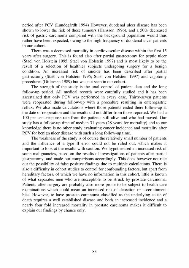
83
period after PCV (Lundegårdh 1994) However, duodenal ulcer disease has been shown to lower the risk of these tumours (Hansson 1996), and a 50% decreased risk of gastric carcinoma compared with the background population would thus rather have been expected, owing to the high frequency of duodenal ulcer patients in our cohort.
There was a decreased mortality in cardiovascular disease within the first 15 years after surgery. This is found also after partial gastrectomy for peptic ulcer (Staël von Holstein 1995; Staël von Holstein 1997) and is most likely to be the result of a selection of healthier subjects undergoing surgery for a benign condition. An increased risk of suicide has been described after partial gastrectomy (Staël von Holstein 1995; Staël von Holstein 1997) and vagotomy procedures (Ditlevsen 1989) but was not seen in our cohort.
The strength of the study is the total control of patient data and the long follow-up period. All medical records were carefully studied and it has been ascertained that only PCV was performed in every case. Thirty-seven patients were reoperated during follow-up with a procedure resulting in enterogastric reflux. We also made calculations where those patients ended there follow-up at the date of reoperation and the results did not differ from those reported. We had a 100 per cent response rate from the patients still alive and who had moved. Our study has a follow-up time of median 31 years (28 years for mortality) and to our knowledge there is no other study evaluating cancer incidence and mortality after PCV for benign ulcer disease with such a long follow-up time.
The weakness of the study is of course the relatively small number of patients and the influence of a type II error could not be ruled out, which makes it important to look at the results with caution. We hypothesised an increased risk of some malignancies, based on the results of investigations of patients after partial gastrectomy, and made our comparisons accordingly. This does however not rule out the possibility of false positive findings due to multiple calculations. There is also a difficulty in cohort studies to control for confounding factors, but apart from hereditary factors, of which we have no information in this cohort, little is known of what separates men who are susceptible to be struck by prostate carcinoma. Patients after surgery are probably also more prone to be subject to health care examinations which could mean an increased risk of detection or ascertainment bias. However, to have prostate carcinoma classified as the underlying cause of death requires a well established disease and both an increased incidence and a nearly four fold increased mortality in prostate carcinoma makes it difficult to explain our findings by chance only.
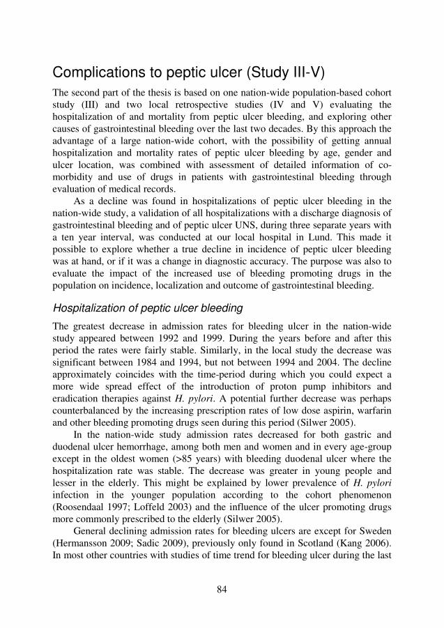
84
Complications to peptic ulcer (Study III-V) The second part of the thesis is based on one nation-wide population-based cohort study (III) and two local retrospective studies (IV and V) evaluating the hospitalization of and mortality from peptic ulcer bleeding, and exploring other causes of gastrointestinal bleeding over the last two decades. By this approach the advantage of a large nation-wide cohort, with the possibility of getting annual hospitalization and mortality rates of peptic ulcer bleeding by age, gender and ulcer location, was combined with assessment of detailed information of co-morbidity and use of drugs in patients with gastrointestinal bleeding through evaluation of medical records.
As a decline was found in hospitalizations of peptic ulcer bleeding in the nation-wide study, a validation of all hospitalizations with a discharge diagnosis of gastrointestinal bleeding and of peptic ulcer UNS, during three separate years with a ten year interval, was conducted at our local hospital in Lund. This made it possible to explore whether a true decline in incidence of peptic ulcer bleeding was at hand, or if it was a change in diagnostic accuracy. The purpose was also to evaluate the impact of the increased use of bleeding promoting drugs in the population on incidence, localization and outcome of gastrointestinal bleeding.
Hospitalization of peptic ulcer bleeding
The greatest decrease in admission rates for bleeding ulcer in the nation-wide study appeared between 1992 and 1999. During the years before and after this period the rates were fairly stable. Similarly, in the local study the decrease was significant between 1984 and 1994, but not between 1994 and 2004. The decline approximately coincides with the time-period during which you could expect a more wide spread effect of the introduction of proton pump inhibitors and eradication therapies against H. pylori. A potential further decrease was perhaps counterbalanced by the increasing prescription rates of low dose aspirin, warfarin and other bleeding promoting drugs seen during this period (Silwer 2005).
In the nation-wide study admission rates decreased for both gastric and duodenal ulcer hemorrhage, among both men and women and in every age-group except in the oldest women (>85 years) with bleeding duodenal ulcer where the hospitalization rate was stable. The decrease was greater in young people and lesser in the elderly. This might be explained by lower prevalence of H. pylori infection in the younger population according to the cohort phenomenon (Roosendaal 1997; Loffeld 2003) and the influence of the ulcer promoting drugs more commonly prescribed to the elderly (Silwer 2005).
General declining admission rates for bleeding ulcers are except for Sweden (Hermansson 2009; Sadic 2009), previously only found in Scotland (Kang 2006). In most other countries with studies of time trend for bleeding ulcer during the last
85
decades the admission rates have been stable or have increased (Higham 2002; Paimela 2002; van Leerdam 2003; Ohmann 2005; Lassen 2006; Post 2006). Although the studies are all from Western Europe they still show quite different results. There could be several reasons to this finding. The prevalence of H. pylori differ between countries (Rothenbacher 2003). Immigration patterns vary a lot between the European countries and immigrants in Western countries are shown to have high H. pylori infection rates (Staat 1996; Heuberger 2003; Loffeld 2003; de Vries 2008). Socio-economic differences might also play a role as low socio-economic class and crowded living conditions in childhood are shown to be related to H. pylori infection (Mendall 1992; Malaty 1994; Staat 1996). Prescription patterns of both ulcer healing and ulcer promoting drugs could be a factor as well as compliance to medication due to costs for medication for the individual patient.
In the local study the anticipated increased co-morbidity and thereby increased use of ulcer and bleeding promoting drugs among peptic ulcer bleeding patients was verified. Especially cardiovascular co-morbidity increased, with the consequence of an increased use of aspirin and warfarin, both as single drugs and in combination. In 2004, four of six peptic ulcer bleeding patients with concurrent low dose aspirin and warfarin use, were found to also take a third bleeding promoting drug in form of another antiplatelet agent (clopidogrel).
However, the prescriptions of these drugs have naturally increased also in the aging back-ground population during this period (Silwer 2005). In comparison with the sales statistics from the Swedish Pharmacy (ApoteketAB) the use of low dose aspirin was around three times higher in age-groups 45 to 74 years and 50% higher in the age-group 75 to 84 years among our peptic ulcer bleeding patients in 2004 than in the general population. Warfarin use was not higher in age-groups between 45 and 75 years but was five times higher in the age-groups over 75 years among ulcer bleeding patients. The use of NSAIDs was higher only in the age-group above 85 years while the use of SSRIs was lower and the use of corticosteroids was the same in ulcer bleeding study patients in comparison with the back-ground population.
Peptic ulcer bleeding patients with a history of previous ulcer or GI bleed were taking significantly less aspirin and other “risk drugs” although they were equally co-morbid as those without a history of previous ulcer or GI bleed. Doctors had prescribed acid reducing therapy to a higher extent (23%) to those with, compared to those without (5%), previous ulcer or GI bleed. According to our finding of a decreased risk of in hospital mortality after peptic ulcer bleeding in aspirin users, it is probably wize to let patients with peptic ulcer take their secondary prophylaxis of low dose aspirin against cardiovascular events perhaps even during a bleeding episode. To increase gastrointestinal safety an increase in prophylactic prescription of PPIs to risk groups and eradication therapies against H. pylori in the presence of infection are however recommended (Lanas 2006b).

86
Hospitalization of gastrointestinal bleeding in total
In Lund we found a declining hospitalization rate of non-variceal gastrointestinal bleed in total between 1984 and 2004 and this was entirely due to a significant decrease in the hospitalization rate of peptic ulcer bleed. Non-ulcer upper GI bleed and lower GI bleed were stable during these 20 years, which is interesting as rather an increase of GI bleed would have been expected during a period when prescription rates of drugs enhancing the risk of GI bleed have increased several fold.
In our study, aspirin was found to be strongest associated with upper GI bleed and peptic ulcer bleed, which is natural as impairment of the prostaglandin synthesis makes the mucosa more vulnerable to the destructive effects of pepsin present in stomach and duodenum. We found a stronger association between SSRIs and non-ulcer GI bleed in comparison with peptic ulcer bleed, which can be compared with the paper by Wessinger et al. who found a significant association between use of SSRIs and lower GI bleed but not upper GI bleed (Wessinger 2006). Steroid use was in our study also associated to a greater extent with non-ulcer GI bleed than with peptic ulcer bleed. This is interesting as peptic ulcer disease previously has been shown to be associated with steroid use (Messer 1983), however disputed (Conn 1994) while more recently published papers have indicated an increased risk of upper (Nielsen 2001) and lower (Jansen 2009) GI bleed among steroid users.
Concomitant use of PPIs among patients on low dose aspirin and NSAIDs has been found to reduce the risk of upper GI bleed (Lanas 2000; Chan 2005; Ibanez 2006; Lanas 2006b) but this preventive strategy has no effect against lower GI bleed. In this study, use of antisecretory drugs was equal among upper GI and lower GI bleeding patients in 2004. However, only 11% of peptic ulcer bleeding patients used acid reducing therapy compared with 35% of non-ulcer upper GI bleeding patients.
Cox-2 inhibitors were found to be very infrequently used among patients with gastrointestinal bleeding. This could be due to two reasons. Cox-2 inhibitors are very seldomly prescribed because of the alarms of the increased risk of cardiovascular events associated with these drugs. Or, cox-2 inhibitors do not increase the risk or may even protect from gastrointestinal bleeding, though adequate analysis of the CLASS study comparing celecoxib, a cox-2 selective inhibitor, versus non-selective NSAIDs showed no difference in GI toxicity (Silverstein 2000; Juni 2002).
Use of risk drugs was found in our study to result in more severe bleedings, especially among non-ulcer GI bleeding patients. This was noticed by lower median serum haemoglobin levels among warfarin, steroid and SSRI users and an increased rate of haemodynamic instability among users of aspirin compared with
87
non-users of these drugs. A similar difference between users and non-users of risk drugs was not seen among peptic ulcer bleeding patients. In these patients the localization of the ulcer and the size of the eroded vessel probably have greater significance.
Mortality from peptic ulcer bleeding and other causes of gastrointestinal bleeding
The standardized 30-day mortality in bleeding ulcer increased in the nation-wide study from 5.3 to 6.2%. Other population-based studies have reported of higher in-hospital or 30-day mortality between 10 to 14% after ulcer bleeding (Rockall 1995; van Leerdam 2003; Lassen 2006). The difference in mortality seen for ulcer hemorrhage is probably due to variations in case mix with different proportions of elderly and co-morbid patients with different nutritional and socio-economic backgrounds in the studies. The lower mortality in Sweden may reflect a better general heath situation in the population in our country. Variations in quality of therapy and care could also play a role.
Thirty-day mortality after bleeding ulcer was in the nation-wide study higher in the older age groups. which is in line with other reports (Rockall 1995; van Leerdam 2003; Kang 2006; Lim 2006). The mortality was also increasing during the study period among the elderly patients while it was stable among patients younger than 65 years.
In the local study the diseased patients were found to be significantly older and they were having more co-morbidity. Causes of mortality after peptic ulcer bleeding are shown to more often be related to cardiac or multiorgan failure rather than the bleeding itself (Sung 2010b). This is reflected in our local study, as in others (Rockall 1995; Blatchford 1997), by a higher mortality among patients who started to bleed while hospitalized for another reason than among patients who started to bleed at home. It may also explain the unchanged or increased mortality found in our studies. The effect of improved therapy against bleeding with intravenous pharmacological treatment and more frequent haemostatic endoscopic procedures as well as improvements in intensive care of patients with haemodynamic instability, were probably outweighed by the fact that patients were older with more severe co-morbidity. An unchanged or even increasing mortality from peptic ulcer bleeding and GI bleeding deserves attention and efforts should be made to change this scenario. In the local study, we found that patients who died from bleeding-related cause, bled from a malignant gastrointestinal tumour. Other deaths were attributed to cardio-pulmonary or multiorgan failure and optimization of management should focus on minimizing the risk of these conditions in addition to haemostasis.
In multivariable analysis it was not surprisingly found that the number of co-morbidities, age above 65 years and haemodynamic instability at admission were
88
independent risk factors of fatal outcome. The use of bleeding promoting drugs did however not increase the risk of fatal outcome after gastrointestinal bleed, corresponding to the results in several other reports (Rockall 1996; Blatchford 1997; van Leerdam 2003; Thomopoulos 2005; Mose 2006), though in contrast with two studies (Lanas 2005; Thomsen 2006).
Aspirin use at admission was in our study even found to reduce the risk of in-hospital mortality after peptic ulcer bleeding, which is in line with the results of a recently published randomised trial (Sung 2010a) where patients after endoscopic haemostasis of a PUB were given low dose aspirin or placebo for 8 weeks. Aspirin recipients had a higher risk of rebleeding but lower mortality, although some deaths in the placebo group were from causes not normally prevented by aspirin. Patients with upper GI bleeding who require secondary cardiovascular prophylaxis would probably benefit from resuming the low-dose aspirin therapy as soon as the cardiovascular risks outweigh the gastrointestinal risks. The usual regimen in our department, when an aspirin or warfarin recipient is admitted for gastrointestinal haemorrhage, is to discontinue the therapy as long as there is a high risk of rebleeding. The anticoagulative effect of warfarin can be reversed by vitamin K but the anti-thrombotic effect of aspirin is prolonged for several days after discontinuation, which could have influenced the case fatality rate in a positive manner among aspirin users in our study.
In our analyses, aspirin was separated from non-aspirin NSAIDs as there is a difference between the drugs in pharmacokinetics and pharmacodynamics. Both drugs impair the capacity of platelets to aggregate by blocking cox-1, but the effect of non-aspirin NSAIDs is reversible while the effect of aspirin is irreversible and consequently persists during the whole life-span of platelets (Bjorkman 1998; Laine 2006). The separate analyses in our study might explain the difference between our results and others, who did not separate aspirin from other NSAIDs (Rockall 1996; Blatchford 1997; van Leerdam 2003).
In the nation-wide study we found 30-day mortality to be stable after gastric ulcer bleeding but it increased after duodenal ulcer hemorrhage, which is in line with the findings in the Scottish study where case fatality rates after gastric ulcer declined but it increased after duodenal ulcer hemorrhage (Kang 2006). Most bleeding ulcer patients are subjected to endoscopic procedures nowadays and most bleedings cease on endoscopic treatment. Duodenal ulcers are often more difficult to reach for the endoscopist with optimal therapy and the rebleeding rate is higher, especially if the ulcer has eroded the gastroduodenal artery, which might explain the increasing mortality rate for duodenal ulcers. For these cases emergency surgery is the rescue therapy and mortality after surgery in these situations is high. As elective ulcer surgery has virtually vanished during the last two decades the decreased experience of gastric operations among surgeons cannot be ruled out as a possible explanatory factor to the increased mortality.
89
General considerations
The strengths of study III is the total coverage of all hospitalizations with significant peptic ulcer bleeding and the restriction to analyzing only discharge diagnoses of bleeding ulcer from the departments with primary responsibility of gastrointestinal bleeding. From clinical experience there was a belief that significant gastrointestinal bleeding was handled at one special unit at each hospital and usually at the Department of Surgery. In the aim of evaluating only the significant bleeders (not including patients with iron deficiency anaemia and positive feces-hemoglobin), a questionnaire was sent to all heads of Department of Surgery in Sweden to verify or disprove the belief. They were asked to which department at their hospital, patients with significant upper gastrointestinal bleeding were admitted or transferred. They were also asked about eventual reorganizations as closing down of adjacent hospitals in their region or other alterations of the routines in handling GI bleeding patients from 1987 to 2005. It turned out that in 2005 there were 63 hospitals in Sweden taking care of emergency cases of gastrointestinal bleed. In all but three hospitals the Departments of Surgery had the primary responsibility of these patients. In our national cohort we therefore included hospitalizations with a main- or co-diagnosis of peptic ulcer and its complications at discharge from all Departments of Surgery and from the other three specific departments at the three specified hospitals.
Another strength is the notice of the misclassification problem of bleeding ulcer as ulcer UNS after the introduction of ICD-10 and that a validation study was conducted with of 96.8% response rate. The extrapolation of the results from the validation study to the ulcer cohort during the analyzed years 1993/1994 and 1999/2000 is thus considered to be fairly accurate. The accuracy of the extrapolation may however decline with time from the index years, and individual annual hospitalization rates must therefore be interpreted with caution although we believe that the overall trends are correct.
In the validation study, the random sampling included cases with diagnoses for acute ulcer without bleeding or perforation (4th position D or .3), chronic ulcer without bleeding or perforation (4th position H or .7) and ulcer not specified as acute or chronic without bleeding or perforation (4th position X or .9). The last code endings are the least specified and the .9 code was more frequent (70%) during the last period compared with the X code (46%) during the first period (p<0.0001). This finding strengthens the idea of a less strict setting of diagnostic codes after the introduction of ICD-10. The forth position .9 was the only one presented in the alphabetical list after 1997.
In the nation-wide validation study a misclassification of bleeding ulcer was found in 34.2% of ulcer UNS cases already before the introduction of ICD-10, rising to nearly 50% after 1997. This is a bit higher than the misclassification rate
90
found in the local evaluation of ulcer UNS in Lund of around 10% in 1984 and 1994 and 27% in 2004, though the same criteria for a misclassified bleeding ulcer was used in both studies. An explanation to this discrepancy might be that there has been a special interest in bleeding ulcer and upper gastrointestinal bleed since many years in Lund which may have resulted in a more correct diagnostic coding. Physicians at University Hospitals in general may also have a greater interest of a more correct diagnostic coding due to own experience of validation problems in register studies in their research.
After extrapolation of the results from the validation study 27% of all hospitalizations for ulcer disease in Sweden were ulcer UNS cases in 2005. This is in line with the results of the nation-wide Dutch study where ulcer UNS rates of 23-30% of total ulcers were found in 2003(Post 2006). However, in Scotland and England the ulcer UNS cases constituted 38-56% of total hospitalizations for ulcer in 1999 (Higham 2002; Kang 2006). This seems medically doubtful as the major reason for hospitalization for ulcer nowadays is an ulcer complication, bleeding or perforation, or in rare cases obstruction. It is therefore a good assumption that also other countries may have misclassifications of diagnostic codes in their National Discharge Registers.
All register studies include problems with potential errors in registry data (misclassifications as well as technical and administrative errors). However, we believe that the Swedish Hospital Discharge Register do not contain more errors than similar National Registers in other countries. The Swedish individual ten-digit personal identification number facilitates registration and follow-up and all emergency hospitals are under public control and audit and patients living within the hospital catchment area are in a very high percentage referred to their local hospital.
Comparisons of hospitalization rates between countries may be difficult because of varying medical traditions and differences in reimbursement of hospitalizations may affect indications for hospital admission. This is however probably a minor problem when studying ulcer complications as bleeding or perforation are serious conditions that require hospital admission in most cases. Differences in diagnostic efforts cannot be ruled out nor those of diagnostic coding.
The strengths of the local study include the coverage of 20 years, the thorough evaluation of records and the validation process of GI bleeding diagnoses. To our knowledge it is only Lanas et al. who previously have made this kind of validation of bleedings from both the upper and the lower GI tract in a current time-trend analysis (Lanas 2009). Their study had the strength of being larger in size compared with ours, but on the other hand we made a validation on every hospitalization for GI bleed (including unspecified peptic ulcer) while they have made a validation of 5-10% of specific codes and 40% of undefined codes cases.
91
Upper GI bleedings were separated from lower GI bleedings by the definitions described in the method section. One can however not be certain that there are no ulcers or other upper GI bleeding sources misclassified as lower GI bleeding, as there were a number of patients not investigated by any diagnostic modality at all in this study. Ten to 15% of bleedings believed to be lower GI bleedings have been shown to have the bleeding source in the upper GI tract (Vernava 1997). In Sweden the diagnostic codes of upper GI bleeding are more specified than those of lower GI bleeding (Table 1). For bleedings from the lower GI tract only bleeding from the anus/rectum has a specified code. Otherwise the code for unspecified GI bleeding is to be used, with the possibility to add the diagnosis behind the bleeding, for example diverticular disease. However, we found that in 2004, the diagnostic code for anorectal bleeding (K62.5) was in half of the cases used as an unspecified code for haematoschizis instead of as a code for a localized bleeding source in the anorectal area, and in 25% of these cases the evaluation revealed that the bleeding source was actually located in the upper GI tract. This finding emphasises the difficulties with retrospective register studies due to errors in diagnostic coding and the importance of validation studies.

92
93
CONCLUSIONS
I. Diversion of enterogastric reflux was not capable of preventing the progression of inflammatory and premalignant mucosal changes (atrophy, intestinal metaplasia, dysplasia) in the gastric remnant after partial gastrectomy for benign peptic ulcer disease. The progression was found to be independent of H. pylori status. Other factors than enterogastric reflux and H. pylori must also be considered in the development of gastric stump carcinoma. The premalignant transformation may have passed a point of no return when the bile diversion occurred.
II. Patients after PCV have a low overall risk profile compared to those after
other types of ulcer surgery that creates duodenogastric reflux. The overall mortality was comparable with that of the general population, but we found an increased incidence and mortality in smoking-related cancer and diseases, in conformity with other long-term follow-up studies after ulcer surgery. Contrary to the results after partial gastrectomy or vagotomy with antrectomy or drainage no increased incidence or mortality in gastric cancer or other gastrointestinal malignancies was found. The elevated incidence and mortality in prostate carcinoma have previously not been shown to be clearly smoking-related, and warrant further investigation.
III. The hospitalization rate for peptic ulcer bleeding has decreased by more
than 40% in Sweden from 1987 to 2005, except for elderly women with duodenal ulcer haemorrhage where hospitalization rates were stable. The decline is probably due to a decreasing prevalence of H. pylori in the population. In this nation-wide analysis an increase in 30-day mortality after peptic ulcer bleeding was observed, due to an increased mortality after duodenal ulcer haemorrhage. This probably reflects more frequent and serious co-morbidity in the elderly population, but the difficulties of endoscopic treatment in the duodenum may also be a factor of importance.
IV. In-hospital mortality was found to be unchanged among peptic ulcer bleeding patients in Lund between 1984 and 2004. Improvements in pharmacological and haemostatic therapy were probably outweighed by older patients suffering from more co-morbidities. There was a several fold increased use of drugs enhancing the risk of ulcer bleed, especially aspirin and warfarin among peptic ulcer bleeding patients. Bleeding promoting

94
drugs did however not influence outcome negatively. Aspirin use was even found to reduce the risk of fatal outcome.
V. The hospitalization rate of peptic ulcer bleed decreased in the local time-
trend analysis in Lund (1984, 1994 and 2004), whereas hospitalizations of non-ulcer upper GI bleed and lower GI bleed were unchanged. The use of aspirin, warfarin and SSRIs increased several fold among both upper and lower GI bleeding patients between years and so did combination therapy of bleeding promoting drugs. Use of non-aspirin NSAIDs and steroids was stable. Age above 65 years, number of co-morbidities and haemodynamic instability were found to be independent risk factors of fatal outcome. Use of bleeding promoting drugs had on the other hand no negative influence on the risk of in-hospital mortality though use of aspirin, warfarin and SSRIs have caused more serious GI bleedings. There was a stronger association between aspirin and peptic ulcer bleed whereas steroids and SSRIs were associated to a greater extent with non-ulcer GI bleed.
95
CLINICAL IMPLICATIONS AND
FUTURE ASPECTS Elective surgery for peptic ulcer has vanished in our part of the World when effective pharmacological treatment became available. Peptic ulcer complications are however still treated by surgery, in most cases of perforated ulcer and in bleeding ulcer when endoscopic therapy is unsucessful. There is lacking evidence in the literature of what kind of surgical procedure is most suitable in those cases, a gastric resection or a local procedure with simple closure or ligation. Further studies are therefore needed, preferably in large cohorts of patients on a nation-wide basis.
The increasing mortality after duodenal ulcer haemorrhage deserves special attention. Duodenal ulcers are often more difficult to reach by the endoscopist and the rebleeding rate is high. Rebleeding is the major risk factor for fatal outcome. With the purpose of trying to reduce mortality a more aggressive approach with earlier intervention should be tested, maybe in a prospective randomized trial with endoscopic therapy +/- prophylactic angiography.
The impact of bleeding promoting drugs on the incidence, severity and outcome of gastrointestinal bleeding should be further addressed. A larger case control study is recommended comparing the use of drugs in gastrointestinal bleeding patients with a sex and age matched control group of patients hospi-talized for a non bleeding cause.
The finding of a decreased risk of in-hospital mortality after peptic ulcer bleeding in aspirin users, but not in non-aspirin NSAID users, needs to be confirmed in a larger cohort study and preferably also in a prospective randomized setting.
In the aim of reducing the risk of gastrointestinal bleedings, doctors should be recommended to try to avoid concomitant prescriptions of NSAIDs, aspirin, warfarin and SSRIs. High-risk patients on aspirin or NSAIDs should be given gastroprotective therapy with PPIs. To reduce mortality after GI bleed, care should be optimized to lower the risk of cardiopulmonary or multiorgan failure in addition of focusing on haemostasis.
The elevated incidence and mortality in prostate carcinoma after PCV warrant further investigations. Studies on other patient groups with ulcer disease and/or hypochlorhydria, for example patients on long-term use of proton pump inhibitors, might be of importance. It would also be interesting to examine the presence of neuroendocrine expression in the prostate cancers in the PCV operated cohort, if tissue is available. Comparison with a control group of patients not subjected to gastric surgery, diagnosed with prostate cancer at the same age, should be performed if adjustment can be made for possible confounding factors.

96

97
POPULÄRVETENSKAPLIG
SAMMANFATTNING Magsårssjukdomen har genomgått radikala förändringar de senaste decennierna beträffande bakomliggande orsaker och behandling.
Magsår kunde endast kureras med kirurgi innan effektiva saltsyrahämmande läkemedel blev tillgängliga. Partiell gastrektomi var den vanligaste operationen och innebar att man tog bort den nedre delen av magsäcken och kopplade ihop magsäcksresten med tolvfingertarmen (Billroth I operation) (Figur 1) eller med tunntarmen (Billroth II operation) (Figur 2). Detta ledde oundvikligen till uppstötning (reflux) av galla till magsäcksresten, vilket kunde ge upphov till inflammation i magsäcken med buksmärtor, illamående och gallkräkningar, sk refluxgastrit. Gallreflux anses också vara den främsta bakomliggande orsaken till den ökade risk för cancer i magsäcksresten som man har funnit hos patienter opererade med partiell gastrektomi. En del patienter opererades därför ånyo med en gallavledande procedur, vanligtvis sk Roux-en-Y rekonstruktion. (Figur 3). Tyvärr har man också funnit en ökad risk för cancer i andra organ, såsom matstrupe, bukspottkörtel, tjocktarm, lungor och manliga könsorgan efter partiell gastrektomi, liksom en ökad risk för död i rökningsrelaterade sjukdomar och i självmord.
1970 introducerades parietalcellsvagotomin (PCV) (Figur 4), en operationsmetod med betydligt färre biverkningar, även om långtidseffekter i form av ökad risk för cancersjukdom och död är ofullständigt utredda. Denna operation innebar att man specifikt delade de ändar av vagusnerven som ger signaler till de saltsyraproducerande cellerna (parietalcellerna) i magsäcken, och produktionen av saltsyra blockerades därmed. Magsäcken i sig lämnades hel och PCV operationen gav således inte upphov till gallreflux.
Upptäckten av magsårsbakterien Helicobacter pylori 1983 gav Warren och Marshall Nobelpriset i Fysiologi eller Medicin 2005. H. pylori har visats vara den främsta orsaken till ett flertal sjukdomstillstånd, såsom magkatarr, magsår och magsäckscancer. Förekomst av H. pylori infektion har minskat i västvärlden i takt med förbättrade levnadsförhållanden.. Med hjälp av antibiotika mot H. pylori samt effektiva saltsyrahämmande läkemedel (t.ex Losec®), kan magsår numera behandlas framgångsrikt utan kirurgi i de flesta fall, och därmed har också behovet för sjukhusvistelse minskat för okomplicerad magsårssjukdom.
Utvecklingen av antalet sjukhusvistelser för brustet eller blödande magsår varierar dock i rapporter från olika länder. Under de senaste decennierna har förskrivningen av läkemedel med magsårs- och blödningsbefrämjande biverk-ningar, såsom lågdos acetylsalicylsyra (ASA) (Trombyl®), anti-inflammatoriska smärtstillande läkemedel (NSAID) (t.ex. Ipren®, Diklofenak®, Naproxen®),
98
blodförtunnande medel (Waran®) och vissa läkemedel mot depressioner (SSRI preparat) ökat drastiskt, och det är oklart vilken betydelse detta har haft för insjuknande och död i blödande magsår och annan magtarmblödning.
Första delen av avhandlingen är ägnad åt att undersöka långtidseffekter av magsårskirurgi. Studie I syftar till att se om Roux-en-Y avledning av gallrefluxen i tidigt skede kan stoppa tillväxten av cellförändringar (förstadier till cancer) i magsäckslemhinnan hos 29 patienter tidigare opererade med partiell gastrektomi pga magsår. Förstadier till cancer (atrofi, intestinal metaplasi, dysplasi) förvärrades dock trots gallavledande kirurgi och utvecklingen var oberoende av förekomst av H. pylori infektion. Andra orsaker än gallreflux och H. pylori måste därför övervägas som orsaker till cancerutveckling i magsäcksresten. Den begynnande canceromvandlingen av slemhinnan i magsäcken kan också ha passerat en ”point of no return” varefter den inte längre går att stoppa.
Studie II utröner sjuklighet och dödlighet hos 383 patienter som opererats med PCV i median nära 30 år tidigare. En ökad risk att insjukna och dö i prostatacancer och i lungcancer/kronisk lungsjukdom sågs hos dessa patienter i jämförelse med normalbefolkningen. Det senare är sannolikt kopplat till den höga andel rökare man ser hos magsårspatienter, än till operationen i sig, då detta fynd visats även hos icke opererade magsårspatienter och efter annan magsårskirurgi. Någon ökad risk för magsäckscancer eller cancer i andra delar av magtarmkanalen sågs inte efter PCV, i motsats till vad man funnit efter magsårskirurgi som ger upphov till gallreflux. Rökning är ingen riskfaktor för insjuknande i prostatacancer och någon riktig förklaring till detta fynd har vi inte. PCV ger i princip upphov till samma fysiologiska tillstånd som långtidsanvänding av protonpumpshämmare (t.ex Losec®) och en undersökning av cancerrisk och död hos dessa patienter kunde vara av värde.
Andra delen av avhandlingen är ägnad åt magsårskomplikationen blödning. Studie III kartlägger utvecklingen av antal vårdtillfällen för och dödsfall i blödande magsår i Sverige under de senaste tjugo åren via en landsomfattande befolkningsbaserad registerstudie inkluderande totalt 58,000 vårdtillfällen för blödande magsår. Vårdtillfällen för blödande magsår i Sverige har minskat med 40% mellan 1987 och 2005, sannolikt beroende på minskad förekomst av H.
pylori i befolkningen. Andelen patienter som dör i samband med blödande magsår är låg i jämförelse med andra länder men den ökar efter blödande magsår beläget i tolvfingertarmen. Magsår i tolvfingertarmen är svåra att komma åt med konventionell endoskopisk blodstillande behandling och det finns stor risk för reblödning. Detta kräver då ofta öppen kirurgi med efterföljande högre dödlighet.
I studie IV och V har en journalgenomgång utförts av samtliga 731 vårdtillfällen för magtarmblödning vid kirurgkliniken i Lund under åren 1984, 1994 och 2004. Syftet var att utröna betydelsen av ålder, kön, andra sjukdomar och bruk av ASA, NSAID, Waran och SSRI preparat för dödligt utfall efter
99
blödande magsår eller annan magtarmblödning. Vi ville också studera dessa läkemedels relation till olika blödningskällor i magtarmkanalen.
Trots ökad läkemedelsanvändning av ASA, Waran och SSRI preparat så minskade antalet vårdtillfällen för magtarmblödning totalt. Detta berodde på minskat antal vårdtillfällen för blödande magsår. Antalet vårdtillfällen för annan övre magtarmblödning respektive nedre magtarmblödning var oförändrat. Ålder över 65 år, cirkulationspåverkan vid blödningsdebut och förekomst av annan sjuklighet var oberoende riskfaktorer för död efter blödande magsår och annan mag-tarmblödning. Bruk av ASA, Waran och SSRI preparat har sannolikt ökat allvarlighetsgraden av magtarmblödningarna men har inte negativt påverkat risken att dö av magtarmblödning. Användning av ASA till och med minskade risken att dö under vårdtillfälle för blödande magsår. ASA visade sig ha en större koppling till blödande magsår emedan kortison och SSRI i större utsträckning visade sig vara relaterade till andra blödningskällor i magtarmkanalen.

100
101
ACKNOWLEDGEMENTS Many have contributed to this work. I would like to express my sincere gratitude to; Associate professor Christer Staël von Holstein, my tutor, for invaluable advice, enthusiasm and support, as well as for endless efforts to share your optimistic view of life with your doctoral candidate, whom from time to time has had a more pessimistic turn of mind. Associate professor Peter Höglund, for your great competence in statistics and ability to explain for a surgeon about statistics in an understandable way, for cooperation with the validation study and the local studies of gastrointestinal bleeding, and for very pleasant discussions about everything and nothing. Professors Weimin Ye and Olof Nyrén as well as epidemiologists Yunxia Lu and Zongli Zheng at the Department of Medical Epidemiology and Biostatistics at Karolinska Institutet, Stockholm for your expertise in epidemiology and for cooperation with the nation-wide population based cohort study of peptic ulcer bleeding. Professor Håkan Olsson, Department of Oncology, Lund University Hospital for good advice and for cooperation with the PCV study. Eric Hammar, MD, PhD, for your impressive competence in gastric pathology and for cooperation with the histological study of mucosal changes in the gastric remnant. Won-Hie Kim, MD, for cooperation with the local study of gastrointestinal bleeding. Annika Edberg at the Swedish National Board of Health and Welfare in Stockholm for cooperation with the validation study. Monica Keidser for invaluable help with some editing of this thesis and for the guidance in the research administration at the Faculty of Medicine. Anna Johansson and Ingrid Konstandelias for useful assistance with the evaluation of medical records.
102
All friends and colleagues at the Department of Surgery and especially my team members; Drs Christian Ingvar, Lisa Rydén, Pia Lindblom, Helena Sveinsdottir, Emma Niméus and Ns Agneta Lindmark, Maria B Magnuson, Maria Lithner, Linda Ågren and Eva Gillsparr, for moral support. The Bengt Ihre Foundation, the Erik and Angelica Sparre Foundation and the Department of Surgery, Lund University Hospital for financial support. On a more personal level…
My dear family, Mum and Ulf, Dad, Ingrid and Elin, Annika, Jean and Emma for encouragement, love and support. Karin and Sigvard, my parents in law, for valuable aid with the household. Anders – for always being there, giving me lots of love and support. You are simply the best!! Malin and Simon, “my bonuskids”, for your support and patience during this spring. You really are a true bonus to me and it´s great to have a big family! Frida – last but not least. My beloved daughter and greatest treasure in life! Thanks for all your support, patience and love when your Mum has been absent minded finalizing this thesis!

103
REFERENCES Abrahamsson, P.-A. (1999). "Neuroendocrine cells in tumour growth of the prostate."
Endocrine-Related Cancer 6: 503-519.
Amdrup, E. and H.-E. Jensen (1970). "Selective vagotomy of the parietal cell mass preserving innervation of the undrained antrum." Gastroenterology 59: 522-527.
ApoteketAB Sales statistics from the Swedish Pharmacy 2005 http://www.apoteket.se/privatpersoner/om/Documents/Om%20Apoteket/Statistik/Receptförskrivet%20per%20ålder%20och%20kön%202005-2007.pdf
Aro, P., T. Storskrubb, et al. (2006). "Peptic ulcer disease in a general adult population: the Kalixanda study: a random population-based study." Am J Epidemiol 163(11): 1025-34.
Arroyo, M. T., M. Forne, et al. (2004). "The prevalence of peptic ulcer not related to Helicobacter pylori or non-steroidal anti-inflammatory drug use is negligible in southern Europe." Helicobacter 9(3): 249-54.
Azevedo, N. F., C. Almeida, et al. (2007). "Coccoid form of Helicobacter pylori as a morphological manifestation of cell adaptation to the environment." Appl Environ Microbiol 73(10): 3423-7.
Bahmanyar, S., W. Ye, et al. (2007). "Long-term risk of gastric cancer by subsite in operated and unoperated patients hospitalized for peptic ulcer." American Journal of Gastroenterology 102: 1185-1191.
Balfour, D. C. (1922). "Factors Influencing the Life Expectancy of Patients Operated on for Gastric Ulcer." Ann Surg 76(3): 405-8.
Bartholomeeusen, S., J. Vandenbroucke, et al. (2007). "Time trends in the incidence of peptic ulcers and oesophagitis between 1994 and 2003." Br J Gen Pract 57(539): 497-9.
Bjarnason, I., J. Hayllar, et al. (1993). "Side effects of nonsteroidal anti-inflammatory drugs on the small and large intestine in humans." Gastroenterology 104(6): 1832-47.
Bjorkman, D. (1998). "Nonsteroidal anti-inflammatory drug-associated toxicity of the liver, lower gastrointestinal tract, and esophagus." Am J Med 105(5A): 17S-21S.
Blaser, M. J. and J. C. Atherton (2004). "Helicobacter pylori persistence: biology and disease." J Clin Invest 113(3): 321-33.
Blatchford, O., L. A. Davidson, et al. (1997). "Acute upper gastrointestinal haemorrhage in west of Scotland: case ascertainment study." Bmj 315(7107): 510-4.
Boni, L., A. Benevento, et al. (2006). "Free radical production in the esophago-gastro-duodenal mucosa in response to acid and bile." Dis Esophagus 19(2): 99-104.
104
Braun, H. (1892). "Ueber die Entero-Anastomose als Ersatz der cirkulären Darmnacht." Dtsch Gesellsch Chir 21: 504-14.
Brodie, B. (1814). "Experiments and observations of the influences of the nerves of the eight pair on the secretions of the stomach." Philos Trans R Soc Lond 104: 102-6.
Brown, R. C., M. J. S. Langman, et al. (1976). "Hospital admissions for peptic ulcer during 1958-72." BMJ 1: 35-37.
Cabrol, J., X. Navarro, et al. (1990). "Bile reflux in postoperative alkaline reflux gastritis." Ann Surg 211(2): 239-43.
Cai, S., L. A. Garcia Rodriguez, et al. (2009). "Uncomplicated peptic ulcer in the UK: trends from 1997 to 2005." Aliment Pharmacol Ther 30(10): 1039-48.
Calvet, X., J. Sanchez-Delgado, et al. (2009). "Accuracy of diagnostic tests for Helicobacter pylori: a reappraisal." Clin Infect Dis 48(10): 1385-91.
Caygill, C. P., M. J. Hill, et al. (1987). "Increased risk of cancer at multiple sites after gastric surgery for peptic ulcer." Gut 28(8): 924-8.
Caygill, C. P., M. J. Hill, et al. (1986). "Mortality from gastric cancer following gastric surgery for peptic ulcer." Lancet 1(8487): 929-31.
Caygill, C. P., M. J. Hill, et al. (1988). "Mortality from colorectal and breast cancer in gastric-surgery patients." Int J Colorectal Dis 3(3): 144-8.
Chan, F. K., J. Y. Ching, et al. (2005). "Clopidogrel versus aspirin and esomeprazole to prevent recurrent ulcer bleeding." N Engl J Med 352(3): 238-44.
Chan, F. K., S. C. Chung, et al. (2001a). "Preventing recurrent upper gastrointestinal bleeding in patients with Helicobacter pylori infection who are taking low-dose aspirin or naproxen." N Engl J Med 344(13): 967-73.
Chan, F. K., L. C. Hung, et al. (2002). "Celecoxib versus diclofenac and omeprazole in reducing the risk of recurrent ulcer bleeding in patients with arthritis." N Engl J Med 347(26): 2104-10.
Chan, F. K., J. J. Sung, et al. (1997a). "Randomised trial of eradication of Helicobacter pylori before non-steroidal anti-inflammatory drug therapy to prevent peptic ulcers." Lancet 350(9083): 975-9.
Chan, F. K., J. J. Sung, et al. (1997b). "Does smoking predispose to peptic ulcer relapse after eradication of Helicobacter pylori?" Am J Gastroenterol 92(3): 442-5.
Chan, F. K., V. W. Wong, et al. (2007). "Combination of a cyclo-oxygenase-2 inhibitor and a proton-pump inhibitor for prevention of recurrent ulcer bleeding in patients at very high risk: a double-blind, randomised trial." Lancet 369(9573): 1621-6.
Chan, H. L., J. C. Wu, et al. (2001b). "Is non-Helicobacter pylori, non-NSAID peptic ulcer a common cause of upper GI bleeding? A prospective study of 977 patients." Gastrointest Endosc 53(4): 438-42.
105
Cheng, S. C., C. R. Sanderson, et al. (1987). "Campylobacter pyloridis in patients with gastric carcinoma." Med J Aust 147(4): 202-3.
Ciociola, A. A., D. J. McSorley, et al. (1999). "Helicobacter pylori infection rates in duodenal ulcer patients in the United States may be lower than previously estimated." Am J Gastroenterol 94(7): 1834-40.
Conn, H. O. and T. Poynard (1994). "Corticosteroids and peptic ulcer: meta-analysis of adverse events during steroid therapy." J Intern Med 236(6): 619-32.
Correa, P. (1988). "A human model of gastric carcinogenesis." Cancer Res 48(13): 3554-60.
Correa, P. (1992). "Human gastric carcinogenesis: a multistep and multifactorial process: first American Society award lecture on cancer epidemiology and prevention." Cancer Research 52: 6735-6740.
Correa, P., J. Fox, et al. (1990). "Helicobacter pylori and gastric carcinoma. Serum antibody prevalence in populations with contrasting cancer risks." Cancer 66(12): 2569-74.
Dalton, S. O., C. Johansen, et al. (2003). "Use of selective serotonin reuptake inhibitors and risk of upper gastrointestinal tract bleeding: a population-based cohort study." Arch Intern Med 163(1): 59-64.
de Abajo, F. J., L. A. Rodriguez, et al. (1999). "Association between selective serotonin reuptake inhibitors and upper gastrointestinal bleeding: population based case-control study." BMJ 319: 1106-1109.
De Corso, E., S. Baroni, et al. (2007). "Bile acids and total bilirubin detection in saliva of patients submitted to gastric surgery and in particular to subtotal Billroth II resection." Ann Surg 245(6): 880-5.
de Vries, A. C., H. F. van Driel, et al. (2008). "Migrant communities constitute a possible target population for primary prevention of Helicobacter pylori -related complications in low incidence countries." Scandinavian Journal of Gastroenterology 43: 403-409.
Dewar, P., R. King, et al. (1982). "Bile acid and lysolecithin concentrations in the stomach in patients with duodenal ulcer before operation and after treatment by highly selective vagotomy, partial gastrectomy, or truncal vagotomy and drainage." Gut 23: 569-577.
Ditlevsen, S. (1989). "Survival after vagotomy: Results of the Aarhus County Vagotomy Trial." World Journal of Surgery 13: 776-781.
Dixon, M. F. (1994). "Pathophysiology of Helicobacter pylori infection." Scand J Gastroenterol Suppl 201: 7-10.
Dixon, M. F. (2001a). "Prospects for intervention in gastric carcinogenesis: reversibility of gastric atrophy and intestinal metaplasia." Gut 49(1): 2-4.
106
Dixon, M. F., R. M. Genta, et al. (1996). "Classification and grading of gastritis. The updated Sydney System. International Workshop on the Histopathology of Gastritis, Houston 1994." Am J Surg Pathol 20(10): 1161-81.
Dixon, M. F., P. M. Neville, et al. (2001b). "Bile reflux gastritis and Barrett's oesophagus: further evidence of a role for duodenogastro-oesophageal reflux?" Gut 49(3): 359-63.
Dixon, M. F., H. J. O'Connor, et al. (1986). "Reflux gastritis: distinct histopathological entity?" J Clin Pathol 39(5): 524-30.
Domellof, L. and K. G. Janunger (1977). "The risk for gastric carcinoma after partial gastrectomy." Am J Surg 134(5): 581-4.
Domellof, L., B. S. Reddy, et al. (1980). "Microflora and deconjugation of bile acids in alkaline reflux after partial gastrectomy." Am J Surg 140(2): 291-5.
Dragstedt, L. and F. Owens (1943). "Supra-diaphragmatic section of the vagus nerves in treatment of duodenal ulcer " Proc Soc Exp Biol Med 53: 152-4.
Drapanas, T. and M. Bethea (1974). "Reflux gastritis following gastric surgery." Ann Surg 179(5): 618-27.
Ekbom, A., G. Lundegårdh, et al. (1998). "Relation of vagotomy to subsequent risk of lung cancer: population based cohort study." British Medical Journal 316: 518-519.
El-Serag, H. B. and A. Sonnenberg (1998). "Opposing time trends of peptic ulcer and reflux disease." Gut 43: 327-333.
Enander, L. K., F. Nilsson, et al. (1982). "The aerobic and anaerobic microflora of the gastric remnant more than 15 years after Billroth II resection." Scand J Gastroenterol 17(6): 715-20.
Eriksson, S. (1983). The operated stomach. Bulletin nr 36. Department of Surgery, Lund University, Sweden.
Fineberg, H. V. and L. A. Pearlman (1981). "Surgical treatment of peptic ulcer in the United States. Trends before and after the introduction of cimetidine." Lancet 1(8233): 1305-7.
Finney, J. M. (1902). "A new method of pyloroplasty." Johns Hopkins Bull 13: 155-61.
Finney, J. M. and E. M. Hanrahan (1929). "The Surgical Treatment of Duodenal Ulcer: End-Results over a Twenty-Five Year Period." Ann Surg 90(5): 904-17.
Finsterer, H. (1918). "Ausgedehnte Magenresektion bei Ulcus duodeni statt der einfachen Duodenalresektion bzw Pylorusausschaltung." Z Chir 45: 434-5.
Fischer, A. B. (1984). "Twenty-five years after Billroth II gastrectomy for duodenal ulcer." World J Surg 8(3): 293-302.
Fisher, S. G., F. Davis, et al. (1994). "Large bowel cancer following gastric surgery for benign disease: a cohort study." Am J Epidemiol 139(7): 684-92.
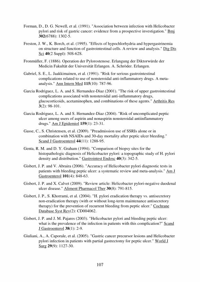
107
Forman, D., D. G. Newell, et al. (1991). "Association between infection with Helicobacter pylori and risk of gastric cancer: evidence from a prospective investigation." Bmj 302(6788): 1302-5.
Freston, J. W., K. Borch, et al. (1995). "Effects of hypochlorhydria and hypergastrinemia on structure and function of gastrointestinal cells. A review and analysis." Dig Dis Sci 40(2 Suppl): 50S-62S.
Fronmüller, F. (1886). Operation der Pylorostenose. Erlangung der Diktorwürde der Medicin Fakultät der Universität Erlangen. A. Schröder. Erlangen.
Gabriel, S. E., L. Jaakkimainen, et al. (1991). "Risk for serious gastrointestinal complications related to use of nonsteroidal anti-inflammatory drugs. A meta-analysis." Ann Intern Med 115(10): 787-96.
Garcia Rodriguez, L. A. and S. Hernandez-Diaz (2001). "The risk of upper gastrointestinal complications associated with nonsteroidal anti-inflammatory drugs, glucocorticoids, acetaminophen, and combinations of these agents." Arthritis Res 3(2): 98-101.
Garcia Rodriguez, L. A. and S. Hernandez-Diaz (2004). "Risk of uncomplicated peptic ulcer among users of aspirin and nonaspirin nonsteroidal antiinflammatory drugs." Am J Epidemiol 159(1): 23-31.
Gasse, C., S. Christensen, et al. (2009). "Preadmission use of SSRIs alone or in combination with NSAIDs and 30-day mortality after peptic ulcer bleeding." Scand J Gastroenterol 44(11): 1288-95.
Genta, R. M. and D. Y. Graham (1994). "Comparison of biopsy sites for the histopathologic diagnosis of Helicobacter pylori: a topographic study of H. pylori density and distribution." Gastrointest Endosc 40(3): 342-5.
Gisbert, J. P. and V. Abraira (2006). "Accuracy of Helicobacter pylori diagnostic tests in patients with bleeding peptic ulcer: a systematic review and meta-analysis." Am J Gastroenterol 101(4): 848-63.
Gisbert, J. P. and X. Calvet (2009). "Review article: Helicobacter pylori-negative duodenal ulcer disease." Aliment Pharmacol Ther 30(8): 791-815.
Gisbert, J. P., S. Khorrami, et al. (2004). "H. pylori eradication therapy vs. antisecretory non-eradication therapy (with or without long-term maintenance antisecretory therapy) for the prevention of recurrent bleeding from peptic ulcer." Cochrane Database Syst Rev(2): CD004062.
Gisbert, J. P. and J. M. Pajares (2003). "Helicobacter pylori and bleeding peptic ulcer: what is the prevalence of the infection in patients with this complication?" Scand J Gastroenterol 38(1): 2-9.
Giuliani, A., A. Caporale, et al. (2005). "Gastric cancer precursor lesions and Helicobacter pylori infection in patients with partial gastrectomy for peptic ulcer." World J Surg 29(9): 1127-30.
108
Goligher, J. C., G. L. Hill, et al. (1978). "Proximal gastric vagotomy without drainage for duodenal ulcer: results after 5-8 years." Br J Surg 65(3): 145-51.
Gong, Z., I. Agalliu, et al. (2008). "Cigarette smoking and prostate cancer-specific mortality following diagnosis in middle-aged men." Cancer Causes Control 19(1): 25-31.
Gonzalez, E. L., P. Patrignani, et al. (2010). "Variability of risk of upper gastrointestinal bleeding among nonsteroidal anti-inflammatory drugs." Arthritis Rheum.
Greenall, M. J. and T. Lehnert (1985). "Vagotomy or gastrectomy for elective treatment of benign gastric ulceration?" Dig Dis Sci 30(4): 353-61.
Griffin, M. R., J. M. Piper, et al. (1991). "Nonsteroidal anti-inflammatory drug use and increased risk for peptic ulcer disease in elderly persons." Ann Intern Med 114(4): 257-63.
Gustavsson, S., K. A. Kelly, et al. (1988). "Trends in peptic ulcer surgery. A population-based study in Rochester, Minnesota, 1956-1985." Gastroenterology 94(3): 688-94.
Gustavsson, S. and O. Nyren (1989). "Time trends in peptic ulcer surgery, 1956 to 1986. A nation-wide survey in Sweden." Ann Surg 210(6): 704-9.
Hallas, J., M. Dall, et al. (2006). "Use of single and combined antithrombotic therapy and risk of serious upper gastrointestinal bleeding: population based case-control study." Bmj 333(7571): 726.
Hansson, L.-E., O. Nyrén, et al. (1996). "The risk of stomach cancer in patients with gastric or duodenal ulcer disease." N Engl J Med 335: 242-9.
Hansson, L. E., J. Baron, et al. (1994). "Tobacco, alcohol and the risk of gastric cancer. A population-based case-control study in Sweden." Int J Cancer 57(1): 26-31.
Hansson, L. E., L. Engstrand, et al. (1993). "Helicobacter pylori infection: independent risk indicator of gastric adenocarcinoma." Gastroenterology 105(4): 1098-103.
Hedberg, M., M. Ogren, et al. (1997). "Pancreatic carcinoma following gastric resection. A case-control study based on 21,660 consecutive clinical necropsies at Malmo University Hospital." Int J Pancreatol 21(3): 219-24.
Helsingen, N. and L. Hillestad (1956). "Cancer development in the gastric stump after partial gastrectomy for ulcer." Ann Surg 143(2): 173-9.
Hermansson, M., A. Ekedahl, et al. (2009). "Decreasing incidence of peptic ulcer complications after the introduction of the proton pump inhibitors, a study of the Swedish population from 1974-2002." BMC Gastroenterology 9(25).
Heuberger, F., D. Pantoflickova, et al. (2003). "Helicobacter pylori infection in Swiss adolescents: prevalence and risk factors." Eur J Gastroenterol Hepatol 15(2): 179-83.

109
Hickey, K., K. A. Do, et al. (2001). "Smoking and prostate cancer." Epidemiol Rev 23(1): 115-25.
Higham, J., J. Y. Kang, et al. (2002). "Recent trends in admissions and mortality due to peptic ulcer in England: increasing frequency of haemorrhage among older subjects." Gut 50(4): 460-4.
Hoare, A. M., A. McLeish, et al. (1978). "Selection of patients for bile diversion surgery: use of bile acid measurement in fasting gastric aspirates." Gut 19(3): 163-5.
Hollands, M. J., I. Filipe, et al. (1989). "Clinical and histological sequelae of Roux-en-Y diversion." Br J Surg 76(5): 481-4.
Holt, J. M., M. W. Gear, et al. (1970). "The role of chronic blood loss in the pathogenesis of postgastrectomy iron-deficiency anaemia." Gut 11(10): 847-50.
Hsu, P. I., K. H. Lai, et al. (2000). "Impact of bacterial eradication on the cell proliferation and p53 protein accumulation in Helicobacter pylori-associated gastritis." Anticancer Res 20(2B): 1221-8.
Huang, J. Q., S. Sridhar, et al. (2002). "Role of Helicobacter pylori infection and non-steroidal anti-inflammatory drugs in peptic-ulcer disease: a meta-analysis." Lancet 359(9300): 14-22.
Ibanez, L., X. Vidal, et al. (2006). "Upper gastrointestinal bleeding associated with antiplatelet drugs." Aliment Pharmacol Ther 23(2): 235-42.
Jaboulay, M. (1892). "La Gastro-enterostomie, la jéjuno-duodénostomie. La résection du pylore." Arch Prov Chir 1: 11-22.
James, M. W. and C. J. Hawkey (2003). "Assessment of non-steroidal anti-inflammatory drug (NSAID) damage in the human gastrointestinal tract." Br J Clin Pharmacol 56: 146-155.
Jamieson, G. (2000). "Current status of indications for surgery in peptic ulcer disease." World Journal of Surgery 24: 256-258.
Jansen, A., S. Harenberg, et al. (2009). "Risk factors for colonic diverticular bleeding: a Westernized community based hospital study." World J Gastroenterol 15(4): 457-61.
Jenkins, J. T., J. R. Duncan, et al. (2007). "Malignant disease in peptic ulcer surgery patients after long term follow-up: a cohort study of 1992 patients." Eur J Surg Oncol 33(6): 706-12.
Johnson, A. (2000). "Proximal gastric vagotomy: Does it have a place in the future management of peptic ulcer?" World Journal of Surgery 24: 259-263.
Johnston, D. and A. R. Wilkinson (1970). "Highly selective vagotomy without a drainage procedure in the treatment of duodenal ulcer." Br J Surg 57(4): 289-96.
Juni, P., A. W. Rutjes, et al. (2002). "Are selective COX 2 inhibitors superior to traditional non steroidal anti-inflammatory drugs?" Bmj 324(7349): 1287-8.
110
Jyotheeswaran, S., A. N. Shah, et al. (1998). "Prevalence of Helicobacter pylori in peptic ulcer patients in greater Rochester, NY: is empirical triple therapy justified?" Am J Gastroenterol 93(4): 574-8.
Kang, J. Y., A. Elders, et al. (2006). "Recent trends in hospital admissions and mortality rates for peptic ulcer in Scotland 1982-2002." Aliment Pharmacol Ther 24(1): 65-79.
Kang, J. Y., A. Tinto, et al. (2002). "Peptic ulceration in general practice in England and Wales 1994-98: period prevalence and drug management." Aliment Pharmacol Ther 16(6): 1067-74.
Karnes, W. E., Jr., I. M. Samloff, et al. (1991). "Positive serum antibody and negative tissue staining for Helicobacter pylori in subjects with atrophic body gastritis." Gastroenterology 101(1): 167-74.
Kemppainen, H., I. Raiha, et al. (1998). "Characteristics of Helicobacter pylori-negative and -positive peptic ulcer disease." Age Ageing 27(4): 427-31.
Kemppainen, H., I. Raiha, et al. (1997). "Clinical presentation of peptic ulcer in the elderly." Gerontology 43(5): 283-8.
Kodama, M., F. Inoue, et al. (1997). "Formation of free radicals from steroid hormones: possible significance in environmental carcinogenesis." Anticancer Res 17(1A): 439-44.
Konturek, S. J., W. Bielanski, et al. (2003). "Helicobacter pylori, non-steroidal anti-inflammatory drugs and smoking in risk pattern of gastroduodenal ulcers." Scand J Gastroenterol 38(9): 923-30.
Kral, J., T. Powley, et al., Eds. (1983). Vagal Nerve Function: Behavioral and methodological considerations, Elsevier Science Publishers B.V. The Netherlands.
Kuipers, E. J., A. S. Pena, et al. (1993). "Seroconversion for Helicobacter pylori." Lancet 342(8867): 328-31.
Kuipers, E. J., J. C. Thijs, et al. (1995a). "The prevalence of Helicobacter pylori in peptic ulcer disease." Aliment Pharmacol Ther 9 Suppl 2: 59-69.
Kuipers, E. J., A. M. Uyterlinde, et al. (1995b). "Long-term sequelae of Helicobacter pylori gastritis." Lancet 345(8964): 1525-8.
Kurata, J. H. and A. N. Nogawa (1997). "Meta-analysis of risk factors for peptic ulcer. Nonsteroidal antiinflammatory drugs, Helicobacter pylori, and smoking." J Clin Gastroenterol 24(1): 2-17.
Kusters, J. G., A. H. van Vliet, et al. (2006). "Pathogenesis of Helicobacter pylori infection." Clin Microbiol Rev 19(3): 449-90.
Laine, L. (2006). "Review article: gastrointestinal bleeding with low-dose aspirin - what's the risk?" Aliment Pharmacol Ther 24(6): 897-908.
111
Lanas, A., E. Bajador, et al. (2000). "Nitrovasodilators, low-dose aspirin, other nonsteroidal antiinflammatory drugs, and the risk of upper gastrointestinal bleeding." N Engl J Med 343(12): 834-9.
Lanas, A., L. A. Garcia-Rodriguez, et al. (2006a). "Risk of upper gastrointestinal ulcer bleeding associated with selective cyclo-oxygenase-2 inhibitors, traditional non-aspirin non-steroidal anti-inflammatory drugs, aspirin and combinations." Gut 55(12): 1731-8.
Lanas, A., L. A. Garcia-Rodriguez, et al. (2009). "Time trends and impact of upper and lower gastrointestinal bleeding and perforation in clinical practice." Am J Gastroenterol 104(7): 1633-41.
Lanas, A. and R. Hunt (2006b). "Prevention of anti-inflammatory drug-induced gastrointestinal damage: benefits and risks of therapeutic strategies." Ann Med 38(6): 415-28.
Lanas, A., M. A. Perez-Aisa, et al. (2005). "A nationwide study of mortality associated with hospital admission due to severe gastrointestinal events and those associated with nonsteroidal antiinflammatory drug use." Am J Gastroenterol 100(8): 1685-93.
Langman, M. J., J. Weil, et al. (1994). "Risks of bleeding peptic ulcer associated with individual non-steroidal anti-inflammatory drugs." Lancet 343(8905): 1075-8.
Lassen, A., J. Hallas, et al. (2006). "Complicated and uncomplicated peptic ulcers in a Danish county 1993-2002: a population-based cohort study." Am J Gastroenterol 101(5): 945-53.
Latarjet, M. (1922). "Resection des nerfs de l'estomac.T*echnique operatoire. Résultats cliniques." Bull Acad Natl Méd 87: 681-91.
Lawrence, W., Jr., P. Vanamee, et al. (1960). "Alterations in fat and nitrogen metabolism after total and subtotal gastrectomy." Surg Gynecol Obstet 10: 601-16.
Lawson, H. H. (1964). "Effect of Duodenal Contents on the Gastric Mucosa under Experimental Conditions." Lancet 1(7331): 469-72.
Leivonen, M., S. Nordling, et al. (1997a). "Does Helicobacter pylori in the gastric stump increase the cancer risk after certain reconstruction types?" Anticancer Res 17(5B): 3893-6.
Leivonen, M. K., C. H. Haglund, et al. (1997b). "Helicobacter pylori infection after partial gastrectomy for peptic ulcer and its role in relapsing disease." Eur J Gastroenterol Hepatol 9(4): 371-4.
Leung, W. K., S. R. Lin, et al. (2004). "Factors predicting progression of gastric intestinal metaplasia: results of a randomised trial on Helicobacter pylori eradication." Gut 53(9): 1244-9.
Lewis, J. D., W. B. Bilker, et al. (2002). "Hospitalization and mortality rates from peptic ulcer disease and GI bleeding in the 1990s: Relationship to sales of Nonsteroidal
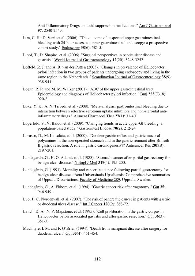
112
Anti-Inflammatory Drugs and acid suppression medications." Am J Gastroenterol 97: 2540-2549.
Lim, C. H., D. Vani, et al. (2006). "The outcome of suspected upper gastrointestinal bleeding with 24-hour access to upper gastrointestinal endoscopy: a prospective cohort study." Endoscopy 38(6): 581-5.
Lipof, T., D. Shapiro, et al. (2006). "Surgical perspectives in peptic ulcer disease and gastritis." World Journal of Gastroenterology 12(20): 3248-3252.
Loffeld, R. J. and A. B. van der Putten (2003). "Changes in prevalence of Helicobacter pylori infection in two groups of patients undergoing endoscopy and living in the same region in the Netherlands." Scandinavian Journal of Gastroenterology 38(9): 938-941.
Logan, R. P. and M. M. Walker (2001). "ABC of the upper gastrointestinal tract: Epidemiology and diagnosis of Helicobacter pylori infection." Bmj 323(7318): 920-2.
Loke, Y. K., A. N. Trivedi, et al. (2008). "Meta-analysis: gastrointestinal bleeding due to interaction between selective serotonin uptake inhibitors and non-steroidal anti-inflammatory drugs." Aliment Pharmacol Ther 27(1): 31-40.
Loperfido, S., V. Baldo, et al. (2009). "Changing trends in acute upper-GI bleeding: a population-based study." Gastrointest Endosc 70(2): 212-24.
Lorusso, D., M. Linsalata, et al. (2000). "Duodenogastric reflux and gastric mucosal polyamines in the non-operated stomach and in the gastric remnant after Billroth II gastric resection. A role in gastric carcinogenesis?" Anticancer Res 20(3B): 2197-201.
Lundegardh, G., H. O. Adami, et al. (1988). "Stomach cancer after partial gastrectomy for benign ulcer disease." N Engl J Med 319(4): 195-200.
Lundegårdh, G. (1991). Mortality and cancer incidence following partial gastrectomy for benign ulcer diseases. Acta Universitatis Upsaliensis, Comprehensive summaries of Uppsala Dissertations. Faculty of Medicine 289. Uppsala, Sweden.
Lundegårdh, G., A. Ekbom, et al. (1994). "Gastric cancer risk after vagotomy." Gut 35: 946-949.
Luo, J., C. Nordenvall, et al. (2007). "The risk of pancreatic cancer in patients with gastric or duodenal ulcer disease." Int J Cancer 120(2): 368-72.
Lynch, D. A., N. P. Mapstone, et al. (1995). "Cell proliferation in the gastric corpus in Helicobacter pylori associated gastritis and after gastric resection." Gut 36(3): 351-3.
Macintyre, I. M. and F. O´Brien (1994). "Death from malignant disease after surgery for duodenal ulcer." Gut 35(4): 451-454.
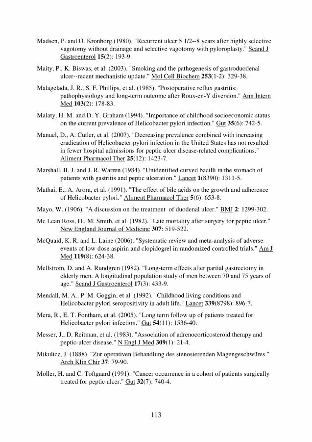
113
Madsen, P. and O. Kronborg (1980). "Recurrent ulcer 5 1/2--8 years after highly selective vagotomy without drainage and selective vagotomy with pyloroplasty." Scand J Gastroenterol 15(2): 193-9.
Maity, P., K. Biswas, et al. (2003). "Smoking and the pathogenesis of gastroduodenal ulcer--recent mechanistic update." Mol Cell Biochem 253(1-2): 329-38.
Malagelada, J. R., S. F. Phillips, et al. (1985). "Postoperative reflux gastritis: pathophysiology and long-term outcome after Roux-en-Y diversion." Ann Intern Med 103(2): 178-83.
Malaty, H. M. and D. Y. Graham (1994). "Importance of childhood socioeconomic status on the current prevalence of Helicobacter pylori infection." Gut 35(6): 742-5.
Manuel, D., A. Cutler, et al. (2007). "Decreasing prevalence combined with increasing eradication of Helicobacter pylori infection in the United States has not resulted in fewer hospital admissions for peptic ulcer disease-related complications." Aliment Pharmacol Ther 25(12): 1423-7.
Marshall, B. J. and J. R. Warren (1984). "Unidentified curved bacilli in the stomach of patients with gastritis and peptic ulceration." Lancet 1(8390): 1311-5.
Mathai, E., A. Arora, et al. (1991). "The effect of bile acids on the growth and adherence of Helicobacter pylori." Aliment Pharmacol Ther 5(6): 653-8.
Mayo, W. (1906). "A discussion on the treatment of duodenal ulcer." BMJ 2: 1299-302.
Mc Lean Ross, H., M. Smith, et al. (1982). "Late mortality after surgery for peptic ulcer." New England Journal of Medicine 307: 519-522.
McQuaid, K. R. and L. Laine (2006). "Systematic review and meta-analysis of adverse events of low-dose aspirin and clopidogrel in randomized controlled trials." Am J Med 119(8): 624-38.
Mellstrom, D. and A. Rundgren (1982). "Long-term effects after partial gastrectomy in elderly men. A longitudinal population study of men between 70 and 75 years of age." Scand J Gastroenterol 17(3): 433-9.
Mendall, M. A., P. M. Goggin, et al. (1992). "Childhood living conditions and Helicobacter pylori seropositivity in adult life." Lancet 339(8798): 896-7.
Mera, R., E. T. Fontham, et al. (2005). "Long term follow up of patients treated for Helicobacter pylori infection." Gut 54(11): 1536-40.
Messer, J., D. Reitman, et al. (1983). "Association of adrenocorticosteroid therapy and peptic-ulcer disease." N Engl J Med 309(1): 21-4.
Mikulicz, J. (1888). "Zur operativen Behandlung des stenosierenden Magengeschwüres." Arch Klin Chir 37: 79-90.
Moller, H. and C. Toftgaard (1991). "Cancer occurrence in a cohort of patients surgically treated for peptic ulcer." Gut 32(7): 740-4.
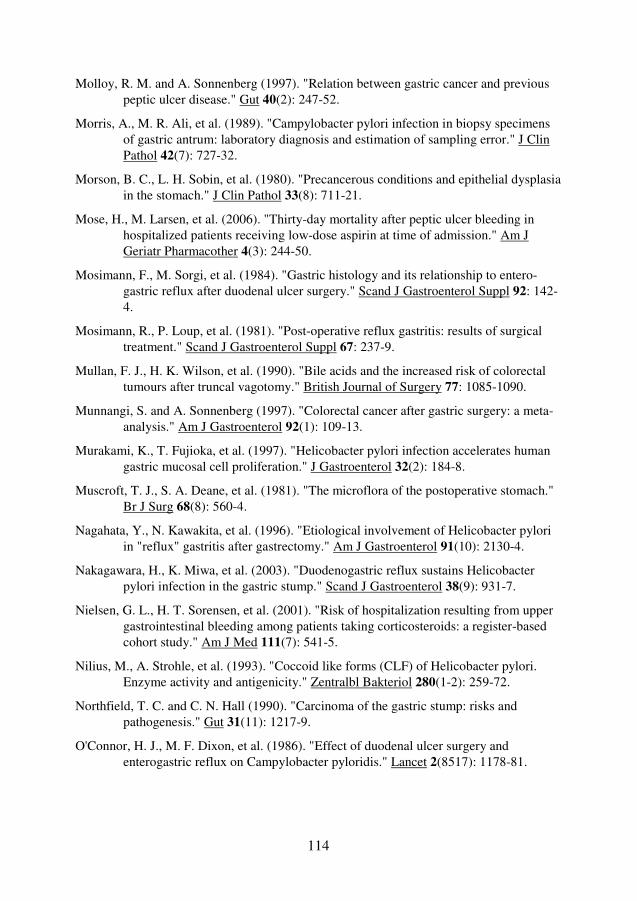
114
Molloy, R. M. and A. Sonnenberg (1997). "Relation between gastric cancer and previous peptic ulcer disease." Gut 40(2): 247-52.
Morris, A., M. R. Ali, et al. (1989). "Campylobacter pylori infection in biopsy specimens of gastric antrum: laboratory diagnosis and estimation of sampling error." J Clin Pathol 42(7): 727-32.
Morson, B. C., L. H. Sobin, et al. (1980). "Precancerous conditions and epithelial dysplasia in the stomach." J Clin Pathol 33(8): 711-21.
Mose, H., M. Larsen, et al. (2006). "Thirty-day mortality after peptic ulcer bleeding in hospitalized patients receiving low-dose aspirin at time of admission." Am J Geriatr Pharmacother 4(3): 244-50.
Mosimann, F., M. Sorgi, et al. (1984). "Gastric histology and its relationship to entero-gastric reflux after duodenal ulcer surgery." Scand J Gastroenterol Suppl 92: 142-4.
Mosimann, R., P. Loup, et al. (1981). "Post-operative reflux gastritis: results of surgical treatment." Scand J Gastroenterol Suppl 67: 237-9.
Mullan, F. J., H. K. Wilson, et al. (1990). "Bile acids and the increased risk of colorectal tumours after truncal vagotomy." British Journal of Surgery 77: 1085-1090.
Munnangi, S. and A. Sonnenberg (1997). "Colorectal cancer after gastric surgery: a meta-analysis." Am J Gastroenterol 92(1): 109-13.
Murakami, K., T. Fujioka, et al. (1997). "Helicobacter pylori infection accelerates human gastric mucosal cell proliferation." J Gastroenterol 32(2): 184-8.
Muscroft, T. J., S. A. Deane, et al. (1981). "The microflora of the postoperative stomach." Br J Surg 68(8): 560-4.
Nagahata, Y., N. Kawakita, et al. (1996). "Etiological involvement of Helicobacter pylori in "reflux" gastritis after gastrectomy." Am J Gastroenterol 91(10): 2130-4.
Nakagawara, H., K. Miwa, et al. (2003). "Duodenogastric reflux sustains Helicobacter pylori infection in the gastric stump." Scand J Gastroenterol 38(9): 931-7.
Nielsen, G. L., H. T. Sorensen, et al. (2001). "Risk of hospitalization resulting from upper gastrointestinal bleeding among patients taking corticosteroids: a register-based cohort study." Am J Med 111(7): 541-5.
Nilius, M., A. Strohle, et al. (1993). "Coccoid like forms (CLF) of Helicobacter pylori. Enzyme activity and antigenicity." Zentralbl Bakteriol 280(1-2): 259-72.
Northfield, T. C. and C. N. Hall (1990). "Carcinoma of the gastric stump: risks and pathogenesis." Gut 31(11): 1217-9.
O'Connor, H. J., M. F. Dixon, et al. (1986). "Effect of duodenal ulcer surgery and enterogastric reflux on Campylobacter pyloridis." Lancet 2(8517): 1178-81.
115
O'Connor, H. J., K. M. Newbold, et al. (1989). "Effect of Roux-en-Y biliary diversion on Campylobacter pylori." Gastroenterology 97(4): 958-64.
Offerhaus, G. J., P. N. Rieu, et al. (1989). "Prospective comparative study of the influence of postoperative bile reflux on gastric mucosal histology and Campylobacter pylori infection." Gut 30(11): 1552-7.
Offerhaus, G. J., A. C. Tersmette, et al. (1988). "Mortality caused by stomach cancer after remote partial gastrectomy for benign conditions: 40 years of follow up of an Amsterdam cohort of 2633 postgastrectomy patients." Gut 29(11): 1588-90.
Ohmann, C., M. Imhof, et al. (2005). "Time-trends in the epidemiology of peptic ulcer bleeding." Scand J Gastroenterol 40(8): 914-20.
Osugi, H., K. Fukuhara, et al. (2004). "Reconstructive procedure after distal gastrectomy to prevent remnant gastritis." Hepatogastroenterology 51(58): 1215-8.
Ovaska, J., T. Havia, et al. (1990). "Roux-en-Y reconstruction for severe postoperative duodenogastric reflux disease." Ann Chir Gynaecol 79(1): 19-22.
Paimela, H., N. K. Oksala, et al. (2004). "Surgery for peptic ulcer today. A study on the incidence, methods and mortality in surgery for peptic ulcer in Finland between 1987 and 1999." Dig Surg 21(3): 185-91.
Paimela, H., L. Paimela, et al. (2002). "Current features of peptic ulcer disease in Finland: incidence of surgery, hospital admissions and mortality for the disease during the past twenty-five years." Scand J Gastroenterol 37(4): 399-403.
Parsonnet, J. (1995). "The incidence of Helicobacter pylori infection." Aliment Pharmacol Ther 9 Suppl 2: 45-51.
Parsonnet, J., G. D. Friedman, et al. (1991). "Helicobacter pylori infection and the risk of gastric carcinoma." N Engl J Med 325(16): 1127-31.
Perez-Aisa, M. A., D. Del Pino, et al. (2005). "Clinical trends in ulcer diagnosis in a population with high prevalence of Helicobacter pylori infection." Aliment Pharmacol Ther 21(1): 65-72.
Petersson, F., K. Borch, et al. (2002a). "Gastric epithelial proliferation and p53 and p21 expression in a general population sample: relations to age, sex, and mucosal changes associated with H. pylori infection." Dig Dis Sci 47(7): 1558-66.
Petersson, F., K. Borch, et al. (2002b). "Prevalence of subtypes of intestinal metaplasia in the general population and in patients with autoimmune chronic atrophic gastritis." Scand J Gastroenterol 37(3): 262-6.
Pienta, K. and P. Esper (1993). "Risk factors for prostate cancer." Annals of Internal Medicine 118(10): 793-803.
Piper, D. W., J. H. McIntosh, et al. (1981). "Analgesic ingestion and chronic peptic ulcer." Gastroenterology 80(3): 427-32.
116
Piper, J. M., W. A. Ray, et al. (1991). "Corticosteroid use and peptic ulcer disease: role of nonsteroidal anti-inflammatory drugs." Ann Intern Med 114(9): 735-40.
Post, P. N., E. J. Kuipers, et al. (2006). "Declining incidence of peptic ulcer but not of its complications: a nation-wide study in The Netherlands." Aliment Pharmacol Ther 23: 1587-1593.
Pounder, R. E. and D. Ng (1995). "The prevalence of Helicobacter pylori infection in different countries." Aliment Pharmacol Ther 9 Suppl 2: 33-9.
Quan, C. and N. J. Talley (2002). "Management of peptic ulcer disease not related to Helicobacter pylori or NSAIDs." Am J Gastroenterol 97(12): 2950-61.
Ritchie, W. P. (1984). "Alkaline reflux gastritis: a critical reappraisal." Gut 25(9): 975-87.
Ritchie, W. P., Jr. (1980). "Alkaline reflux gastritis. An objective assessment of its diagnosis and treatment." Ann Surg 192(3): 288-98.
Robles-Campos, R., J. A. Lujan-Mompean, et al. (1993). "Role of Helicobacter pylori infection and duodenogastric reflux in the pathogenesis of alkaline reflux gastritis after gastric operations." Surg Gynecol Obstet 176(6): 594-8.
Rockall, T. A., R. F. Logan, et al. (1995). "Incidence and mortality from acute upper gastrointestinal haemorrhage in the United Kingdom. Steering committee and members of the National Audit of Acute Upper Gastrointestinal Haemorrhage " BMJ 311(6999): 222-226.
Rockall, T. A., R. F. Logan, et al. (1996). "Risk assessment after acute upper gastrointestinal haemorrhage." Gut 38(3): 316-21.
Roosendaal, R., E. J. Kuipers, et al. (1997). "Helicobacter pylori and the birth cohort effect: evidence of a continuous decrease of infection rates in childhood." Am J Gastroenterol 92(9): 1480-1482.
Rothenbacher, D. and H. Brenner (2003). "Burden of Helicobacter pylori and H. pylori-
related diseases in developed countries: recent developments and future implications." Microbes and Infection 5: 693-703.
Roux, C. (1897). "De la gastroenterostomie. ." Rev Gynecol Chir Abd 1: 67.
Ruddell, W. S., E. S. Bone, et al. (1976). "Gastric-juice nitrite. A risk factor for cancer in the hypochlorhydric stomach?" Lancet 2(7994): 1037-9.
Rydygier, L. (1882). "Die erste Magenresektion beim Magengeschwür." Berl Klin Wochensch 19: 39-41.
Sadic, J., A. Borgström, et al. (2009). "Bleeding peptic ulcer - time trends in incidence, treatment and mortality in Sweden." Aliment Pharmacol Ther 30: 392-398.
Salomoni, M., E. Zuccato, et al. (1989). "Effect of bile salts on carbonic anhydrase from rat and human gastric mucosa." Scand J Gastroenterol 24(1): 28-32.
117
Schrumpf, E., A. Serck-Hanssen, et al. (1977). "Mucosal changes in the gastric stump 20-25 years after partial gastrectomy." Lancet 2(8036): 467-9.
Silwer, L. and C. S. Lundborg (2005). "Patterns of drug use during a 15 year period: data from a Swedish county, 1988--2002." Pharmacoepidemiol Drug Saf 14(11): 813-820.
Silverstein, F. E., G. Faich, et al. (2000). "Gastrointestinal toxicity with celecoxib vs nonsteroidal anti-inflammatory drugs for osteoarthritis and rheumatoid arthritis: the CLASS study: A randomized controlled trial. Celecoxib Long-term Arthritis Safety Study." Jama 284(10): 1247-55.
Silverstein, F. E., D. Y. Graham, et al. (1995). "Misoprostol reduces serious gastrointestinal complications in patients with rheumatoid arthritis receiving nonsteroidal anti-inflammatory drugs. A randomized, double-blind, placebo-controlled trial." Ann Intern Med 123(4): 241-9.
Sipponen, P. (2002). "Gastric cancer: pathogenesis, risks, and prevention." J Gastroenterol 37 Suppl 13: 39-44.
Slomiany, B. L., M. Aono, et al. (1984). "Effect of bile acids on the glycoprotein constituent of gastric mucus." J Appl Biochem 6(5-6): 308-13.
Sobala, G. M., H. J. O'Connor, et al. (1993). "Bile reflux and intestinal metaplasia in gastric mucosa." J Clin Pathol 46(3): 235-40.
Sorensen, H. T., L. Mellemkjaer, et al. (2000). "Risk of upper gastrointestinal bleeding associated with use of low-dose aspirin." Am J Gastroenterol 95(9): 2218-24.
Staat, M. A., D. Kruszon-Moran, et al. (1996). "A population-based serologic survey of Helicobacter pylori infection in children and adolescents in the United States." J Infect Dis 174(5): 1120-3.
Stack, W. A., J. C. Atherton, et al. (2002). "Interactions between Helicobacter pylori and other risk factors for peptic ulcer bleeding." Aliment Pharmacol Ther 16(3): 497-506.
Staël von Holstein, C. C. S., H. Anderson, et al. (1997). "The significance of ulcer disease on late mortality after partial gastric resection." European Journal of Gastroenterology & Hepatology 9: 33-40.
Staël von Holstein, C. C. S., H. Anderson, et al. (1995). "Mortality after remote surgery for benign gastroduodenal disease." Gut 37: 617-622
Staël von Holstein, C. C. S., H. Graffner, et al. (1987). "One hundred patients ten years after parietal cell vagotomy." British Journal of Surgery 74: 101-103.
Stalsberg, H. and S. Taksdal (1971). "Stomach cancer following gastric surgery for benign conditions." Lancet 2(7735): 1175-7.
118
Stein, H. J., J. Esplugues, et al. (1989). "Direct cytotoxic effect of oxygen radicals on the gastric mucosa." Surgery 106(2): 318-23: discussion 323-4.
Suerbaum, S. and P. Michetti (2002). "Helicobacter pylori infection." N Engl J Med 347(15): 1175-86.
Sugiyama, Y., H. Sohma, et al. (1987). "Regurgitant bile acids and mucosal injury of the gastric remnant after partial gastrectomy." Am J Surg 153(4): 399-403.
Sung, J. J., E. J. Kuipers, et al. (2009). "Systematic review: the global incidence and prevalence of peptic ulcer disease." Aliment Pharmacol Ther 29(9): 938-46.
Sung, J. J., J. Y. Lau, et al. (2010a). "Continuation of low-dose aspirin therapy in peptic ulcer bleeding: a randomized trial." Ann Intern Med 152(1): 1-9.
Sung, J. J., W. K. Leung, et al. (1997). "One-week antibiotics versus maintenance acid suppression therapy for Helicobacter pylori-associated peptic ulcer bleeding." Dig Dis Sci 42(12): 2524-8.
Sung, J. J., K. K. Tsoi, et al. (2010b). "Causes of mortality in patients with peptic ulcer bleeding: a prospective cohort study of 10,428 cases." Am J Gastroenterol 105(1): 84-9.
Take, S., M. Mizuno, et al. (2007). "Baseline gastric mucosal atrophy is a risk factor associated with the development of gastric cancer after Helicobacter pylori eradication therapy in patients with peptic ulcer diseases." J Gastroenterol 42 Suppl 17: 21-7.
Tannenbaum, S. R. (1983). "N-nitroso compounds: a perspective on human exposure." Lancet 1(8325): 629-32.
Tascilar, M., B. P. van Rees, et al. (2002). "Pancreatic cancer after remote peptic ulcer surgery." J Clin Pathol 55(5): 340-5.
Tatsuta, M., H. Yamamura, et al. (1982). "Gastrin protection against chemically induced gastric adenocarcinomas in Wistar rats: histopathology of the glandular stomach and incidence of gastric adenocarcinoma." J Natl Cancer Inst 69(1): 59-66.
Tersmette, A. C., F. M. Giardiello, et al. (1995). "Carcinogenesis after remote peptic ulcer surgery: The long-term prognosis of partial gastrectomy." Scandinavian Journal of Gastroenterology 30 Suppl 212: 96-99.
Thomas, R., M. Hellmich, et al. (2003). "Role of gastrointestinal hormones in the proliferation of normal and neoplastic tissues." Endocrine Reviews 24(5): 571-599.
Thomopoulos, K. C., K. P. Mimidis, et al. (2005). "Acute upper gastrointestinal bleeding in patients on long-term oral anticoagulation therapy: endoscopic findings, clinical management and outcome." World J Gastroenterol 11(9): 1365-8.

119
Thomsen, R. W., A. Riis, et al. (2006). "Outcome of peptic ulcer bleeding among users of traditional non-steroidal anti-inflammatory drugs and selective cyclo-oxygenase-2 inhibitors." Aliment Pharmacol Ther 24(10): 1431-8.
Toftgaard, C. (1989). "Gastric cancer after peptic ulcer surgery. A historic prospective cohort investigation." Ann Surg 210(2): 159-64.
Toye, D. K. and J. A. Williams (1965). "Post-gastrectomy bile vomiting." Lancet 2(7411): 524-6.
van Heerden, J. A., S. F. Phillips, et al. (1975). "Postoperative reflux gastritis." Am J Surg 129(1): 82-8.
van Leerdam, M. E. (2008). "Epidemiology of acute upper gastrointestinal bleeding." Best Pract Res Clin Gastroenterol 22(2): 209-24.
van Leerdam, M. E., E. M. Vreeburg, et al. (2003). "Acute upper GI bleeding: did anything change? Time trend analysis of incidence and outcome of acute upper GI bleeding between 1993/1994 and 2000." Am J Gastroenterol 98(7): 1494-9.
Wang, Y. R., J. E. Richter, et al. (2010). "Trends and outcomes of hospitalizations for peptic ulcer disease in the United States, 1993 to 2006." Ann Surg 251(1): 51-8.
Watt, P. C., J. M. Sloan, et al. (1983). "Histology of the postoperative stomach before and after diversion of bile." Br Med J (Clin Res Ed) 287(6403): 1410-2.
Watt, P. C. H., C. C. Patterson, et al. (1984). "Late mortality after vagotomy and drainage for duodenal ulcer." British Medical Journal 288: 1335-1338.
Weil, J., D. Colin-Jones, et al. (1995). "Prophylactic aspirin and risk of peptic ulcer bleeding." Bmj 310(6983): 827-30.
Wells, C. and R. Welbourn (1951). "Post-gastrectomy syndromes; a study in applied physiology." Br Med J 1(4706): 546-54.
Vernava, A. M., 3rd, B. A. Moore, et al. (1997). "Lower gastrointestinal bleeding." Dis Colon Rectum 40(7): 846-58.
Wessinger, S., M. Kaplan, et al. (2006). "Increased use of selective serotonin reuptake inhibitors in patients admitted with gastrointestinal haemorrhage: a multicentre retrospective analysis." Aliment Pharmacol Ther 23(7): 937-44.
Wiese, E. (1929). "Theodor Billroth, scholar, musician, master surgeon. ." Ann Med Hist 10: 278-86.
Wilcox, C. M., L. N. Alexander, et al. (1997). "Nonsteroidal antiinflammatory drugs are associated with both upper and lower gastrointestinal bleeding." Dig Dis Sci 42(5): 990-7.
Viste, A., E. Bjornestad, et al. (1986). "Risk of carcinoma following gastric operations for benign disease. A historical cohort study of 3470 patients." Lancet 2(8505): 502-5.
120
Wolfler, A. (1881). "Gastroenterostomie." Z Chir 8: 705.
Xia, H. H., N. Phung, et al. (2000). "Demographic and endoscopic characteristics of patients with Helicobacter pylori positive and negative peptic ulcer disease." Med J Aust 173(10): 515-9.
Xia, H. H., B. C. Wong, et al. (2001). "Clinical and endoscopic characteristics of non-Helicobacter pylori, non-NSAID duodenal ulcers: a long-term prospective study." Aliment Pharmacol Ther 15(12): 1875-82.
You, W. C., L. M. Brown, et al. (2006). "Randomized double-blind factorial trial of three treatments to reduce the prevalence of precancerous gastric lesions." J Natl Cancer Inst 98(14): 974-83.
Zittel, T. T., E. C. Jehle, et al. (2000). "Surgical management of peptic ulcer disease today--indication, technique and outcome." Langenbecks Arch Surg 385(2): 84-96.
Åhsberg Johannesson, K., E. Hammar, et al. (2003). "Mucosal changes in the gastric remnant: long-term effects of bile reflux diversion and Helicobacter pylori infection." European Journal of Gastroenterology & Hepatology 15: 35-40.