Embed Size (px)
Citation preview

Resection and reconstruction for periacetabular tumors is one of the most difficult challenges for the orthopedic oncologist. The primary goal of the procedure is to control the tumor locally by complete resection. The secondary goal is to reconstruct the periacetabular defect to restore as much pelvic stability and hip joint function as possible. Reconstructive options after periacetabular resection can be divided into 2 categories depending on the preservation of the acetabular-femoral articulation. Nonpreservation reconstruction includes fusion, saddle prosthesis insertion,
hip transposition, and pseudoarthrosis.1-4) Preservation reconstruction includes the use of a custom-made pros-thesis and allograft or recycled autograft-prosthesis com-posite.5-11) When internal pelvectomy was introduced as an alternative to hindquarter amputation, iliofemoral co-aptation or ischiofemoral arthrodesis was performed with the compromise of limb length shortening and hip insta-bility.2,12) The use of a saddle prosthesis was considered a temporary compromise to address the issue of flail hip; however, this prosthesis posed the risk of cranialization or luxation of the implant.4,13) Therefore, to achieve pelvic continuity and durable hip function, acetabular-femoral articulation-preserving reconstruction was devised.7,11,14-16) This provided promising results in a substantial propor-tion of patients, but short- and long-term complications were significant.6,17-19) In this regard, pseudoarthrosis or hip transposition, which may avoid problems related to
Comparison of Pasteurized Autograft-Prosthesis Composite Reconstruction and Resection Hip
Arthroplasty for Periacetabular TumorsSeung Yong Lee, MD, Dae-Geun Jeon, MD, Wan Hyeong Cho, MD,
Won Seok Song, MD, Chang-Bae Kong, MD
Department of Orthopedic Surgery, Korea Cancer Center Hospital, Seoul, Korea
Background: Because of the high complication rate of anatomical reconstruction after periacetabular resection, the strategy of resection alone has been revisited. However, in terms of complications and functional outcome, whether resection hip arthroplasty (RHA) shows a superior result to that of pelvic ring reconstruction remains controversial. Methods: We compared 24 RHAs and 16 pasteurized autograft-prosthesis composite (PPC) reconstructions regarding the compli-cation rates, operative time, blood loss, and functional outcome. Results: Compared to 16 PPC hips, 24 RHA hips showed lower major and minor complication rates (p < 0.001), shorter surgical time (p < 0.001), and superior Musculoskeletal Tumor Society scores (p < 0.001). Of the 24 RHA hips, bony neo-acetabulum was identified in 7 on computed tomography and partial neo-acetabulum in 9; the remaining 8 had no bony acetabular structure. The average time to bony neo-acetabulum formation was 7 months (range, 4 to 13 months). Conclusions: RHA for periacetabular tumors can be an excellent alternative to anatomical reconstruction. It offers short surgical time, low complication rates, and functional results comparable to those of other reconstruction methods. However, this procedure is indicated for patients who can accept some limb shortening, and a tumor should be confined to the periacetabular area. Keywords: Acetabulum, Reconstructive surgery, Treatment outcome
Original Article Clinics in Orthopedic Surgery 2017;9:374-385 • https://doi.org/10.4055/cios.2017.9.3.374
Copyright © 2017 by The Korean Orthopaedic AssociationThis is an Open Access article distributed under the terms of the Creative Commons Attribution Non-Commercial License (http://creativecommons.org/licenses/by-nc/4.0)
which permits unrestricted non-commercial use, distribution, and reproduction in any medium, provided the original work is properly cited.Clinics in Orthopedic Surgery • pISSN 2005-291X eISSN 2005-4408
Received September 20, 2016; Accepted March 4, 2017Correspondence to: Dae-Geun Jeon, MD Department of Orthopedic Surgery, Korea Cancer Center Hospital, 75 Nowon-ro, Nowon-gu, Seoul 01812, KoreaTel: +82-2-970-1242, Fax: +82-2-970-2403E-mail: [email protected]

375
Lee et al. Resection Hip Arthroplasty for Periacetabular TumorsClinics in Orthopedic Surgery • Vol. 9, No. 3, 2017 • www.ecios.org
pelvic ring reconstruction, is being revisited.1,20-22) A recent report of resection hip arthroplasty (RHA) in 27 patients has reconfirmed the usefulness of this approach, a similar technique of which was addressed in a study involving 5 patients in 1978.2,23) Hu et al.23) postulated that RHA would show fewer complications, shorter surgical time, less blood loss, and better functional results than those of pelvic ring reconstruction methods. However, in terms of complica-tions and functional outcomes, whether RHA is superior
to anatomical pelvic ring reconstruction is still controver-sial. Proponents of anatomical reconstruction speculate that functional results in patients with failed pelvic ring reconstruction would not be inferior to those of primary RHA.
In this study, we compared the results of 24 cases of RHA and 16 pasteurized bone-prosthesis composite reconstructions in terms of complication rates, operative time, blood loss, and ultimate functional outcome. Addi-
Table 1. Demographic Data of 24 Patients with Resection Hip Arthroplasty
Case Age (yr)/sex Diagnosis Tumor
stage TV (cc)Extent of
iliac lesion (cm)
Resection type
Surgical margin
Surgical time (hr)
RBC transfusion
(pint)*LR Meta. Final
statusF/U
(mo)
1 21/F GCT Benign 115 2.3 II m 2.2 7 + - NED 54
2 17/F OS IIB 145 1.2 II + III w 4.8 1 - - CDF 70
3 17/F CS IIB 648 1 II + III w 4.8 10 - - CDF 182
4 32/M GCT Benign 66 0 II + III† m 2.7 2 + - NED 118
5 43/F FS IIB 111 1.7 II + III w 3.9 4 - + DOD 41
6 47/F CS IIB 172 0 II + III w 5.1 2 - - CDF 61
7 68/M CS IIB 103 2.5 II + III m 4.8 11 + - AWD 66
8 34/M CS IIB 66 4.5 I† + II w 1.8 2 - - CDF 37
9 43/M CS IIB 165 4.9 I† + II w 1.7 0 - - CDF 35
10 46/M CS IIA 87 3.7 I† + II w 2.0 0 - - CDF 32
11 47/M CS IIA 67 3.4 I† + II w 1.7 3 - - CDF 29
12 15/M SS III 165 1.7 I† + II + III m 2.7 4 - - CDF 37
13 21/M SS III 147 2.5 I† + II + III m 4.7 9 - - CDF 280
14 22/M OS IIB 330 0.9 I† + II + III w 8.8 12 - - CDF 69
15 27/M OS IIB 125 3.3 I† + II + III m 5.4 7 - - CDF 43
16 30/M GCT Benign 144 5 I† + II + III† w 5.5 10 - - CDF 132
17 32/M OS IIB 204 2.2 I† + II + III w 5.0 11 - - CDF 26
18 34/F CS IIB 414 2 I† + II + III w 5.8 8 - - CDF 301
19 36/F OS IIB 383 4.4 I† + II + III m 5.2 20 + + DOD 36
20 39/M CS IIB 116 0 I† + II + III w 5.1 4 - - CDF 56
21 41/M CS IIB 212 3.4 I† + II + III m 2.9 6 + + NED 26
22 46/M CS IIB 725 8 I† + II + III i 3.5 12 + - NED 42
23 61/M CS IIB 565 1.6 I† + II + III m 5.9 15 - - CDF 35
24 65/M CS IIB 162 3.4 I† + II + III w 5.5 5 - - CDF 69
TV: tumor volume, iliac lesion: measured from the top of the femoral head, RBC: red blood cell, LR: local recurrence, Meta.: metastasis, GCT: giant cell tumor, m: marginal, +: local recurrence confirmed, NED: no evidence of disease, OS: osteosarcoma, w: wide, CDF: continuous disease-free, CS: chondrosarcoma, FS: fibrosarcoma, DOD: died of disease, AWD: alive with disease, SS: synovial sarcoma, i: intralesional.*RBC 1 pint: 400 cc. †Partial resection of involved bone.

376
Lee et al. Resection Hip Arthroplasty for Periacetabular TumorsClinics in Orthopedic Surgery • Vol. 9, No. 3, 2017 • www.ecios.org
tionally, in cases of RHA, we described the pattern of neo-hip joint formation according to the extent of iliac bone resection or the mode of postoperative management.
METHODS
Between January 1990 and March 2015, 99 patients with pelvic bone and soft tissue tumors underwent periacetabu-lar resection at Korea Cancer Center Hospital. According to the pelvic resection category described by Enneking and Dunham,12) 43 were type I + II, 28 were type I + II + III, 25 were type II + III, and 3 were type II resections. Among these, we selected 24 patients who underwent RHA and 16 patients who underwent pasteurized autograft-prosthesis composite (PPC) reconstruction. Exclusion criteria were: (1) iliac resection at the sacroiliac joint (37 patients); (2) reconstruction with saddle prosthesis (8 patients); and (3) less than 2 years of follow-up without an event (14 patients). Finally, 23 male and 17 female patients with an
average age of 35.3 years (range, 15 to 68 years) were in-cluded. Follow-up duration was a minimum of 13 months (average, 101 months; range, 13 to 301 months).
The criteria for RHA were: (1) tumors located mainly in region II and III (some partially involved region I) and (2) preoperative magnetic resonance imaging (MRI) showing no sign of femoral head involvement by tumor. PPC reconstruction was indicated for tumors when at least 1 of 2 landmarks on plain pelvic anteroposterior radio-graphs (ilioischial and iliopectineal lines) was intact.
Of the 24 patients who underwent RHA, 18 had pri-mary malignant bone tumors, 3 had soft tissue sarcomas, and 3 had giant cell tumors (Table 1). Of the 16 patients who underwent PPC reconstruction, 11 had primary malignant bone tumors, 3 had benign aggressive bone tu-mors, and 2 had metastatic tumors (Table 2). Preoperative staging included plain radiography and MRI of the pelvis, computed tomography (CT) of the chest, and whole body technetium bone scan. Staging was determined accord-
Table 2. Demographic Data of 16 Patients with Pasteurized Autograft-Prosthesis Composite Reconstruction
Case Age (yr)/sex Diagnosis Tumor
stage TV (cc) Resection type
Surgical margin
Surgical time (hr)
RBC transfusion
(pint)*LR Meta. Final
statusF/U
(mo)
1 35/M OS IIB 52 II w 7 6 - + DOD 24
2 19/F GCT Benign 188 II + III w 5.8 8 - - CDF 271
3 20/F OS IB 63 II + III m 7 7 - - CDF 246
4 49/F HES IB 187 II + III† w 6.4 6 - - CDF 243
5 52/M CS IIB 251 II + III w 8 9 - - CDF 125
6 54/F CS IIB 110 II + III w 6.3 9 - - CDF 108
7 18/F ES IIB 201 I† + II m 7.3 7 + + DOD 13
8 20/F OS IIB 377 I† + II m 7.6 16 - - CDF 150
9 21/M MFH IIB 31 I† + II w 7.3 5 - - CDF 254
10 24/F DF Benign 82 I† + II m 6.8 9 - - CDF 120
11 34/F MC III 79 I† + II w 7.9 13 + - NED 224
12 54/F MC III 26 I† + II w 6.6 7 - - CDF 161
13 25/M CS IIB 653 I† + II + III† m 9.3 15 - - CDF 141
14 30/M OS IIB 226 I† + II + III w 7.3 4 + + DOD 69
15 31/F CS IIB 63 I† + II + III† w 6.8 8 - - CDF 63
16 45/M CS IIB 141 I† + II + III w 7.3 10 + + DOD 21
TV: tumor volume, RBC: red blood cell, LR: local recurrence, Meta.: metastasis, OS: osteosarcoma, w: wide, +: local recurrence confirmed, DOD: died of disease, GCT: giant cell tumor, CDF: continuous disease-free, m: marginal, HES: hemangioendothelioma, CS: chondrosarcoma, ES: Ewing sarcoma, MFH: malignant fibrous histiocytoma, DF: desmoplastic fibrosarcoma, MC: metastatic carcinoma, NED: no evidence of disease.*RBC 1 pint: 400 cc. †Partial resection of involved bone.

377
Lee et al. Resection Hip Arthroplasty for Periacetabular TumorsClinics in Orthopedic Surgery • Vol. 9, No. 3, 2017 • www.ecios.org
ing to Enneking's criteria.24) Neoadjuvant and adjuvant chemotherapy were administered to 13 patients. Initial tumor volume, pathologic diagnosis, extent of iliac lesion on MRI, osteotomy level from iliac crest, resection type, surgical time, surgical margin, and amount of transfusion were recorded. The tumor volume was calculated using 3 parameters (height, width, and depth) by the ellipsoid for-mula: [V = (4π/3)×a×b×c].25) The height, width, and depth of a tumor were measured on the coronal and axial plane MRI scans. The resection and reconstruction included the following procedures. An ilioinguinal approach was used for the main incision, and a satellite incision was made from the anterosuperior iliac spine to the greater trochan-ter, if necessary. In 12 of the 24 patients who underwent RHA, 2 or 3 osteotomies were made to preserve the iliac bone-hip flexor or abductor continuity, and to displace the bone block-muscle complex inferiorly and laterally. Pelvic resection was classified according to the system of Ennek-ing and Dunham.12) In 16 cases of PPC reconstruction, 6 cases were type I + II, 5 were type II + III, 4 were type I + II + III, and 1 was a type II resection. In 24 cases of RHA, 13 were type I + II + III, 6 were type II + III, 4 were type I + II, and 1 was a type II resection. The margin in the 40 patients was wide in 25, marginal in 14, and intralesional in 1, according to Enneking's criteria.24) The preparation of pasteurized bone and reconstruction using a total hip prosthesis was performed as described previously.9) In RHA, the average osteotomy length from the iliac crest was 9.5 cm (range, 4 to 14.5 cm). After tumor removal, the femoral head was pushed up to the inferior aspect of the resected ilium. In 12 cases, the femoral head was fixed to the remaining ilium with wire. In the remaining 12 cases, no fixation was undertaken. In 12 cases of RHA, previously detached iliac bone block-muscle complex was
repositioned to the iliac wing with wire. Postoperatively, patients who underwent PPC reconstruction were usually immobilized for 8–10 weeks in a one-and-a-half hip spica cast, which was followed by crutch walking until radio-graphic union was achieved. In patients who underwent RHA, only 3 were immobilized in a hip spica cast. The remaining 21 were allowed to ambulate when drains were removed (around 2 weeks after the surgery). Monthly plain anteroposterior and bilateral oblique radiographic examinations were performed until 2 years after the index operation. In patients who underwent RHA, trimonthly pelvic CT was performed to evaluate the formation of bony neo-acetabulum. More than 3/4 of circular bony neo-acetabulum formation was defined as complete, and less than 1/2 of a circle as partial (Fig. 1).
At final follow-up, 28 patients were event-free, 6 died of their disease, 5 had no evidence of disease, and 1 was alive with disease. Nine patients (22.5%) developed local recurrence, and their surgical margin status was mar-ginal in 6, wide in 2, and intralesional in 1. Functional re-sults were assessed by the Musculoskeletal Tumor Society (MSTS) system.26) Scores were recorded at the last visit for patients with intact PPC and who underwent RHA. For PPC reconstruction patients whose graft was removed, the functional scores before and after graft removal were recorded. Final limb length discrepancy on radiographs and the shoe heel height to compensate for limb shorten-ing were recorded. Reconstruction failure was defined as the removal of the composite or iliac wing-femoral head coaptation due to complications. Time to failure (months) was defined as the time elapsed between the first surgery and the date of reconstruction removal. A major complica-tion was defined as the one that eventually necessitated re-moval of the graft or prosthesis as a revision procedure. A
A B
Fig. 1. (A) The axial pelvic computed tomography (CT) shows complete bony neo-acetabulum formation 9 months postoperatively. (B) The axial CT shows partial bony neo-acetabulum formation (less than 1/2 of the femoral head circumference).

378
Lee et al. Resection Hip Arthroplasty for Periacetabular TumorsClinics in Orthopedic Surgery • Vol. 9, No. 3, 2017 • www.ecios.org
Table 3. Comparison of Tumor Characteristics and Surgical Outcome between Resection Hip Arthroplasty (n = 24) and Pasteurized Autograft-Prosthesis Composite Reconstruction (n = 16)
Variable Resection hip arthroplasty (%)
Pasteurized autograft-prosthesis composite reconstruction (%) p-value
Age (yr) 0.505 ≤ 40 14 (58.3) 11 (68.8) > 40 10 (41.7) 5 (31.2)Sex 0.037 Male 17 (70.8) 6 (37.5) Female 7 (29.2) 10 (62.5)Initial tumor volume (cc) Mean (range) 226.5 (66–725) 170.6 (26–653) 0.334 ≤ 150 12 (50.0) 9 (56.3) 0.698 > 150 12 (50.0) 7 (43.7)Resection type 0.581 I + II + III 17 (70.8) 10 (62.5) II + III 7 (29.2) 6 (37.5)Surgical time (hr) Mean (range) 4.2 (1.7–8.8) 7.2 (5.8–9.3) < 0.001 ≤ 6 23 (95.8) 1 (6.3) < 0.001 > 6 1 (4.2) 15 (93.8)Transfusion (pint) Mean (range) 6.9 (0–20) 8.7 (4–16) 0.219 ≤ 6 12 (50.0) 4 (25.0) 0.114 > 6 12 (50.0) 12 (75.0)Surgical margin 0.505 Wide 14 (58.3) 11 (68.8) Marginal, intralesional 10 (41.7) 5 (31.2)Major complication < 0.001 Present 0 12 (75.0) Absent 24 (100) 4 (25.0)Minor complication < 0.001 Present 2 (8.3) 15 (6.3) Absent 22 (91.7) 1 (93.7)Final leg length discrepancy (cm) Mean (range) 3.7 (1.3–6.4) 3.7 (0–7) 0.943 ≤ 3.0 10 (41.7) 6 (37.5) 0.792 > 3.0 14 (58.3) 10 (62.5)MSTS score Mean (range) 23.5 (15–28) 14.8 (8–26) < 0.001 ≤ 20 6 (25.0) 13 (81.3) 0.001 > 20 18 (75.0) 3 (18.8)Local recurrence 1.000 Recurred 6 (25.0) 4 (25.0) Not recurred 18 (75.0) 12 (75.0)Distant metastasis 0.407 Occurred 3 (12.5) 4 (25.0) Not occurred 21 (87.5) 12 (75.0)Final outcome 0.138 Alive 23 (95.8) 12 (75.0) Dead 1 (4.2) 4 (25.0)Total 24 (100) 16 (100)
MSTS: Musculoskeletal Tumor Society.
379
Lee et al. Resection Hip Arthroplasty for Periacetabular TumorsClinics in Orthopedic Surgery • Vol. 9, No. 3, 2017 • www.ecios.org
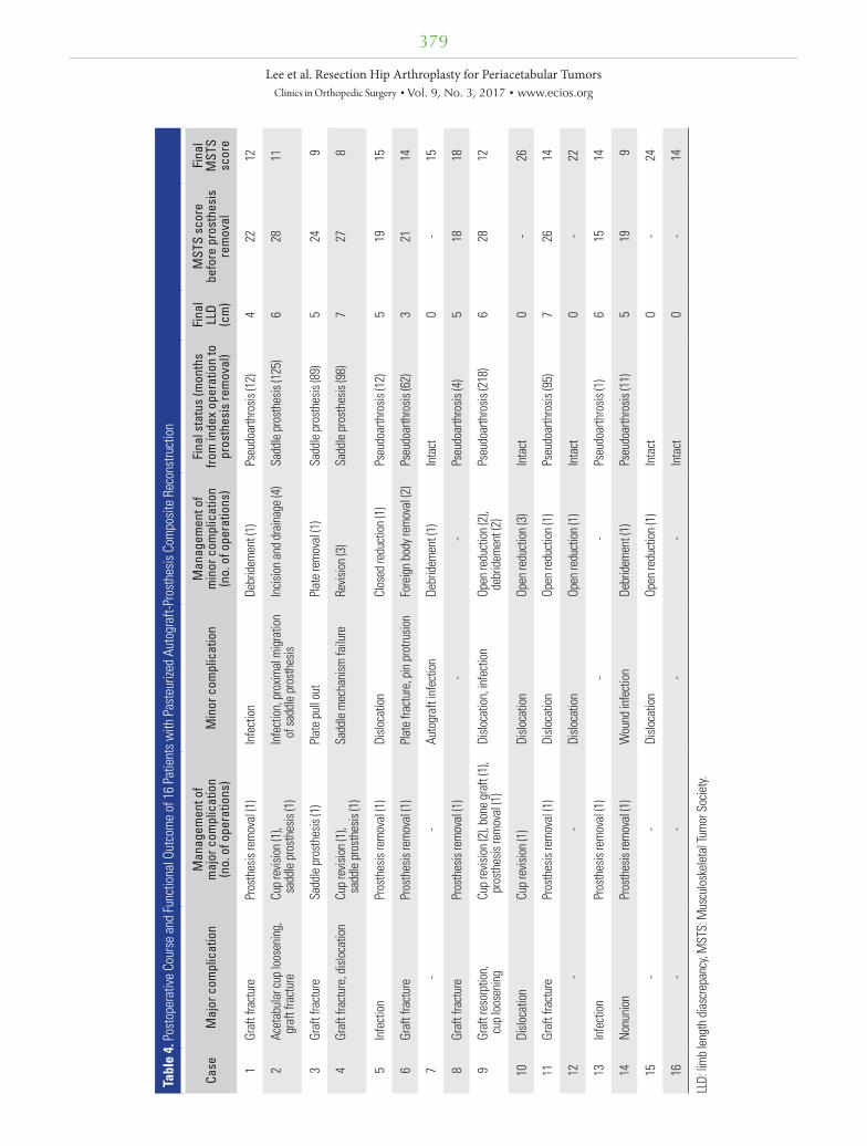
Tabl
e 4.
Pos
tope
rativ
e Co
urse
and
Func
tiona
l Out
com
e of
16
Patie
nts w
ith P
aste
urize
d Au
togr
aft-P
rost
hesis
Com
posit
e Re
cons
truct
ion
Case
Maj
or c
ompl
icat
ion
Man
agem
ent o
f m
ajor
com
plic
atio
n
(no.
of o
pera
tions
)M
inor
com
plic
atio
nM
anag
emen
t of
min
or c
ompl
icat
ion
(n
o. o
f ope
ratio
ns)
Fina
l sta
tus
(mon
ths
from
inde
x op
erat
ion
to
pros
thes
is re
mov
al)
Fina
l LL
D
(cm
)
MST
S sc
ore
be
fore
pro
sthe
sis
re
mov
al
Fina
l M
STS
scor
e
1Gr
aft f
ract
ure
Pros
thes
is re
mov
al (1
)In
fect
ion
Debr
idem
ent (
1)Ps
eudo
arth
rosis
(12)
422
12
2Ac
etab
ular
cup
loos
enin
g,
graf
t fra
ctur
eCu
p re
visio
n (1
),
sadd
le p
rost
hesis
(1)
Infe
ctio
n, p
roxim
al m
igra
tion
of sa
ddle
pro
sthe
sis
Incis
ion
and
drai
nage
(4)
Sadd
le p
rost
hesis
(125
)6
2811
3Gr
aft f
ract
ure
Sadd
le p
rost
hesis
(1)
Plat
e pu
ll ou
t Pl
ate
rem
oval
(1)
Sadd
le p
rost
hesis
(89)
524
9
4Gr
aft f
ract
ure,
disl
ocat
ion
Cup
revis
ion
(1),
sa
ddle
pro
sthe
sis (1
)Sa
ddle
mec
hani
sm fa
ilure
Re
visio
n (3
)Sa
ddle
pro
sthe
sis (9
8)7
27 8
5In
fect
ion
Pros
thes
is re
mov
al (1
)Di
sloca
tion
Clos
ed re
duct
ion
(1)
Pseu
doar
thro
sis (1
2)5
1915
6Gr
aft f
ract
ure
Pros
thes
is re
mov
al (1
)Pl
ate
fract
ure,
pin
pro
trusio
nFo
reig
n bo
dy re
mov
al (2
) Ps
eudo
arth
rosis
(62)
321
14
7-
-Au
togr
aft i
nfec
tion
Debr
idem
ent (
1)In
tact
0-
15
8Gr
aft f
ract
ure
Pros
thes
is re
mov
al (1
)-
-Ps
eudo
arth
rosis
(4)
518
18
9Gr
aft r
esor
ptio
n,
cup
loos
enin
gCu
p re
visio
n (2
), bo
ne g
raft
(1),
pros
thes
is re
mov
al (1
)Di
sloca
tion,
infe
ctio
nOp
en re
duct
ion
(2),
de
brid
emen
t (2)
Pseu
doar
thro
sis (2
18)
628
12
10Di
sloca
tion
Cup
revis
ion
(1)
Dislo
catio
nOp
en re
duct
ion
(3)
Inta
ct0
-26
11Gr
aft f
ract
ure
Pros
thes
is re
mov
al (1
)Di
sloca
tion
Open
redu
ctio
n (1
)Ps
eudo
arth
rosis
(95)
726
14
12-
-Di
sloca
tion
Open
redu
ctio
n (1
)In
tact
0-
22
13In
fect
ion
Pros
thes
is re
mov
al (1
)-
-Ps
eudo
arth
rosis
(1)
615
14
14No
nuni
onPr
osth
esis
rem
oval
(1)
Wou
nd in
fect
ion
Debr
idem
ent (
1)Ps
eudo
arth
rosis
(11)
519
9
15-
-Di
sloca
tion
Open
redu
ctio
n (1
)In
tact
0-
24
16-
--
-In
tact
0-
14
LLD:
lim
b le
ngth
dia
scre
panc
y, M
STS:
Mus
culo
skel
etal
Tum
or S
ocie
ty.

380
Lee et al. Resection Hip Arthroplasty for Periacetabular TumorsClinics in Orthopedic Surgery • Vol. 9, No. 3, 2017 • www.ecios.org
Tabl
e 5.
Pos
tope
rativ
e Co
urse
and
Func
tiona
l Out
com
e of
24
Patie
nts w
ith R
esec
tion
Hip
Arth
ropl
asty
Case
Oste
otom
y le
vel
from
ilia
c cr
est
(cm
)Fi
xatio
n (fe
mor
al
head
to il
ium
)Po
stop
erat
ive
imm
obili
zatio
nBo
ny n
eo-a
ceta
bulu
m/ti
me
to
neo-
acet
abul
um fo
rmat
ion
(m
o)Co
mpl
icat
ion
Lim
b le
ngth
di
scre
panc
y (c
m)
Shoe
hee
l he
ight
(cm
)W
alki
ng
aid
MST
S sc
ore
110
.5No
ne*
None
Com
plet
e/9
-2.
6No
neNo
ne28
29
None
None
Com
plet
e/13
-3
1No
ne25
311
Wire
Hip
spica
cast
None
/NA
-3
1.5
None
24
414
.5No
neNo
nePa
rtial
/8-
1.3
None
None
28
510
.7W
ireNo
nePa
rtial
/6-
31.
5No
ne25
610
.7No
neNo
nePa
rtial
/7Re
-exp
lora
tion
3.3
2No
ne25
78.
2No
neNo
nePa
rtial
/6-
2.4
None
None
24
89.
2No
ne*
None
Com
plet
e/6
-3.
12
None
26
99.
3W
ire*
None
Parti
al/7
-4.
33
1 Ca
ne19
1010
None
*No
nePa
rtial
/6-
3.5
2No
ne25
1110
None
*No
nePa
rtial
/5Fla
p ne
cros
is2.
4No
neNo
ne27
129.
3W
ire*
None
Com
plet
e/4
-2
None
None
28
1310
.5W
ireHi
p sp
ica ca
stNo
ne/N
A-
4.7
31
Cane
19
1411
.2W
ireNo
neCo
mpl
ete/
7-
4.7
3No
ne25
159
Wire
*No
neCo
mpl
ete/
6-
4.5
3No
ne26
169
None
None
None
/NA
-5.
83
1 Ca
ne19
179.
4W
ire*
None
Com
plet
e/6
-2.
5No
neNo
ne27
189.
6W
ireHi
p sp
ica ca
stPa
rtial
/13
-5.
23
None
22
196.
6W
ire*
None
None
/NA
Chro
nic p
ain
6.4
31
Crut
ch17
209
None
None
None
/NA
-6
41
Crut
ch25
218.
8W
ire*
None
None
/NA
-4.
53
1 Ca
ne18
224
None
*No
neNo
ne/N
A-
5.5
31
Crut
ch15
2310
Wire
*No
nePa
rtial
/5
Re-e
xplo
ratio
n2.
4No
neNo
ne24
248.
8No
neNo
neNo
ne/N
A-
3.7
2No
ne23
MST
S: M
uscu
losk
elet
al T
umor
Soc
iety,
NA:
not
ass
esse
d, R
e-ex
plor
atio
n: d
urin
g fo
llow
-up,
exp
lora
tion
of a
lesio
n su
spici
ous
of lo
cal r
ecur
renc
e on
mag
netic
reso
nanc
e im
agin
g tu
rned
out
to b
e a
new
bon
e in
ne
o-ac
etab
ulum
form
atio
n.*D
urin
g su
rger
y, 2
or 3
ilia
c win
g os
teot
omie
s wer
e ca
rried
out
to p
rese
rve
the
iliac
cres
t-hip
flex
or o
r abd
ucto
r mus
cle co
ntin
uity.

381
Lee et al. Resection Hip Arthroplasty for Periacetabular TumorsClinics in Orthopedic Surgery • Vol. 9, No. 3, 2017 • www.ecios.org
minor complication was defined as a problem other than those described above, which necessitated an additional surgical procedure or conservative management. Demo-graphic and treatment variables in the 2 study groups were compared using the t-test and Fisher exact test. Analyses were performed using SPSS ver. 13.0 (SPSS Inc., Chicago, IL, USA), and p-values less than 0.05 were considered sig-nificant.
RESULTS
Compared to the 16 cases of PPC reconstruction, the 24 cases of RHA showed lower major and minor complica-tion rates (p < 0.001), shorter surgical time (p < 0.001), and superior MSTS score (p < 0.001) (Table 3). No pa-tients who underwent RHA experienced disruption of the femoral head-iliac wing articulation. However, 11 of the 16 PPCs (69%) were removed at an average of 66 months
(range, 1 to 218 months). Causes of PPC failure were graft fracture in 8, infection in 2, and nonunion in 1. The 11 failed PPCs were converted to pseudoarthrosis in 8 and a saddle prosthesis in 3. Minor complications in patients who used PPC included wound infection, plate failure, and dislocation. Overall, 16 patients who used PPC underwent 17 major and 24 minor additional procedures after the index operation (Table 4). The average functional score of the 11 failed PPC patients was 22.4 (74%) until removal of the construct, and their average score deteriorated to 12.4 (41%) after removal. Mean leg length discrepancy of the 11 patients with failed PPC was 5.4 cm (range, 3 to 7 cm). One each RHA case showed flap necrosis and chronic pain. Another 2 patients who had RHA underwent re-ex-ploration for suspicious recurrent lesions; however, these proved to be new bones in neo-acetabulum formation.
Of the 24 patients who underwent RHA, circular bony neo-acetabulum on CT was identified in 7 and par-
A B C
Fig. 2. (A) The preoperative plain radiograph shows a mixed osteolytic and sclerotic lesion in the right ilium and acetabulum in a 34-year-old male patient with chondrosarcoma (case 8). (B) The postoperative plain radiograph shows Enneking type II + I (partial) resection and repositioning of the previously detached iliac bone block-muscle complex with wire. (C) The follow-up plain radiograph shows complete neo-hip joint formation; the patient is fully active with shortening by 3 cm.
A B C
Fig. 3. (A) The plain radiograph shows an osteolytic lesion in the right acetabulum in a 41-year-old patient with chondrosarcoma (case 21). (B) The postoperative radiograph demonstrates Enneking type I (partial) + II + III (partial) resection and the femoral head fixed to the remaining iliac wing with a single wire. (C) At 6 months postoperatively, because of the high iliac osteotomy level, only partial bony neo-acetabulum had formed. The patient had no pain and could walk with one cane.

382
Lee et al. Resection Hip Arthroplasty for Periacetabular TumorsClinics in Orthopedic Surgery • Vol. 9, No. 3, 2017 • www.ecios.org
tial neo-acetabulum in 9; the remaining 8 patients did not show a bony acetabular structure (Table 5). Average time to bony neo-acetabulum formation was 7 months (range, 4 to 13 months) (Fig. 2). Excluding 2 patients who had a hip spica cast postoperatively, all 13 patients with an osteotomy > 9 cm from the iliac crest showed partial or complete bony neo-acetabulum formation, while only 3 of 9 patients with osteotomy level < 9 cm demonstrated bony neo-acetabulum (Fig. 3). The average MSTS functional score in 9 patients with < 9 cm of the remaining iliac wing was 21 (70%), while that of 15 patients with > 9 cm of the ilium was 25 of 30 points (83%). Average limb shortening in 24 patients who underwent RHA was 3.7 cm (range, 1.3 to 6.4 cm).
DISCUSSION
Excision of periacetabular tumors usually leaves a large skeletal defect, and attempts at reconstruction by arthrod-esis or pseudoarthrosis often result in considerable limb shortening and poor function.17) In this regard, anatomical reconstruction of the hip and hemipelvis by biological or mechanical means were suggested to provide improved functional outcomes and walking ability.1,11,14,27-29) How-ever, most reconstructions had high complication and fail-ure rates. Therefore, a strategy of resection alone has been revisited.6,7,23,28,30) In our comparative study of anatomical reconstruction and RHA, we confirmed that RHA is a reli-able primary procedure for periacetabular tumors, with low complication rates, good functional results, and short surgical time. Moreover, in patients who underwent RHA, less iliac wing resection and early postoperative mobiliza-tion seemed to facilitate early stable bony neo-acetabulum formation.
This study has several limitations. First, there are many confounding factors in relatively small comparative cohort groups. We acknowledge the heterogeneity due to fac-tors such as the amount of bone and soft tissue resection, differences in postoperative management, use of chemo-therapy, and the nonrandomized choice of reconstruction type. In addition, because we compared our recent cases of RHA with past PPC reconstruction cases, improvement in surgical skill may have influenced the complication rate. However, between 2 groups, no differences were found in tumor size, pathologic diagnosis, resection type, local recurrence, or metastasis rate. Furthermore, the high pro-portion of male patients in the RHA group may be related with the superior functional outcome. However, this factor cannot offset the time-related failure pattern in the PPC group. Tabl
e 6.
Com
paris
on w
ith P
revio
us S
tudi
es
Stud
yN
o. o
f pa
tient
sTy
pe o
f pel
vic
reco
nstru
ctio
n (n
)De
ep in
fect
ion
(%)
Ampu
tatio
n du
e to
in
fect
ion
(%)
Reco
nstru
ctio
n
failu
re (%
)Lo
cal
recu
rren
ce (%
)M
ean
MST
S
scor
e/LL
D (c
m)
Curre
nt se
ries
40PP
C (1
6)RH
A (2
4) 2
/16
(12)
00 0
11/
16 (6
9)0
3/1
6 (1
8) 6
/24
(25)
14.8
/3.7
23.5
/3.7
Hu e
t al.
(201
2)23
) 27
RHA
00
00
22.6
/5
Ange
lini e
t al.
(201
4)28
)27
0No
reco
nstru
ctio
n (1
33)
Allo
graf
t (57
)AP
C (5
9)Pe
lvic p
rost
hesis
(21)
20/
133
(15)
16/
57 (2
8) 1
2/59
(20)
7/2
1 (3
3)
5/5
5 (9
)16
/137
(11)
Rec
onst
ruct
ion
rem
oved
for i
nfec
tion
NANA
Gebe
rt et
al.
(201
1)1)
62Hi
p tra
nspo
sitio
n 2
0/62
(32)
1/6
2 (1
)25
/62
(40)
Had
revis
ion
6/6
2 (9
.6)
18.6
/5
Dona
ti et
al.
(201
1)11
)35
APC
8/3
5 (2
2)0
15/
35 (4
2) 3
/35
(8.5
) 21
.6/N
A
Jaisw
al e
t al.
(200
8)7)
98Pe
lvic p
rost
hesis
17/
98 (1
8) 1
/98
(1)
22/
98 (2
3.7)
29/
98 (3
1)59
.4%
(TES
S)
MST
S: M
uscu
losk
elet
al Tu
mor
Soc
iety,
LLD:
leg
leng
th d
iscre
panc
y, PP
C: p
aste
urize
d au
togr
aft-p
rost
hesis
com
posit
e, R
HA: r
esec
tion
hip
arth
ropl
asty,
APC
: allo
graf
t pro
sthe
sis co
mpo
site,
NA:
not
ass
esse
d, T
ESS:
To
ront
o Ex
trem
ity S
alva
ge S
core
.

383
Lee et al. Resection Hip Arthroplasty for Periacetabular TumorsClinics in Orthopedic Surgery • Vol. 9, No. 3, 2017 • www.ecios.org
Our comparative study and previous reports show that RHA is a valuable primary procedure after periace-tabular resection, with much reduced complication rates or need for further surgery (Table 6). Chronologically, patients with pelvic resection face 2 major complications: infection and mechanical failure. Infection is a devastat-ing event that may lead to removal of the reconstruction hardware or hindquarter amputation. However, pelvic reconstruction was also reported as an independent con-tributory factor to infection.28) Lower rates of infection in the resection alone group may be explained by the shorter operative time, no foreign body, and reduced dead space by permitting proximal migration of the femoral head. Ensuing problems are mechanical, and include nonunion or fracture of biologic material and loosening or breakage of the prosthesis.7,28) These late mechanical complications also necessitate the removal of the construct in a sub-stantial proportion of patients, and the functional results of failed cases after intervention are worse than those of primary RHA. Conceptually, because either RHA or failed reconstruction is a pseudoarthrosis, failed reconstruction is assumed to have a functional score similar to that of resection alone. However, 2 factors are related to superior outcome of RHA. One critical factor is the integrity of the femoral head. Patients with failed reconstruction invari-ably lose the femoral head, and this leads to an additional shortening of around 5 cm (the usual height of the femoral head), compared to RHA patients. Moreover, this loss of femoral head precludes the development of neo-acetab-ulum. In this regard, in patients who can accept initial shortening of the affected limb, RHA would be a valuable procedure with long-term durability and low risk of com-plications.
RHA after periacetabular resection is not new. As early as 1978, one study reported the procedure as a satis-factory substitute for hindquarter amputation in 5 patients with chondrosarcoma.2) Since then, several series have reported the usefulness of RHA after pelvic resection with some variation in technique and concept.18,20,30) However, for better results, there are points to consider with regard to optimal indications, surgical technique, and postopera-tive care. To create a bony or fibrous “neo hip joint,” the femoral head should not be involved by the tumor and a substantial portion of the iliac wing should be saved. Pa-
tients with less iliac wing resection (preferably iliac osteot-omy level > 9 cm from the iliac crest) show minimal limb shortening and an increased percentage of bony neo-ace-tabulum formation. At the time of surgical approach, the origins of hip abductors and hip flexors are detached in-feriorly and laterally through osteotomies made along the iliac crest and anterior iliac spine. This approach seems to facilitate repair after resection and functional recovery. To maintain the iliac wing-femoral head contact and to mini-mize the external rotation of the femoral head, a single wire was tied between the femoral head and ilium in half of our patients. However, as the case number increased, we found that this wire fixation was not necessary. In a pre-vious report, to control the location of the femoral head postoperatively, skin traction with rotation-proof shoes or skeletal traction was applied (ambulation was started 4–6 weeks later).23) In our series, except for the 3 early cases with postoperative hip spica cast, all patients were encour-aged to ambulate around 2 weeks after surgery. Early ac-tive exercise seemed to promote the formation of a neo-hip joint. In active young patients, walking without aid and neo-hip joint formation was observed around 6 months postoperatively; however, at older ages, independent walk-ing took up to 1 year. The average MSTS functional score in patients with neo-acetabulum formation was 25.3, while that of patients without neo-acetabulum was 20.1.
In conclusion, our comparative study confirmed that RHA for periacetabular tumors can be an excellent alternative to anatomical reconstruction. RHA offers a short surgical time, low complication rates, and functional results comparable to those of other reconstruction meth-ods. However, this procedure is indicated for patients who can accept some shortening of the limb, and the tumor should be confined to the periacetabular area.
CONFLICT OF INTEREST
No potential conflict of interest relevant to this article was reported.
ACKNOWLEDGEMENTS
The authors thank Ji Young Yoo, MD for radiological eval-uation in this study.
REFERENCES
1. Gebert C, Wessling M, Hoffmann C, et al. Hip transposition as a limb salvage procedure following the resection of peri-
acetabular tumors. J Surg Oncol. 2011;103(3):269-75.
2. Steel HH. Partial or complete resection of the hemipelvis:

384
Lee et al. Resection Hip Arthroplasty for Periacetabular TumorsClinics in Orthopedic Surgery • Vol. 9, No. 3, 2017 • www.ecios.org
an alternative to hindquarter amputation for periacetabu-lar chondrosarcoma of the pelvis. J Bone Joint Surg Am. 1978;60(6):719-30.
3. Fuchs B, O'Connor MI, Kaufman KR, Padgett DJ, Sim FH. Iliofemoral arthrodesis and pseudarthrosis: a long-term functional outcome evaluation. Clin Orthop Relat Res. 2002;(397):29-35.
4. Aljassir F, Beadel GP, Turcotte RE, et al. Outcome after pel-vic sarcoma resection reconstructed with saddle prosthesis. Clin Orthop Relat Res. 2005;438:36-41.
5. Satcher Jr RL, O'Donnell RJ, Johnston JO. Reconstruction of the pelvis after resection of tumors about the acetabulum. Clin Orthop Relat Res. 2003;(409):209-17.
6. Guo W, Li D, Tang X, Ji T. Surgical treatment of pelvic chondrosarcoma involving periacetabulum. J Surg Oncol. 2010;101(2):160-5.
7. Jaiswal PK, Aston WJ, Grimer RJ, et al. Peri-acetabular re-section and endoprosthetic reconstruction for tumours of the acetabulum. J Bone Joint Surg Br. 2008;90(9):1222-7.
8. Tsuchiya H, Wan SL, Sakayama K, Yamamoto N, Nishida H, Tomita K. Reconstruction using an autograft contain-ing tumour treated by liquid nitrogen. J Bone Joint Surg Br. 2005;87(2):218-25.
9. Jeon DG, Kim MS, Cho WH, Song WS, Lee SY. Reconstruc-tion with pasteurized autograft-total hip prosthesis compos-ite for periacetabular tumors. J Surg Oncol. 2007;96(6):493-502.
10. Langlais F, Lambotte JC, Thomazeau H. Long-term results of hemipelvis reconstruction with allografts. Clin Orthop Relat Res. 2001;(388):178-86.
11. Donati D, Di Bella C, Frisoni T, Cevolani L, DeGroot H. Alloprosthetic composite is a suitable reconstruction after periacetabular tumor resection. Clin Orthop Relat Res. 2011;469(5):1450-8.
12. Enneking WF, Dunham WK. Resection and reconstruction for primary neoplasms involving the innominate bone. J Bone Joint Surg Am. 1978;60(6):731-46.
13. Aboulafia AJ, Buch R, Mathews J, Li W, Malawer MM. Re-construction using the saddle prosthesis following excision of primary and metastatic periacetabular tumors. Clin Or-thop Relat Res. 1995;(314):203-13.
14. Bell RS, Davis AM, Wunder JS, Buconjic T, McGoveran B, Gross AE. Allograft reconstruction of the acetabulum after resection of stage-IIB sarcoma: intermediate-term results. J Bone Joint Surg Am. 1997;79(11):1663-74.
15. Delloye C, Banse X, Brichard B, Docquier PL, Cornu O. Pelvic reconstruction with a structural pelvic allograft after resection of a malignant bone tumor. J Bone Joint Surg Am.
2007;89(3):579-87.
16. Ozaki T, Hoffmann C, Hillmann A, Gosheger G, Lindner N, Winkelmann W. Implantation of hemipelvic pros-thesis after resection of sarcoma. Clin Orthop Relat Res. 2002;(396):197-205.
17. Abudu A, Grimer RJ, Cannon SR, Carter SR, Sneath RS. Reconstruction of the hemipelvis after the excision of ma-lignant tumours: complications and functional outcome of prostheses. J Bone Joint Surg Br. 1997;79(5):773-9.
18. Hillmann A, Hoffmann C, Gosheger G, Rodl R, Winkel-mann W, Ozaki T. Tumors of the pelvis: complications after reconstruction. Arch Orthop Trauma Surg. 2003;123(7):340-4.
19. Ozaki T, Hillmann A, Bettin D, Wuisman P, Winkelmann W. High complication rates with pelvic allografts: experience of 22 sarcoma resections. Acta Orthop Scand. 1996;67(4):333-8.
20. Hoffmann C, Gosheger G, Gebert C, Jurgens H, Winkel-mann W. Functional results and quality of life after treat-ment of pelvic sarcomas involving the acetabulum. J Bone Joint Surg Am. 2006;88(3):575-82.
21. Griesser MJ, Gillette B, Crist M, et al. Internal and exter-nal hemipelvectomy or flail hip in patients with sarcomas: quality-of-life and functional outcomes. Am J Phys Med Rehabil. 2012;91(1):24-32.
22. Kusuzaki K, Shinjo H, Kim W, Nakamura S, Murata H, Hi-rasawa Y. Resection hip arthroplasty for malignant pelvic tumor: outcome in 5 patients followed more than 2 years. Acta Orthop Scand. 1998;69(6):617-21.
23. Hu YC, Huang HC, Lun DX, Wang H. Resection hip ar-throplasty as a feasible surgical procedure for periacetabular tumors of the pelvis. Eur J Surg Oncol. 2012;38(8):692-9.
24. Enneking WF, Spanier SS, Goodman MA. A system for the surgical staging of musculoskeletal sarcoma. Clin Orthop Relat Res. 1980;(153):106-20.
25. Bieling P, Rehan N, Winkler P, et al. Tumor size and prog-nosis in aggressively treated osteosarcoma. J Clin Oncol. 1996;14(3):848-58.
26. Enneking WF, Dunham W, Gebhardt MC, Malawar M, Pritchard DJ. A system for the functional evaluation of reconstructive procedures after surgical treatment of tu-mors of the musculoskeletal system. Clin Orthop Relat Res. 1993;(286):241-6.
27. Guo W, Li D, Tang X, Yang Y, Ji T. Reconstruction with modular hemipelvic prostheses for periacetabular tumor. Clin Orthop Relat Res. 2007;461:180-8.
28. Angelini A, Drago G, Trovarelli G, Calabro T, Ruggieri P. Infection after surgical resection for pelvic bone tumors: an analysis of 270 patients from one institution. Clin Orthop Relat Res. 2014;472(1):349-59.

385
Lee et al. Resection Hip Arthroplasty for Periacetabular TumorsClinics in Orthopedic Surgery • Vol. 9, No. 3, 2017 • www.ecios.org
29. Ueda T, Kakunaga S, Takenaka S, Araki N, Yoshikawa H. Constrained total hip megaprosthesis for primary periace-tabular tumors. Clin Orthop Relat Res. 2013;471(3):741-9.
30. Carmody Soni EE, Miller BJ, Scarborough MT, Parker Gibbs C. Functional outcomes and gait analysis of patients after periacetabular sarcoma resection with and without ischiofe-moral arthrodesis. J Surg Oncol. 2012;106(7):844-9.