Embed Size (px)
Citation preview
188
Original Article
Journal of Lasers in Medical Sciences Volume 5 Number 4 Autumn 2014
Comparison of Alexandrite and Diode Lasers for Hair Removal in Dark and Medium Skin: Which is Better?
Farhad Hamad Mustafa1, Mohamad Suhimi Jaafar2, Asaad Hamid Ismail3, Kussay Nugamesh Mutter4
1Medical Physics and Radiation Science, College of Pharmacy, Hawler Medical University, 44002 Erbil, Iraq2Medical Physics and Laser Research Group, School of Physics, Universiti Sains Malaysia, 11800 Penang, Malaysia
3Radiation Science and Medical Physics, Education College, Physics Department, Salahaddin University, 44002 Erbil, Iraq4Engineering Physics, School of Physics, Universiti Sains Malaysia, 11800 Penang, Malaysia
Abstract:
Introduction: To improve laser hair removal (LHR) for dark skin, the fluence rate reaching the hair follicle in LHR is important. This paper presents the results of a comparative study examining the function of wavelength on dark skin types using 755 nm alexandrite and 810 nm diode lasers.Methods: The structure of the skin was created using a realistic skin model by the Advanced Systems Analysis Program.Result: In this study, the alexandrite laser (755 nm) and diode laser (810 nm) beam–skin tissue interactions were simulated. The simulation results for both lasers differed. The transmission ratio of the diode laser to the dark skin dermis was approximately 4% more than that of the alexandrite laser for the same skin type. For the diode laser at skin depth z = 0.67 mm, the average transmission ratios of both samples were 36% and 27.5%, but those for the alexandrite laser at the same skin depth were 32% and 25%.Conclusion: Both lasers were suitable in LHR for dark skin types, but the diode laser was better than the alexandrite laser because the former could penetrate deeper into the dermis layer.Keywords: lasers; simulation; skin; diode laser
IntroductionGiven the potential use of lasers in various medical
applications, including laser hair removal (LHR) and orthopedic treatments, the mechanism of laser-tissue interaction has been widely studied. LHR is currently the most commonly requested cosmetic procedure in the world, particularly for female clients. Thirty years ago, the ability of lasers to damage hair follicles was noted1. Physicians involved in simulation investigations need to assure that the fluence rates of the alexandrite and diode lasers for hair removal in deep parts of the skin have an effect on situations leading to skin darkening. Managing human skin in a cosmetic center for investigational
purposes on the laser fluence rate is difficult, so different types of phantoms are used to simulate the properties of tissue. Skin-simulating phantoms have been developed to analyze bio-optical instrumentation and techniques for several purposes. Data reported in previous papers2-4 indicated that the computer model can be used to accurately calculate the light fluence rate even at the deepest parts of the skin layer. Kareten reported that the epidermal contents affect the fluence rate entering the skin depth3.
Various simulations and modalities of light-based technology are currently used in the field of laser skin interaction. These simulations include MCL5, Monte Carlo, and Advanced Systems Analysis Program
Please cite this article as follows:Mustafa FH, Jaafar MS, Ismail AH, Mutter KN. Comparison of Alexandrite and Diode Lasers for Hair Removal in Dark and Medium Skin: Which is Better? J Lasers Med Sci 2014;5(4):188-93
Corresponding Author: Farhad Hamed Mustafa, PhD; Medical Physics and Radiation Science, College of Pharmacy, Hawler Medical University, 44002 Erbil, Iraq. Tel: +964-7504514644; Fax: +964- 662273382; E-mail: [email protected]
Alexandrite and Diode Lasers in Hair Removal
189Journal of Lasers in Medical Sciences Volume 5 Number 4 Autumn 2014
(ASAP®) software.LHR system is the most effective for people who have
light skin and dark hair. A recent study reported that both the skin color and hair color influence the success of LHR5,6. As of this writing, few studies have evaluated patient satisfaction and complications after LHR among people of color7-11. The penetration of laser beam through human skin for hair removal is extremely dependent on the optical properties of skin12. The absorption coefficient of the epidermis varies with the volume fraction of melanosomes and amount of eumelanin concentration in the epidermis3. The absorption and scattering of the light in the skin layers determine the fluence rate of the light reaching the intended treatment site. To effectively destroy the target, losses caused by reflection, scattering and absorption must be considered4. However, the idea of laser fluence rate for hair removal, including the appropriate approaches for dark skin types and laser sources, has not yet been optimized with the simulation method.
Different types of lasers mainly vary in terms of wavelength13; different laser wavelengths target different skin issues. Therefore, various lasers are needed to treat a variety of skin concerns5,14,15. An investigation of different laser wavelengths for hair removal should be conducted to address all the problems that these procedures may have. An explanation of the differences between these different laser types may be very lengthy, technical, and rather confusing, so we focused on the diode and alexandrite lasers as optimal options for LHR of candidates with dark skin types. The fluence rates of the alexandrite and diode lasers in dark skin were compared using computational simulated skin from the Realistic Skin Model (RSM) part of the ASAP® software from Breault Research.
Methodology
1. Implementation of the simulation model by Advanced Systems Analysis Program (ASAP)
Biomedical researchers and optical engineers have long used modeling software to efficiently develop new products and applications16,17. The ASAP program is the gold standard for simulation in the present study. The version of ASAP V1R1 2009 creates realistic tissue phantoms for the investigation of the optical properties of skin. This ASAP version is a new technique in the optical simulation program that permits the simulation of photon propagation and power density recording of laser in the skin layers17,18. This technique is also useful for predicting radiation transfer and fluence rate delivery into the target17. This model is roughly divided into four major parts. Part I describes the principles of ASAP simulations of building system, units, and the sampling model, and how the simulation can be realized in software. Part II provides detailed instructions for creating, using, and modifying a source. Part III creates ray tracing and some computational results and verifications, and exhibits the analysis of results. Part IV, the final part of the model, introduces some basic analytical tools and concepts. This part is generally the most interesting step in the modeling procedure because questions about the optical behavior of the model can be answered in this step.
2. Creating the skin
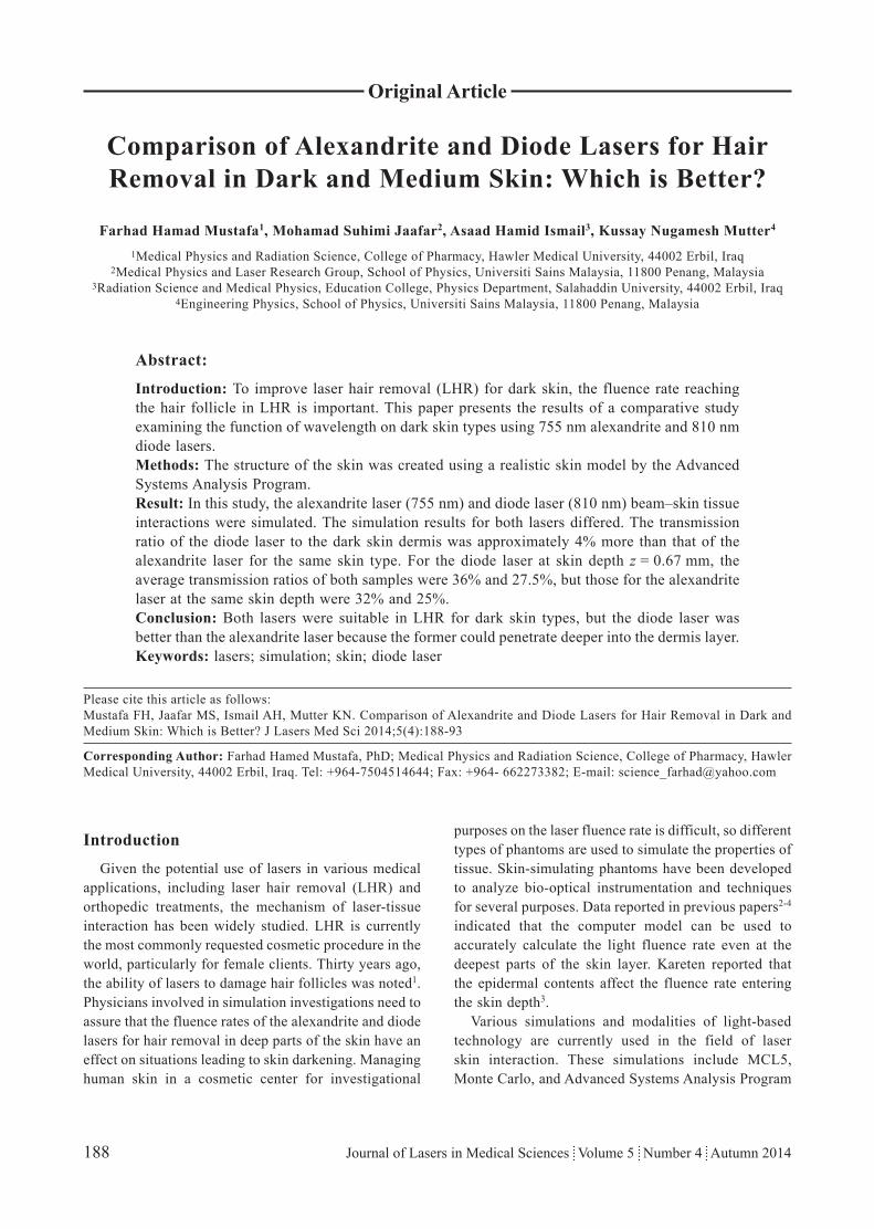
Figure 1 shows the geometry of the RSM. The arbitrary part of the human skin is chosen, and the area of the skin is 100 mm2. This area is modeled as a three-dimensional
Figure 1. Model specifying the layers of skin, laser beam, hair parameter, and VOXEL element.
Alexandrite and Diode Lasers in Hair Removal
190 Journal of Lasers in Medical Sciences Volume 5 Number 4 Autumn 2014
medium divided into four layers. The details of the skin structure are presented in Table 1. The first layer is the stratum corneum, a 0.01 mm-thick top layer mainly containing keratin and dead cells. The second layer is the epidermis, a 0.0875 mm-thick layer that mainly contains living cells. Aside from containing primarily living cells, this layer holds a fraction of chromophore and melanin. The third layer is the dermis, a 1.8 mm-thick layer mainly containing oxyhemoglobin and deoxyhemoglobin. The fourth layer is the hypodermis, a 3 mm-thick layer containing fat cells.
In this study, the only difference between the skin samples was the volume fraction of melanosomes and melanin concentrations in the epidermal layer. Differences in the epidermal thickness were not explicitly considered. Two types of skin were created by changing the volume fraction of melanosomes in the epidermis (Table 1). The RSM parameters for the eumelanin and pheomelanin concentrations in the epidermis were 80 and 12 g/L, respectively. The eumelanin and pheomelanin concentrations were maintained at the above values for both skin types.
Table 1 shows a detailed description of the parameter input of chromophore concentrations for skin layers. In this study, the parameter input methods of using default parameters and visual characteristics in ASAP software library were applied (Table 1).
3. Hair modeling
Hair modeling, which is part of the RSM in the ASAP software, was used to create and model hair on the skin surface. Two types of skin were created in this study: medium and dark skin with the same hair density (50 no. hair cm−2), same hair diameter (0.1 mm), same hair color (light brown), and same hair angle and length (60° and 5 mm) (Figure 1).
4. Laser sources
Diode and alexandrite lasers with wavelengths of 810 and 755 nm, respectively, were used in this study. An irradiation power of 1000 mW with a beam diameter of 5 mm was used in the model. The distance between the laser beam and target was set to 5 mm. An irradiation beam with ray traces of 1,000,000 rays transferred the power from the source to the skin (Figure 1). The fluence rate of the laser was obtained with a VOXEL (volume picture or pixel elements) command. VOXEL in ASAP was used to capture the energy that passed through the volume of the skin.
The simulation model was applied on both types of skin. We applied both the alexandrite and diode lasers according to the same power and spot diameters, and the fluence rates were calculated in different depths of skin layers. For the first simulation, the alexandrite laser at 755 nm was used for dark and medium skin. For the second simulation, the diode laser at 810 nm was used for medium and dark skin.
Results and discussion
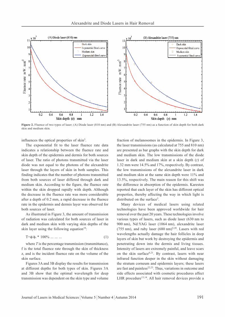
The data and fitting results of the two types of laser sources for the two types of skin are shown in Figure 2. One case shows the diode laser, and the other case shows the alexandrite laser. Simulations of the transmitted diode laser and alexandrite laser from the layers of skin with two different types of skin were performed. Photons penetrated into the skin layer and advanced randomly. In each layer of skin, the photon was absorbed by the chromophore concentrations. The results show that the fluence rate was influenced by both the wavelength and composition of the tissue. Given that melanin and water have a wavelength-dependent absorption coefficient, the distribution of these components (melanin and water)
Skin layers Stratum corneum Epidermis Dermis HypodermisIndex of refraction 1.55 1.5 1.4 1.44An isotropic factor 0.9 0.79 0.82 0.9Thickness (mm) 0.015 0.0875 1.8 3Eumelanin concentrations (g/L) — 80 — —Pheomelanin concentrations (g/L) — 12 — —Volume fraction of water 0.13 0.67 0.8 —Volume fraction of oxyhemoglobin — — 0.72 —Volume fraction of blood in dermis — — 0.01 —Hemoglobin blood concentration in dermis (g/L) — — 150 —Beta carotene concentrations (g/L) 0.00021 0.00021 0.00007 —Bilirubin concentrations (g/L) — — 0.005 —
Table 1. Optical properties and chromophore concentrations for the different layers in the model.
Alexandrite and Diode Lasers in Hair Removal
191Journal of Lasers in Medical Sciences Volume 5 Number 4 Autumn 2014
influences the optical properties of skin3.The exponential fit to the laser fluence rate data
indicates a relationship between the fluence rate and skin depth of the epidermis and dermis for both sources of laser. The ratio of photons transmitted via the laser diode was not equal to the photons of the alexandrite laser through the layers of skin in both samples. This finding indicates that the number of photons transmitted from both sources of laser differed through dark and medium skin. According to the figure, the fluence rate within the skin dropped rapidly with depth. Although the decrease in the fluence rate was more considerable after a depth of 0.2 mm, a rapid decrease in the fluence rate in the epidermis and dermis layer was observed for both sources of laser.
As illustrated in Figure 3, the amount of transmission of radiation was calculated for both sources of laser in dark and medium skin with varying skin depths of the skin layer using the following equation19:
T=ϕ/ϕ° * 100% ... ... ... (1)
where T is the percentage transmission (transmittance), f is the total fluence rate through the skin of thickness z, and is the incident fluence rate on the volume of the skin surface.
Figures 3A and 3B display the results for transmission at different depths for both types of skin. Figures 3A and 3B show that the optimal wavelength for deep transmission was dependent on the skin type and volume
fraction of melanosomes in the epidermis. In Figure 3, the laser transmissions (as calculated at 755 and 810 nm) are presented as bar graphs with the skin depth for dark and medium skin. The low transmissions of the diode laser in dark and medium skin at a skin depth (z) of 1.32 mm were 14.5% and 17%, respectively. By contrast, the low transmissions of the alexandrite laser in dark and medium skin at the same skin depth were 11% and 13.5%, respectively. The main reason for this shift was the difference in absorption of the epidermis. Karesten reported that each layer of the skin has different optical properties, thereby affecting the way in which light is distributed on the surface3.
Many devices of medical lasers using related technologies have been approved worldwide for hair removal over the past 20 years. These technologies involve various types of lasers, such as diode laser (630 nm to 900 nm), Nd:YAG laser (1064 nm), alexandrite laser (755 nm), and ruby laser (680 nm)5,20. Lasers with red wavelengths actually damage the hair follicles in deep layers of skin but work by destroying the epidermis and penetrating down into the dermis and living tissues. Intensity of lasers are extremely painful, and leave scars on the skin surface6,21. By contrast, lasers with near infrared function deeper in the skin without damaging the stratum corneum and epidermis layers; these lasers are fast and painless22,23. Thus, variations in outcome and side effects associated with cosmetic procedures affect LHR procedure13,14. All hair removal devices provide a
Figure 2. Fluence of two types of laser; (A) Diode laser (810 nm) and (B) Alexandrite laser (755 nm) as a function of skin depth for both dark skin and medium skin.

Alexandrite and Diode Lasers in Hair Removal
192 Journal of Lasers in Medical Sciences Volume 5 Number 4 Autumn 2014
significant opportunity for epidermal injury during the process. By studying the types of laser and skin with the simulation system, determining which type of skin will show the best results with LHR is easy.
Our simulation results show that a low fluence rate should be delivered to the target for a given amount of power for the diode laser used on light brown hair in dark skin. For the alexandrite laser, which has a short wavelength, a higher fluence rate may be needed to reach the same dose effect because of the high risk of thermal damage to the surrounding tissue, especially the epidermis. However, a high fluence rate is not
recommended.The results in Figures 2 and 3 show the effects from
different lasers, which suggest that the diode laser at 810 nm was the most effective laser for hair removal in dark and medium skin. These results suggest that the number of photons absorbed by the target increased as the number of photons passing into the dermis increased. This increase was also consistent with the increase in z. Thus, a high number of absorbed photons could result in great damage to hair follicles.
The results of our simulation study in the discussion support the positive results observed in previous studies24.
Figure 3. Variation of laser transmission ratio as a function of laser source and skin depth for two types of skin: (A) Dark skin and (B) Medium skin.

Alexandrite and Diode Lasers in Hair Removal
193Journal of Lasers in Medical Sciences Volume 5 Number 4 Autumn 2014
Further clinical trials were conducted to evaluate the safety and effectiveness of a diode laser for unwanted hair in 201223, and showed that the wavelength of 810 nm can be safely applied on dark skin to achieve complete hair reduction without the risk of adverse thermal damage. In 2011, a case of burning on the upper extremity caused by an alexandrite laser was reported10. Thus, laser wavelengths should be considered to improve efficacy for both types of skin, namely, medium and dark skin.
Conclusions
This study confirmed that diode and alexandrite lasers had no similar outcomes in simulations with medium and dark skin types. Results show that the diode laser at 810 nm was a better option for hair removal than the alexandrite laser at 755 nm.
Acknowledgement
This study was conducted under a grant from the Hawler Medical University, Ministry of Scientific Research and High Education in Erbil. This investigation was carried out under the auspices of the University Science Malaysia (USM). The authors thank the School of Physics in USM for the invaluable cooperation in providing the ASAP software during this study.
References
1. Grossman MC, Dierickx C, Farinelli W, Flotte T, Anderson RR. Damage to hair follicles by normal-mode ruby laser pulses. J Am Acad Dermatol 1996;35(6):889-94.
2. Mustafa FH, Jaafar MS, Ismail AH, Omar AF, Timimi ZA, Houssein HA. Control Light Delivery in PDT by Taking Account the Optical Properties of Hair Density on the Skin Surface. Modern Appl Sci 2011;5(2): 149-55.
3. Karsten A, Singh A. Quantifying the influence of the epidermal optical properties on laser treatment parameters. in European Conferences on Biomedical Optics. 2013: International Society for Optics and Photonics.
4. Mustafa F, Jaafar M. Comparison of wavelength-dependent penetration depths of lasers in different types of skin in photodynamic therapy. Indian J Phys 2013; 87(3):203–9.
5. Battle E. Laser hair removal for darker skin types. In: Andrew F. Alexis and Victoria H. Barbosa (eds.), Skin of Color: A Practical Guide to Dermatologic Diagnosis and Treatment (New York: Springer, 2013), 237–46.
6. Lanigan SW. Incidence of side effects after laser hair removal. J Am Acad Dermatol 2003; 49(5): 882–6.
7. Casey AS, Goldberg D. Guidelines for laser hair removal. J Cosmet Laser Ther 2008; 10(1): 24–33.
8. Garcia C, Alamoudi H, Nakib M, Zimmo S. Alexandrite Laser Hair Removal is Safe for Fitzpatrick Skin Types IV-VI. Dermatol Surg 2000; 26(2): 130–4.
9. Ibrahimi OA, Avram MM, Hanke CW, Kilmer SL, Anderson RR. Laser hair removal. Dermatol Ther 2011; 24(1): 94–107.
10. SeverC,ŞahinaC,BayrambY,UyguraF,KülahçıaY.Unusual Complication Caused by Laser Hair Remova:skin burnsl. J Exp Clin Med 2012; 29(1):74-6.
11. Nanni CA, Alster TS. Long-pulsed alexandrite laser-assisted hair removal at 5, 10, and 20 millisecond pulse durations. Lasers Surg Med 1999; 24(5): 332–7.
12. Gan SD, Graber EM. Laser Hair Removal: A Review. Dermatol Surg 2013;39(6): 823-38.
13. Amin SP, Goldberg DJ. Clinical comparison of four hair removal lasers and light sources. J Cosmet Laser Ther 2006; 8(2): 65–8.
14. Toosi P, Sadighha A, Sharifian A, Razavi GM. A comparison study of the efficacy and side effects of different light sources in hair removal. Lasers Med Sci 2006; 21(1): 1–4.
15. Klein A, Steinert S, Baeumler W, Landthaler M, Babilas P. Photoepilation with a diode laser vs. intense pulsed light: a randomized, intrapatient left-to-right trial. Br J Dermatol 2013;168(6):1287-93.
16. Michel B, Beck TJ. Raytracing in Medical Applications. Laser + Photonik 5 (2005): 38-40
17. Karsten AE, Singh A, Braun MW. Experimental verification and validation of a computer model for light–tissue interaction. Lasers Med Sci 2012;27(1): 79–86.
18. Karsten A. What is the effect of different skin types on the required dose for photodynamic therapy? Biophotonics Group, NLC, CSIR, 2008.
19. Kolari PJ. Penetration of unfocused laser light into the skin. Arch Dermatol Res 1985; 277(4): 342–4.
20. Gold MH. An Update on Lasers and Light Sources for the Removal of Unwanted Hair. Prime 2012.
21. Fontana CR, Bonini D, Bagnato VS. A 12-month follow-up of hypopigmentation after laser hair removal. J Cosmet Laser Ther 2013; 15(2): 80–4.
22. Rao K, Sankar TK. Long-pulsed Nd: YAG laser-assisted hair removal in Fitzpatrick skin types IV–VI. Lasers Med Sci 2011; 26(5): 623–6.
23. Wanitphakdeedecha R, Thanomkitti K, Sethabutra P, Eimpunth S, Manuskiatti W. A split axilla comparison study of axillary hair removal with low fluence high repetition rate 810 nm diode laser vs. high fluence low repetition rate 1064 nm Nd: YAG laser. J Eur Acad Dermatol Venereol 2012; 26(9): 1133–6.
24. Rogachefsky AS, Silapunt S, Goldberg DJ. Evaluation of a new super-long-pulsed 810 nm diode laser for the removal of unwanted hair: The concept of thermal damage time. Dermatol Surg 2002; 28(5): 410–4.


![JOURNAL - tu-plovdiv.bg- 6 - and studied such mode of operation for the two modern lasers - CW diode-pumped Yb-doped crystal lasers and the CW red diode lasers [7-9]](https://img.dokumen.tips/doc/110x75/5e6fd350b25a843bd51ea44f/journal-tu-6-and-studied-such-mode-of-operation-for-the-two-modern-lasers.jpg)


![A Little Light Relief - Amazon S3 · Femtosecond lasers [two-photon excitation, micro-laser scalpel] Cosmetic. Various lasers,including those above, plus ruby, alexandrite, used for](https://img.dokumen.tips/doc/110x75/5ec2da27e0f92628945f34e6/a-little-light-relief-amazon-s3-femtosecond-lasers-two-photon-excitation-micro-laser.jpg)