Embed Size (px)
Citation preview

Published: July 11, 2011
r 2011 American Chemical Society 4088 dx.doi.org/10.1021/pr2002726 | J. Proteome Res. 2011, 10, 4088–4104
ARTICLE
pubs.acs.org/jpr
Combining High-Energy C-Trap Dissociation and Electron TransferDissociation for Protein O-GlcNAc Modification Site AssignmentPeng Zhao,†,‡ Rosa Viner,§ Chin Fen Teo,†,|| Geert-Jan Boons,†,‡ David Horn,§ and Lance Wells*,†,‡,||
†Complex Carbohydrate Research Center, University of Georgia, Athens, Georgia 30602, United States‡Chemistry, University of Georgia, Athens, Georgia 30602, United States§Thermo Fisher Scientific, San Jose, California, United States
)Biochemistry and Molecular Biology, University of Georgia, Athens, Georgia 30602, United States
bS Supporting Information
’ INTRODUCTION
Glycosylation on serine and threonine by a single O-linkedβ-N-acetylglucosamine (O-GlcNAc) moiety is a widespreadpost-translational modification observed on cytosolic and nucle-ar proteins. O-GlcNAc modification is a nutrient/stress-sensingmodification that regulates proteins involved in a wide array ofbiological processes, including transcription, signal transduction, andmetabolism.1,2 Cycling of O-GlcNAc is regulated by the concertedactions of O-GlcNAc transferase (OGT) and O-GlcNAcase(OGA),3 and thefluctuation ofO-GlcNAc levels has been associatedwith the etiology of type II diabetes, cardiovascular disease, andneurodegenerative disorders.4�7 Elucidating the molecular structureof O-GlcNAc modified proteins not only is crucial in revealing theirsite-specific functional roles but also is necessary in facilitating furtherdiscovery of the involvement of O-GlcNAc in major biologicalnetworks.
To compensate for the substoichiometric occupancy ofO-GlcNAc modification,3,8,9 numerous techniques have beendeveloped for the detection and enrichment of O-GlcNAcmodified proteins, such as immunoblotting,10�12 lectin affinity
chromatography,13,14 and chemoenzymatic approach.15,16 Facili-tated by the advances in analytical technology, the identificationof O-GlcNAc modified proteins following specific enrichmenttechniques are mostly accomplished by tandem mass spectro-metry. However, since some of the enrichment techniques areperformed at the protein level, O-GlcNAc modified peptidesoften remain underrepresented in a proteolyzed mixture during a“bottom-up” proteomic experiment. Furthermore, due to thesusceptibility of β-O-glycosidic bond to gas-phase collisionalfragmentation,17�19 the GlcNAc residue is readily cleavableunder collision-induced dissociation (CID) generating dominantneutral loss ions that suppress the production of peptide back-bone fragments and renders the site of modification unknown.Especially when analyzing a peptide mixture that may containpeptides with multiple sites of O-GlcNAc modification, a neutralloss MS approach cannot accurately characterize a modifiedpeptide without introducing unnecessary ambiguity in anticipating
Received: March 24, 2011
ABSTRACT: Mass spectrometry-based studies of proteins thatare post-translationally modified by O-linked β-N-acetylglucosa-mine (O-GlcNAc) are challenged in effectively identifying the sitesof modification while simultaneously sequencing the peptides.Here we tested the hypothesis that a combination of high-energyC-trap dissociation (HCD) and electron transfer dissociation(ETD) could specifically target the O-GlcNAc modified peptidesand elucidate the amino acid sequence while preserving theattached GlcNAc residue for accurate site assignment. By takingadvantage of the recently characterized O-GlcNAc-specific IgGmonoclonal antibodies and the combination of HCD and ETDfragmentation techniques, O-GlcNAc modified proteins wereenriched from HEK293T cells and subsequently characterizedusing the LTQ Orbitrap Velos ETD (Thermo Fisher Scientific) mass spectrometer. In our data set, 83 sites of O-GlcNAcmodification are reported with high confidence confirming that the HCD/ETD combined approach is amenable to the detectionand site assignment of O-GlcNAc modified peptides. Realizing HCD triggered ETD fragmentation on a linear ion trap/Orbitrapplatform for more in-depth analysis and application of this technique to other post-translationally modified proteins are currentlyunderway. Furthermore, this report illustrates that the O-GlcNAc transferase appears to demonstrate promiscuity with regards tothe hydroxyl-containing amino acid modified in short stretches of primary sequence of the glycosylated polypeptides.
KEYWORDS:O-GlcNAc, HCD, ETD, tandem mass spectrometry, site assignment, post-translational modification, glycosylation
4089 dx.doi.org/10.1021/pr2002726 |J. Proteome Res. 2011, 10, 4088–4104
Journal of Proteome Research ARTICLE
the number of modification sites. Therefore, a reliable character-ization of O-GlcNAc modified proteins, especially in terms ofgenerating high quality peptide fragmentation that includes thesites of modification, cannot be easily achieved in typical CID-oriented MS experiments. To date, proteomic analyses haveidentified more than 700 O-GlcNAc modified proteins in diversefunctional classes;5 however, only a small percentage (<12%) ofthose proteins were assigned with modification sites. Recentadvances in mass spectrometry, such as the introduction of high-energy C-trap dissociation (HCD) coupled with the Orbitrap andelectron transfer dissociation (ETD) have provided the capability toperform unambiguous characterization of peptides with variousmodifications. When applied to post-translationally modified pep-tides, HCD tend to generate characteristic immonium or oxoniumions at a lowm/z region, such as phosphotyrosine immonium ion20
and HexNAc oxonium ion,18,19,21 which can serve as diagnostictools for certain types of modification, whereas ETD producessufficient c- and z-ions for confident peptide sequencing while oftenpreserving the modified residue for accurate site assignment.
In our study, we took advantage of the recently characterizedO-GlcNAc-specific IgG monoclonal antibodies10 and the com-bination of HCD and ETD fragmentation techniques andmapped a total of 83 O-GlcNAc modification sites followinghigh-stringency filtering from the enriched HEK293T cell ex-tracts. The HCD/ETD combined approach is easily amenable tothe detection and site localization of O-GlcNAcmodification andprovides insight into O-GlcNAc transferase (OGT) site utiliza-tion suggesting that the enzyme displays a degree of promiscuity.Applicability of the HCD/ETD approach to other type ofglycosylated peptides, such as O-Mannose and O-GalNAcmodified peptides, were also investigated in our study.
’EXPERIMENTAL PROCEDURES
Monoclonal AntibodiesThe three monoclonal antibodies used in our current study,
18B10.C7(3), 9D1.E4(10) and 1F5.D6(14), were generated andcharacterized as described in our previous study.10
Standard and Synthetic PeptidesIn our experiments, three O-GlcNAc modified standard peptides
were used, which are CREB [TAPTs(GlcNAc)TIAPG], CKII [PG-GSTPVs(GlcNAc)SANMM], and BPP [PSVPVs(GlcNAc)GSA-PGR]; and three O-Mannose and O-GalNAc modified synthet-ic peptides were used, which are Ac-IRt(Man)t(Man)t(GalNAc)-SGVPR-NH2, Ac-PTTt(GalNAc)PLK-NH2, and Ac-RIRTTt(Man)SGVPR-NH2.
Protein DigestionOnemg of bovine serum albumin (Fisher) was incubated with
10mMDTT and 40mMNH4CO3 at 56 �C for 1 h. After cooling toroom temperature, the suspension was incubated for 45 min in darkwith 55 mM iodoacetamide dissolved in 40 mM NH4CO3. Afterdenaturing and alkylation, the samplewas digested overnight at 37 �Cusing sequencing grade modified trypsin (Promega). The reactionwas quenched by 0.1% trifluoroacetic acid, and the resulting peptideswere divided into 4 aliquots (250μg each), desalted usingVydacC18Silica spin columns (Nest Group) and dried in SpeedVac.22
Preparation of Multiple-Antibody-Enriched HEK293T CellExtract
The O-GlcNAc proteome enrichment from HEK293T cellpellets were prepared as previously described by Teo et al.10
Briefly, HEK293T cells were obtained from ATCC and main-tained in Dulbecco’s modified Eagle’s medium (4.5 g/L glucose;Cellgro/Mediatech, Inc.) supplemented with 10% fetal bovineserum (GIBCO/Invitrogen) in 37 �C incubator humidified with5% CO2. Two 15-cm plates of HEK293T cells were treated with50 μMof O-GlcNAcase inhibitor, PUGNAc, for 24 h, washed offof the plates with ice-cold PBS and stored as a pellet at �80 �Cuntil used. To prepare the nucleocytosolic fraction for immuno-precipitation, the PUGNAc-treated HEK293T cell pellets wereresuspended in 4 volumes of hypotonic buffer (5 mM Tris-HCl,pH 7.5, Protease inhibitor cocktail) and transferred into a 2 mLhomogenizer. After incubating on ice for 10 min, the cellsuspension was subjected to dounce homogenization followedby 5 min incubation on ice. One volume of hypertonic buffer(0.1 M Tris-HCl, pH 7.5, 2 M NaCl, 5 mM EDTA, 5 mM DTT,Protease inhibitor cocktail) was then added to the lysate. Thelysate was incubated on ice for 5 min followed by another roundof dounce homogenization. The resulting lysates were trans-ferred to microcentrifuge tubes containing PUGNAc (final con-centration 10 μM) and centrifuged at 18 000� g for 25 min at4 �C. Protein concentration was determined using Bradfordprotein assay (Bio-Rad, Hercules, CA). Prior to immunopreci-pitation, the lysates were supplemented with Igepal CA-630(1%) and SDS (0.1%), and precleared with a mixture of normalmouse IgG AC and protein A/G PLUS agarose at 4 �C for30 min. The precleared supernatant was incubated at 4 �C in thepresence of antibodies of interest (conjugated MAbs 18B10.C7(3), 9D1.E4(10) and 1F5.D6(14)) for 4 h at 4 �C. Afteradding protein A/G PLUS agarose, the samples were incubatedfor another 2 h at 4 �C and extensively washed with IP washbuffer (10 mM Tris-HCl, pH 7.5, 150 mM NaCl, 1% Igepal CA-630, 0.1% SDS). To elute proteins from the agarose, glycine(0.1 M, pH 2.5) was added and the eluates were immediatelyneutralized with Tris-HCl (1M, pH 8.0). The samples were thenreduced and alkylated as described above and subjected to LysC(Roche) digestion at 37 �C for 24 h. After digestion, the sampleswere desalted and dried as described above.
LC�MS/MS Analysis of the Peptide MixturesTwo peptide mixtures were analyzed in the experiment. The
first mixture was produced by mixing 2 nmol of each O-GlcNAcmodified standard peptides (sequence as described above) withan equal molar concentration of bovine serum albumin digest.The second mixture was produced by mixing equal molaramounts of three synthetic O-Mannose and O-GalNAc modifiedpeptides (sequence as described above). Each peptide mixturewas resuspended in 1 μL of solvent B (0.1% formic acid/80%acetonenitrile) and 39 μL of solvent A (0.1% formic acid) andloaded on a 75 μm i.d. � 105 mm C18 reverse phase column(packed in house, YMC GEL ODS-AQ120 Å�S-5, Waters) bynitrogen bomb. Peptides were eluted directly into the nanospraysource of an LTQOrbitrap XL (Thermo Fisher Scientific) with a160-min linear gradient consisting of 5�100% solvent B over 100min at a flow rate of approximately 250 nL/min. The sprayvoltage was set to 2.0 kV and the temperature of the heatedcapillary was set to 200 �C. Full MS scans were acquired fromm/z 150 to 2000 at a resolution of 60 000 (fwhm at m/z 400),with a maximum ion injection time of 100 ms, and an automaticgain control (AGC) setting of 700 000 ions. AGC was set to30 000 ions for MS/MS analysis (CID and ETD modes) in theion trap and to 500 000 ions for the MS/MS analysis (HCDmode) in the Orbitrap. The HCD normalized collision energy

4090 dx.doi.org/10.1021/pr2002726 |J. Proteome Res. 2011, 10, 4088–4104
Journal of Proteome Research ARTICLE
was set to 35%, and fragment ions were detected in the Orbitrapat a resolution of 7500 (fwhm at m/z 400) using 1 microscan,with a maximum injection time of 100 ms. For ion trap CID MS/MS, isolation of 2 amu, 1 microscan with a maximum injection timeof 100mswere used; for ion trapETDMS/MS, isolation of 2 amu, 1microscan with a maximum injection time of 300 ms were used.ETD fragmentations were performed based on charge state with theanion AGC target set at 300 000. A dynamic exclusion window wasapplied which prevents the same m/z value from being selected for6 s after its acquisition.Data acquisitionwas conducted in the fashionof anOrbitrapMS followed by top 4 data-dependentOrbitrapHCDMS/MS, ion trap ETD MS/MS, and ion trap CID MS/MS triple-play using Xcalibur (ver. 2.0.7, Thermo Fisher Scientific).
LC�MS/MS Analysis of the Enriched HEK293T Cell ExtractTwo LC�MS/MS experiments were performed on an LTQ
Orbitrap Velos ETD mass spectrometer (Thermo Fisher Scien-tific) with nano-ESI source that was coupled to a Surveyor MSPumpwith a flow splitter. Peptides were separated on a 75μm i.d.�200 mm spraytip Magic C18 column (Michrom Bioresources)with a 240-min linear gradient consisting of elution by solvent Bfrom 5 to 20% over 120 min and from 20 to 40% over 70 min at aflow rate of approximately 300 nL/min. The LTQOrbitrap Velosmass spectrometer was operated at S-lenses setting of 50%, theheated capillary temperature of 200 �C, resolution of 60 000(fwhm atm/z 400) in fullMS, with amaximum ion injection timeof 300 ms, and AGC setting of 1 000 000 ions. AGC was set to10 000 ions forMS/MS analysis in the ion trap and to 50 000 ionsfor the MS/MS analysis in the Orbitrap. HCD normalizedcollision energy was set to 35% and fragment ions were detectedin theOrbitrap at a resolution of 7500 (fwhm atm/z 400) using 1microscan, with a maximum injection time of 200 ms. ETDreactions were performed based on charge state with the anionAGC target set at 200 000. Three different LC�MS/MS acquisi-tion methods were performed: (1) Orbitrap MS followed by top5 data-dependent Orbitrap HCD MS/MS and ion trap ETDMS/MS double-play; (2) Orbitrap MS followed by top 5 data-dependent Orbitrap HCD MS/MS. Data were acquired usingXcalibur (ver. 2.0.7, Thermo Fisher Scientific).
Data AnalysisFor the peptide mixtures, the raw spectra were interpreted
manually. For the enriched HEK293T sample, database searcheswere performed using SEQUEST (ProteomeDiscoverer ver. 1.2,Thermo Fisher Scientific), with the following search parameters:20-ppm tolerance for monoisotopic precursor mass, and 1.2-Daor 0.01-Da tolerance for monoisotopic fragment masses of iontrap MS/MS or Orbitrap MS/MS spectra, respectively; LysC(fully digested) specified as the enzyme; a maximum of 3 missedcleavage sites, 3 differential amino acids per modification and 3differential modifications per peptide allowed. Each raw spectralfile was searched twice against different databases with corre-sponding settings of dynamic modifications (Figure 1): the firstsearch was performed against Uniprot human database (70 337entries, updated at Dec. 21, 2009), allowing for methionineoxidation (+15.995 Da) and cysteine carbamidomethylation-(+57.021 Da); the second search was performed against theoutput exported from the first search after being filtered at 5%peptide-level FDR, allowing for methionine oxidation (+15.995 Da),cysteine carbamidomethylation (+57.021 Da), and HexNAcmodification on serine/threonine (203.079 Da). Random data-base searches were performed against the corresponding reverseddatabases using the same program. The output from the first search
was filtered at 1% peptide-level FDR, and the single-hit proteinassignments within the filtered result were eliminated to generatethe list of identified proteins; the output from the second search wasfiltered at 1% peptide-level FDR, and all the assignments ofO-GlcNAc modification sites were manually validated.
A multiple-engine database search strategy was applied to thesame raw spectra files obtained from theHEK293T cell sample toincrease the sensitivity of data analysis. In this strategy (FigureS1, Supporting Information), the database searches were per-formed in parallel with three search engines: SEQUEST(Proteome Discoverer ver. 1.2, Thermo Fisher Scientific), Mas-cot (ver. 2.3, www.matrixscience.com), and ProteinProspector(v5.7.3, prospector.ucsf.edu). With each search engine, databasesearches comprised of two consecutive sections: the first search wasperformed against the same human database as described above,allowing for N-terminal and cysteine carbamidomethylation(+57.021 Da), methionine oxidation (+15.995 Da) and dethio-methylation (�48.003 Da) as dynamic modifications; the secondsearch was against the exported database from the first search with5% peptide-level FDR filter, and allowed for HexNAc modificationon serine/threonine (+203.079Da) and phosphorylation on serine/threonine/tyrosine (+79.966 Da) in addition to the 3 dynamicmodifications specified in the first search. Other search parametersand random database searches were set up as described above. Thelist of identified proteins was generated only from the seconddatabase searches and was filtered at 1% peptide-level FDR. The listof O-GlcNAc modified peptides was the combination of multiple-engine database search results and each modification site wasfurther verified manually.
Figure 1. Database search strategy for the enriched HEK293T sample.Each raw file was searched twice against respective databases usingSEQUEST. The first search was against the entire human databasewithout allowing for HexNAc modification, the output of which wasinitially filtered at 5% peptide-level FDR to generate a limited proteindatabase and further filtered at 1% protein-level FDR for high-con-fidence protein identification. The final list of identified proteins wasproduced after eliminating the 1-hit protein assignments from the 1%FDR-filtered result. The second search was performed against theexported database from the first search allowing for HexNAc modifica-tion, the output of which was filtered at 1% peptide-level FDR togenerate a list of O-GlcNAc peptides. All the assignments of O-GlcNAcmodification on the list were manually validated to produce the final listof O-GlcNAc sites.

4091 dx.doi.org/10.1021/pr2002726 |J. Proteome Res. 2011, 10, 4088–4104
Journal of Proteome Research ARTICLE
Figure 2. Continued
4092 dx.doi.org/10.1021/pr2002726 |J. Proteome Res. 2011, 10, 4088–4104
Journal of Proteome Research ARTICLE
Figure 2. Continued

4093 dx.doi.org/10.1021/pr2002726 |J. Proteome Res. 2011, 10, 4088–4104
Journal of Proteome Research ARTICLE
Figure 2. Continued

4094 dx.doi.org/10.1021/pr2002726 |J. Proteome Res. 2011, 10, 4088–4104
Journal of Proteome Research ARTICLE
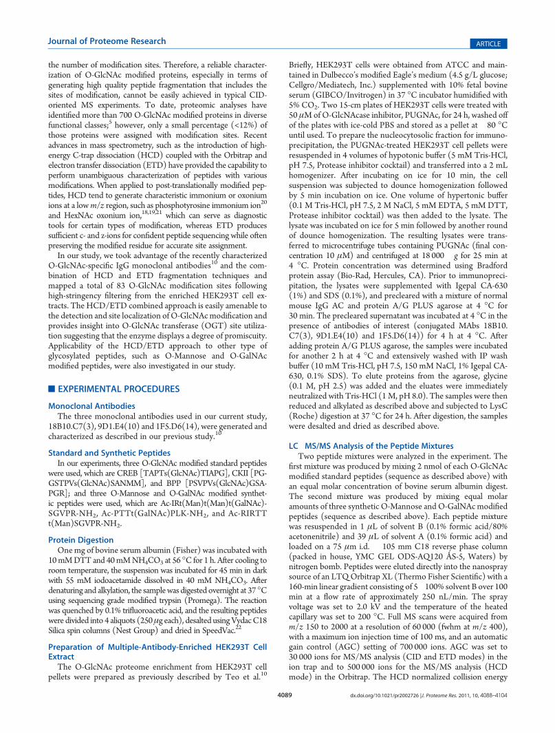
Figure 2. Respective CID, ETD, and HCD spectra of standard O-GlcNAc modified peptides CKII and BPP. (A�B) CID and ETD spectra ofO-GlcNAcmodifiedCKII peptide, respectively; (C�D)HCD and zoomed inHCD spectra of O-GlcNAcmodifiedCKII peptide; (E�F)CID and ETDspectra of O-GlcNAc modified BPP peptide, respectively; (G�H) HCD and zoomed in HCD spectra of O-GlcNAc modified CKII peptide. Note: “-HexNAc” or “-H2O” indicates the loss of HexNAc or H2O. Low m/z range HCD displays a distinctive pattern of HexNAc fragments (D and H).

4095 dx.doi.org/10.1021/pr2002726 |J. Proteome Res. 2011, 10, 4088–4104
Journal of Proteome Research ARTICLE
’RESULTS AND DISCUSSION
The MS approach employed in this study for sequencingO-GlcNAc modified peptides and assigning the modificationsites is a combination of HCD and ETD fragmentation. Whenanalyzing O-GlcNAc modified peptides, the unique advantage ofHCD fragmentation is the generation of distinct HexNAcoxonium ions (m/z 204.09)18,19,21 and a series of HexNAcfragments (m/z 186, m/z 168, m/z 144, m/z 138 and m/z126) which are significant to the diagnosis of O-GlcNAcmodified peptides yet not necessarily observed in ion traptandem mass spectra because of the dependence of ion trap scanrange on precursor m/z values. Moreover, the product ionsformed during HCD are detected in the Orbitrap analyzer;therefore, not only do they overcome the 1/3 cutoff rule of anion trap, but they also exhibit higher mass accuracy and lowerchemical noise. However, HCD spectra alone are not as infor-mative as those acquired in the linear ion trap pertaining topeptide sequence-related ion products, which has been attributedto the increased collision energy leading to ion scattering and alack of peptide backbone fragments.23
To provide more informative spectra for peptide sequencingas well as the site assignment of O-GlcNAc modification, ETD wasemployed in combination with HCD. By transferring an electronfrom a radical anion to a protonated peptide, ETD has been provenadvantageous for analyzing relatively large, nontryptic peptidescompared to CID. In the particular case of post-translationallymodified peptides, ETD has the capacity to preserve labile mod-ifications attached to peptide backbones, such as phosphorylationand glycosylation, allowing for the detection of multiple modifica-tions within the context of one another, as well as producing almostcomplete series of peptide backbone fragments for peptide sequen-cing at the same time.24 Especially when facilitated by supplemen-tary collisional activation converting the nondissociative electrontransfer products into c- and z- type fragment ions, the effectprecursor ion charge states have on dissociation efficiency, whichis one of the limitations imposed by the intrinsic charge-reducingprocess in electron-based fragmentationmethods, has been remark-ably reduced.25
By utilizing the HexNAc signature ions generated by HCDand the extensive peptide sequence information provided by
Table 1. Novel O-GlcNAc Proteins Identified in HEK293T Cells
UniProt accession protein name gene name coverage by #AA (%) # peptides
Q9Y265�1 Isoform 1 of RuvB-like 1 RUVBL1 21.05 14
P35268 60S ribosomal protein L22 RPL22 21.88 12
P22061�1 Isoform 1 of Protein-L-isoaspartate(D-aspartate) O-methyltransferase PCMT1 9.69 10
P62081 40S ribosomal protein S7 RPS7 21.65 10
P62263 40S ribosomal protein S14 RPS14 18.54 9
Q07021 Complement component 1 Q subcomponent-binding protein, mitochondrial C1QBP 15.6 9
P26196 Probable ATP-dependent RNA helicase DDX6 DDX6 7.45 8
P62750 60S ribosomal protein L23a RPL23A 17.95 8
C9JU56 Putative uncharacterized protein RPL31 RPL31 35.65 7
P25398 40S ribosomal protein S12 RPS12 41.67 7
P60866 40S ribosomal protein S20 RPS20 19.33 7
P62829 60S ribosomal protein L23 RPL23 28.57 7
C9JLW8 HCG1818442, isoform CRA_c hCG 1818442 11.34 6
P30050�1 Isoform 1 of 60S ribosomal protein L12 RPL12 17.58 6
P62913�2 Isoform 2 of 60S ribosomal protein Lll RPL11 23.73 6
Q9NYF8�3 Isoform 3 of Bcl-2-associated transcription factor 1 BCLAF1 11.51 6
C9J0W3 Putative uncharacterized protein SMARCE1 SMARCE1 17.2 5
P24534 Elongation factor 1-beta EEF1B2 12 5
Q6MZS5 Putative uncharacterized protein DKFZp686A13234 DKFZp686A13234 8.32 5
Q92922 SWI/SNF complex subunit SMARCC1 SMARCC1 4.07 5
Q9Y3I0 UPF0027 protein C22orf28 C22orf28 5.94 5
095881 Thioredoxin domain-containing protein 12 TXNDC12 16.86 4
P62266 40S ribosomal protein S23 RPS23 15.38 4
Q9NPF5 DNA methyltransferase 1-associated protein 1 DMAP1 8.57 4
P13929�3 Isoform 3 of Beta-enolase EN03 10.49 3
P49207 60S ribosomal protein L34 RPL34 22.22 3
Q5T4L4 Ribosomal protein S27 RPS27 16.67 3
Q8IXM2 Uncharacterized potential DNA-binding protein C17orf49 C17orf49 26.16 3
Q96K80 Zinc finger CCCH domain-containing protein 10 ZC3H10 8.99 3
C9JMM0 Putative uncharacterized protein CBX3 CBX3 48.39 2
P30040 Endoplasmic reticulum protein ERp29 ERP29 8.43 2
P37108 Signal recognition particle 14 kDa protein SRP14 16.18 2
P50991 T-complex protein 1 subunit delta CCT4 5.38 2
Q8TAQ2�2 Isoform 2 of SWI/SNF complex subunit SMARCC2 SMARCC2 1.77 2
4096 dx.doi.org/10.1021/pr2002726 |J. Proteome Res. 2011, 10, 4088–4104
Journal of Proteome Research ARTICLE
ETD, we analyzed glycosylated standard peptides to investigatethe applicability of HCD/ETD method in identification and sitelocalization of O-GlcNAc modified peptides. Furthermore, weapplied the same MS approach to a complex biological sample toprove the effectiveness and robustness of this method.
Characterization of O-GlcNAc Modified Standard PeptidesO-GlcNAc modified standard peptides in the mixture were
analyzed by an alternating CID/ETD/HCD fragmentation meth-od. As indicated in Figure 2, the most dominant peaks formedduringCID fragmentation of glycopeptidesCKII andBPP resultedfrom the loss of HexNAc residue from respective precursor ions(Figure 2A and E). As a result of the intensive HexNAc-loss ions,b- and y-type ions generated by backbone fragmentation that arerequired for peptide sequencing and site assignment are severelysuppressed, making it difficult to accurately and confidentlycharacterize O-GlcNAc peptides. In Figure 2B and F, almost acomplete series of c- or z-type ions were observed in the ETDspectrum allowing for the elucidation of both respective peptidesequence and modifications sites. During the HCD fragmenta-tion of CKII and BPP glycopeptides (Figure 2C and G), the highm/z fragment ions were either missing or at very low intensity.However, the HexNAc oxonium ion (m/z 204) and its fragments(m/z 186, m/z 168, m/z 144, m/z 138, and m/z 126) wereproduced at pronounced high intensity (Figure 2D and H,Table S1, Supporting Information). This distinctive fragmentpattern ofHexNAc residue allows for unambiguous identificationof O-GlcNAc modified peptides which are generally under-represented in protein mixtures extracted from biologicalsamples.3,8,9
WhenO-GlcNAc modified peptides are subjected to CID, it isprobable, yet not necessary, that the GlcNAc residue is cleavedfrom the peptide backbone and forms an oxonium ion at m/z204.09. When undergoing HCD, the GlcNAc residue will likelyalways be cleaved and generate the oxonium ion at m/z 204.09.This characteristic ion product can be used as a diagnostic tool toidentify the O-GlcNAc modified peptides. Moreover, not onlywill theHexNAc oxonium ion be present in theHCD spectrum, aseries of its fragment ions will also be produced that are indicativeof HexNAc residue, such as m/z 186, m/z 168, m/z 144, m/z138, and m/z 126.26 The presence of HexNAc oxonium ion and
the series of its fragment ions can be used to target O-GlcNAcmodified peptides during an MSn experiment and selectivelytrigger an equal order CID or ETD fragmentation of the sameprecursor ion for more detailed peptide characterization. Com-pared to the dominant neutral loss ions created upon the loss ofHexNAc residues during CID, which suppress the formation ofpeptide sequencing-related ions, ETD has the capability topreserve labile modifications and render more complete ionseries for peptide sequencing and site assignment. Using theHexNAc oxonium ion as an indicator to target O-GlcNAcmodified peptides with modification favorable ETD fragmenta-tion, peptide sequencing and site localization can be achievedwith improved selectivity and sensitivity.
Characterization of O-GlcNAc Modified Proteins Enrichedfrom HEK293T Cell Extract
186 proteins were identified at 1% pepFDR from the multiple-antibody enriched HEK293T cell extract (Table S2, SupportingInformation). In comparison with the recently published workfrom our group10 that used the same sample, 67% (124/186) of theidentified proteins were consistent with the result from previousexperiment, including heterogeneous nuclear ribonucleoproteins,BAT2 domain-containing protein 1, ribosomal proteins, heat shockproteins, and nuclear pore complex proteins. Of the 33% (62/186)of the data set that was only observed in our current study,17 proteins were supported in the literature2�6,14,27 as beingO-GlcNAc modified and 45 were identified as novel O-GlcNAcproteins (Table 1). Furthermore, while the previous experiment didnot discover any O-GlcNAc modification sites, our current studyidentified 83O-GlcNAc sites on 172 glycopeptides from13proteins(Table S3, Supporting Information). By comparison to theliterature,8,13�15,28�33 13 of the 83 sites had been previouslyassigned leaving 70 novel O-GlcNAc sites (Table 2); and 11 of
Table 2. Novel O-GlcNAc Sites Identified in HEK293T Cells
UniProt accession protein name novel sites
O95487�2 Isoform 2 of Protein transport protein Sec24B T327;T341
P35658�2 Isoform 2 of Nuclear pore complex protein Nup214 S1202; S1904; S1905; S1907; T1915; S1916
P49790 Nuclear pore complex protein NuplS3 T535; S893; S895; S1017; S1018; T1026; T1041
P51610�1 Isoform 1 of Host cell factor T405; S620; S622; S623; T625; S628; S638; T640; T651; T652; T658
P52594�2 Isoform 2 of Arf-GAP domain and FG repeats-containing protein 1 S291
P52948�4 Isoform 4 of Nuclear pore complex protein Nup98-Nup96 S262; T264
Q14157�4 Isoform 4 of Ubiquitin-associated protein 2-like S445
Q2KHR3�1 Isoform 1 of Glutamine and serine-rich protein 1 T1271
Q5T6F2 Ubiquitin-associated protein 2 T487; S494
Q5T8P6�3 Isoform 3 of RNA-binding protein 26 S657; S667
Q6MZP7�1 Isoform 1 of Protein lin-54 homologue T109
Q7Z589 Isoform 1 of Protein EMSY S228; T264; T272
Q8IWZ3�1 Isoform 1 of Ankyrin repeat and KH domain-containing protein 1 S1817
Q9H4A3�2 Isoform 2 of Serine/threonine-protein kinase WNK1 S1849
Q9Y520 Isoform 7 of BAT2 domain-containing protein 1 S2196
Table 3. Comparison of Two Methods for O-GlcNAc SiteMapping
method O-GlcNAc peptides
manually
confirmed sites
presence of
HexNAc signature ions
HCD/ETD 172 83 100%
HCD-alone 37 7 100%
4097 dx.doi.org/10.1021/pr2002726 |J. Proteome Res. 2011, 10, 4088–4104
Journal of Proteome Research ARTICLE
the 13 characterized proteins were also previously observed asmodified but not site-mapped in the previously work from our group.
By comparing the results from the two respective LC�MS/MSexperiments, 37O-GlcNAc peptides were observed by theHCD-alone method, and 168 glycopeptides were found by the HCD/ETD method (Table 3). After manual validation, 7 O-GlcNAcsites were assigned in the HCD-alone analysis, and 83 sites wereassigned in the HCD/ETD analysis.
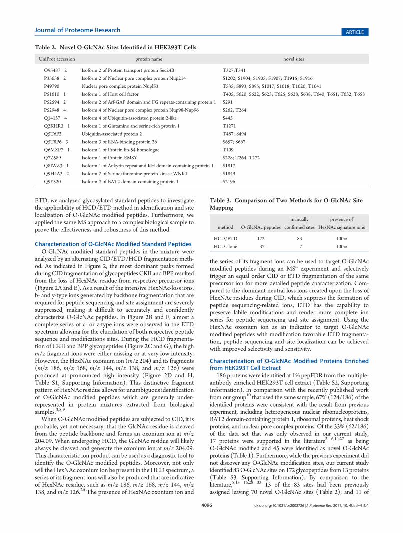
We also evaluated the abundance of ionized and fragmentedO-GlcNAc peptides in the sample by analyzing the occurrence ofHexNAc oxonium ions in the spectral data collected from HCD-alone and HCD/ETD experiments. Generally, the respective ionchromatograms at m/z 204.086 (bottom panel) of the HCD-alone (Figure S1, Supporting Information) and HCD/ETD(Figure 3) experiments were extracted and compared to thetotal ion chromatograms (top panel). The extracted ion chro-matograms illustrates the retention time profiles of theO-GlcNAc peptides analyzed by the mass spectrometer andindicates the acquisition of HCD spectra of those peptides. Inboth figures (Figure 3 and S1, Supporting Information), thenoticeable difference of more than 2 orders of magnitude inintensity between the extracted and total ion chromatogramsdemonstrated the low abundance of O-GlcNAc modifiedpeptides in the ionized mixture, and further suggests the sensi-tivity of detection by HCD and the importance of selectivelytargeting O-GlcNAc peptides for fragmentation as a manner ofenrichment.
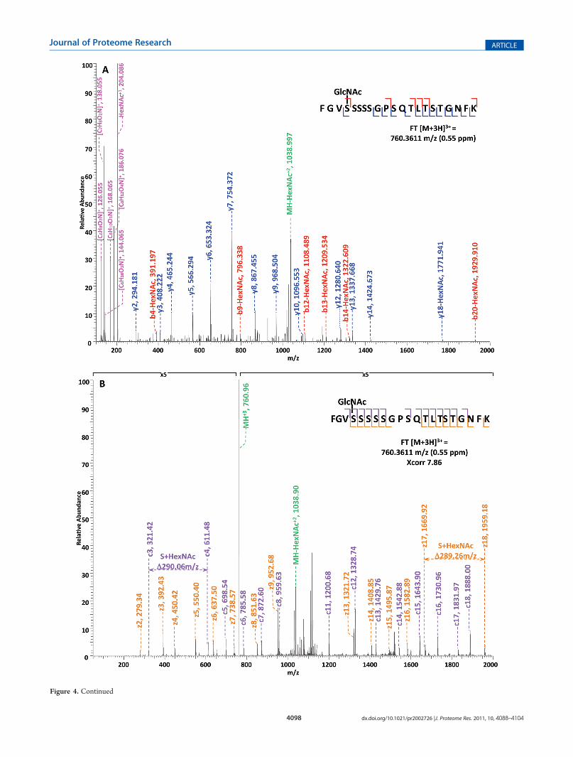
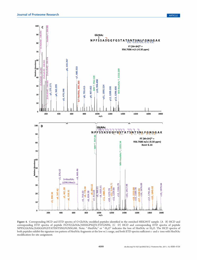
Specifically, in the HCD-alone experiment, each HCD spec-trum of the eluted O-GlcNAc peptides showed the oxonium ionsof HexNAc atm/z 204 and some other signature ions atm/z 186,m/z 168, etc., including the spectra of the 37 peptides assigned bythe database search program that survived 1% FDR filtering.However, because of the lack of primary fragment ions caused bythe high-energy during the fragmentation, most O-GlcNAcpeptides, even when being indicated by the HexNAc signatureions in the spectra, were not sequenced by the database searchalgorithm due to the poor ion match ratios. The 37 O-GlcNAcpeptides in our data set that were sequenced still suffer from lowratio of ion match and therefore low scores (Table S4, Support-ing Information), especially because of the loss of GlcNAc duringHCD, the majority of the glycopeptides do not have indicative b-or y- ions in their spectra for the manual validation of O-GlcNAcsite localization. This observation suggests that even thoughHCD is powerful in generating high quality spectral data toindicate the presence of O-GlcNAc peptides, it is not capable ofproviding sufficient information for accurately mapping the sitesof modification. In the HCD/ETD experiment, where 172O-GlcNAc peptides were identified and 83 sites were mappedon them, all of the O-GlcNAc peptides were characterized by theETD spectra and each of those ETD spectra corresponds with anHCD spectrum observing a pattern of HexNAc signature ions. Asan example, in Figure 4, panel A and panel B are the consecutivelyacquired HCD and ETD spectra of the same triple-chargedprecursor m/z 760.3611. As noted in Figure 4A, most b- and
Figure 3. Comparison between the extracted ion chromatogram at m/z 204.086 and the total ion chromatogram of the HCD/ETD LC�MS/MSexperiment on the enriched HEK293T sample. The extracted ion chromatogram atm/z 204.086 (bottom panel) delineates the retention time profile ofO-GlcNAc modified peptides eluted over the entire gradient. In comparison with the total ion chromatogram, the normalized peak height of theextracted ion chromatogram indicates the substoichiometric nature of O-GlcNAc modification and therefore suggests the importance of selectivefragmentation on O-GlcNAc peptides.
4098 dx.doi.org/10.1021/pr2002726 |J. Proteome Res. 2011, 10, 4088–4104
Journal of Proteome Research ARTICLE
Figure 4. Continued
4099 dx.doi.org/10.1021/pr2002726 |J. Proteome Res. 2011, 10, 4088–4104
Journal of Proteome Research ARTICLE
Figure 4. Corresponding HCD and ETD spectra of O-GlcNAc modified peptides identified in the enriched HEK293T sample. (A�B) HCD andcorresponding ETD spectra of peptide FGVS(GlcNAc)SSSSGPSQTLTSTGNFK; (C�D) HCD and corresponding ETD spectra of peptideNPFS(GlcNAc)SASGGFGSTATSNTSNLFGNSGAK. Note: “-HexNAc” or “-H2O” indicates the loss of HexNAc or H2O. The HCD spectra ofboth peptides exhibit the signature ion pattern of HexNAc fragments at the lowm/z range, and both ETD spectra sufficient c- and z- ions with HexNAcmodification for site assignment.

4100 dx.doi.org/10.1021/pr2002726 |J. Proteome Res. 2011, 10, 4088–4104
Journal of Proteome Research ARTICLE
y- ions were generated without the attachment of GlcNAc (b-HexNAc or y-HexNAc) and were at relatively low intensity,therefore can not be used to assign the site of modification. At thelow m/z range of the spectrum, a strong HexNAc oxonium ionpeak was observed at m/z 204.086, and the other five signatureions of HexNAcwere also found at high intensities, indicating thepresence of O-GlcNAc modification on the peptide. InFigure 4B, an almost complete series of c- and z- ions wereproduced in the ETD spectrum of the same peptide and the siteof O-GlcNAc modification was unambiguously indicated bycorresponding c- and z- ion pairs.
We further investigated the prevalence of each HexNAcsignature ion by summarizing the occurrence of them in the172 O-GlcNAc peptides identified by the HCD/ETD analysis(Table 4). As presented in Table 4, the HexNAc oxonium ion atm/z 204 and its fragments at m/z 138 and m/z 126 wereobserved in the spectra of all 172 peptides; signature ions atm/z 186 and m/z 168 were also present in over 90% of thespectra, whereas the signature ion atm/z 144 was observed in lessthan 75% of the spectra. By examining the patterns of theHexNAc signature ions observed from the 172 O-GlcNAcpeptides (Table 5), the most prevalent pattern is the presenceof all 6 signature ions, which was observed in approximately 85%of the spectra; the second most prevalent pattern is the presenceof 5 signature ions withm/z 144 missing, indicating thatm/z 144is an unfavorable product of HexNAc fragmentation. The patternsthat have more than one signature ionmissing were observed at lowprevalence, suggesting that most fragments from HexNAc underHCD are stable and constant. Further comparison between theextracted ion chromatograms at respective signature ions of Hex-NAc (m/z 204, m/z 186, m/z 168, m/z 144, etc.) is illustrated inFigure S2 (Supporting Information), where the general difference inintensity between the chromatograms is consistent with the pre-valence of each signature ion, for example, the intensity of theextracted ion chromatogram at m/z 204.086 is the highest amongthe 6 chromatograms whereas that at m/z 144.064 is the lowest.
The consistent and reliable production of HexNAc signatureions by HCD suggests the possibility and plausibility of targeting
O-GlcNAc modified peptides with selective fragmentation; thetwo examples presented in Figure 4B and D confirmed thecapability of ETD in characterizing O-GlcNAc modified peptides.On the basis of the result of analysis on the occurrence of 6HexNAc signature ions, we further propose that while it is capableto target O-GlcNAc peptides by only the HexNAc oxonium ion atm/z 204, it would increase the accuracy of selection and decreasethe possibility of false positives by monitoring more than onesignature ions, such as the combination of m/z 204, m/z 138 andm/z 126, which were observed in all 172 O-GlcNAc peptidesdetected in the HCD/ETD experiment.
We further analyzed the same raw spectra files obtained fromthe HEK293T sample using a multiple-engine database searchapproach to improve the sensitivity of data analysis (Figure S3,Supporting Information). Basically, database search programsassign an MSn spectrum with the most probable peptide match,and the assigned spectra generally constitute only a fraction of allthe spectra acquired in an LC�MSn analysis. Variations indatabase searching algorithms for assigning peptides to MSn
spectra have been known to provide different identificationresults.34 By combining search results from different searchengines have been proven to lead to a larger number of proteinidentification with an increased rate of peptide assignments.35�37
By processing the files in this manner, 200 proteins wereidentified at 1% pepFDR after combining and validating theoutput from three independent database searches. In comparisonwith the result from our recently published work10 that used thesame sample, 66% (132/200) of the identified proteins wereconsistent with the previous experiment. Among the rest 34%(68/200) of the data set which had only been observed in ourcurrent study, 18 proteins were supported in the literature asbeing O-GlcNAc modified and 50 were identified as novelO-GlcNAc proteins. Furthermore, the multiple-engine searchstrategy indentified 165 O-GlcNAc sites on 178 glycopeptidesfrom 40 proteins (Table S5, Supporting Information), includingthe 83 sites that were assigned by SEQUEST-only database search.By comparison to the literature,8,13�15,28�33 29 of the 165 sites havebeen previously assigned leaving 136 novel O-GlcNAc sites; and 22of the 40 characterized proteins were also observed in the previouslywork from our group,10 whereas 18 of the 40 proteins are novel. Bytaking advantage of the multiple-engine database search methodol-ogy, the numbers of identified proteins, peptides, and modificationsites have been greatly increased and the sensitivity of the acquiredraw data has been dramatically improved.
Clustered O-GlcNAc Sites on Single Peptide Sequence Sug-gests Promiscuity of O-GlcNAc Transferase (OGT)
O-GlcNAc modification is analogous to serine/threoninephosphorylation in many respects.15,32 However, unlike phos-phorylation, which is catalyzed by almost 500 kinases encoded inthe human genome,38 O-GlcNAcmodification is catalyzed by theproducts of a single human gene.39,40 Studies have shown thatOGT glycosylation is quite specific,41 and furthermore that thecatalytic subunit of OGT achieves both high specificity and aremarkable diversity of substrates through forming a complexwith a variety of targeting proteins via its tetratricopeptide repeat(TPR) protein�protein interaction domains.42�44 In our dataset, we discovered several cases of clustered sites of O-GlcNAcmodification exhibited by coeluted peptides, such as proteinsNup214, Nup153, host cell factor and EMSY (Table S3, Sup-porting Information). One such example, presented in Figure 5,shows multiple isobaric glycopeptides were eluted at the same
Table 4. HexNAc Signature Ion Occurrence in HCD/ETDAnalysis
204 186 16 144 138 16
O-GlcNAc Peptides 172 164 164 148 172 172
Corresponding Sites 83 76 77 70 83 83
Prevalence 100% 95.35% 93.02% 74.42% 100% 100%
Table 5. HexNAc Signature Ion Patterns in HCD/ETDAnalysisa
signaure ion patterns
204 186 168 144 138 126 prevalence
O-GlcNAc
peptides
corresponding
sites√ √ √ √ √ √
84.88% 146 68√�
√ √ √ √1.16% 2 4√
�√ √ √ √
9.30% 16 13√ √� �
√ √1.16% 2 4√
� � �√ √
3.49% 6 6a√
Observed; � Not observed.

4101 dx.doi.org/10.1021/pr2002726 |J. Proteome Res. 2011, 10, 4088–4104
Journal of Proteome Research ARTICLE
time and had acquired the same charge state. In one ETDspectrum, there were possibly three mono-GlcNAc modifiedpeptides being coeluted, and the only difference between them isthe site of the modified serine residue on the peptide (S7 in red,S8 in gray, and S9 in blue). By interpreting the spectrum, wefound that the ions at m/z 969 and m/z 1056 correspond to thez10 and z11 ions of the S7-modified peptide, and the ions atm/z1172 andm/z 1259 correspond to the z10 and z11 ions of the S9-modified peptide. In fact, all the ions observed in the spectrumcan be explained as the fragments of S7-modified and S9-modified peptides, confirming the presence of both. However,since the ions atm/z 969 andm/z 1259 can also be interpreted asz10 and z11 ions of the S8-modified peptide and there is nounique ion to exclude its presence, the coeluting mixture can haveup to three O-GlcNAc modified peptides. Thus, there are threesequential sites on this single peptide that can all be recognized byOGT, and throughout our data set, this peptide was not found tobe modified by di- or tri-GlcNAc. This observation of the diversityin peptide substrates was observed in multiple cases and suggeststhe promiscuity of OGT (Table S3, Supporting Information).
Signature Ion Patterns Observed from Other Types ofGlycosylated Peptides under HCD
In order to explore the applicability of HCD/ETD MSapproach on other types of O-glycosylation, synthetic O-Man-nose and O-GalNAc modified peptides were analyzed in thesame fashion as O-GlcNAc modified peptides using the
combined HCD/ETD fragmentation. Indicative ion patterns atlow m/z range were observed, respectively, when O-Mannoseand O-GalNAc peptides underwent HCD: the O-GalNAc mod-ified peptide exhibited similar signature ions at m/z 204, m/z186, m/z 168, m/z 144, m/z 138, and m/z 126 (Figure S4A andB, Supporting Information); the O-Mannosylated peptide ex-hibited the Hexose oxonium ion at m/z 163, and a series of itsfragments at m/z 145, m/z 127, m/z 115, and m/z 109 (FigureS4A and C, Table S1, Supporting Information). The utility ofHCD has been demonstrated for the characterization of proteintyrosine-phosphorylation,20 protein N-glycosylation,45 and thequantification of iTRAQ-labeled phosphopeptides.46,47 Our ob-servation of the distinctive ion patterns of O-GlcNAc, O-Man-nose, andO-GalNAc peptides proved the applicability of HCD intargeting O-glycosylated peptides. Although, based on our data,it appears that O-GlcNAc and O-GalNAc cannot be distin-guished from each other solely by their signature ion patterns.Furthermore, the signature ion-trigger strategy has the potentialto be applied to the analysis of post-translational modificationsbesides glycosylation and phosphorylation, such as methylation,bromylation, hydroxylation and other modifications that havepreviously been shown to produce specific fragment ions.48
’CONCLUSION
The signature ion patterns of HexNAc and Hexose revealedunder HCD condition can be utilized to selectively target
Figure 5. ETD spectrum of coeluted mono-O-GlcNAc modified peptides GFDTSSSSs(GlcNAc)NSAASSSFK and GFDTSSs(GlcNAc)SSNSAASSSFK. Spectrum illustrates heterogeneity of the singly O-GlcNAc modified peptide suggestive of O-GlcNAc transferase having promiscuityin terms of site of modification.

4102 dx.doi.org/10.1021/pr2002726 |J. Proteome Res. 2011, 10, 4088–4104
Journal of Proteome Research ARTICLE
HexNAc and/or Hexose modified proteins, and when combinedwith ETD, which preserves labile post-translational modificationon proteins, the reliability and accuracy in glycoprotein identi-fication and site localization can be greatly improved. In ourstudy, we investigated the applicability of the combined HCD/ETD MS approach in characterizing O-GlcNAc modified pro-teins from a complex biological sample and successfully identifieda minimum of 83 modification sites. Additionally, with a multi-ple-engine database search method, we were able to increase thesensitivity of our discovery drastically to reach a total of 165 sitesof O-GlcNAc modification.
By analyzing the data from two parallel experiments per-formed respectively by HCD-alone and HCD/ETD method, wewere able to prove the consistency and stability of signature ionsproduced from O-GlcNAc peptides under HCD, and demon-strate the superior sensitivity and accuracy of the HCD/ETDmethod in sequencing and site-mapping of O-GlcNAc modifiedpeptides. Considering that the neutral loss of HexNAc fromO-GlcNAc peptides under HCDmay also be used as a trigger forselective fragmentation, the signature ion-trigger mechanism hasthe advantage of producing the constant m/z values of signatureions regardless of the occupancy of themodification or the chargestates of the peptides especially in a complex mixture, which willgenerate ambiguity in a neutral loss-triggered approach byanticipating the masses for monitoring.
A more detailed analysis on the occurrence of specificsignature ions of HexNAc revealed some stable candidates fora product-ion-monitoring model, such as the combination ofthree ions at m/z 204, m/z 138, and m/z 126, which can beimplemented with the HCD-trigger-ETD approach via a deci-sion tree mechanism in the future.
We further demonstrated the capability of the HCD/ETDmethod in characterizing O-GalNAc and O-Mannose modifiedpeptides as well, suggesting a more general applicability of thefuture HCD-trigger-ETD approach. Along with the advance-ment of both hardware and software in mass spectrometry, weanticipate that anHCD-product-ion-triggered-ETD approach ona hybrid linear ion trap/Orbitrap platform will greatly improvethe sensitivity of detection and the accuracy in assigning theglycosylation sites on peptides from a complex mixture. Further-more, this study sheds light on the action of the O-GlcNActransferase in that it appears to be promiscuous with regards towhich Ser/Thr residue is used in clustered regions of hydroxyl-containing amino acids in substrate proteins.
’ASSOCIATED CONTENT
bS Supporting InformationFigure S1. Comparison between the extracted ion chromato-
gram atm/z 204.086 and the total ion chromatogram of theHCD-alone LC�MS/MS experiment on the enriched HEK293T sam-ple. Figure S2. Comparison between the total ion chromatogramand the respective extracted ion chromatogram at m/z 204.086,m/z 186.076, m/z 168.065, m/z 144.065, m/z 138.054, and m/z126.054 from the HCD/ETD LC-MS/MS experiment of theenriched HEK293T sample. Figure S3. Multiple-engine databasesearch strategy for the analysis on the enriched HEK293Tsample. Figure S4. HCD and ETD spectra of O-Mannose andO-GalNAc modified peptides. Table S1. Fragments of HexNAcand Hexose oxonium ions. Table S2. List of proteins identified inthe enriched HEK293T sample by HCD/ETD approach. TableS3. List of validated O-GlcNAc modified peptides identified in
the enriched HEK293T sample by HCD/ETD approach. TableS4. List of validated O-GlcNAc modified peptides identified inthe enriched HEK293T sample by HCD-alone approach. TableS5. List of validated O-GlcNAc modified peptides identified inthe enriched HEK293T sample by HCD/ETD approach usingmultiple-engine database searches. This material is available freeof charge via the Internet at http://pubs.acs.org.
’AUTHOR INFORMATION
Corresponding Author*Lance Wells, 315 Riverbend Road, CCRC, UGA, Athens, GA,30602. Phone: (706) 542-7806. Fax: (706) 542-4412. E-mail:[email protected].
’ACKNOWLEDGMENT
This work was supported by a P41 grant from NCRR(P41RR018502, L.W., senior investigator) and an R01 grantfrom NIDDK (R01DK075069, L.W.). We thank Dr. Gerald W.Hart at Johns Hopkins University School of Medicine forproviding the O-GlcNAc modified standard peptides and Dr.David Live at the University of Georgia for the synthesis ofO-Mannose and O-GalNAc modified peptides.
’ABBREVIATIONS:
AGC, automatic gain control; CID, collision induced dissocia-tion; DTT, dithiothreitol; ETD, electron transfer dissociation;FDR, false discovery rate; fwhm, full width at half-maximum;GalNAc, N-acetylgalactosamine; GlcNAc, N-acetylglucosamine;HCD, high-energy C-trap dissociation; Hex, hexose; HexNAc,N-acetylhexosamine; LC, liquid chromatography;Man, mannose;MS, mass spectrometry;OGA, O-linked β-N-acetylglucosamini-dase;OGT, O-linked β-N-acetylglucosamine transferase; pepFDR,peptide-level FDR;TRP domain, tetratricopeptide repeat domain
’REFERENCES
(1) Love, D. C.; Hanover, J. A. The hexosamine signaling pathway:deciphering the “O-GlcNAc code. Sci. STKE 2005, 2005 (312), re13.
(2) Zachara, N. E.; Hart, G. W. O-GlcNAc a sensor of cellular state:the role of nucleocytoplasmic glycosylation in modulating cellularfunction in response to nutrition and stress. Biochim. Biophys. Acta2004, 1673 (1�2), 13–28.
(3) Hart, G. W.; Housley, M. P.; Slawson, C. Cycling of O-linkedbeta-N-acetylglucosamine on nucleocytoplasmic proteins. Nature 2007,446 (7139), 1017–22.
(4) Laczy, B.; Hill, B. G.; Wang, K.; Paterson, A. J.; White, C. R.;Xing, D.; Chen, Y. F.; Darley-Usmar, V.; Oparil, S.; Chatham, J. C.Protein O-GlcNAcylation: a new signaling paradigm for the cardiovas-cular system. Am. J. Physiol. Heart Circ. Physiol. 2009, 296 (1), H13–28.
(5) Copeland, R. J.; Bullen, J. W.; Hart, G. W. Cross-talk betweenGlcNAcylation and phosphorylation: roles in insulin resistance andglucose toxicity. Am. J. Physiol. Endocrinol. Metab. 2008, 295 (1),E17–28.
(6) Dias, W. B.; Hart, G. W. O-GlcNAc modification in diabetes andAlzheimer’s disease. Mol. Biosyst. 2007, 3 (11), 766–72.
(7) Lefebvre, T.; Guinez, C.; Dehennaut, V.; Beseme-Dekeyser, O.;Morelle, W.; Michalski, J. C. Does O-GlcNAc play a role in neurode-generative diseases? Expert Rev. Proteomics 2005, 2 (2), 265–75.
(8) Hu, P.; Shimoji, S.; Hart, G. W. Site-specific interplay betweenO-GlcNAcylation and phosphorylation in cellular regulation. FEBS Lett.2010, 584 (12), 2526–38.
4103 dx.doi.org/10.1021/pr2002726 |J. Proteome Res. 2011, 10, 4088–4104
Journal of Proteome Research ARTICLE
(9) Haynes, P. A.; Aebersold, R. Simultaneous detection and identi-fication of O-GlcNAc-modified glycoproteins using liquid chromatog-raphy-tandem mass spectrometry. Anal. Chem. 2000, 72 (21), 5402–10.(10) Teo, C. F.; Ingale, S.; Wolfert, M. A.; Elsayed, G. A.; Not, L. G.;
Chatham, J. C.;Wells, L.; Boons, G. J. Glycopeptide-specificmonoclonalantibodies suggest new roles forO-GlcNAc.Nat. Chem. Biol. 2010, 6 (5),338–43.(11) Comer, F. I.; Vosseller, K.; Wells, L.; Accavitti, M. A.; Hart,
G. W. Characterization of a mouse monoclonal antibody specific forO-linked N-acetylglucosamine. Anal. Biochem. 2001, 293 (2), 169–77.(12) Snow, C. M.; Senior, A.; Gerace, L. Monoclonal antibodies
identify a group of nuclear pore complex glycoproteins. J. Cell Biol. 1987,104 (5), 1143–56.(13) Chalkley, R. J.; Thalhammer, A.; Schoepfer, R.; Burlingame,
A. L. Identification of protein O-GlcNAcylation sites using electrontransfer dissociation mass spectrometry on native peptides. Proc. Natl.Acad. Sci. U.S.A. 2009, 106 (22), 8894–9.(14) Vosseller, K.; Trinidad, J. C.; Chalkley, R. J.; Specht, C. G.;
Thalhammer, A.; Lynn, A. J.; Snedecor, J. O.; Guan, S.; Medzihradszky,K. F.; Maltby, D. A.; Schoepfer, R.; Burlingame, A. L. O-linkedN-acetylglucosamine proteomics of postsynaptic density preparationsusing lectin weak affinity chromatography and mass spectrometry. Mol.Cell. Proteomics 2006, 5 (5), 923–34.(15) Wang, Z.; Udeshi, N. D.; Slawson, C.; Compton, P. D.; Sakabe,
K.; Cheung, W. D.; Shabanowitz, J.; Hunt, D. F.; Hart, G. W. Extensivecrosstalk between O-GlcNAcylation and phosphorylation regulatescytokinesis. Sci. Signal. 2010, 3 (104), ra2.(16) Khidekel, N.; Ficarro, S. B.; Clark, P. M.; Bryan, M. C.; Swaney,
D. L.; Rexach, J. E.; Sun, Y. E.; Coon, J. J.; Peters, E. C.; Hsieh-Wilson,L. C. Probing the dynamics of O-GlcNAc glycosylation in the brain usingquantitative proteomics. Nat. Chem. Biol. 2007, 3 (6), 339–48.(17) Jebanathirajah, J.; Steen, H.; Roepstorff, P. Using optimized
collision energies and high resolution, high accuracy fragment ionselection to improve glycopeptide detection by precursor ion scanning.J. Am. Soc. Mass Spectrom. 2003, 14 (7), 777–84.(18) Chalkley, R. J.; Burlingame, A. L. Identification of GlcNAcyla-
tion sites of peptides and alpha-Crystallin using Q-TOF mass spectro-metry. J. Am. Soc. Mass Spectrom. 2001, 12 (10), 1106–13.(19) Huddleston, M. J.; Bean, M. F.; Carr, S. A. Collisional frag-
mentation of glycopeptides by electrospray ionization LC/MS and LC/MS/MS: methods for selective detection of glycopeptides in proteindigests. Anal. Chem. 1993, 65 (7), 877–84.(20) Olsen, J. V.; Macek, B.; Lange, O.; Makarov, A.; Horning, S.;
Mann, M. Higher-energy C-trap dissociation for peptide modificationanalysis. Nat. Methods 2007, 4 (9), 709–12.(21) Carr, S. A.; Huddleston, M. J.; Bean, M. F. Selective identifica-
tion and differentiation of N- and O-linked oligosaccharides in glyco-proteins by liquid chromatography-mass spectrometry. Protein Sci. 1993,2 (2), 183–96.(22) Lim, J. M.; Sherling, D.; Teo, C. F.; Hausman, D. B.; Lin, D.;
Wells, L. Defining the regulated secreted proteome of rodent adipocytesupon the induction of insulin resistance. J. Proteome Res. 2008, 7 (3),1251–63.(23) Scherl, A.; Shaffer, S. A.; Taylor, G. K.; Hernandez, P.; Appel,
R. D.; Binz, P. A.; Goodlett, D. R. On the benefits of acquiring peptidefragment ions at highmeasuredmass accuracy. J. Am. Soc. Mass Spectrom.2008, 19 (6), 891–901.(24) Mikesh, L. M.; Ueberheide, B.; Chi, A.; Coon, J. J.; Syka, J. E.;
Shabanowitz, J.; Hunt, D. F. The utility of ETD mass spectrometry inproteomic analysis. Biochim. Biophys. Acta 2006, 1764 (12), 1811–22.(25) Swaney, D. L.; McAlister, G. C.; Wirtala, M.; Schwartz, J. C.;
Syka, J. E.; Coon, J. J. Supplemental activation method for high-efficiency electron-transfer dissociation of doubly protonated peptideprecursors. Anal. Chem. 2007, 79 (2), 477–85.(26) Peterman, S. M.; Mulholland, J. J. A novel approach for
identification and characterization of glycoproteins using a hybrid linearion trap/FT-ICRmass spectrometer. J. Am. Soc. Mass Spectrom. 2006, 17(2), 168–79.
(27) Zachara, N. E.; Hart, G. W. O-GlcNAc modification: a nutri-tional sensor that modulates proteasome function. Trends Cell Biol.2004, 14 (5), 218–21.
(28) Zachara, N. E.; Molina, H.; Wong, K. Y.; Pandey, A.; Hart,G. W. The dynamic stress-induced “O-GlcNAc-ome” highlights func-tions for O-GlcNAc in regulating DNA damage/repair and other cellularpathways. Amino Acids 2011, 40 (3), 793–808.
(29) Zeidan, Q.; Wang, Z.; De Maio, A.; Hart, G. W. O-GlcNAccycling enzymes associate with the translational machinery andmodify core ribosomal proteins. Mol. Biol. Cell 2010, 21 (12),1922–36.
(30) Zachara, N. E.; Cheung, W. D.; Hart, G. W. Nucleocytoplasmicglycosylation, O-GlcNAc: identification and site mapping.Methods Mol.Biol. 2004, 284, 175–94.
(31) Wells, L.; Vosseller, K.; Cole, R. N.; Cronshaw, J. M.; Matunis,M. J.; Hart, G.W.Mapping sites ofO-GlcNAcmodification using affinitytags for serine and threonine post-translational modifications.Mol. Cell.Proteomics 2002, 1 (10), 791–804.
(32) Wang, Z.; Gucek, M.; Hart, G. W. Cross-talk between GlcNA-cylation and phosphorylation: site-specific phosphorylation dynamics inresponse to globally elevated O-GlcNAc. Proc. Natl. Acad. Sci. U.S.A.2008, 105 (37), 13793–8.
(33) Wang, Z.; Park, K.; Comer, F.; Hsieh-Wilson, L. C.; Saudek,C. D.; Hart, G. W. Site-specific GlcNAcylation of human erythrocyteproteins: potential biomarker(s) for diabetes. Diabetes 2009, 58 (2),309–17.
(34) Boutilier, K.; Ross, M.; Podtelejnikov, A. V.; Orsi, C.; Taylor,R.; Taylor, P.; Figeys, D. Comparison of different search engines usingvalidated MS/MS test datasets. Anal. Chim. Acta 2005, 534, 10.
(35) Yu, W.; Taylor, J. A.; Davis, M. T.; Bonilla, L. E.; Lee, K. A.;Auger, P. L.; Farnsworth, C. C.; Welcher, A. A.; Patterson, S. D.Maximizing the sensitivity and reliability of peptide identification inlarge-scale proteomic experiments by harnessing multiple search en-gines. Proteomics 2010, 10 (6), 1172–89.
(36) Carrascal, M.; Gay, M.; Ovelleiro, D.; Casas, V.; Gelpi, E.; Abian,J. Characterization of the human plasma phosphoproteome using linearion trap mass spectrometry and multiple search engines. J. Proteome Res.2010, 9 (2), 876–84.
(37) Searle, B. C.; Turner, M.; Nesvizhskii, A. I. Improving sensitiv-ity by probabilistically combining results from multiple MS/MS searchmethodologies. J. Proteome Res. 2008, 7 (1), 245–53.
(38) Manning,G.;Whyte,D. B.;Martinez, R.;Hunter, T.; Sudarsanam,S. The protein kinase complement of the human genome. Science 2002, 298(5600), 1912–34.
(39) Nolte, D.; Muller, U. Human O-GlcNAc transferase (OGT):genomic structure, analysis of splice variants, fine mapping in Xq13.1.Mamm. Genome 2002, 13 (1), 62–4.
(40) Shafi, R.; Iyer, S. P.; Ellies, L. G.; O’Donnell, N.; Marek, K. W.;Chui, D.; Hart, G. W.; Marth, J. D. The O-GlcNAc transferase generesides on the X chromosome and is essential for embryonic stem cellviability and mouse ontogeny. Proc. Natl. Acad. Sci. U.S.A. 2000, 97 (11),5735–9.
(41) Lubas, W. A. Hanover, J. A., Functional expression of O-linkedGlcNAc transferase. Domain structure and substrate specificity. J. Biol.Chem. 2000, 275 (15), 10983–8.
(42) Cheung, W. D.; Sakabe, K.; Housley, M. P.; Dias, W. B.; Hart,G. W. O-linked beta-N-acetylglucosaminyltransferase substrate specifi-city is regulated by myosin phosphatase targeting and other interactingproteins. J. Biol. Chem. 2008, 283 (49), 33935–41.
(43) Iyer, S. P.; Hart, G. W. Roles of the tetratricopeptide repeatdomain in O-GlcNAc transferase targeting and protein substrate speci-ficity. J. Biol. Chem. 2003, 278 (27), 24608–16.
(44) Kreppel, L. K.; Hart, G.W. Regulation of a cytosolic and nuclearO-GlcNAc transferase. Role of the tetratricopeptide repeats. J. Biol.Chem. 1999, 274 (45), 32015–22.
(45) Segu, Z. M.; Mechref, Y. Characterizing protein glycosylationsites through higher-energy C-trap dissociation. Rapid Commun. MassSpectrom. 2010, 24 (9), 1217–25.

4104 dx.doi.org/10.1021/pr2002726 |J. Proteome Res. 2011, 10, 4088–4104
Journal of Proteome Research ARTICLE
(46) Zhang, Y.; Ficarro, S. B.; Li, S.; Marto, J. A. Optimized OrbitrapHCD for quantitative analysis of phosphopeptides. J. Am. Soc. MassSpectrom. 2009, 20 (8), 1425–34.(47) Boja, E. S.; Phillips, D.; French, S. A.; Harris, R. A.; Balaban,
R. S. Quantitative mitochondrial phosphoproteomics using iTRAQ onan LTQ-Orbitrap with high energy collision dissociation. J. Proteome Res.2009, 8 (10), 4665–75.(48) Carr, S. A.; Annan, R. S.; Huddleston, M. J. Mapping post-
translational modifications of proteins by MS-based selective detection:application to phosphoproteomics. Methods Enzymol. 2005, 405,82–115.