Embed Size (px)
Citation preview

Tendon Transfer and Combined Nerve Palsy

Biomechanics of Tendon Transfers
• Positive Factors – Balance – Strength – Mobility
• Negative Factors – Stiffness – Deformity – Disuse

Balance
“In the management of paralysis, balance is more important than strength.”
Brand, 1988

Balance
• Must redistribute muscle power that remains in forearm and hand
• Does not mean equal power on each side of a joint – Normal hand
• Digital extensors weaker than flexors – Do not contract against each other – Work in harmony (flexors relax as extensors fire)
• Wrist extensors must be strong enough to balance finger flexors
– Contract simultaneously against each other on opposite sides of wrist joint
– Must produce equal torque on each side of wrist joint to provide stability for distal activity
• Synergism

Synergism
• Active Synergism – Occurs when muscles have to contract together in
order to augment the effectiveness of each muscle – Needed when a sequence of joints must be controlled
• Sequential synergism – Active contraction of one muscle stretches an
opposing muscle – Energy in stretched position released when opposing
muscle contracts

Synergism
!!!!!!!!!
(Clinical Mecanics of the Hand, 1985)

Blix Curve
!!!!!!!
(Skand Arch Physiol, 1891)

Synergism
• When limb is rested with some muscles slack and some stretched – Slack muscles absorb sarcomere units – Stretched muscles add sarcomere units – Ultimately, all fibers reach their original resting tension – True of other tissues as well
• Joint capsule • Ligaments • Skin
(Hand Clinics, 1988)
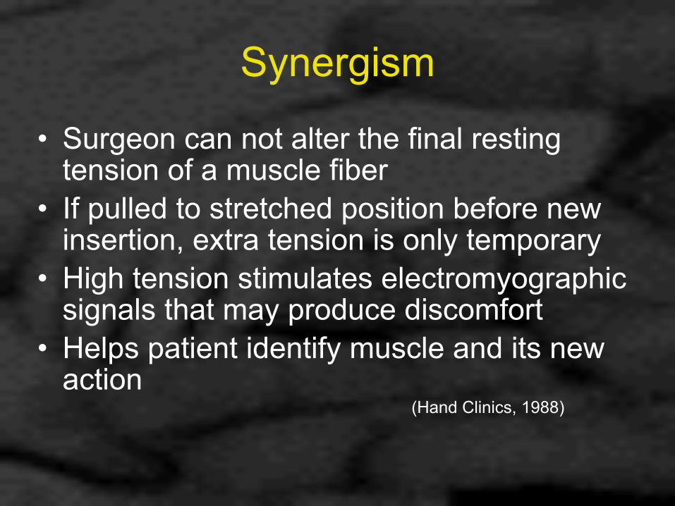
Synergism
• Surgeon can not alter the final resting tension of a muscle fiber
• If pulled to stretched position before new insertion, extra tension is only temporary
• High tension stimulates electromyographic signals that may produce discomfort
• Helps patient identify muscle and its new action
(Hand Clinics, 1988)

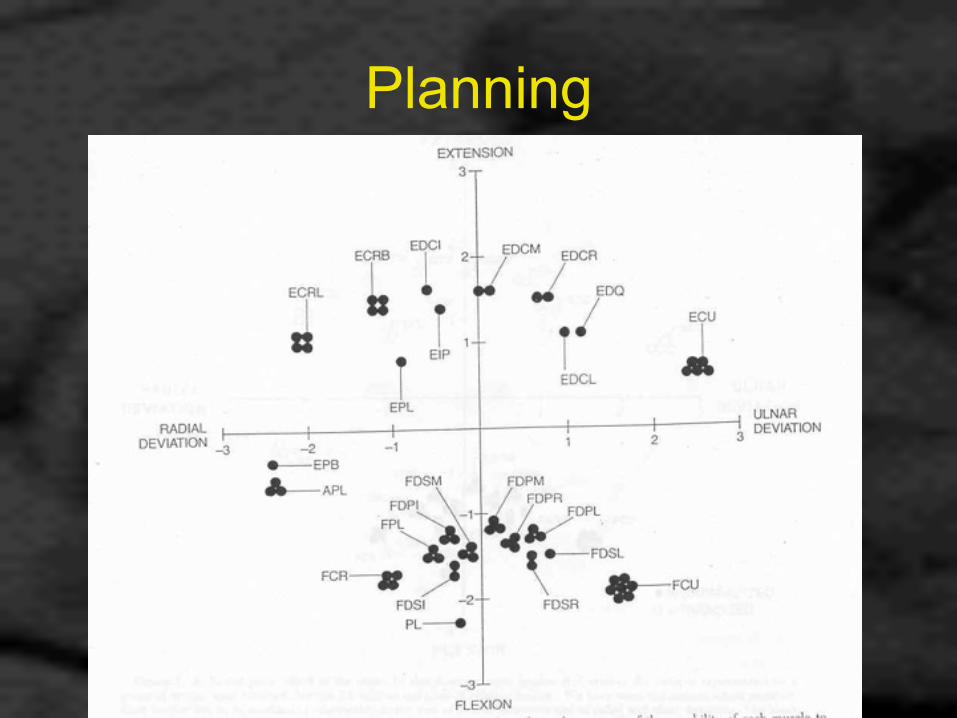
Planning
• Each muscle needs an opposing muscle to achieve its own best output
• Must consider this balance when planning transfers
• Do not just list “paralyzed” muscles and “available” muscles

Planning
• Factors to consider: – Patient needs – Available muscles for transfer
• Unparalyzed • Least deficit • Strength • Excursion

Strength vs. Excursion
• Stength – Potential for creating tension – Lack of strength can be compensated for with therapy
• Excursion – Distance through which tension can be maintained – If too short, can not lengthen with therapy – Can compensate (extrafunctional excursion) by flexing
one joint in series while extending others
Planning

Planning

Planning

Planning

Change in Muscle Following Transfer
• Myth: Muscle strength is decreased following transfer !
• Maximum tension capability – Based on cross-sectional area of all fibers – Not changed by transfer

Change in Muscle Following Transfer
• What may change is range of excursion – Affected by adhesions – Scarring leads to resistance

Resistance
• Only when tendons pass around curves do they acquire synovial sheath
• Elsewhere, surrounded by connective tissue attached to nonmoving tissues by scar
• Nature of nonmoving tissues greatly affects results – Bone, fascia ➔ poor result – Subcutaneous fat ➔ better result

Resistance
• Scar adhesion is the most common cause of failure of tendon transfers – Use only blunt instruments to tunnel – Avoid “raw” surfaces – When scar forms to compliant tissue, still get
excursion

Too Many Joints, Too Few Muscles
• In severe paralysis, don’t be too ambitious • Trying to restore motion to all joints results
in weakness an secondary deformity • Better to achieve a few well supported
actions • Tailor to patient’s needs • Consider joint fusion to provide stability
Remember
• When one muscle is used as a motor: – All insertions move together every time one of
them moves – All insertions move the same distance
• Example: PT to ECRB and ECU – ECRB must move twice as far for given wrist motion as
ECU (twice the moment arm) – ECRB will become slack as soon as wrist extension
intiated (held back by ECU) – Wrist will always ulnar deviate with extension

General Principles of Tendon Transfer
• Restore sensibility first – Primary nerve repair – Nerve grafting – Ensure recovery in progress prior to initiating
reconstruction by tendon transfer • Gain tissue equilibrium in the extremity
– Edema – Scar – Joint stiffness

Principles of Tendon Transfer for Combined Nerve Injuries
• Individualize, don’t “cookbook” • Carefully plan multiple operations • Combine multiple transfers so that
postoperative rehabilitation can be unified by a single treatment goal – Transfer muscles with reasonably similar pre-
and post-transfer function

Principles of Tendon Transfer for Combined Nerve Injuries
• Avoid crossing tendons if possible • If must cross, consider Hunter rod at time
of initial transfer with crossing tendon transfer done through newly created sheath at later stage
(Eversmann, 1988)

Principles of Tendon Transfer for Combined Nerve Injuries
• Do not use functional muscles to stabilize while awaiting reinnervation – i.e. do not use “internal splints” – Fewer muscle-tendon units available
• Avoid use of reinnervated muscle for tendon transfer
• Avoid spastic muscle

Principles of Tendon Transfer for Combined Nerve Injuries
• In general, results from tendon transfers for multiple nerve injuries are not as good as those for single nerve injuries
• Goal of reconstruction is a good helper hand

Dynamic Tenodesis
• Excursion of a tendon transfer can be augmented if that transfer crosses the wrist and can be enhanced by wrist motion
Low (Distal) Median-Ulnar Nerve Palsy
• Most common combined nerve injury in upper extremity
• Complete intrinsic muscle paralysis – Claw hand deformity
• Volar sensory loss all digits

Low (Distal) Median-Ulnar Nerve Palsy
• Flat transverse palmar arch
• MP hyperextension • PIP hyperflexion • Adduction contracture first
web • Patient flexes wrist to get
finger extension (functional tenodesis) may result in flexion contracture of wrist
(J Hand Surg, 1983)

Low (Distal) Median-Ulnar Nerve Palsy
• Basic requirements – Key pinch
• Strong thumb adductor – Thumb abduction/opposition – Tip pinch between thumb and index – PIP power flexion – Coordination of MP and IP motion – Sensibility for key or tip pinch
• Repair median nerve

Low (Distal) Median-Ulnar Nerve Palsy
• Radial-innervated muscles available for transfer – ECRB – ECRL – BR – EIP – EDM
• Only if EDC to small is present – APL
• FDS may also be available for transfer, but tendons often lacerated with initial injury – Repair tendons first, then harvest later for transfer

Low (Distal) Median-Ulnar Nerve Palsy
• Restore sensation first • Splinting to avoid
– Adduction contracture of thumb – PIP flexion contracture of fingers
• All transfers for low median-ulnar nerve palsy can be done at one time

Low (Distal) Median-Ulnar Nerve Palsy

Low (Distal) Median-Ulnar Nerve Palsy
• Tailor reconstruction to patient needs • Emphasis on strength of grasp, pinch or
effective finger position (more intrinsic plus)

Main Functional Loss - Grasp
• Easy fatiguability • Difficulty opening jars • Most effective transfer
– ECRL transfer using 4-tailed free tendon graft – Pass grafts volar to transverse metacarpal lig – Insert into flexor sheath (A2 pulley) or radial side of
proximal phalanx (bone insertion) – Provides power grip – With wrist flexion, no extension at MP – If undesirable, FDS to A1 pulley (Zancolli Lasso)
(J Bone Joint Surg, 1973)

Main Functional Loss - Pinch
• Difficulty pushing buttons • Difficulty turning keys, opening soda cans • Need two intrinsic transfers to regain
control of thumb – Often a third to restore adduction, opposition
and short flexor function

Main Functional Loss - Pinch
• Adductor transfer – FDS (ring) to adductor insertion base of thumb
proximal phalanx – Passed deep to flexor tendons – If FDS (ring) not available:
• FDS (middle) • ECU with tendon graft • BR with tendon graft passed between index and
middle metacarpals • ECRB with tendon graft passed between middle
and ring metacarpals inserted on APB tendon

Main Functional Loss - Pinch
• Opposition – EIP – Rerouted around ulnar border of hand
(pisiform pulley), across thenar eminence, inserted on APB, or looped around thumb extensors
– So effective, hard to recommend alternative • FDS (ring) to APB • PL to APB


5 days post-op abduction & opposition

4 week post-op abduction & opposition

Main Functional Loss - Pinch
• Thumb MP fusion – Negates need for short flexor in intrinsically
deprived thumb – Provides stability for adductor and opponens
transfers, which can be dampened by thumb MP flexion

Main Functional Loss - Pinch
• Radial stability to index finger – If ECRL 4-tailed graft insufficient – BR extended with tendon graft to radial side of
index finger (Plast Reconstr Surg, 1984)

High (Proximal) Median-Ulnar Nerve Palsy
“The hand will rarely be used for precision activities following this severe injury, even if minimal muscle balance is resored.” !
Green’s Operative Hand Surgery Fourth Edition

High (Proximal) Median-Ulnar Nerve Palsy
• Goals of reconstruction: – Helper hand – Simple grasp – Some ability to pinch
• Mostly key pinch
• Utilize dynamic tenodesis – Requires full wrist range of motion

High (Proximal) Median-Ulnar Nerve Palsy

High (Proximal) Median-Ulnar Nerve Palsy
• Finger flexion – ECRL to FDP
• Anastomose tendons of FDP together before inserting ECRL
• Resting cascade with index slightly more extended than middle and so on
• Transfer proximal to carpal tunnel in distal 1/3 of forearm with ECRL rerouted around radial side of forearm

ECRL to FDP

High (Proximal) Median-Ulnar Nerve Palsy
• Thumb flexion – BR to FPL
• Disadvantage: crosses multiple joints • Can fixate to bone distal to elbow joint to eliminate
mechanical disadvantage • Set tension to again take advantage of tenodesis
effect of wrist extension • Arthrodesis of thumb MP strengthens both tip and
key pinch

High (Proximal) Median-Ulnar Nerve Palsy
• Wrist flexion – ECU to FCU
• To provide some wrist flexion • ECU excursion 1/3 that of FCU normally • Do not compromise wrist extension needed for
dynamic tenodesis

High (Proximal) Median-Ulnar Nerve Palsy
• Opposition – EIP around pisiform
pulley to APB and EPL insertions (Riordan’s insertion) (Bull Hosp Jt Dis, 1984)
– Pass through tunnel in denervated thenar muscles to prevent bowstringing
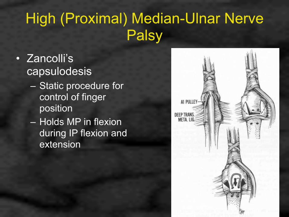
High (Proximal) Median-Ulnar Nerve Palsy
• Zancolli’s capsulodesis – Static procedure for
control of finger position
– Holds MP in flexion during IP flexion and extension

High (Proximal) Median-Ulnar Nerve Palsy
• Sensibility – Fillet flap, index finger
• Superficial radial branch brings protective sensation to first web space (J Bone Joint Surg, 1969)
– Neurovascular island flap
• FDMA + superficial radial nerve
• Can provide protective sensation to thumb pulp

High (Proximal) Ulnar-Radial Nerve Palsy
• Retain radio-volar sensibility • Few available muscle-tendon units to
reconstruct functional deficit • Staged reconstruction • Difficult rehab
– Restore both finger flexion and extension – Restore both thumb adduction and abduction

High (Proximal) Ulnar-Radial Nerve Palsy

High (Proximal) Ulnar-Radial Nerve Palsy
• Wrist extension – PT to ECRL/ECRB
• First described by Jones in 1917 ( Notes on Military Orthopedics, 1917)
• Problem: – With loss of FCU (sparing FCR) get significant radial
imbalance at wrist • Solutions:
– ECRB only insertion » More motion
– Yoke insertion to ECRL and ECRB » Less radial deviation

PT to ECRB

Yoke
• Definition: “1. (n) a wooden frame
or bar with loops or bows at either end, fitted around the necks of a pair of oxen, etc. for harnessing them together…
2. (v) to join together, link.”
Webster’s New World Dictionary, Second Edition

Yoke Insertion
• Detach ECU at musculotendinous junction • Resuture ECU at slightly greater tension
than ECRL to PT transfer • More balanced dorsiflexion position than
simple transfer to ECRB • Less motion than PT to ECRB transfer
(Hand Surgery, 2nd Ed., 1975)

Yoke Insertion
• If injury to ulnar nerve is distal to branch to FCU, yoke insertion of PT is not necessary !
(Orthop Clin North Am, 1974)

High (Proximal) Ulnar-Radial Nerve Palsy
• Finger and thumb extension – FDS (index,middle) to EDC through interosseous
membrane • Must excise 9cm of interosseous membrane • Avoid anterior interosseous nerve and artery
– EDC (middle) anastomosed to EPL – Can use PL for transfer to EPL and APL if present
• Thumb adduction – FDS (ring) to adductor pollicis – Thumb MP arthrodesis

High (Proximal) Ulnar-Radial Nerve Palsy
• Finger flexion – Side-to-side anastomosis of FDP ring and
small to FDP middle – Leave FDP index to have some independent
motion

High (Proximal) Median-Radial Nerve Palsy
• Vast loss of strength and stability of fingers and thumb
• Sensibility only ulnar ½ ring and small fingers
• Reconstruction results in only marginally functional hand only slightly better than prosthesis

High (Proximal) Median-Radial Nerve Palsy
• Must attempt to restore continuity of median nerve
• All wrist motors lost except FCU • Wrist stability can only be gained by
arthrodesis at expense of motion • Finger flexion not enhanced by wrist
extension, so total ROM limited • Wrist arthrodesis provides FCU for use as
tendon transfer

High (Proximal) Median-Radial Nerve Palsy

High (Proximal) Median-Radial Nerve Palsy
• FDP sutured side-to-side for ulnar-innervated mass action – Index and long under
greater tension – Resting fingers in
“straight” rather than “oblique” transverse line

High (Proximal) Median-Radial Nerve Palsy
• Digital extension – FCU to EDC and EPL

Summary
• Nearly all patients with combined nerve palsies will require multiple operations to restore function
• Procedures must be well thought out and carefully planned
• Must design reconstructive plan to meet patient’s specific needs
• Goal is to redistribute assets that are available, not recreate a normal hand
• The more complex the surgical plan, the more likely it is to fail