Embed Size (px)
Citation preview
l s 2
a
e26 d e n t a l m a t e r i a
gel 15 days after treatment. No significant differences werefound between the tested remineralizing gels.
Conclusions: It was concluded that neutral bleaching gelsignificantly reduced enamel microhardness 15 days afterbleaching and the use of remineralizing gels did not signifi-cantly enhance the microhardness of bleached enamel.
doi:10.1016/j.dental.2011.08.461
59Polymerization stress and cuspal deflection of low-shrinkagecommercial composites
L.C.C. Boaro 1,∗, N.R. Froes-Salgado 1, V.E.S. Gajewski 1, A.A.Bicalho 2, A.D.C.M. Valdivia 2, C.J. Soares 2, C.S.C. Pfeifer 3, R.R.Braga 1, W.G. Miranda 1
1 University of São Paulo, Brazil2 University of Uberlandia, Brazil3 University of Colorado, United States
Objectives: The aim of this study was to compare thepolymerization stress and cuspal deflection of low-shrinkagecommercial composites with those of conventional compos-ites.
Materials and methods: Five BisGMA-based composites(Filtek Z250/3M ESPE, Heliomolar/Ivoclar Vivadent, Aelite LSPosterior/Bisco, Filtek Supreme/3M ESPE, ELS/Saremco), aurethane-based (Venus Diamond/Heraeus-Kulzer) and onesilorane-based (Filtek LS/3M ESPE) were tested. Polymeriza-tion stress (n = 5) was determined in 1.5 mm tall specimens,inserted between two glass rods. Nominal stress was cal-culated by dividing the maximum contraction force by thecross-sectional area of the rod. Cuspal deflection (n = 9) wasmeasured using the strain-gage method. Human pre-molarreceived MOD preparations with 4.1(±0.4) mm of width and3.2 (±0.2) mm of height, and 1.5 mm occlusal box. Lingual andbuccal cuspal movement was monitored for 10 min. For bothtests composite was inserted in bulk and photoactivated witha radiant exposure of 18 J/cm2. Data for cuspal deflection wereanalysed using one-way ANOVA/Tukey test, and for stressusing Kruskal–Wallis/Tukey test. In both cases, the global sig-nificance level was 5%.
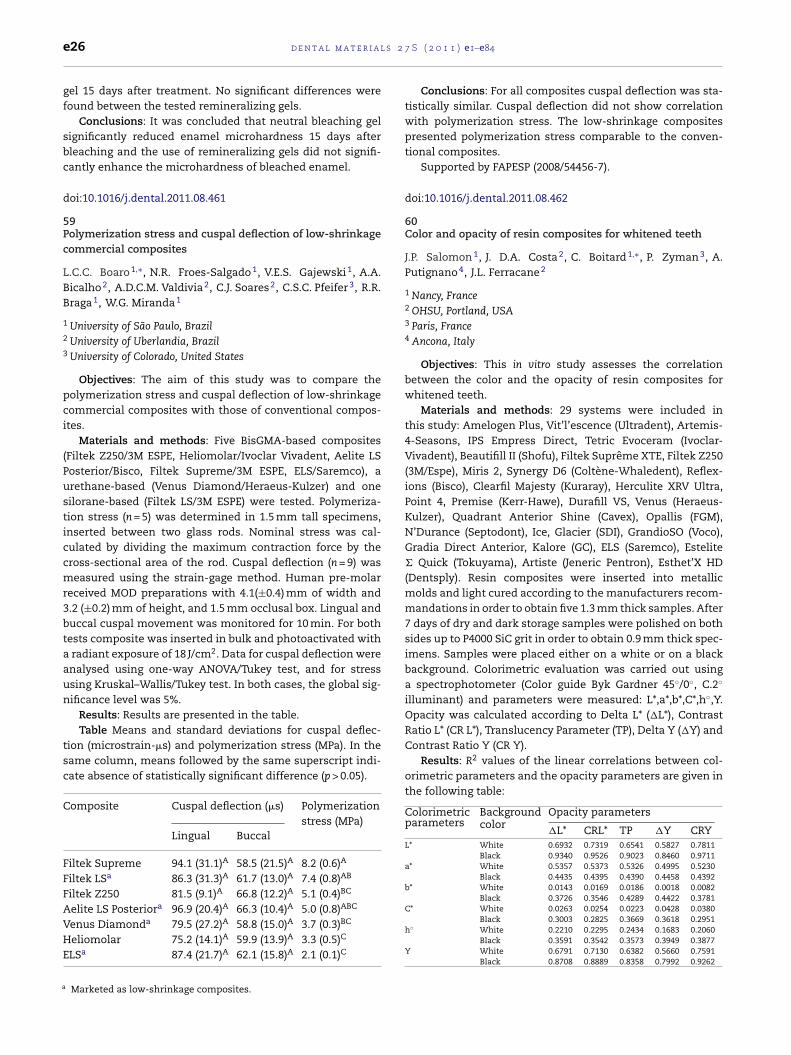
Results: Results are presented in the table.Table Means and standard deviations for cuspal deflec-
tion (microstrain-�s) and polymerization stress (MPa). In thesame column, means followed by the same superscript indi-cate absence of statistically significant difference (p > 0.05).
Composite Cuspal deflection (�s) Polymerizationstress (MPa)
Lingual Buccal
Filtek Supreme 94.1 (31.1)A 58.5 (21.5)A 8.2 (0.6)A
Filtek LSa 86.3 (31.3)A 61.7 (13.0)A 7.4 (0.8)AB
Filtek Z250 81.5 (9.1)A 66.8 (12.2)A 5.1 (0.4)BC
Aelite LS Posteriora 96.9 (20.4)A 66.3 (10.4)A 5.0 (0.8)ABC
Venus Diamonda 79.5 (27.2)A 58.8 (15.0)A 3.7 (0.3)BC
Heliomolar 75.2 (14.1)A 59.9 (13.9)A 3.3 (0.5)C
ELSa 87.4 (21.7)A 62.1 (15.8)A 2.1 (0.1)C
Marketed as low-shrinkage composites.
7 S ( 2 0 1 1 ) e1–e84
Conclusions: For all composites cuspal deflection was sta-tistically similar. Cuspal deflection did not show correlationwith polymerization stress. The low-shrinkage compositespresented polymerization stress comparable to the conven-tional composites.
Supported by FAPESP (2008/54456-7).
doi:10.1016/j.dental.2011.08.462
60Color and opacity of resin composites for whitened teeth
J.P. Salomon 1, J. D.A. Costa 2, C. Boitard 1,∗, P. Zyman 3, A.Putignano 4, J.L. Ferracane 2
1 Nancy, France2 OHSU, Portland, USA3 Paris, France4 Ancona, Italy
Objectives: This in vitro study assesses the correlationbetween the color and the opacity of resin composites forwhitened teeth.
Materials and methods: 29 systems were included inthis study: Amelogen Plus, Vit’l’escence (Ultradent), Artemis-4-Seasons, IPS Empress Direct, Tetric Evoceram (Ivoclar-Vivadent), Beautifill II (Shofu), Filtek Suprême XTE, Filtek Z250(3M/Espe), Miris 2, Synergy D6 (Coltène-Whaledent), Reflex-ions (Bisco), Clearfil Majesty (Kuraray), Herculite XRV Ultra,Point 4, Premise (Kerr-Hawe), Durafill VS, Venus (Heraeus-Kulzer), Quadrant Anterior Shine (Cavex), Opallis (FGM),N’Durance (Septodont), Ice, Glacier (SDI), GrandioSO (Voco),Gradia Direct Anterior, Kalore (GC), ELS (Saremco), Estelite� Quick (Tokuyama), Artiste (Jeneric Pentron), Esthet’X HD(Dentsply). Resin composites were inserted into metallicmolds and light cured according to the manufacturers recom-mandations in order to obtain five 1.3 mm thick samples. After7 days of dry and dark storage samples were polished on bothsides up to P4000 SiC grit in order to obtain 0.9 mm thick spec-imens. Samples were placed either on a white or on a blackbackground. Colorimetric evaluation was carried out usinga spectrophotometer (Color guide Byk Gardner 45◦/0◦, C.2◦
illuminant) and parameters were measured: L*,a*,b*,C*,h◦,Y.Opacity was calculated according to Delta L* (�L*), ContrastRatio L* (CR L*), Translucency Parameter (TP), Delta Y (�Y) andContrast Ratio Y (CR Y).
Results: R2 values of the linear correlations between col-orimetric parameters and the opacity parameters are given inthe following table:
Colorimetricparameters
Backgroundcolor
Opacity parameters
�L* CRL* TP �Y CRYL* White 0.6932 0.7319 0.6541 0.5827 0.7811
Black 0.9340 0.9526 0.9023 0.8460 0.9711a* White 0.5357 0.5373 0.5326 0.4995 0.5230
Black 0.4435 0.4395 0.4390 0.4458 0.4392b* White 0.0143 0.0169 0.0186 0.0018 0.0082
Black 0.3726 0.3546 0.4289 0.4422 0.3781C* White 0.0263 0.0254 0.0223 0.0428 0.0380
Black 0.3003 0.2825 0.3669 0.3618 0.2951h◦ White 0.2210 0.2295 0.2434 0.1683 0.2060
Black 0.3591 0.3542 0.3573 0.3949 0.3877
Y White 0.6791 0.7130 0.6382 0.5660 0.7591Black 0.8708 0.8889 0.8358 0.7992 0.9262
2 7 S
cttsb
d
6Ri
AP
flie
esgb3ifidbsie
saiddbntg
rttgmo
d
d e n t a l m a t e r i a l s
Conclusions: Within the limitations of this in vitro study wean conclude that: 1, only L* and Y are significantly correlatedo the 5 opacity parameters; 2, the couple L* black/CR Y gavehe highest value of R2 (=0.97) and must be considered as atandard for future investigations; 3, R2 values are influencedy the background color.
oi:10.1016/j.dental.2011.08.463
1emineralizing agents on microhardness of sound and dem-
neralized bleached enamel
.B. Borges ∗, C.A. Guimarães, C.J. Ramos, A.L.S. Borges, C.R.ucci, C.R.G. Torres
Universidade Estadual Paulista, UNESP, São José dos Campos, Brazil
Objectives: To evaluate the effect of calcium anduoride addition in 35% hydrogen peroxide (HP) bleach-
ng gel on microhardness of sound and demineralizednamel.
Materials and methods: Cylindrical specimens of bovinenamel (3 mm × 2 mm) were divided in two groups (n = 30):ound enamel (SE) and demineralized enamel (DE). Eachroup was divided in three subgroups, according to theleaching gel modified by manufacturer1: HP 35%; HP5% + calcium gluconate; HP 35% + sodium fluoride. Bleach-ng procedures were performed ex vivo. The specimens werexed in intraoral devices used by 10 volunteers during 7ays in order to evaluate the effect of human saliva onleached enamel. Surface Knoop Microhardness (SMH) ofuperficial enamel was measured before and after bleach-ng treatments, as well as after 1 and 7 days of salivaxposure.
Results: Data were analyzed by RM-ANOVA (5%) andhowed significant difference for time factor, both for SEnd DE groups (p = 0.001). The application of Tukey’s testn SE groups revealed significant lower SMH mean imme-iately after bleaching compared to baseline and after 7ays. For DE specimens, the lowest means were obtained foraseline period. Bleaching with HP + calcium increased sig-ificantly the SMH means immediately after bleaching andhe exposure to saliva resulted in increasing of SMH for allroups.
Conclusions: Bleaching with 35% HP caused significanteduction in SE microhardness and did not exacerbatehe demineralization of initial artificial caries. The addi-ion of potentially remineralizing agents in the bleachingels can play an important role in the maintenance oficrohardness values of SE and to induce remineralization
f DE.
oi:10.1016/j.dental.2011.08.464
1 FGM Produtos Odontológicos.
( 2 0 1 1 ) e1–e84 e27
62A novel at-home bleaching technique modified by a CPP-ACPpaste
B.C. Borges 1,∗, J.S. Borges 2, C.D. Melo 2, I.V. Pinheiro 2, A.J.Santos 2, R. Braz 1, M.A. Montes 1
1 University of Pernambuco, Camaragibe, Brazil2 Federal University of Rio Grande do Norte, Natal, Brazil
Objectives: This study was designed to evaluate in vitrothe efficacy of a novel at-home bleaching technique using 10%or 16% carbamide peroxides modified by a CPP-ACP paste (MIPaste, GC Corporation) and its influence on the microhardnessof bleached enamel.
Materials and methods: Forty bovine incisors were dividedinto four groups (n = 10) according to the bleaching agent uti-lized: 10% carbamide peroxide only; a blend of 10% carbamideperoxide and MI paste; 16% carbamide peroxide only; and ablend of 16% carbamide peroxide and MI Paste. During the14-day bleaching regimen, the samples were stored in artifi-cial saliva. The Vickers microhardness and color of the teethwere assessed at baseline (T0) and immediately after thebleaching regimen (T14) using a microhardness tester and aspectrophotometer, respectively. The degree of color changewas determined by the CIEL*a*b* system (�E, �L*, �a* and �b*)and Vita shade guide parameters. The data was analyzed byanalysis of variance and Tukey’s test (p < 0.05).
Results: The teeth that were bleached with a blend of perox-ides (10% or 16%) and the CPP-ACP paste presented increasedmicrohardness values at T14 compared with T0, whereas thesamples that were bleached with peroxides only did not showany differences in their microhardness values. All the bleach-ing agents were effective at whitening the teeth and did notshow a statistically significant difference using the CIEL*a*b*system (�E, �L*, �a* and �b*) or the Vita shade guide param-eters.
Conclusions: The use of a CPP-ACP paste with carbamideperoxide bleaching agents increased the bleached enamel’smicrohardness and did not have an influence on whiteningefficacy. This improves safety and might even reduce in vivotooth sensitivity during the bleaching process.
doi:10.1016/j.dental.2011.08.465
63Polymerization stress assessment by crack analysis andmechanical testing
R.R. Braga 1,∗, T. Yamamoto 2, K. Tyler 3, L.C. Boaro 1, J.L.Ferracane 4, M.V. Swain 3
1 University of São Paulo, Brazil2 Tsurumi University, Japan3 University of Sydney, Australia4 Oregon Health & Science University, USA
Objectives: The aim of this study was to verify the workinghypothesis that two methods for determining polymerizationcontraction stress, analysis of cracks from hardness indents
and axial loading, would rank a series of commercial restora-tive composites in a similar order, and that both tests would