Embed Size (px)
Citation preview
Doctoral thesis for the Degree of Doctor of Philosophy, Faculty of Medicine
Colonic Barrier Function in Ulcerative Colitis
Interactions Between Ion and Mucus Secretion
Jenny K. Gustafsson
Institute of Neuroscience and Physiology Department of Physiology Sahlgrenska Academy University of Gothenburg 2012
Jenny Gustafsson
2
A doctoral thesis at a University in Sweden is produced either as a monograph or as a collection of papers. In the latter case, the introductory part constitutes the formal thesis, which summarizes the accompanying papers. These have already been published or are manuscripts at various stages (in press, submitted, or manuscript).
ISBN: 978-91-628-8442-0 URL: http://hdl.handle.net/2077/28487 © Jenny K. Gustafsson University of Gothenburg Institute of Neuroscience and Physiology Department of Physiology Sahlgrenska Academy SWEDEN Printed by Kompendiet Gothenburg, Sweden 2012 Cover illustration: Top left: scanning electron micrograph of a human colonic crypt. Top right: mouse colon stained with Calcein violet blue: Bottom left: Human colon stained against MUC2. Bottom right: Transmission electron micrograph of a goblet cell.
Colonic barrier function in ulcerative colitis
3
Till Farmor
Jenny Gustafsson
4
ABSTRACT
Jenny K. Gustafsson Institutes of Neuroscience and Physiology and Biomedicine, Departments of Physiology and Medical Biochemistry and Cell Biology, Sahlgrenska Academy at University of Gothenburg Anion and mucus secretions have traditionally been looked upon as two separate parts of the epithelial defense system. The importance of anion secretion has been attributed to its role in creating the driving force for fluid secretion that flushes the epithelium from bacteria, while mucus secretion ensures protection via the mucus layer that forms a physical barrier between the bacteria and the epithelium. In addition to its role in fluid secretion it is becoming increasingly clear that anion secretion contributes to the regulation of mucus properties. This opens up for the possibility that alterations in epithelial transport can regulate the colonic barrier also via its effects on the mucus layer. The aim of the present thesis was to clarify how epithelial anion secretion regulates the intestinal mucus layer, and to delineate how these two systems are affected in Ulcerative colitis, the most common chronic inflammatory bowel disease.
By using an in house developed ex vivo method for the study of mucus properties, it was shown that CFTR mediated bicarbonate secretion regulates many aspects of mucus properties in the mouse small intestine, including mucus growth, adherence and penetrability. In the colon, baseline mucus growth was CFTR independent whereas secretagogue (carbachol) induced mucus growth required a functioning CFTR channel. The impaired mucus growth seen in mice lacking a functional CFTR channel was probably not due to reduced mucus secretion since the exocytosis response to carbachol was unaffected. In WT colon, carbachol induced mucus exocytosis required functioning basolateral transport via NKCC1 and K+ channels.
To test how epithelial transport and mucus properties were affect by inflammation, the barrier properties of the colonic mucus were studied in various murine colitis models (IL10-/-, TLR5-/-, NHE3-/-, C1GalT-/- and DSS induced colitis) and in UC patients. The results showed that all tested colitis models had signs of a defective mucus barrier, defined as abnormal amounts of bacteria in contact with the epithelium. Alterations in the mucus layer were also found in the human colon. Colonic biopsies from control patients secreted mucus that separated beads the size of bacteria from the epithelium, whereas biopsies from UC patients with acute disease secreted mucus that was penetrable to the beads. The majority of UC patients in remission secreted mucus with normal penetrability, while a subset of patients secreted mucus that was permeable to the beads. Also epithelial anion secretion was normal in the distal colon of UC patients in remission, but in the proximal colon the reactivity to secretagogues was shifted towards an increased forskolin response and a decreased carbachol response.
In summary, the results from this thesis show that acute colitis makes the colonic mucus layer unable to physically separate bacteria from the epithelium. In the small intestine, CFTR mediated secretion regulates most aspect of mucus properties while in the colon only secretagogue-induced mucus growth seems to be CFTR dependent. In ulcerative colitis in remission, the epithelium of the proximal colon becomes more reactive to stimulation of the CFTR system, which may be a defense mechanism to reduce the degree of contact between bacteria and epithelium. Key words: intestine, mucus, ion transport, colitis, CFTR
Colonic barrier function in ulcerative colitis
5
LIST OF PAPERS
This thesis is based on the following papers, published or as manuscripts which are referred to in the text by their roman numerals:
I. Gustafsson JK, Ermund A, Johansson MEV, Schütte A, Hansson GC, and Sjövall H. An ex vivo method for studying mucus formation, properties and thickness in human colonic biopsies and mouse small and large intestinal explants Am J Physiol Gastrointest Liver Physiol. 2012 Feb;302(4):G430-8
II. Gustafsson JK, Alwan A, Scholte BJ, Hansson GC, Lindén SK and Sjövall H
Relation between carbachol induced anion and mucus secretion in the murine colon Manuscript
III. Gustafsson JK, Ermund A, Ambort D, Johansson MEV, Nilsson HE, Thorell K,
Hebert H, Sjövall H and Hansson GC Bicarbonate and functional CFTR channel are required for proper mucin secretion and link cystic fibrosis with its mucus phenotype Submitted to Journal of Experimental Medicine
IV. Gustafsson JK, Hansson GC and Sjövall H
Ulcerative colitis patients in remission have an altered secretory capacity in the proximal colon despite a macroscopically normal mucosa Submitted to Neurogastroenterology and Motility
V. Johansson MEV, Gustafsson JK, Holmén-Larsson J, Jabbar KS, Xia L, Xu H,
Ghishan FK, Carvalho FA, Gewirtz AT, Sjövall H and Hansson GC Bacteria penetrate the inner colon mucus layer in both murine colitis models and in patients with ulcerative colitis Manuscript
Reprints were made with permission from the publisher
Jenny Gustafsson
6
Colonic barrier function in ulcerative colitis
7
CONTENTS
HISTORICAL PERSPECTIVE ......................................................................................................................................................................... 9
BACKGROUND ............................................................................................................................................................................................. 10
The colonic epithelium ............................................................................................................................................................................... 11
The enteric nervous system ........................................................................................................................................................................ 11
Regulation of secretomotor function ..................................................................................................................................................... 11
Epithelial transport ..................................................................................................................................................................................... 12
Absorption ............................................................................................................................................................................................ 13
Secretion ............................................................................................................................................................................................... 13
The intestinal mucus layer .......................................................................................................................................................................... 16
The MUC2 mucin ................................................................................................................................................................................. 17
Mucus secretion .................................................................................................................................................................................... 17
Regulation of mucus properties – lessons from cystic fibrosis .............................................................................................................. 20
The mucosal immune system...................................................................................................................................................................... 21
Ulcerative colitis......................................................................................................................................................................................... 23
Pathophysiology .................................................................................................................................................................................... 24
AIM OF THESIS ............................................................................................................................................................................................. 27
METHODOLOGICAL CONSIDERATIONS ................................................................................................................................................. 28
Materials and ethics .................................................................................................................................................................................... 28
Human subjects (Paper I, IV, V) ........................................................................................................................................................... 28
Animal studies (Paper I-V) ................................................................................................................................................................... 29
Methodology .............................................................................................................................................................................................. 31
Ex vivo measurements of mucus properties (Paper I, II, III and V) ...................................................................................................... 31
Ussing chamber in combination with square wave current analysis (Paper II, IV) ................................................................................ 33
In situ hybridization (Paper V) .............................................................................................................................................................. 36
Immunohistochemistry (Paper I, III, V) ................................................................................................................................................ 36
Electron microscopy (Paper I, III) ......................................................................................................................................................... 36
RESULTS AND COMMENTS ....................................................................................................................................................................... 38
Colonic mucus secretion – regulation by ion transport (Paper II) ............................................................................................................... 38
Regulation of mucus properties in the small and large intestine (Paper I and III) ....................................................................................... 40
Role of CFTR and bicarbonate in the regulation of intestinal mucus properties (Paper II and III) ............................................................. 42
Carbachol induced anion and mucus secretion in the normal and diseased human colon (Paper IV) ......................................................... 46
The colonic mucus layer and inflammation (Paper V) ................................................................................................................................ 48
GENERAL DISCUSSION .............................................................................................................................................................................. 52
CONCLUDING REMARKS ........................................................................................................................................................................... 55
FUTURE PERSPECTIVES ............................................................................................................................................................................. 56
POPULÄRVETENSKAPLIG SAMMANFATTNING ................................................................................................................................... 57
ADDITIONAL BIBLIOGRAPHY .................................................................................................................................................................. 58
ACKNOWLEDGMENTS ............................................................................................................................................................................... 59
REFERENCES ................................................................................................................................................................................................ 61
Jenny Gustafsson
8
ABBREVIATIONS
Ach acetylcholine C1GalT core 1 glycosyltransferase CaCC calcium activated chloride channel CCh carbachol CF cystic fibrosis CFTR cystic fibrosis transmembrane conductance regulator ChAT choline acetyltransferase Cp epithelial capacitance CTX charybdotoxin DIDS 4,4'-Diisothiocyano-2,2'-stilbenedisulfonic acid DSS dextran sodium sulphate ENaC epithelial sodium channel 5-HT serotonin IgA immunoglobulin A
IFN-γ interferon-gamma IL interleukin Im net membrane current Isc short circuit current ISN intrinsic sensory neuron KO knock out LPS lipopolysaccharide MUC mucin NBC Na+-HCO3
- co-transporter NHE sodium-hydrogen exchanger NKCC1 Na+-K+-Cl- co-transporter PD transepithelial potential difference PGE2 prostaglandin E2 PKC protein kinase C Rp epithelial resistance Rs subepithelial resistance RT total tissue resistance SCFA short chain fatty acid Slc9a3 solute carrier family 9 member 3 TEM transmission electron microscopy
TGF-β transforming growth factor-beta Th T helper cell TLR toll-like receptor Treg regulatory T cell UC ulcerative colitis V0 voltage response at time zero VIP vasoactive intestinal peptide VPAC vasoactive intestinal polypeptide receptor WT wild type
Colonic barrier function in ulcerative colitis
9
HISTORICAL PERSPECTIVE
The biological function of the colon was for a long time mainly considered to be to act as a storage unit for digestive waste products. In the early 1900s physicians began to shift their views towards the possibility that the functions of the colon were more complex than previously thought. Studies had shown that the colonic mucosa actively reabsorbed fluids, and it was suggested that the colonic bacteria may contribute to the digestive process rather than being solely harmful and pathogenic (Goodhart, 1913). In the 1920s the role of the colonic mucosa in fluid absorption was well established and reports started to mention the colonic mucus as being important for lubrication and mucosal protection. The idea that the mucus layer was important for epithelial protection emerged from observations that colonic irritants and mechanical stress induced mucus secretion (Hurst, 1922). Until the 1960s most of the knowledge in the field of colonic function in humans was obtained from case reports describing different aspects of colonic physiology and pathophysiology, including motility patterns during diarrhea and constipation and the histological characteristics of ulcerative colitis (Bonoff, 1939; Truelove et al., 1955). In the late 1960s and 1970s more detailed information was obtained regarding regulation of ion transport. At the same time the intestinal barrier emerged as a concept and experimental studies using animal models of colitis were described (Watt & Marcus, 1973). In the following years studies of regulation of intestinal mucus secretion were performed which established that both mucus and anion secretion were regulated by the enteric nervous system (Specian & Neutra, 1980; Dharmsathaphorn & Pandol, 1986; Kuwahara et al., 1989). Despite the interest in the intestinal mucus in the 1980s this aspect of colonic function fell into oblivion after the initial characterization, while studies of the molecular aspects of anion secretion continued and are still being extensively pursued. During the last decade a renewed interest has emerged around the colonic mucus layer as an important part of the colonic defense system after studies had shown that mice lacking the main component of the colonic mucus layer, the Muc2 mucin, develop spontaneous colitis (Van der Sluis et al., 2006).
Although we know more about the regulation of colonic function today than one century ago there are still a large number of questions regarding the different aspects of colonic physiology that remain unanswered.
Jenny Gustafsson
10
BACKGROUND
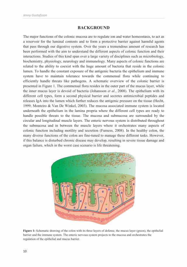
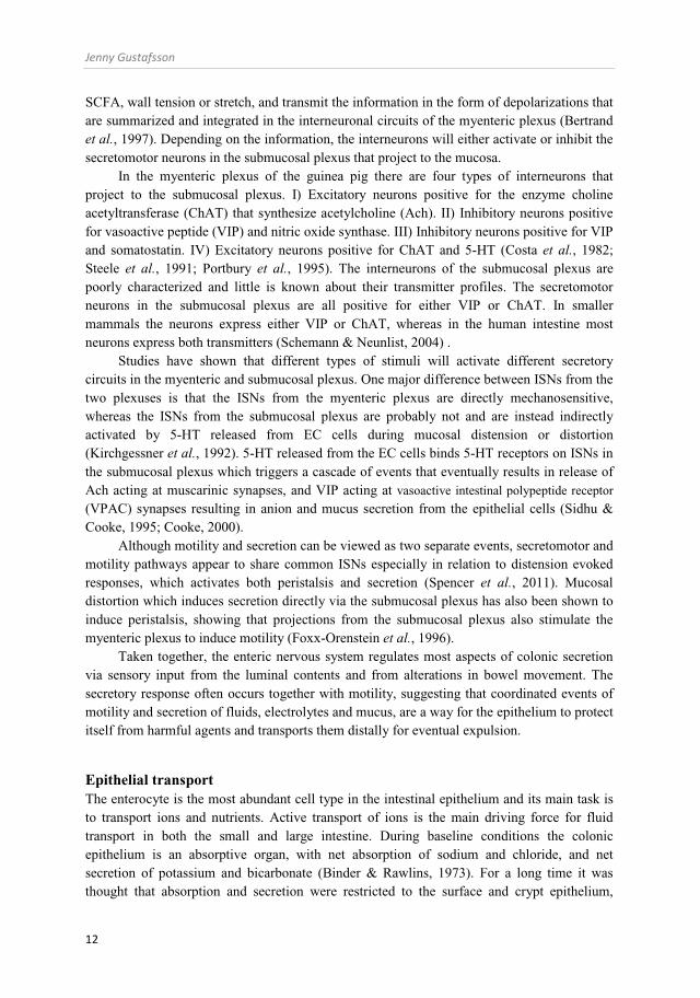
The major functions of the colonic mucosa are to regulate ion and water homeostasis, to act as a reservoir for the luminal contents and to form a protective barrier against harmful agents that pass through our digestive system. Over the years a tremendous amount of research has been performed with the aim to understand the different aspects of colonic function and their interactions. Studies of this kind span over a large variety of disciplines such as microbiology, biochemistry, physiology, neurology and immunology. Many aspects of colonic functions are related to the ability to coexist with the huge amount of bacteria that reside in the colonic lumen. To handle the constant exposure of the antigenic bacteria the epithelium and immune system have to maintain tolerance towards the commensal flora while continuing to efficiently handle threats like pathogens. A schematic overview of the colonic barrier is presented in Figure 1. The commensal flora resides in the outer part of the mucus layer, while the inner mucus layer is devoid of bacteria (Johansson et al., 2008). The epithelium with its different cell types, form a second physical barrier and secretes antimicrobial peptides and releases IgA into the lumen which further reduces the antigenic pressure on the tissue (Hecht, 1999; Monteiro & Van De Winkel, 2003). The mucosa associated immune system is located underneath the epithelium in the lamina propria where the different cell types are ready to handle possible threats to the tissue. The mucosa and submucosa are surrounded by the circular and longitudinal muscle layers. The enteric nervous system is distributed throughout the submucosa and in between the muscle layers where it orchestrates many aspects of colonic function including motility and secretion (Furness, 2008). In the healthy colon, the many diverse functions of the colon are fine-tuned to manage these different tasks. However, if this balance is disturbed chronic disease may develop, resulting in severe tissue damage and organ failure, which in the worst case scenario is life threatening.
Figure 1: Schematic drawing of the colon with its three layers of defense, the mucus layer (green), the epithelial barrier and the immune system. The enteric nervous system projects to the mucosa and orchestrates the regulation of the epithelial and mucus barrier.
Colonic barrier function in ulcerative colitis
11
The colonic epithelium The intestinal epithelium is a rapidly self-renewing tissue maintained by a population of stem cells that resides at the base of the crypt. These stem cells divide and differentiate into five separate cell types; columnar cells, goblet cells, enterochromaffin cells, tuft cells and Paneth cells (Cheng & Leblond, 1974; Barker et al., 2007; Gerbe et al., 2011). All cell types except Paneth cells migrate from the proliferative zone at the base of the crypt towards the surface epithelium. During the migration towards the surface the cells differentiate and reach terminal differentiation at the crypt neck. The cell types that migrate towards the surface epithelium have an average life span of 4-5 days after which they undergo apoptosis and are shed into the intestinal lumen (Chang & Leblond, 1971). The Paneth cells are mainly found in the small intestine and to some extent in the proximal colon, where they are found scattered in between the stem cells at the bottom of the crypts (Sato et al., 2011). In the colon, secretory cells can be found at the same position as the Paneth cells and it was recently shown that these cells have a similar role in regulation of stem cell proliferation and survival that previously has been shown for the Paneth cells in the small intestine (Sato et al., 2011; Rothenberg et al., 2012). The structural and functional integrity of the intestine is largely dependent on a constant movement and maturation of all these different cell types along the crypt surface axis. Regulation of epithelial functions is to a large extent mediated via the enteric nervous system through: secretion of fluids and electrolytes from the columnar cells; mucus secretion from the goblet cells and secretion of antimicrobial peptides from the Paneth cells (Specian & Neutra, 1980; Ouellette, 1999; Osbak et al., 2007). At the same time the epithelium signals to the enteric nervous system via secretion of serotonin (5-HT) from the enterochromaffin (EC) cells (Cooke, 2000).
The enteric nervous system The enteric nervous system is a key regulator of most intestinal functions such as motility and secretion and is organized in two plexuses; the Myenteric (Auerbach’s) and submucosal (Meissner’s) plexus. The Myenteric plexus is located in between the longitudinal and circular muscle and its primary function is to regulate motility. The submucosal plexus is the primary regulator of fluid, electrolyte and mucus secretion and is located in the submucosa. Although the submucosal plexus contains complete secretory circuits some stimuli require activation of myenteric ganglia for a full secretory response and these types of stimuli often trigger both secretion and peristalsis (Goyal & Hirano, 1996).
Regulation of secretomotor function In the colon, most aspects of secretion are regulated by the enteric nervous system which sets both the basal tone and induces an appropriate response to various types of physiological stimuli, like short chain fatty acids (SCFA), bacterial toxins, distension and mucosal distortion (Yajima, 1988; Diener & Rummel, 1990; Christofi et al., 2004; Alzamora et al., 2011). The secretory reflex response involves three types of neurons, the intrinsic sensory neurons (ISNs), the interneurons and the secretomotor neuron (Cooke, 1998). The ISNs that have their cell bodies in the myenteric plexus respond directly to physiological stimuli such as
Jenny Gustafsson
12
SCFA, wall tension or stretch, and transmit the information in the form of depolarizations that are summarized and integrated in the interneuronal circuits of the myenteric plexus (Bertrand et al., 1997). Depending on the information, the interneurons will either activate or inhibit the secretomotor neurons in the submucosal plexus that project to the mucosa.
In the myenteric plexus of the guinea pig there are four types of interneurons that project to the submucosal plexus. I) Excitatory neurons positive for the enzyme choline acetyltransferase (ChAT) that synthesize acetylcholine (Ach). II) Inhibitory neurons positive for vasoactive peptide (VIP) and nitric oxide synthase. III) Inhibitory neurons positive for VIP and somatostatin. IV) Excitatory neurons positive for ChAT and 5-HT (Costa et al., 1982; Steele et al., 1991; Portbury et al., 1995). The interneurons of the submucosal plexus are poorly characterized and little is known about their transmitter profiles. The secretomotor neurons in the submucosal plexus are all positive for either VIP or ChAT. In smaller mammals the neurons express either VIP or ChAT, whereas in the human intestine most neurons express both transmitters (Schemann & Neunlist, 2004) .
Studies have shown that different types of stimuli will activate different secretory circuits in the myenteric and submucosal plexus. One major difference between ISNs from the two plexuses is that the ISNs from the myenteric plexus are directly mechanosensitive, whereas the ISNs from the submucosal plexus are probably not and are instead indirectly activated by 5-HT released from EC cells during mucosal distension or distortion (Kirchgessner et al., 1992). 5-HT released from the EC cells binds 5-HT receptors on ISNs in the submucosal plexus which triggers a cascade of events that eventually results in release of Ach acting at muscarinic synapses, and VIP acting at vasoactive intestinal polypeptide receptor
(VPAC) synapses resulting in anion and mucus secretion from the epithelial cells (Sidhu & Cooke, 1995; Cooke, 2000).
Although motility and secretion can be viewed as two separate events, secretomotor and motility pathways appear to share common ISNs especially in relation to distension evoked responses, which activates both peristalsis and secretion (Spencer et al., 2011). Mucosal distortion which induces secretion directly via the submucosal plexus has also been shown to induce peristalsis, showing that projections from the submucosal plexus also stimulate the myenteric plexus to induce motility (Foxx-Orenstein et al., 1996).
Taken together, the enteric nervous system regulates most aspects of colonic secretion via sensory input from the luminal contents and from alterations in bowel movement. The secretory response often occurs together with motility, suggesting that coordinated events of motility and secretion of fluids, electrolytes and mucus, are a way for the epithelium to protect itself from harmful agents and transports them distally for eventual expulsion.
Epithelial transport The enterocyte is the most abundant cell type in the intestinal epithelium and its main task is to transport ions and nutrients. Active transport of ions is the main driving force for fluid transport in both the small and large intestine. During baseline conditions the colonic epithelium is an absorptive organ, with net absorption of sodium and chloride, and net secretion of potassium and bicarbonate (Binder & Rawlins, 1973). For a long time it was thought that absorption and secretion were restricted to the surface and crypt epithelium,
Colonic barrier function in ulcerative colitis
13
respectively. However, this paradigm had to be revised after compelling evidence that both the surface epithelium and the crypts are able to secrete and absorb ions and fluids (Geibel, 2005). Although spatial differences in transport function do occur, this pattern can be shifted with the appropriate stimuli (Jakab et al., 2011).
Absorption One of the major functions of the colonic mucosa is to reabsorb fluids secreted from the more proximal parts of the gastrointestinal tract. In the colon, fluid absorption depends on active transport of electrolytes and solutes that creates the osmotic gradients for water absorption. Absorption can occur via electroneutral or electrogenic uptake. Electroneutral absorption operates via Na+/H+ exchange (i.e. NHE3 and NHE2) coupled to either Cl-/HCO3
- exchange or SCFA/HCO3
- exchange, whereas electrogenic absorption involves the epithelial sodium channel (ENaC) in combination with Cl-/HCO3
- exchange or passive uptake of chloride via the paracellular shunt (Garty & Palmer, 1997; Kunzelmann & Mall, 2002; Zachos et al., 2005). Since neither of the apical sodium transporters are able to operate against an electrochemical gradient, net sodium absorption requires active transport via the basolateral sodium-potassium ATPase (Na+/K+-ATPase). The Na+/K+-ATPase transports three sodium ions out of the cell in exchange for two potassium ions at the expense of one ATP molecule (Kirk et al., 1980). This process maintains a low intracellular sodium concentration and high intracellular potassium concentration, resulting in an electronegative cell interior compared to the extracellular fluid. This low intracellular sodium concentration and electronegative negative cell interior favors uptake of the positive sodium ion over the apical membrane. To maintain the electronegative state of the cell, potassium is recirculated over the basolateral membrane via K+ channels (Sandle et al., 1994). Electroneutral absorption is the dominating route for absorption in the proximal colon, while electrogenic absorption via ENaC is the dominating pathway in the distal colon (Rask-Madsen & Hjelt, 1977; Clauss et al., 1985). This results in a more alkaline luminal pH in the distal colon, due to increasing domination of net bicarbonate secretion via Cl/HCO3
- exchange (Kawamata et al., 2006).
Secretion Colonic anion secretion is an established part of the epithelial defense system. By creating the driving force for fluid secretion it helps to protect the mucosa from damage by flushing it from harmful agents. In the colon, anion secretion is primarily regulated by the enteric nervous system via cholinergic and VIPergic neurons, but is also highly reactive to mediators from the immune system such as prostaglandins and histamine, and to 5-HT secreted from enterochromaffin cells (Keely et al., 1995; Kokubo et al., 2005; Collins et al., 2009; Bekkali et al., 2011). As mentioned earlier, the colonic mucosa is absorptive at baseline but can shift towards net anion secretion with the right stimuli. Upon activation of for example muscarinic and VPAC receptors, secretion is induced via the second messengers Ca2+ and cAMP.
Jenny Gustafsson
14
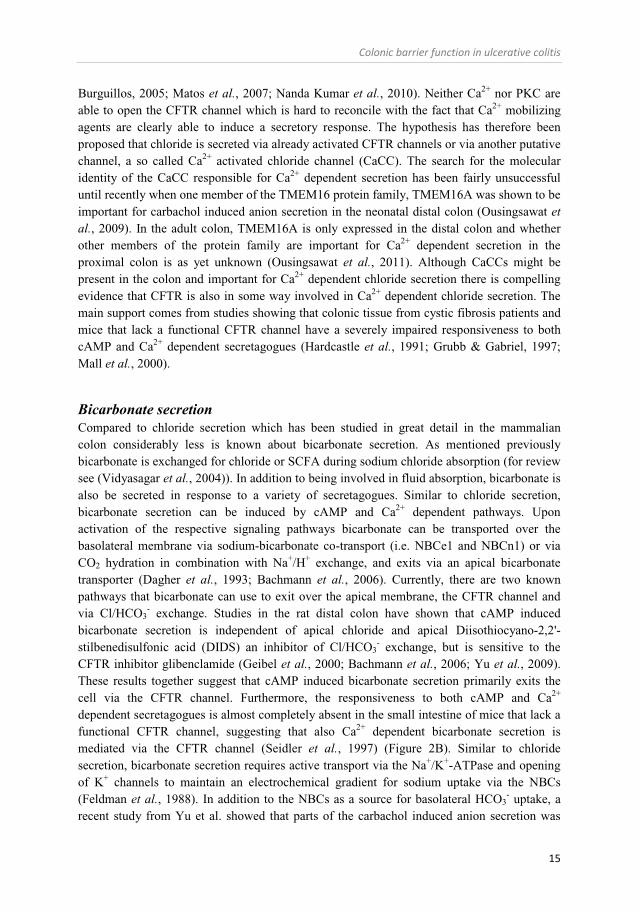
Chloride secretion Electrogenic chloride secretion has been studied in great detail in cultured epithelial cells, rodent colon and to some extent also in human colon (Keely & Barrett, 2000). The transepithelial secretory machinery involves chloride uptake across the basolateral membrane via the Na+/K+/Cl- co-transporter (NKCC1), activation of apical and basolateral potassium channels, and activation of an apical chloride channel (Dharmsathaphorn & Pandol, 1986) (Figure 2A). Activation of K+ channels maintains a negative intracellular membrane potential that favors chloride exit via apical chloride channels. Similar to sodium absorption, the process requires active transport via the Na+/K+-ATPase that maintains a low intracellular sodium concentration that favors basolateral uptake via the NKCC1 (Silva et al., 1977). Depending on the stimulus/agonist, secretion is induced via cAMP or Ca2+ dependent pathways. cAMP dependent secretion involves activation of an adenylyl cyclase that increases intracellular levels of cAMP, resulting in opening of cAMP gated K+ channels, activation of the NKCC1 and subsequent opening of the cystic fibrosis transmembrane conductance regulator (CFTR) via phosphorylation dependent pathways (Boige et al., 1984; Loo & Kaunitz, 1989; Cheng et al., 1991; Reynolds et al., 2007). The signaling cascade results in a sustained secretory response that persists as long as the agonist is present.
Figure 2: Systems behind electrogenic anion secretion. A: Both Ach and VIP induced chloride secretion involves basolateral uptake via NKCC1 and opening of Ca2+ and cAMP gated K+ channels. Secretion over the apical membrane is mediated via the CFTR channel and possibly via the CaCC TMEM16A in the case of Ach. B: Bicarbonate secretion involves basolateral uptake via NBCe1, opening of K+ channels and secretion via the CFTR channel.
The Ca2+ dependent pathway acts via phospholipase C and production of diacyl glycerol and the Ca2+ mobilizing agent inositol 1,4,5 trisphosphate, resulting in increased levels of intracellular Ca2+ and activation of protein kinase C (PKC) (Hirota & McKay, 2006). The elevated Ca2+ levels increases the open probability of Ca2+ dependent K+ channels, which results in hyperpolarization of the plasma membrane that favors chloride exit over the apical membrane (Dharmsathaphorn & Pandol, 1986; Loo & Kaunitz, 1989; del Castillo &
NBCe1
VIP Ach
m3
Na+K+
HCO3- K+
K+
K+Na+
Ca2+
ATPase
HCO3-
NKCC1
VIP Ach
m3
Na+K+
Cl- K+
K+
K+K+ Cl-Na+
Cl-
Ca2+cAMP
ATPase
CaC
C
CFTR
CFTR
cAMP Ca2+
A B
Colonic barrier function in ulcerative colitis
15
Burguillos, 2005; Matos et al., 2007; Nanda Kumar et al., 2010). Neither Ca2+ nor PKC are able to open the CFTR channel which is hard to reconcile with the fact that Ca2+ mobilizing agents are clearly able to induce a secretory response. The hypothesis has therefore been proposed that chloride is secreted via already activated CFTR channels or via another putative channel, a so called Ca2+ activated chloride channel (CaCC). The search for the molecular identity of the CaCC responsible for Ca2+ dependent secretion has been fairly unsuccessful until recently when one member of the TMEM16 protein family, TMEM16A was shown to be important for carbachol induced anion secretion in the neonatal distal colon (Ousingsawat et al., 2009). In the adult colon, TMEM16A is only expressed in the distal colon and whether other members of the protein family are important for Ca2+ dependent secretion in the proximal colon is as yet unknown (Ousingsawat et al., 2011). Although CaCCs might be present in the colon and important for Ca2+ dependent chloride secretion there is compelling evidence that CFTR is also in some way involved in Ca2+ dependent chloride secretion. The main support comes from studies showing that colonic tissue from cystic fibrosis patients and mice that lack a functional CFTR channel have a severely impaired responsiveness to both cAMP and Ca2+ dependent secretagogues (Hardcastle et al., 1991; Grubb & Gabriel, 1997; Mall et al., 2000).
Bicarbonate secretion Compared to chloride secretion which has been studied in great detail in the mammalian colon considerably less is known about bicarbonate secretion. As mentioned previously bicarbonate is exchanged for chloride or SCFA during sodium chloride absorption (for review see (Vidyasagar et al., 2004)). In addition to being involved in fluid absorption, bicarbonate is also be secreted in response to a variety of secretagogues. Similar to chloride secretion, bicarbonate secretion can be induced by cAMP and Ca2+ dependent pathways. Upon activation of the respective signaling pathways bicarbonate can be transported over the basolateral membrane via sodium-bicarbonate co-transport (i.e. NBCe1 and NBCn1) or via CO2 hydration in combination with Na+/H+ exchange, and exits via an apical bicarbonate transporter (Dagher et al., 1993; Bachmann et al., 2006). Currently, there are two known pathways that bicarbonate can use to exit over the apical membrane, the CFTR channel and via Cl/HCO3
- exchange. Studies in the rat distal colon have shown that cAMP induced bicarbonate secretion is independent of apical chloride and apical Diisothiocyano-2,2'-stilbenedisulfonic acid (DIDS) an inhibitor of Cl/HCO3
- exchange, but is sensitive to the CFTR inhibitor glibenclamide (Geibel et al., 2000; Bachmann et al., 2006; Yu et al., 2009). These results together suggest that cAMP induced bicarbonate secretion primarily exits the cell via the CFTR channel. Furthermore, the responsiveness to both cAMP and Ca2+ dependent secretagogues is almost completely absent in the small intestine of mice that lack a functional CFTR channel, suggesting that also Ca2+ dependent bicarbonate secretion is mediated via the CFTR channel (Seidler et al., 1997) (Figure 2B). Similar to chloride secretion, bicarbonate secretion requires active transport via the Na+/K+-ATPase and opening of K+ channels to maintain an electrochemical gradient for sodium uptake via the NBCs (Feldman et al., 1988). In addition to the NBCs as a source for basolateral HCO3
- uptake, a recent study from Yu et al. showed that parts of the carbachol induced anion secretion was
Jenny Gustafsson
16
bicarbonate dependent and mediated via the goblet cell specific transporter Bestrophin 2. This observation suggests the existence of parallel systems, with expression of the CFTR in the enterocyte and bestrophin 2 in the goblet cell (Yu et al., 2010).
Regulation of anion secretion An interesting aspect of colonic anion secretion is that chloride and bicarbonate secretion are induced by the same stimuli. Some of the signaling pathways are shared by the two systems but there are also important difference in particular regarding the time courses of the responses. Stimulation with carbachol induces a rapid transient increase in membrane current that peaks within two minutes of stimulation. In the proximal mouse colon the response is transient and returns towards baseline values, whereas in the distal colon of the same species there is a sustained plateau phase (Yu et al., 2009; Yu et al., 2010). The secretory process involves an acute phase of activation and recruitment of NKCC1 to the basolateral plasma membrane followed by endocytosis and degradation of the transporter, the latter process being mediated via PKC (Reynolds et al., 2007). Simultaneously with this process, NBCe1 is inserted into the apical plasma membrane via PKC dependent pathways. The result is that a sustained increase in bicarbonate secretion is induced that peaks within 20 min of stimulation (Bachmann et al., 2006). Thus, it appears that chloride secretion is a rapid transient event that precedes a sustained bicarbonate secretion. In the proximal colon, the electrogenic secretory response is entirely transient suggesting that in this segment bicarbonate secretion is largely electroneutral. Surprisingly, the plateau phase of carbachol induced anion secretion, that is mediated via the goblet cell specific transporter Bestrophin 2 is absent in CFTR deficient mice, pointing to a cross talk between the enterocytes and the goblet cells since the CFTR is not expressed in goblet cells (Yu et al., 2010).
In contrast to the characteristic transient response of Ca2+ dependent secretion, activation of the cAMP pathway induces a sustained increase in chloride and bicarbonate secretion that follows a similar time course (Bachmann et al., 2006). Regarding regulation of the secretory response by recirculation of transporters in the apical and basolateral membranes, both NBCe1 and CFTR have been shown to be inserted into the basolateral and apical membranes, respectively after forskolin stimulation (Bertrand & Frizzell, 2003; Reynolds et al., 2007; Yu et al., 2009).
The intestinal mucus layer One feature that all mucosal surfaces have in common is that they are covered by a mucus layer that lubricates and protects the underlying epithelium. Depending on different needs and conditions in the respective organs, the physical properties of the mucus have been adapted to fit these local requirements. In the gastrointestinal tract, mucus properties differ markedly between the stomach and the small and large intestine. The mucus is thick and adherent in the stomach, where it forms a diffusion barrier loaded with buffering bicarbonate that is secreted to protect the epithelium from the acidic luminal contents (Phillipson et al., 2008). In the small intestine, the mucus layer is loose to allow for nutrient uptake and to assist gradual diffusion of antibacterial peptides (Atuma et al., 2001; Johansson & Hansson, 2011;
Colonic barrier function in ulcerative colitis
17
Vaishnava et al., 2011). The loose structure also allows for the binding of bacteria and other harmful agents and their transportation in the distal direction for expulsion. In the distal colon, the mucus instead forms a two layer system with an outer layer that is the habitat of the microbiota and an inner layer that forms a physical barrier impeding the vast majority of bacteria from reaching the epithelial surface (Johansson et al., 2008).
The MUC2 mucin The intestinal mucus layer is generated by the goblet cells that produce and secrete the MUC2 mucin, the core component of the mucus in both the small and large intestine. The MUC2 mucin is a large heavily O-glycosylated protein characterized by two central mucin domains composed of amino acid sequences rich in proline, threonine and serine (Johansson et al., 2011a). Upon synthesis, the protein is translocated to the endoplasmatic reticulum where it is folded and forms disulfide bonded dimers in the far C-terminal end (Lidell et al., 2003). In the Golgi apparatus the MUC2 dimers are O-glycosylated at the threonine and serine residues within the mucin domains, to later form oligomers via its N-termini (Godl et al., 2002). Following oligomerization the protein is tightly packed in secretory granules under low pH and high Ca2+ conditions. The dense glycosylation of the protein backbone protects against proteolytic degradation and is important for the gel-forming properties by being able to bind large amounts of water. Upon secretion the mucin molecules are unfolded and expand approximately a 1000 fold to form the hydrated mucus gel (Ambort et al., 2012).
Mucus secretion Intestinal mucus secretion can be divided into two separate processes, baseline secretion and compound exocytosis. Both systems use the regulated secretory pathways and secrete mucins that are stored in mature secretory granules (Forstner, 1995). A recent study from our lab showed that the surface epithelium is the main source for baseline mucus secretion, while the intestinal crypts seems to be the main site for compound exocytosis induced by substances such as Ach (Johansson, unpublished) (Specian & Neutra, 1980; Phillips et al., 1984).
The regulated secretory pathway and baseline mucus secretion Upon synthesis the mucin molecules are stored in secretory granules in the goblet cell theca. The mature secretory granules migrate along microtubules towards the apical surface where they fuse with the plasma membrane. Studies have shown that granules stored at the lateral side of the theca are preferentially secreted under baseline conditions (Specian & Neutra, 1984). However, the number of studies on baseline mucus secretion in the intestine is limited and these studies are primarily based on histological observations. In contrast, studies in other secretory cells and tissues have identified a number of protein families that are important for the exocytosis process. One of these protein families is the SNARE group of proteins which are expressed in the vesicle membrane and are essential for vesicle - vesicle fusion and fusion with the plasma membrane (Jahn & Scheller, 2006). During baseline conditions, the SNARE proteins are in a closed formation that requires opening to form a core complex with adjacent
Jenny Gustafsson
18
membranes. Munc13 has been shown to be one of the proteins that open up SNAREs on a target membrane which allows interaction between SNAREs from different vesicles, leading to formation of the so called core complex that is required for vesicle fusion (Burgoyne & Morgan, 2007). Munc13-2 null mice have been shown to have mucus accumulation in both the small and large intestine, suggesting that this protein is important for baseline intestinal mucus secretion (Zhu et al., 2008). Although the processes of vesicle formation and fusion with the plasma membrane have been studied to some extent there are still a number of questions that remain regarding regulation of the final release step. The classical view of exocytosis is that it occurs via formation of a secretion pore and release of the vesicle content via diffusion. During intestinal mucus secretion, single granule fusion with the plasma membrane has been observed as well as exocytosis of large granules that protrude out of the cell (Specian & Oliver, 1991). Due to the viscous properties of the mucins, the final release step is not likely to occur via simple diffusion. It has therefore been suggested that the release step is coordinated with a process that facilitates release of the secreted mucins into the lumen (Forstner, 1995). Since both the small and large intestine are constantly exposed to a basal tone of mucus secretagogues such as acetylcholine, it is likely that the balance between single vesicle release and compound exocytosis varies depending on the strength of the stimuli.
Compound exocytosis of mucin granules Compound exocytosis of mucins enables release of almost the entire granule pool of the goblet cell. This process has been shown to be regulated by intracellular Ca2+ levels, thus Ca2+ mobilizing agents such as acetylcholine and histamine are potent inducers of mucus secretion in both the small and large intestine (MacDermott et al., 1974; Specian & Neutra, 1980). The increased intracellular calcium levels promote a signaling cascade that involves fusion of centrally located granules that migrate towards the apical part of the cell, fuse with the plasma membrane and release their contents into the lumen (Specian & Neutra, 1980; Specian & Oliver, 1991). Following release of one large vesicle, adjacent vesicles can fuse with the remaining membrane from the previous vesicle. By continuation of this process the entire mucin granule content can be released within a matter of minutes. The exact mechanisms for compound exocytosis in the colon are not known. However, in the airways compound exocytosis of mucins requires vesicle fusion mediated via the SNARE protein VAMP 8, that also is expressed in the mucin granule membrane of human colon (Jones et al., 2012; Rodriguez-Pineiro et al., 2012).
Although little is known about mechanisms that regulate mucus secretion one feature that most mucus secretagogues have in common is that they also are potent inducers of anion secretion. The neurotransmitter acetylcholine is the most studied mucus secretagogue in the intestine and has been shown to induce mucus secretion in the small and large intestine of mouse, rat and rabbit and human colon (Specian & Neutra, 1980; Neutra et al., 1984; Halm & Halm, 2000; Gustafsson et al., 2011). Histamine is another substance that induces mucus secretion in rodents and human colon (Neutra et al., 1982; Halm & Halm, 2000). VIP and prostaglandin E2 (PGE2) have been shown to induce mucus secretion in rat colon but had no effect in rabbit colon (Neutra et al., 1982; Halm et al., 1995; Plaisancie et al., 1998). Whether VIP induces mucus secretion in human colon is not known, but PGE2 which operates via the
Colonic barrier function in ulcerative colitis
19
same system did not induce mucus secretion in isolated human colonic crypts (Halm & Halm, 2000). Related to immune regulation of mucus secretion, goblet cell hyperplasia and increased mucus secretion is a central component of the immune response during clearing of gut parasites and interferon-gamma (IFN-γ) has also been shown to drive mucus secretion during salmonella infection in the caecum (Khan et al., 1995; Songhet et al., 2011).
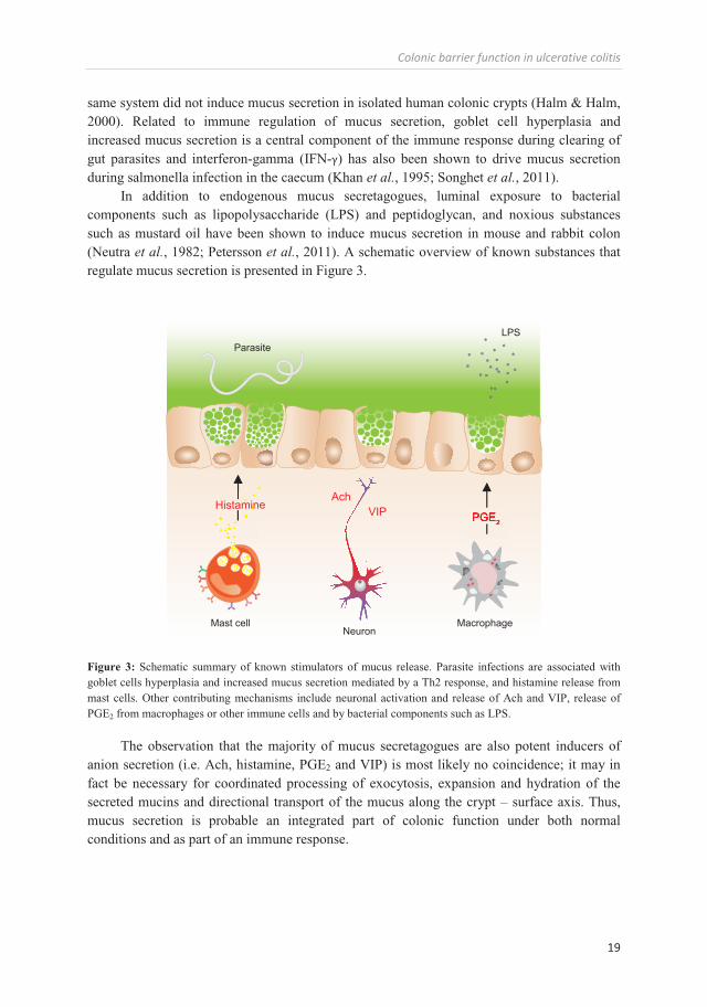
In addition to endogenous mucus secretagogues, luminal exposure to bacterial components such as lipopolysaccharide (LPS) and peptidoglycan, and noxious substances such as mustard oil have been shown to induce mucus secretion in mouse and rabbit colon (Neutra et al., 1982; Petersson et al., 2011). A schematic overview of known substances that regulate mucus secretion is presented in Figure 3.
Figure 3: Schematic summary of known stimulators of mucus release. Parasite infections are associated with goblet cells hyperplasia and increased mucus secretion mediated by a Th2 response, and histamine release from mast cells. Other contributing mechanisms include neuronal activation and release of Ach and VIP, release of PGE2 from macrophages or other immune cells and by bacterial components such as LPS.
The observation that the majority of mucus secretagogues are also potent inducers of anion secretion (i.e. Ach, histamine, PGE2 and VIP) is most likely no coincidence; it may in fact be necessary for coordinated processing of exocytosis, expansion and hydration of the secreted mucins and directional transport of the mucus along the crypt – surface axis. Thus, mucus secretion is probable an integrated part of colonic function under both normal conditions and as part of an immune response.
PGE2
NeuronMacrophage
Ach
LPS
PGE2
Parasite
VIP
Mast cell
Histamine
Jenny Gustafsson
20
Regulation of mucus properties – lessons from cystic fibrosis Upon secretion the mucin molecules expand in volume and together with associated proteins they form the mucus gel (Johansson et al., 2009). As mentioned previously, the properties of the mucus gel differ greatly between the different levels of the gastrointestinal tract although the core component of the mucus gel can be the same like in the case of MUC2 in the small and large intestine. How the same protein can form a gel with so vastly different properties remains an enigma.
The role of ion transport in the regulation of mucus properties became evident when it was discovered that the recessive genetic disease cystic fibrosis (CF) was caused by mutations in the gene coding for the CFTR channel (Kerem et al., 1989; Riordan et al., 1989; Rommens et al., 1989). CF manifests itself mainly as a severe lung disease, characterized by mucus accumulation, bacterial overgrowth, recurrent lung infections and eventual organ failure. In addition to involvement of the airways, most organs that express CFTR and gel forming mucins are affected by the disease i.e. the pancreas, the biliary system, the salivary glands, the small intestine, the gall bladder and the urogenital tract (Quinton, 1999). Strangely, mouse models of CF do not develop lung disease. They instead develop an intestinal phenotype similar to the human distal intestinal obstructive syndrome (DIOS) which is caused by accumulation of viscous mucus and fecal material in the terminal ileum and caecum (Grubb & Gabriel, 1997; Wyllie, 1999). Due to their intestinal phenotype CF mice have become an essential model in gastrointestinal CF research. As mentioned above, most organs that express CFTR are severely affected in CF. However, one exception to the rule is the colon which expresses CFTR and the gel-forming mucin MUC2, but does not seems to be affected by the disease. One possible explanation is that the protective function of the colonic mucus is based on the thick adherent layer that keeps bacteria separated from the epithelial surface. Thus, an increased adherence of the mucus to epithelial surface does not necessarily have to have a negative effect on the protective function of the colonic mucus layer.
Although the connection between defective ion transport and mucus pathology is acknowledged by most people in the field of mucus and CF research, the molecular mechanisms behind the altered mucus properties are not fully understood. At this time there are two major hypotheses regarding the origin of the viscous sticky mucus. Both hypotheses are built around the transport defect, but focus on either hyperabsorption or hyposecretion. The hyperabsorption theory is based on the assumption that over-absorption of sodium results in dehydration of the liquid that lines the airway surface resulting in reduced mucociliary clearance (Matsui et al., 1998). In support of this theory, studies have shown that CF patients have an increased amiloride sensitive current (i.e. increased sodium absorption) in the airways, and mice over-expressing ENaC suffer from mucus accumulation in the airways (Boucher et al., 1986; Zhou et al., 2011). The hyposecretion theory is instead based on the assumption that anion secretion and specifically bicarbonate secretion is essential for expansion and formation of a normal mucus layer and when secretion is impaired like in CF the mucus becomes sticky (Quinton, 2008, 2010). In favor of this hypothesis is that bicarbonate secretion has been shown to be essential for formation of a normal mucus layer in the small intestine and in the cervix (Garcia et al., 2009; Muchekehu & Quinton, 2010). Furthermore, it was recently shown that the transport defect in CF piglets that do develop lung
Colonic barrier function in ulcerative colitis
21
disease was associated with hyposecretion rather than hyperabsorption (Chen et al., 2010). Although the role of ion transport in regulation of mucus properties is not fully understood, it is likely that an altered ionic milieu during mucus secretion regulates the properties of the formed mucus gel.
In addition to endogenous regulation of mucus properties via epithelial transport, exposure to bacteria affects the properties of the colonic mucus layer. Accordingly, studies have shown that mice raised under germ free conditions have a very thin adherent mucus layer in the colon compared to conventionally raised mice (Johansson et al., 2008; Petersson et al., 2011).
The mucosal immune system Due to the massive amount of bacteria that reside in our gastrointestinal tract the colonic mucosa has to constantly maintain homeostasis between the microbiota and the host. In the distal colon the epithelium manages this task by limiting the antigenic pressure on the tissue by secreting a dense mucus layer that separates the vast majority of the bacteria from the epithelium (Johansson et al., 2011b). This mucus layer is not impervious to all bacteria and both specialized mucus-degrading commensals and some pathogens are able to penetrate the mucus layer, reach the epithelium and initiate an immune response (Bergstrom et al., 2010). Also bacterial components such as LPS, peptidoglycan and flagellin may diffuse through the mucus layer and modulate immunity. The antigenic nature of the bacteria, bacterial components and other biological and chemical substances requires a well regulated mucosal immune response which manages to handle threats to the barrier, restore homeostasis and maintain tolerance. In the colon, the mucosal defense can be divided into three parts: the mucus layer, the underlying epithelial barrier and the immune system. The colon does not have Peyers’ patches like the small intestine, but is instead supplied with a large number of isolated lymphoid follicles with a similar structure that can sample antigens, present them to antigen presenting cells (dendritic cells and macrophages) and modulate immunity (Owen et al., 1991). The mucosal immune system can be divided into an innate and an adaptive component. The innate part of the cellular defense includes among other players the epithelial cells, neutrophils, macrophages and dendritic cells (Smith et al., 2005; Iwasaki & Medzhitov, 2010). These cells respond to general features of microbes and constitute the first lines of defense. The epithelial cells secrete mucus and antimicrobial peptides to prevent against bacterial colonization (Boman, 2003). The neutrophils and macrophages express large amounts of antibacterial agents that are either released to kill a target (neutrophils) or act through phagocytosis (neutrophils and macrophages) and destroy their victims within intracellular granules (Amulic et al., 2011). Resident macrophages in the lamina propria play an important role in local immune homeostasis by phagocytosing bacteria that have managed to cross the intestinal barrier. They are the same time secreting the anti-inflammatory cytokines interleukin 10 (IL-10) and transforming growth factor-beta (TGF-β) which prevent further immune activation and recruitment of effector cells to the site of exposure, thus preventing initiation of an inflammatory response (Mowat & Bain, 2011).
Expression of pathogen pattern recognition receptors such as toll-like receptors (TLR) enable the epithelial cells to respond to microbial virulence factors such as flagellin which
Jenny Gustafsson
22
initiates a signaling cascade involving secretion of cytokines and chemokines for recruitment of effector cells like neutrophils to the site of exposure (Swamy et al., 2010).
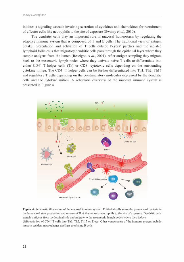
The dendritic cells play an important role in mucosal homeostasis by regulating the adaptive immune system that is composed of T and B cells. The traditional view of antigen uptake, presentation and activation of T cells outside Peyers’ patches and the isolated lymphoid follicles is that migratory dendritic cells pass through the epithelial layer where they sample antigens from the lumen (Rescigno et al., 2001). After antigen sampling they migrate back to the mesenteric lymph nodes where they activate naïve T cells to differentiate into either CD4+ T helper cells (Th) or CD8+ cytotoxic cells depending on the surrounding cytokine milieu. The CD4+ T helper cells can be further differentiated into Th1, Th2, Th17 and regulatory T cells depending on the co-stimulatory molecules expressed by the dendritic cells and the cytokine milieu. A schematic overview of the mucosal immune system is presented in Figure 4.
Figure 4: Schematic illustration of the mucosal immune system. Epithelial cells sense the presence of bacteria in the lumen and start production and release of IL-8 that recruits neutrophils to the site of exposure. Dendritic cells sample antigens from the luminal side and migrate to the mesenteric lymph nodes where they induce differentiation of CD4+ T cells into Th1, Th2, Th17 or Tregs. Other components of the immune system include mucosa resident macrophages and IgA producing B cells.
Th1
Th2Th17
TregTh0
IL-8
Macrophage
Dendritic cell
B cell
Neutrophil
T cell differentation
IgA
Mesenteric lymph node
Colonic barrier function in ulcerative colitis
23
The Th1 response is driven by IL-12/IL-23 and is crucial for proper handling of intracellular pathogens (Romagnani, 1995). The Th2 response is essential for handling extracellular bacteria and clearing parasite infections, and is characterized by production of IL-4 and IL13 (McKenzie et al., 1998). The Th17 cells are suggested to take care of threats which the classic Th1 and Th2 responses are unable to handle, and are characterized by secretion of IL-17 (Korn et al., 2009). The last Th type, the regulatory T cell, is a suppressive cell type driven by IL-10 and TGF-β production that maintains mucosal homeostasis by regulating the other T-cell populations (Coombes et al., 2005).
The epithelial cells themselves have been ignored in this process mainly due to the spatial separation between the epithelium and the naïve T cells in the mesenteric lymph nodes. However, this paradigm is starting to shift with studies showing that the epithelium does play an active part in the modulation of adaptive immunity (Swamy et al., 2010). Epithelial cells have accordingly been shown to process and present antigens directly to dendritic cells and T-cells, and to secrete a large number of cytokines and chemokines that affect the features of the immune response (Telega et al., 2000; Rimoldi et al., 2005). Furthermore, it was recently shown that in the distal small intestine, antigens are taken up by the goblet cells and presented to resident dendritic cells in the lamina propria, which is a completely novel mode of antigen presentation (McDole et al., 2012).
B cells are the central mediators of humoral immunity in the intestine that can neutralize a pathogen with pathogen specific antibodies. In the intestine the vast majority of all plasma cells secrete IgA (Brandtzaeg et al., 1999). Although IgA production and secretion is by definition part of the adaptive immune system it is also regarded as an important part in innate immunity due to it its role in the first line of mucosal defense. Mucosal IgA is secreted by plasma cells in the lamina propria and bind to the polymeric IgA receptor on the basolateral side of epithelial cells. The IgA-secretory factor-receptor complex is actively transported through the cell and free IgA bound to the secretory factor is released to the luminal side after proteolytic cleavage from the receptor. On the luminal side IgA can bind pathogens and prevent them from reaching the epithelium (Monteiro & Van De Winkel, 2003)
Ulcerative colitis Ulcerative colitis is a chronic relapsing inflammatory disease of unknown etiology. The inflammation is restricted to the colonic mucosa and is characterized by a continuous inflammation that always involves the rectum and progresses varying distances proximally. Three common distribution patterns have been indentified that include isolated proctitis, left-sided colitis and total colitis (Nikolaus & Schreiber, 2007). In Northern Europe and North America, the prevalence of UC increased rapidly during the first half of the 20th century, and continued to increase until around 10 to 20 years ago when the incidence of disease started to stabilize. In other parts of the world such as Asia and the Middle East where the incidence of UC has been traditionally low, the opposite pattern is observed with an increasing incidence during the last 10 to 20 years (Hanauer, 2006). This pattern coincides with adaptation to a Western lifestyle. Furthermore, studies have shown that people who migrate from a low risk area to high risk areas increase their relative risk for developing disease (Barreiro-de Acosta et al., 2011). These types of rapid changes spanning over one generation cannot be explained
Jenny Gustafsson
24
by genetic adaptations and indicate that environmental factors are important in UC, which also fits with the relatively low concordance rate in monozygotic twins ~10% (Tysk et al., 1988). However, one cannot exclude that improved healthcare systems and more accurate diagnosis in certain parts of the world contribute to the increasing numbers of identified new cases. The current hypothesis for the pathogenesis of UC is that the disease develops in a genetically predisposed individual with environmental factors triggering an inappropriate immune response directed towards the commensal flora. The assumption that the inflammation is directed towards the commensal flora is based on observations showing that acute Crohn’s colitis heals after diversion of the fecal stream through an ileostomy, and that probiotics may improve the disease activity in certain patient groups (Winslet et al., 1994; Mack, 2011). Furthermore, there is some evidence in favor of antibiotics being effective in induction of remission, and the fact that no single pathogen has been positively associated with UC suggests that the commensal flora itself is being the targeted by the immune system (Khan et al., 2011). Patients with UC have also been shown to have a reduced bacterial complexity (Lepage et al., 2011) The most compelling evidence in favor of the commensal flora being important for development of colitis comes from animal studies showing that the majority of murine colitis models do not develop disease when raised as germ free (Dianda et al., 1997; Matharu et al., 2009). Together, these results suggest that the inflammation is directed towards the commensal flora and that there is an altered balance in the microbial community. Alternatively, increased exposure to the commensal flora and loss of tolerance might be involved in triggering inflammation. The large number of colitis models and the huge amount of divergent data from patients strongly suggest that the pathogenesis of colitis is far more complex than previously thought, and the challenge now facing the scientific community is to find the mechanisms behind the abnormal bacterial – immune interaction.
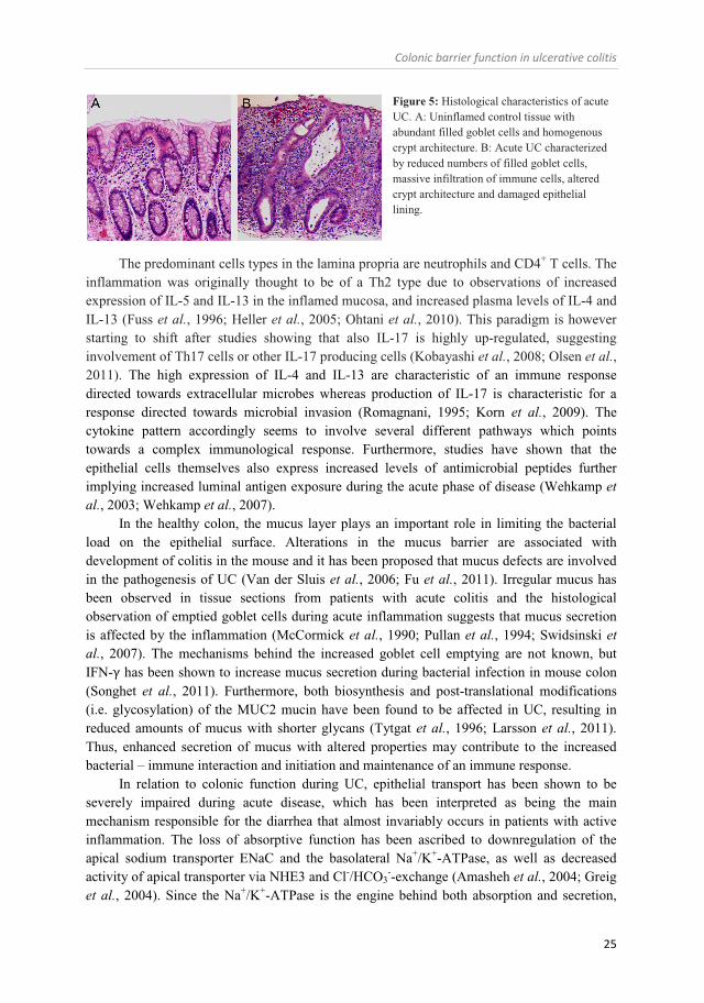
Pathophysiology The pathophysiology of UC may involve every single aspect of colonic function including alterations in the luminal flora, the mucus layer, epithelial transport, epithelial integrity, the innate and adaptive immune system and the enteric nervous system. Since the main problem in UC is the aggressive immune response resulting in tissue damage and severe loss of organ function, the main focus has been to understand the characteristics of the established inflammation. The histological appearance of the acutely inflamed colonic mucosa is characterized by massive infiltration of neutrophils, macrophages, T-cells and B-cells. The epithelial lining is often thin and depleted of mucin containing goblet cells. Cryptitis and crypt abscesses are common during moderate and severe inflammation, and ulcerations and denuded epithelium represent the most severe form of tissue damage (Figure 5) (McCormick et al., 1990; Nikolaus & Schreiber, 2007). High mucus content of the stool is common in patients with acute UC, therefore the histological observation of goblet cell depletion may well be increased mucus secretion rather than specific loss of goblet cells.
Colonic barrier function in ulcerative colitis
25
Figure 5: Histological characteristics of acute UC. A: Uninflamed control tissue with abundant filled goblet cells and homogenous crypt architecture. B: Acute UC characterized by reduced numbers of filled goblet cells, massive infiltration of immune cells, altered crypt architecture and damaged epithelial lining.
The predominant cells types in the lamina propria are neutrophils and CD4+ T cells. The inflammation was originally thought to be of a Th2 type due to observations of increased expression of IL-5 and IL-13 in the inflamed mucosa, and increased plasma levels of IL-4 and IL-13 (Fuss et al., 1996; Heller et al., 2005; Ohtani et al., 2010). This paradigm is however starting to shift after studies showing that also IL-17 is highly up-regulated, suggesting involvement of Th17 cells or other IL-17 producing cells (Kobayashi et al., 2008; Olsen et al., 2011). The high expression of IL-4 and IL-13 are characteristic of an immune response directed towards extracellular microbes whereas production of IL-17 is characteristic for a response directed towards microbial invasion (Romagnani, 1995; Korn et al., 2009). The cytokine pattern accordingly seems to involve several different pathways which points towards a complex immunological response. Furthermore, studies have shown that the epithelial cells themselves also express increased levels of antimicrobial peptides further implying increased luminal antigen exposure during the acute phase of disease (Wehkamp et al., 2003; Wehkamp et al., 2007).
In the healthy colon, the mucus layer plays an important role in limiting the bacterial load on the epithelial surface. Alterations in the mucus barrier are associated with development of colitis in the mouse and it has been proposed that mucus defects are involved in the pathogenesis of UC (Van der Sluis et al., 2006; Fu et al., 2011). Irregular mucus has been observed in tissue sections from patients with acute colitis and the histological observation of emptied goblet cells during acute inflammation suggests that mucus secretion is affected by the inflammation (McCormick et al., 1990; Pullan et al., 1994; Swidsinski et al., 2007). The mechanisms behind the increased goblet cell emptying are not known, but IFN-γ has been shown to increase mucus secretion during bacterial infection in mouse colon (Songhet et al., 2011). Furthermore, both biosynthesis and post-translational modifications (i.e. glycosylation) of the MUC2 mucin have been found to be affected in UC, resulting in reduced amounts of mucus with shorter glycans (Tytgat et al., 1996; Larsson et al., 2011). Thus, enhanced secretion of mucus with altered properties may contribute to the increased bacterial – immune interaction and initiation and maintenance of an immune response.
In relation to colonic function during UC, epithelial transport has been shown to be severely impaired during acute disease, which has been interpreted as being the main mechanism responsible for the diarrhea that almost invariably occurs in patients with active inflammation. The loss of absorptive function has been ascribed to downregulation of the apical sodium transporter ENaC and the basolateral Na+/K+-ATPase, as well as decreased activity of apical transporter via NHE3 and Cl-/HCO3
--exchange (Amasheh et al., 2004; Greig et al., 2004). Since the Na+/K+-ATPase is the engine behind both absorption and secretion,

Jenny Gustafsson
26
most aspects of epithelial transport will be affected by reduced activity of this transporter. Simultaneously with downregulation of ion transport, the lamina propria cells produce high concentrations of secretagogues such as prostaglandins which promote anion secretion (Rampton et al., 1980). Accordingly, the highly regulated balance between absorption and secretion may be shifted in the secretory direction. Loss of epithelial function also results in more acidic luminal contents due to reduced bicarbonate secretion (Caprilli et al., 1986). This pH shift will potentially affect the properties of the colonic mucus layer due to reduced unfolding of the MUC2 mucin (Ambort et al., 2012).
Most studies of epithelial transport during UC have focused on studying the inflamed tissue during active disease. However, up to 30% of patients still suffer from symptoms such as abdominal pain and diarrhea during remission, despite absence of ongoing inflammation. (Isgar et al., 1983; Simren et al., 2002). The sustained diarrheal symptoms imply that there may be a sustained imbalance between absorption and secretion also in remission. The exact mechanisms behind these remaining symptoms are not known, but observations of restored absorptive functions during remission and increased levels of secretagogues in the tissue suggest that the pathogenesis may be different from that during acute disease (Hawker et al., 1980; Rampton et al., 1980).
In summary, ulcerative colitis is a complex disease that affects most aspects of colonic function. When untreated it may become life-threatening. The pathways responsible for the acute inflammatory response are to some extent known, but it is still not known how and why the inflammatory reaction is initiated. Since anti-inflammatory treatment invariably leads to immune suppression with the risk for side-effects, identification of the triggering factor and targeting of this factor for treatment would be of immense medical value.
Colonic barrier function in ulcerative colitis
27
AIM OF THESIS Overall aim The overall aim of this thesis work is to understand the function and regulation of the intestinal barrier with a special focus on the colonic barrier in Ulcerative colitis. Specific aims
I. To develop an ex vivo method for studies of mucus properties in mouse small and large intestine and in human colonic biopsies
II. To develop a method for simultaneous measurement of ion and mucus secretion
and to analyze the interaction between these two systems in mouse distal colon
III. To determine the molecular mechanisms behind mucus pathology in mice lacking the CFTR channel
IV. To study the secretory physiology of the colonic epithelium in patients with
ulcerative colitis in remission
V. To study the properties of the colonic mucus in mouse and human colon in relation to inflammation
Jenny Gustafsson
28
METHODOLOGICAL CONSIDERATIONS
Materials and ethics
Human subjects (Paper I, IV, V) All experiments involving human tissue were approved by the ethical committee of the Sahlgrenska University Hospital and written informed consent was obtained from all study subjects. In the present thesis, a large number of subjects were included in the various studies. The control material consisted of patients referred for colonoscopy for reasons such as bleedings of unspecified origin, polyp surveillance, diverticulitis or altered bowel habit an in whom the colonoscopy and the macroscopical appearance of the mucosa was normal. In addition to the control group, a large number of UC patients were included in the different studies. The UC patients were either referred to colonoscopy as a part of their disease surveillance program or for clinical reasons related to their disease. The disease activity was evaluated in two ways, first via the endoscopic Mayo score as judged by the gastroenterologist performing the colonoscopy (Lewis et al., 2008), and secondly by the pathologist evaluating the clinical biopsies taken from the respective segments; caecum, ascending colon, right colon, transverse colon, left colon, descending colon, sigmoid colon, and rectum. The UC patients were divided into two groups; remission and acute inflammation. The remission patients had an endoscopic Mayo score of 0 and the histological evaluation showed maximally altered crypt architecture and a slight increase in infiltrating immune cells (eosinophils, neutrophils, and plasma cells). Patients with acute inflammation had Mayo scores of 1 to 3 and the histological profile was characterized by altered crypt architecture, infiltration of immune cells, cryptitis or crypt abscesses and in the most severe cases ulcerations or denuded epithelium. In paper IV only patients that were diagnosed with pancolitis (initial inflammation in the entire colon) were included to ensure that both tested segments (ascending and sigmoid colon) had previously been exposed to the inflammation. In paper V the extent of mucosal disease was not taken into account when including the patients. Comments: All patients underwent the colonoscopy due to clinical symptoms or known disease and the only additional procedure that was related to the research studies was the taking of extra biopsies (maximum 16). Data obtained from patients were collected in such a way that the patients’ identity was kept confidential. Since our control group consisted of patients that were referred for colonoscopy for various medical reasons it is possible that this patient group diverge from a control group composed of healthy volunteers with no history of bowel disorders. On the other hand, there are other advantages with using a patient population of this type (“disease controls”). The material was fairly heterogeneous and the risk for error due to a common disease-denominator in the control group is therefore very small. Regarding the gender and age distribution of our patient material, the control patients were older than the UC patients and the UC patients showed a male predominance. Although population studies only show a small tendency towards a male predominance in UC, it appears that our patient cohort differs from the general UC population in terms of gender distribution. We cannot explain why we obtained this skewed distribution but tentative analysis in our material did not reveal any obvious gender differences in the functional studies.
Colonic barrier function in ulcerative colitis
29
Animal studies (Paper I-V) All animal experiments were approved by the animal ethics committee at the University of Gothenburg. The experiments were performed using male and female wild type (WT), Muc-/-, CftrΔ508 and IL-10-/- mice, all on a C57/Bl6 background between 8 and 16 weeks old. The WT mice were either purchased from Taconic (Denmark) or were obtained from our in house breeding programs. The Muc2-/- mice were obtained from Dr Velcich (Velcich et al., 2002), the CftrΔF508 strain was obtained from Dr Scholte (van Doorninck et al., 1995) and the IL-10-/- mice were originally from Dr Müller (Kuhn et al., 1993). To maintain the CftrΔ F508 mice, they were treated with an osmotic laxative in the drinking water (polyethylene glycol 4000 18 mM, KCl 10 mM, Na2SO4 40 mM, NaHCO3 84 mM and NaCl 25 mM ) and were transferred to normal tap water 2-3 days prior to the experiment. In paper V we also used tissue sections from Slc9a3-/-, TLR5-/-, C1GalT-/- mice which were obtained from Drs. Hua, Gerwitz and Xia, respectively. The ethical approvals for these experiments were obtained from the respective universities, Arizona, Emroy and Oklahoma. In paper V dextran sodium sulphate (DSS) treated mice were used as described previously (Johansson et al., 2010).
Muc2-/- mice The importance of the colonic mucus layer in colonic barrier function became clear with the finding that mice lacking the Muc2 mucin develop spontaneous colitis around the time of weaning. The inflammation is most prominent in the distal colon and is characterized by crypt elongation, infiltration of immune cells and intracellular bacteria (Van der Sluis et al., 2006; Johansson et al., 2008). Comment: In the present work, the Muc2 deficient mice were used to determine whether the carbachol effect on epithelial capacitance was related to mucin granule exocytosis. The advantage of using these animals is that they do not compensate for the loss of Muc2 production by up-regulation of other gel-forming mucins, thus the exocytosis response is expected to be reduced. One of the disadvantages with these animals is the ongoing inflammation which might affect the responsiveness to carbachol induced exocytosis. To overcome this problem, the response was studied in both the proximal and distal colon of the Muc2-/- mice and in the distal colon of Muc2+/- mice which do not develop colitis.
CftrΔF508 mice In both the small and large intestine, electrogenic anion secretion is crucially dependent on the apical CFTR channel. In humans, loss of CFTR mediated transport causes the disease cystic fibrosis, which manifests itself as a severe lung disease characterized by mucus accumulation, bacterial overgrowth and eventually inflammatory organ failure. Mice models of CF do not develop lung disease but instead exhibit a severe small intestinal phenotype characterized by mucus accumulation, bacterial overgrowth and in the worst case septicemia. This phenotype is similar to the human disease DIOS (distal intestinal obstructive syndrome) that many CF patients also suffer from (Grubb & Gabriel, 1997; Riedel, 1997).
Jenny Gustafsson
30
Comment: CftrΔF508 mice were used to investigate the role of CFTR mediated transport in carbachol induced mucin granule exocytosis in the colon, and the role of CFTR in regulation of mucus properties in the small intestine and colon. The CftrΔF508 model was chosen since this mutation is the most common mutation in the human CF population and their intestinal phenotype resembles that of the human intestinal disease DIOS (Davis, 2006). These mice do express the protein and have a small residual CFTR current.
IL-10-/- mice The IL-10 deficient mouse is one of the earliest and most well characterized murine colitis models (Kuhn et al., 1993). Studies have shown that the inflammation is mediated by CD4+ T-cells via production of IL-23, IL-17 and IL-6 and that the inflammatory response is directed towards the commensal flora (Davidson et al., 1996). Related to the clinical relevance of the model it has been shown that the cytokine profile of the UC mucosa also contains high levels of IL-17, IL23 and IL-6 (Olsen et al., 2011). Comment: The IL-10 deficient mice were used to study the relation between mucus properties and development of colitis. When raised in our animal facility, the IL-10-/- mice show a mild phenotype compared to published data. This mild phenotype may be due to the absence of Helicobacter hepaticus in our facility, a bacteria known to module the inflammatory process (Matharu et al., 2009). Furthermore, the low age of the animals is likely to contribute to the mild inflammation since the disease is known to develop at around four months of age. The mild phenotype facilitates interpretation of the results since any observed changes in mucus properties are likely to be directly linked to the loss of IL-10, rather than being secondary to an ongoing inflammatory process.
Slc9a3, TRL5 and GalT1 deficient mice and dextran sodium sulphate (DSS) induced colitis In this thesis work one of the major questions was whether alterations in the mucus layer is a general feature of colitis. To address this question the barrier function of the mucus layer was studies in three additional murine colitis models that develop spontaneous inflammation, the Slc9a3-/-, TLR5-/- and the C1GalT-/- mice.
The solute carrier family 9 member 3 (Slc9a3, NHE3) deficient mice develop a distal colitis, despite the fact that the main route for sodium uptake in the distal colon is via ENaC and not via the NHE3 (Laubitz et al., 2008). The exact mechanisms behind the inflammation are not known but its occurrence shows that a maintained epithelial transport is essential for normal colonic homeostasis.
The second model that was used, the TRL5 deficient mice show a heterogeneous phenotype where around 25% of the mice develop colitis whereas the other 75% develop the metabolic syndrome. The colitis of these mice is characterized by infiltration of immune cells, crypt loss, edema and goblet cells depletion. The mechanisms behind the inflammation are also here not fully understood but it is suggested that an altered bacterial composition and increased bacterial burden trigger the inflammatory response. It is also assumed that the loss of TLR5 signaling from the epithelial cells is the main initiator of the altered immune
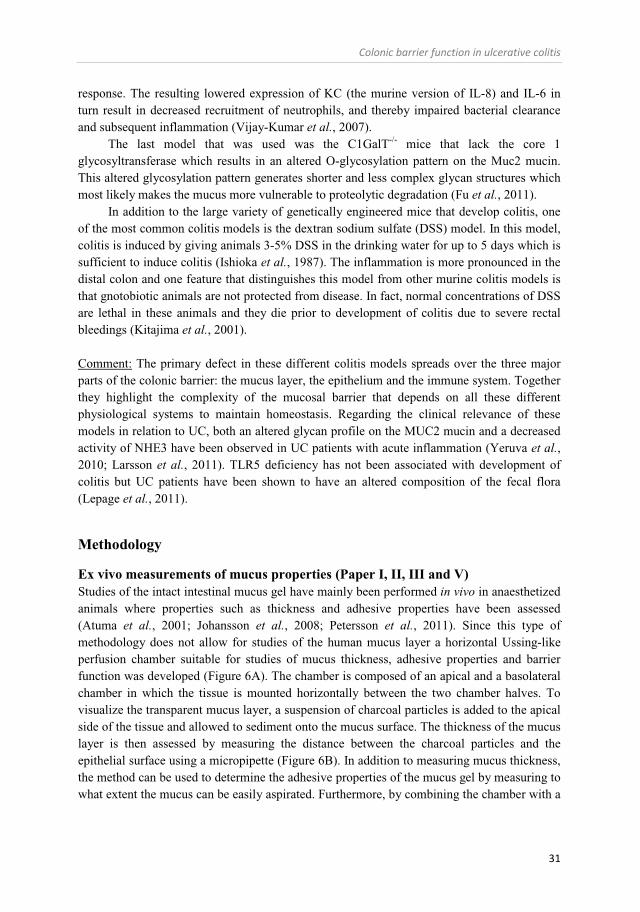
Colonic barrier function in ulcerative colitis
31
response. The resulting lowered expression of KC (the murine version of IL-8) and IL-6 in turn result in decreased recruitment of neutrophils, and thereby impaired bacterial clearance and subsequent inflammation (Vijay-Kumar et al., 2007).
The last model that was used was the C1GalT-/- mice that lack the core 1 glycosyltransferase which results in an altered O-glycosylation pattern on the Muc2 mucin. This altered glycosylation pattern generates shorter and less complex glycan structures which most likely makes the mucus more vulnerable to proteolytic degradation (Fu et al., 2011).
In addition to the large variety of genetically engineered mice that develop colitis, one of the most common colitis models is the dextran sodium sulfate (DSS) model. In this model, colitis is induced by giving animals 3-5% DSS in the drinking water for up to 5 days which is sufficient to induce colitis (Ishioka et al., 1987). The inflammation is more pronounced in the distal colon and one feature that distinguishes this model from other murine colitis models is that gnotobiotic animals are not protected from disease. In fact, normal concentrations of DSS are lethal in these animals and they die prior to development of colitis due to severe rectal bleedings (Kitajima et al., 2001).
Comment: The primary defect in these different colitis models spreads over the three major parts of the colonic barrier: the mucus layer, the epithelium and the immune system. Together they highlight the complexity of the mucosal barrier that depends on all these different physiological systems to maintain homeostasis. Regarding the clinical relevance of these models in relation to UC, both an altered glycan profile on the MUC2 mucin and a decreased activity of NHE3 have been observed in UC patients with acute inflammation (Yeruva et al., 2010; Larsson et al., 2011). TLR5 deficiency has not been associated with development of colitis but UC patients have been shown to have an altered composition of the fecal flora (Lepage et al., 2011).
Methodology
Ex vivo measurements of mucus properties (Paper I, II, III and V) Studies of the intact intestinal mucus gel have mainly been performed in vivo in anaesthetized animals where properties such as thickness and adhesive properties have been assessed (Atuma et al., 2001; Johansson et al., 2008; Petersson et al., 2011). Since this type of methodology does not allow for studies of the human mucus layer a horizontal Ussing-like perfusion chamber suitable for studies of mucus thickness, adhesive properties and barrier function was developed (Figure 6A). The chamber is composed of an apical and a basolateral chamber in which the tissue is mounted horizontally between the two chamber halves. To visualize the transparent mucus layer, a suspension of charcoal particles is added to the apical side of the tissue and allowed to sediment onto the mucus surface. The thickness of the mucus layer is then assessed by measuring the distance between the charcoal particles and the epithelial surface using a micropipette (Figure 6B). In addition to measuring mucus thickness, the method can be used to determine the adhesive properties of the mucus gel by measuring to what extent the mucus can be easily aspirated. Furthermore, by combining the chamber with a
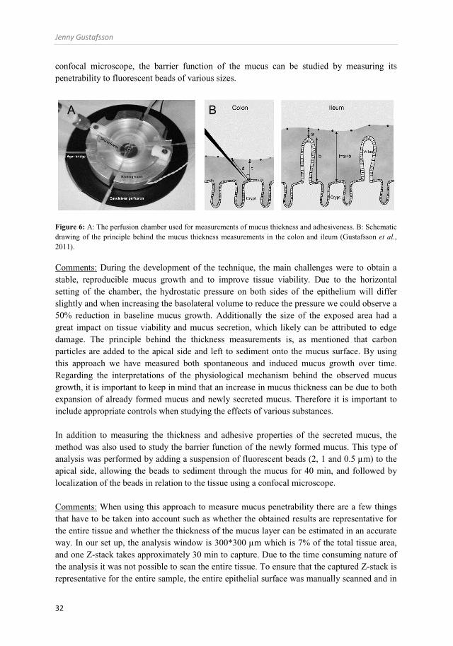
Jenny Gustafsson
32
confocal microscope, the barrier function of the mucus can be studied by measuring its penetrability to fluorescent beads of various sizes.
Figure 6: A: The perfusion chamber used for measurements of mucus thickness and adhesiveness. B: Schematic drawing of the principle behind the mucus thickness measurements in the colon and ileum (Gustafsson et al., 2011).
Comments: During the development of the technique, the main challenges were to obtain a stable, reproducible mucus growth and to improve tissue viability. Due to the horizontal setting of the chamber, the hydrostatic pressure on both sides of the epithelium will differ slightly and when increasing the basolateral volume to reduce the pressure we could observe a 50% reduction in baseline mucus growth. Additionally the size of the exposed area had a great impact on tissue viability and mucus secretion, which likely can be attributed to edge damage. The principle behind the thickness measurements is, as mentioned that carbon particles are added to the apical side and left to sediment onto the mucus surface. By using this approach we have measured both spontaneous and induced mucus growth over time. Regarding the interpretations of the physiological mechanism behind the observed mucus growth, it is important to keep in mind that an increase in mucus thickness can be due to both expansion of already formed mucus and newly secreted mucus. Therefore it is important to include appropriate controls when studying the effects of various substances. In addition to measuring the thickness and adhesive properties of the secreted mucus, the method was also used to study the barrier function of the newly formed mucus. This type of analysis was performed by adding a suspension of fluorescent beads (2, 1 and 0.5 µm) to the apical side, allowing the beads to sediment through the mucus for 40 min, and followed by localization of the beads in relation to the tissue using a confocal microscope. Comments: When using this approach to measure mucus penetrability there are a few things that have to be taken into account such as whether the obtained results are representative for the entire tissue and whether the thickness of the mucus layer can be estimated in an accurate way. In our set up, the analysis window is 300*300 µm which is 7% of the total tissue area, and one Z-stack takes approximately 30 min to capture. Due to the time consuming nature of the analysis it was not possible to scan the entire tissue. To ensure that the captured Z-stack is representative for the entire sample, the entire epithelial surface was manually scanned and in
A B
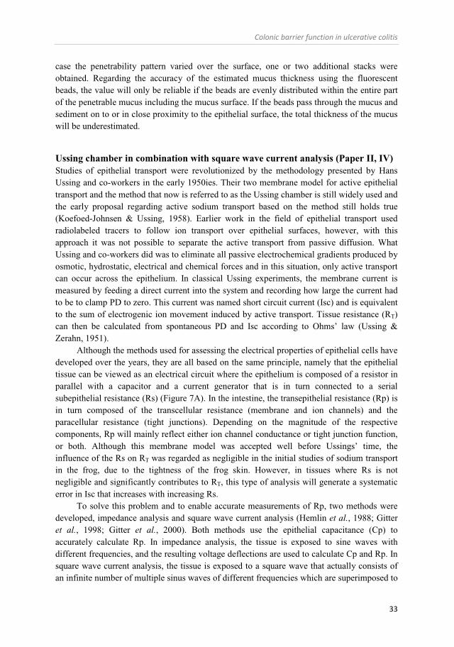
Colonic barrier function in ulcerative colitis
33
case the penetrability pattern varied over the surface, one or two additional stacks were obtained. Regarding the accuracy of the estimated mucus thickness using the fluorescent beads, the value will only be reliable if the beads are evenly distributed within the entire part of the penetrable mucus including the mucus surface. If the beads pass through the mucus and sediment on to or in close proximity to the epithelial surface, the total thickness of the mucus will be underestimated.
Ussing chamber in combination with square wave current analysis (Paper II, IV) Studies of epithelial transport were revolutionized by the methodology presented by Hans Ussing and co-workers in the early 1950ies. Their two membrane model for active epithelial transport and the method that now is referred to as the Ussing chamber is still widely used and the early proposal regarding active sodium transport based on the method still holds true (Koefoed-Johnsen & Ussing, 1958). Earlier work in the field of epithelial transport used radiolabeled tracers to follow ion transport over epithelial surfaces, however, with this approach it was not possible to separate the active transport from passive diffusion. What Ussing and co-workers did was to eliminate all passive electrochemical gradients produced by osmotic, hydrostatic, electrical and chemical forces and in this situation, only active transport can occur across the epithelium. In classical Ussing experiments, the membrane current is measured by feeding a direct current into the system and recording how large the current had to be to clamp PD to zero. This current was named short circuit current (Isc) and is equivalent to the sum of electrogenic ion movement induced by active transport. Tissue resistance (RT) can then be calculated from spontaneous PD and Isc according to Ohms’ law (Ussing & Zerahn, 1951).
Although the methods used for assessing the electrical properties of epithelial cells have developed over the years, they are all based on the same principle, namely that the epithelial tissue can be viewed as an electrical circuit where the epithelium is composed of a resistor in parallel with a capacitor and a current generator that is in turn connected to a serial subepithelial resistance (Rs) (Figure 7A). In the intestine, the transepithelial resistance (Rp) is in turn composed of the transcellular resistance (membrane and ion channels) and the paracellular resistance (tight junctions). Depending on the magnitude of the respective components, Rp will mainly reflect either ion channel conductance or tight junction function, or both. Although this membrane model was accepted well before Ussings’ time, the influence of the Rs on RT was regarded as negligible in the initial studies of sodium transport in the frog, due to the tightness of the frog skin. However, in tissues where Rs is not negligible and significantly contributes to RT, this type of analysis will generate a systematic error in Isc that increases with increasing Rs.
To solve this problem and to enable accurate measurements of Rp, two methods were developed, impedance analysis and square wave current analysis (Hemlin et al., 1988; Gitter et al., 1998; Gitter et al., 2000). Both methods use the epithelial capacitance (Cp) to accurately calculate Rp. In impedance analysis, the tissue is exposed to sine waves with different frequencies, and the resulting voltage deflections are used to calculate Cp and Rp. In square wave current analysis, the tissue is exposed to a square wave that actually consists of an infinite number of multiple sinus waves of different frequencies which are superimposed to
Jenny Gustafsson
34
form a uniform pulse. The advantages of impedance analysis are that the method uses a well defined spectrum of frequencies that gives more detailed information about the epithelial properties, and that the method can be used to study local differences in electrical properties along the crypt-surface axis (Gitter et al., 2000). The disadvantage of the technique is that the calculations are based on the specific components in the membrane model and small errors in the model can have a large effect on the results (Gitter et al., 1998). The square wave current analysis has the advantage of being more robust, and small diversions from the membrane model will not have the same impact.
In this work, the square wave current analysis was used to measure epithelial resistance, capacitance and net membrane current in mouse and human colon. The technique is based on the observation that when a square wave current is fed through an Rp-Cp element, the resulting voltage change will be delayed, with a time course that depends on the relation between Rp and Cp. Likewise, on termination of the current pulse, the voltage reduction will be delayed, with an approximately monoexponential decay. The rapid voltage response will be due to the subepithelial resistance (Rs) and the delayed part of the curve will be due to charging of the epithelial Rp-Cp element (Figure 7B). By plotting the voltage logarithmically versus time one can generate a linear fit that can be extrapolated back to time zero (V0), i.e. the end of the current pulse. By following the linear part of the discharge event one can specifically analyze the Rp-Cp element i.e. the epithelium. The epithelial resistance (Rp) can be calculated from Ohm’s law by dividing the voltage response at time zero (V0) by the applied current. The epithelial capacitance can be calculated from the time constant of the Rp-Cp element.
Figure 7: Principle behind the square wave current analysis. A: The epithelial cell as an electrical circuit with a parallel current generator (Im), resistor (Rp) and a capacitor (Cp). B: Voltage deflection following the square pulse. The rapid increase in voltage depends on the subepithelial resistance (Rs) whereas the delayed part of the curve depends on the charging of the Rp-Cp element. The formulae used in the analyses are as follows:
V(t) = Vt x e-t/(R x C)
V(0) = V0 x e-0/(R x C) V(0) = V0 x 1 V0
V0 = I x Rp
V (t) = V0 x e-t/(R x C) C = (t2 – t1)/(R x ln(V1/V2))
Colonic barrier function in ulcerative colitis
35
V1 and V2 are the voltages at two selected times t1 and t2 of the mono exponential discharge event. By measuring the transepithelial voltage (PD) after each pulse, the net membrane current (Im) can also be calculated using Ohm’s law:
Im = PD / Rp
Comments, resistance: Measurements of epithelial resistance have been extensively used to monitor tissue integrity in various species and organs (Schmitz et al., 1999; Resta-Lenert & Barrett, 2003; Howe et al., 2005; Reims et al., 2006; McGilligan et al., 2007). In general terms, total tissue resistance has been almost exclusively used as a measurement of tight junction conductance in studies on intact tissue specimens. This assumption may be more or less correct depending on the relation between paracellular and transcellular resistances in the specific organ. When the paracellular resistance is higher than the transcellular resistance, the epithelial resistance will mainly reflect ion channel permeability, whereas if the transcellular permeability is higher than the paracellular permeability, the epithelial resistance will mainly reflect tight junction function. In our studies of mouse and human colon, it has been observed that baseline Rp is highly sensitive to, for example pharmacological inhibition of K+ channels and also drops rapidly in response to various types of secretagogues. Against this background, our interpretation of the colonic Rp is that it mainly reflects changes in ion channel permeability. This observation is in line with previous studies in mouse colon which showed that the paracellular resistance was 20 times higher than the transcellular resistance (Gitter et al., 2000). Thus, in the normal mouse colon, the epithelial resistance will probably mainly reflect transcellular permeability. Whether this applies also to inflamed mucosa remains unknown. Comment, capacitance: Measurements of epithelial capacitance have been extensively used in single cells and cultured epithelial monolayers to study exocytosis of mucins, histamine, serotonin and insulin (Kalinowski & Figaszewski, 1992; Bertrand et al., 1998; Bertrand et al., 1999; Gopel et al., 2004). Studies have shown that the capacitance of an unfolded biological membrane is approximately 1 µF/cm2, but due to the microvilli of the epithelial cells and the folding of the intestinal epithelium the actual surface area is much larger than that. Our results showed that the capacitance of mouse colon is approximately 20 µF/cm2 compared to 30 and 50 µF/cm2 in the proximal and distal human colon, respectively. Others using impedance analysis has found that the capacitance of mouse colon is approximately 8 µF/cm2, which is less than our observations but still in the same range (Gitter et al., 2000). In this thesis work, changes in capacitance were used to monitor mucin granule exocytosis. This type of analysis is based on the proportional relation between the capacitance of a membrane and its area, thus an increase in membrane area can be quantified as an increase in capacitance. Although capacitance measurements are routinely used to study changes in membrane area, one drawback with the technique is that it is not possible to isolate one specific exocytosis process. Many other compounds and transporters undergo vesicular trafficking, e.g. circulate between the intracellular compartment and the membrane. When using membrane capacitance as a marker for mucin exocytosis one therefore has to perform control experiments to confirm
Jenny Gustafsson
36
that the assumption is correct. To validate our approach the response was studied also in cell systems and animals that do not express gel forming mucins. Comments membrane current: Of the three parameters discussed here, the membrane current is the one used routinely among researchers in the field of epithelial transport. The standard way of measuring this current is to short-circuit the tissue and measure the required current, the so called short-circuit current (Isc). In the intestine, electrogenic sodium absorption and chloride secretion are the main contributors to the net membrane current. As mentioned above, the (correct) method used for measuring the epithelial resistance will influence the magnitude of the membrane current. Our values of baseline Im are approximately -300 µA/cm2 in the proximal human colon and -150 µA/cm2 in the distal colon. In mouse colon, baseline Im is in the range of -200µA/cm2 in both proximal and distal colon. These values are high in comparison to measurements of traditional colonic Isc, which can be explained by our more correct way of measuring Rp. One thing to take into consideration when interpreting the Im signal is that enhanced absorption of positive ions and secretion of negative ions will generate a response with the same polarity. Therefore it is important to investigate which transport processes that are involved in the response and not make assumptions based on only the magnitude and polarity of the response.
In situ hybridization (Paper V) In situ hybridization using a 16S rRNA probe was used to analyze the barrier function of the mouse colonic mucus. The 16S rRNA probe used was a general bacteria probe labeling most bacteria (EU622773) (Amann et al., 1990).
Immunohistochemistry (Paper I, III, V) In the present work, immunostaining of the Muc2 mucin was performed using the C3 antisera which stain the mature form of the Muc2 mucin. The specificity of the antisera has previously been verified by peptide block and lack of staining in the Muc2 deficient mice (Johansson et al., 2008).
Electron microscopy (Paper I, III) Transmission electron microscopy (TEM) is a powerful tool for studies of epithelial morphology, and has been an essential technique in early studies of the characteristics of intestinal mucus secretion (Specian & Neutra, 1980). The method is based on the tissue being stained using electron dense substances and cut in ultra thin sections (50 nm). Our protocol included sequential staining using osmium, tannic acid and uranyl acetate which generated very good cell morphology and stained the intestinal mucus both in the cells and after secretion. The method was used to study the morphology of the tissue specimens after incubation in the perfusion chamber and to study the mucus phenotype in WT and CF small intestine.
Colonic barrier function in ulcerative colitis
37
Although TEM is a powerful tool in morphological analyses of epithelial tissue, the high magnification makes is difficult for a reader to determine whether the presented results are representative for the entire tissue. In paper I, TEM was used in combination with light microscopy to show that tissue integrity was intact after 1 h incubation in the perfusion chamber. In paper III fluorescence microscopy was used in combination with TEM to visualize mucus adherence in CF and WT mouse ileum. By combining high and low resolution imaging one can avoid possible bias in the analysis.
Jenny Gustafsson
38
RESULTS AND COMMENTS
The intestinal epithelium is a complex structure composed of many different cell types with sometimes different roles in the integrated physiology of the small and large intestine. In this thesis, the focus has been on studying the relations between mucus physiology and epithelial physiology in normal and inflamed mucosa. In both the small intestine and colon, activation of secretomotor neurons of the enteric nervous system induces a coordinated secretory process involving ion secretion from the columnar cells and mucus secretion from the goblet cells (Specian & Neutra, 1980; Osbak et al., 2007). There is also release of hormones and antimicrobial peptides from the Paneth cells of the small intestine and from enterochromaffin cells of both the small and large intestine (Ouellette, 1999; Braun et al., 2007). The molecular regulation of ion transport has been studied in great detail, while much less is known about regulation of mucus secretion and mucus properties. This gap of knowledge can to a large extent be explained by lack of useful tools for studies of goblet cell function and mucus properties, particularly in human biopsies. To overcome some of the technical difficulties, an ex vivo method for studies of mucus properties in human colonic biopsies was developed, and this technique was also used in mouse small and large intestinal explants. A modified Ussing chamber approach based on square wave current analysis was also used to study the corresponding epithelial secretory events and the control of mucin exocytosis. In this setup, membrane current and epithelial capacitance was used as substitute markers for anion secretion and mucin granule exocytosis, respectively.
Colonic mucus secretion – regulation by ion transport (Paper II) As mentioned above, activation of cholinergic pathways is likely to induce coordinated secretion of ions, fluid and mucus from the intestinal mucosa. Coordination of ion and mucus secretion may be necessary for proper expansion and hydration of the secreted mucins. In the airways and in the small intestine, epithelial chloride and bicarbonate transport have been shown to affect both mucus secretion and formation of a normal mucus layer. However, how these processes interact in the colon is unknown, both under control conditions and in a state of colonic inflammation.
Epithelial capacitance as a measurement of mucin granule exocytosis Epithelial capacitance has previously been used to measure mucin granule exocytosis in cultured cells (Bertrand et al., 1999). However, due to the low number of studies we started out by validating this approach using two different epithelial cell lines; one enterocytes model (Caco-2) and one goblet cell containing model (LS513). The results showed that serosal stimulation with carbachol induced a transient increase in epithelial capacitance in the goblet cell containing cell line (LS513), and failed to induce any response in the enterocyte model (Caco-2). To further test whether the response did indeed reflect exocytosis, the LS513 cells were pretreated with the exocytosis inhibitors primaquine or nocodazole followed by stimulation with carbachol. The results showed that nocodazole significantly reduced the carbachol effect on capacitance whereas primaquine had a partial effect. Thus, the carbachol
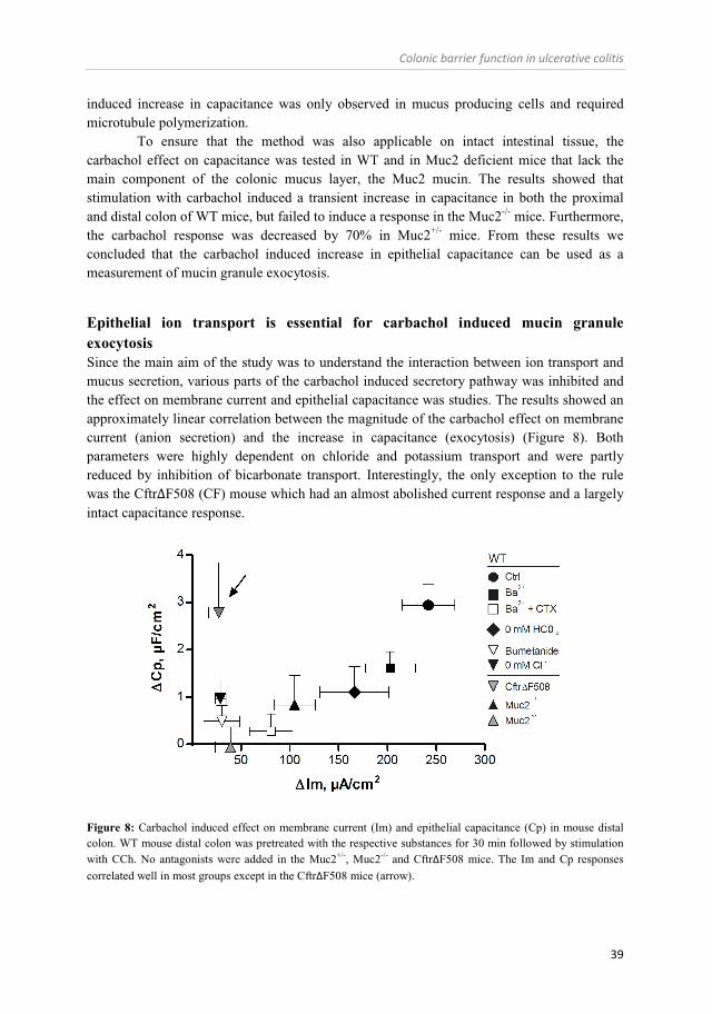
Colonic barrier function in ulcerative colitis
39
induced increase in capacitance was only observed in mucus producing cells and required microtubule polymerization.
To ensure that the method was also applicable on intact intestinal tissue, the carbachol effect on capacitance was tested in WT and in Muc2 deficient mice that lack the main component of the colonic mucus layer, the Muc2 mucin. The results showed that stimulation with carbachol induced a transient increase in capacitance in both the proximal and distal colon of WT mice, but failed to induce a response in the Muc2-/- mice. Furthermore, the carbachol response was decreased by 70% in Muc2+/- mice. From these results we concluded that the carbachol induced increase in epithelial capacitance can be used as a measurement of mucin granule exocytosis.
Epithelial ion transport is essential for carbachol induced mucin granule exocytosis Since the main aim of the study was to understand the interaction between ion transport and mucus secretion, various parts of the carbachol induced secretory pathway was inhibited and the effect on membrane current and epithelial capacitance was studies. The results showed an approximately linear correlation between the magnitude of the carbachol effect on membrane current (anion secretion) and the increase in capacitance (exocytosis) (Figure 8). Both parameters were highly dependent on chloride and potassium transport and were partly reduced by inhibition of bicarbonate transport. Interestingly, the only exception to the rule was the CftrΔF508 (CF) mouse which had an almost abolished current response and a largely intact capacitance response.
Figure 8: Carbachol induced effect on membrane current (Im) and epithelial capacitance (Cp) in mouse distal colon. WT mouse distal colon was pretreated with the respective substances for 30 min followed by stimulation with CCh. No antagonists were added in the Muc2+/-, Muc2-/- and CftrΔF508 mice. The Im and Cp responses correlated well in most groups except in the CftrΔF508 mice (arrow).
Jenny Gustafsson
40
To determine whether the same principle applied to the goblet cell line LS513, the cells were pretreated with the NKCC1 inhibitor bumetanide or with the CFTR inhibitor Gly-H101, followed by stimulation with CCh. The results showed that the carbachol effect on the capacitance was reduced by pretreatment with bumetanide but was unaffected by the CFTR inhibitor. Thus, regulation of the Cp response appears to be similar in mouse distal colon and the goblet cell containing cell line LS513. Comment: These results imply that epithelial transport is not only important for mucus expansion and hydration, it is also crucial for mucin granule exocytosis. Interestingly, the process seems to involve the basolateral NKCC1 and K+ channels but is seemingly independent of the apical CFTR channel. Thus, electrogenic anion secretion per se is not the main driving force for mucin granule exocytosis. The intact capacitance response in the CF mice is in accordance with previous findings showing that both NKCC1 activity and K+ secretion are normal in these mice (Mall et al., 2000; Bachmann et al., 2003). These results also fit with studies from the airways showing that inhibition of chloride and bicarbonate transport reduces VIP, carbachol and histamine induced mucus secretion, whereas activation of potassium channels increases the secretory response (Choi et al., 2007; Kondo et al., 2012). The coordinated process of mucus and potassium secretion has been observed for a long time in the extreme cases of villus adenomas where the tissue secretes massive amounts of mucus in the range of 1 to 2 liters per 24 hour which can severely deplete plasma potassium (Rowe, 1964).
Although our observations imply that ion transport is required for mucin secretion, they still do not explain at what stage of the exocytosis process the interaction occurs. Increased levels of Ca2+ should induce vesicle fusion despite inhibition of ion transport, and one possibility is that vesicle release is in some way regulated by ion transport. In enterochromaffin cells and platelets, ion transport has been shown to be essential for creating the intra vesicular pressure that is required for vesicle rupture. In these cells, the vesicle membrane becomes permeable to anions after fusion with the plasma membrane. Anions secreted from the cells (i.e. OH-) will pass over the vesicle membrane, and when the osmotic pressure inside the vesicle becomes too high the vesicle ruptures and releases its content (i.e. 5-HT) (Pollard et al., 1977). In support of such a model, the mucin granule content is highly viscous and this type of event might help to expel the mucins that cannot just diffuse into the lumen. Furthermore, partly expanded mucins can be seen inside the mucin vesicles prior to release, suggesting that the expansion process starts already inside the intact mucins granules (Ambort et al., 2012).
Regulation of mucus properties in the small and large intestine (Paper I and III) The current knowledge regarding the properties and regulation of the intact intestinal mucus gel mainly comes from in vivo studies in anaesthetized rodents, perfusion studies of intestinal segments or to a limited extent from cultured tissue specimens (Pullan et al., 1994; Atuma et al., 2001; Johansson et al., 2008; Fyderek et al., 2009). These studies have shown that mucus
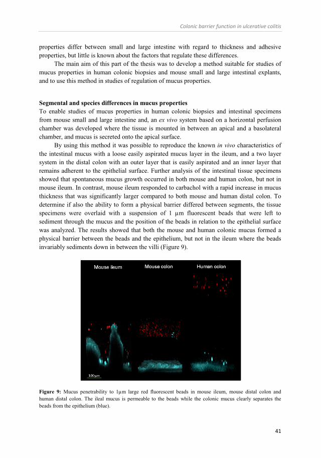
Colonic barrier function in ulcerative colitis
41
properties differ between small and large intestine with regard to thickness and adhesive properties, but little is known about the factors that regulate these differences.
The main aim of this part of the thesis was to develop a method suitable for studies of mucus properties in human colonic biopsies and mouse small and large intestinal explants, and to use this method in studies of regulation of mucus properties.
Segmental and species differences in mucus properties To enable studies of mucus properties in human colonic biopsies and intestinal specimens from mouse small and large intestine and, an ex vivo system based on a horizontal perfusion chamber was developed where the tissue is mounted in between an apical and a basolateral chamber, and mucus is secreted onto the apical surface.
By using this method it was possible to reproduce the known in vivo characteristics of the intestinal mucus with a loose easily aspirated mucus layer in the ileum, and a two layer system in the distal colon with an outer layer that is easily aspirated and an inner layer that remains adherent to the epithelial surface. Further analysis of the intestinal tissue specimens showed that spontaneous mucus growth occurred in both mouse and human colon, but not in mouse ileum. In contrast, mouse ileum responded to carbachol with a rapid increase in mucus thickness that was significantly larger compared to both mouse and human distal colon. To determine if also the ability to form a physical barrier differed between segments, the tissue specimens were overlaid with a suspension of 1 µm fluorescent beads that were left to sediment through the mucus and the position of the beads in relation to the epithelial surface was analyzed. The results showed that both the mouse and human colonic mucus formed a physical barrier between the beads and the epithelium, but not in the ileum where the beads invariably sediments down in between the villi (Figure 9).
Figure 9: Mucus penetrability to 1µm large red fluorescent beads in mouse ileum, mouse distal colon and human distal colon. The ileal mucus is permeable to the beads while the colonic mucus clearly separates the beads from the epithelium (blue).
Jenny Gustafsson
42
Comment: These results show that some key features of mucus secretion and mucus properties differ between the small and large intestine, although the core component of the mucus, the Muc2 mucin, is the same in both segments. These segmental differences likely reflect the different demands on the ileum and colon. The rapid secretory response to carbachol in the mouse ileum may be related to the fact that the ileum is intermittently exposed to a substantial amount of bacteria, and has to be able to efficiently secrete mucus and transport the bacteria distally into the colon that is better equipped to handle large bacterial loads. As expected, the mucus properties were similar in mouse and human colon, with the exception that the human mucus layer was thicker. These results confirm that the human colonic mucus forms a physical barrier similar to that observed in mouse colon and that mouse distal colon is a suitable model for human distal colon with respect to both baseline and induced mucus growth.
Role of CFTR and bicarbonate in the regulation of intestinal mucus properties (Paper II and III) Loss of CFTR mediated transport results in disease in the small intestine of both CF patients and CF mouse models, but CF patients do not have an obvious phenotype in the colon. The small intestinal pathology is associated with intestinal obstruction due to mucus accumulation, bacterial overgrowth and inflammation (Norkina et al., 2004). Due to the abnormal mucus accumulation, CFTR mediated transport has been suggested to play a decisive role in regulation of mucus properties in the small intestine. One of the hypotheses regarding how CFTR affects mucus properties is via its ability to transport bicarbonate that regulates the viscosity of the secreted mucus (Poulsen et al., 1994; Garcia et al., 2009). However, the exact mechanisms behind this effect of bicarbonate on mucus properties are unknown. It is also unclear why this phenomenon does not seem to have any consequences in the colon, despite the fact that the two systems secrete the same gel-forming mucin, the Muc2 mucin.
In this part of the thesis the role of CFTR mediated transport in the regulation of mucus properties in both the small and large intestine was investigated, with the aim to understand how ion transport via the CFTR affects mucus properties.
Ileal mucus properties are regulated by CFTR mediated transport and bicarbonate Baseline mucus properties (i.e. thickness, adhesion and penetrability) were studied in the ileum of WT and CF mice. In the WT ileum, the mucus was transparent, easily aspirated and penetrable to 2 µm large fluorescent beads. In CF ileum, the initial mucus thickness was similar to that observed in the WT tissue but different in other important respects: the mucus was opaque, remained adherent to the epithelial surface, and was impenetrable to 2 µm large beads.
Since one of the current hypotheses regarding the mechanisms behind the mucus phenotype in CF is loss of CFTR mediated bicarbonate secretion, it was tested whether administration of high concentrations of bicarbonate to the apical solution could normalize the ileal mucus phenotype. The results showed that indeed the addition of 5 times the normal
Colonic barrier function in ulcerative colitis
43
bicarbonate concentration led to expansion of the mucus, reversed the adherent phenotype and increased its penetrability to the 2 µm large beads towards normal levels, i.e. restored the properties of WT ileal mucus. In contrast, high concentrations of bicarbonate did not affect the mucus thickness in WT mouse ileum. The observation that the CF mucus expanded when exposed to high bicarbonate concentrations suggests high mucin protein content. To determine whether this was the case, the glycoprotein content of mucus collected from WT and CF ileum was measured. The results showed that the CF mucus contained 2.6 times more protein than the WT mucus. This pattern was also associated with an increased number of goblet cells in the crypts of the CF mice compared to WT mice.
Because the dominating location of the CFTR is in the intestinal crypts and the main targets for compound exocytosis are the goblet cells in the small intestinal crypts, the mucus phenotype after secretagogue induced mucus secretion was also studied. In WT ileum, stimulation with a combination of carbachol and PGE2 induced a rapid increase in mucus thickness, and the secreted mucus was easily aspirated. In CF ileum, stimulation with carbachol and PGE2 also induced a rapid increase in mucus thickness similar to that observed in WT ileum but this mucus remained adherent to the epithelial surface. To determine whether the adherent phenotype could be reversed by high concentrations of bicarbonate as was the case for the spontaneously released mucus, the experiments was repeated in the presence of 5 times the bicarbonate concentration on the apical side. Similarly to the effect on the initial mucus, increasing bicarbonate concentration reversed the adherent mucus phenotype and the mucus was easily aspirated. However, this was not the case when the same high concentration of bicarbonate was added to the basolateral side, suggesting that bicarbonate exerts its effect on the apical side of the epithelial lining.
Since high concentrations of bicarbonate normalized the adherent phenotype in CF ileum, we proceded by testing whether removal of bicarbonate could also induce a CF phenotype in WT ileum. Bicarbonate was removed from the basolateral perfusate and mucus secretion was stimulated using carbachol and PGE2. The results showed that removal of bicarbonate from the basolateral solution indeed generated adherent mucus in WT ileum, similar to that seen in CF ileum.
One of the proposed mechanisms for how bicarbonate affects mucus properties is by its ability to chelate Ca2+ that is bound to the densely stored mucins, thus enabling expansion of the mucin molecule upon secretion. To test this hypothesis, the Ca2+ chelating agent EDTA was added to the apical solution and mucus secretion was stimulated from CF ileum using carbachol and PGE2. The results showed that EDTA reversed the adherent mucus phenotype to the same degree as high concentrations of bicarbonate.
Accordingly, these results together strongly support the hypothesis that bicarbonate secreted via the CFTR facilitates mucus expansion by chelating Ca2+.
Comment: Our observations from WT and CF mouse ileum support the hypothesis that impaired bicarbonate secretion is the missing link between the mucoviscidosis seen in CF and that the role of bicarbonate is to chelate calcium and permit expansion of the mucin molecules upon secretion. Previous studies by Garcia et al. 2009 showed that bicarbonate is necessary to release mucus from the small intestine (Garcia et al., 2009). Our results further support this finding by demonstrating that CF ileum responds to secretagogues with an increased mucus
Jenny Gustafsson
44
thickness, but with the important difference that the secreted mucus now remains attached to the epithelial surface. In mouse ileum, CFTR contributes approximately 30% of baseline bicarbonate secretion and apparently the remaining 70% are not enough to form a normal mucus layer (Seidler et al., 1997). One possible explanation might be increased demands on the tissue since CF ileum secreted more dense mucus that had 2.6 times higher glycoprotein content compared to the WT ileal mucus. Previous studies have accordingly shown that CF ileum exhibits goblet cell hyperplasia (van Doorninck et al., 1995).
Our results also showed that an adherent mucus phenotype could be induced in WT ileum by removing the bicarbonate from the basolateral perfusate, thus showing that there is a direct link between bicarbonate secretion and mucus properties. The bicarbonate free buffer contained high concentrations of both phosphate and chloride, suggesting that neither of these anions can create mucus with normal physical properties.
In contrast to the fairly low contribution of CFTR to baseline ileal bicarbonate secretion, secretagogue induced bicarbonate secretion is almost exclusively dependent on the CFTR (Seidler et al., 1997). The results presented here revealed that stimulation with carbachol and PGE2 induced a similar increase in mucus thickness in WT and CF ileum, but generated a mucin with different adhesiveness. The fact that CF ileum responded to stimulation with an increased mucus growth shows that the small intestinal mucus is actually able to expand partly even in the absence of CFTR activity, but that the final properties of this secreted mucus become defective. How the mucus is attached to the epithelium is still not known, but histological analysis of the tissue suggests that the mucus remains attached both to the goblet cells and to the surface epithelium. It is possible that partly expanded mucins can actually adhere to the glycocalyx or other parts of the epithelial surface.
Taken together, these results show that external application of bicarbonate to CF ileum can reverse most of the mucus pathology associated with CF.
Baseline colonic mucus growth is CFTR independent while secretagogue induced mucus growth is dependent on a functioning CFTR channel To determine whether CFTR mediated transport is involved in regulating mucus properties also in the colon, spontaneous and secretagogues induced mucus growth was measured in WT and CF mouse distal colon. Baseline mucus growth was recorded for 1 h and neither the initial thickness nor the baseline growth rate differed between WT and CF colon. In contrast, stimulation with carbachol induced a rapid increase in mucus thickness in WT colon, but failed to induce any significant response in CF colon (Figure 10). Comment: These results show that baseline mucus growth in the distal colon is independent of CFTR mediated transport, whereas formation of a normal mucus layer following carbachol induced secretion depends on CFTR mediated transport. Interestingly, CF patients do not have any obvious colonic phenotype, suggesting that the reduced ability to expand the colonic mucus following induced mucus secretion does not have a negative impact on the colonic barrier. The main source of baseline mucus secretion is probably the surface epithelium, which also is the main location for baseline bicarbonate secretion via Cl/HCO3
--exchange (Vidyasagar et al., 2004). In contrast, CFTR is mainly expressed in the colonic crypts were it
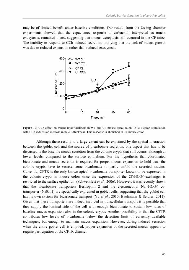
Colonic barrier function in ulcerative colitis
45
may be of limited benefit under baseline conditions. Our results from the Ussing chamber experiments showed that the capacitance response to carbachol, interpreted as mucin exocytosis, remained intact, suggesting that mucus exocytosis still occurred in the CF mice. The inability to respond to CCh induced secretion, implying that the lack of mucus growth was due to reduced expansion rather than reduced exocytosis.
Figure 10: CCh effect on mucus layer thickness in WT and CF mouse distal colon. In WT colon stimulation with CCh induces an increase in mucus thickness. This response is abolished in CF mouse colon.
Although these results to a large extent can be explained by the spatial interaction
between the goblet cell and the source of bicarbonate secretion, one aspect that has to be discussed is the baseline mucus secretion from the colonic crypts that still occurs, although at lower levels, compared to the surface epithelium. For the hypothesis that coordinated bicarbonate and mucus secretion is required for proper mucus expansion to hold true, the colonic crypts have to secrete some bicarbonate to partly unfold the secreted mucins. Currently, CFTR is the only known apical bicarbonate transporter known to be expressed in the colonic crypts in mouse colon since the expression of the Cl-/HCO3
--exchanger is restricted to the surface epithelium (Schweinfest et al., 2006). However, it was recently shown that the bicarbonate transporters Bestrophin 2 and the electroneutral Na+-HCO3
- co-transporter (NBCn1) are specifically expressed in goblet cells, suggesting that the goblet cell has its own system for bicarbonate transport (Yu et al., 2010; Bachmann & Seidler, 2011). Given that these transporters are indeed involved in transcellular transport it is possible that they supply the luminal side of the cell with enough bicarbonate to sustain low rates of baseline mucus expansion also in the colonic crypts. Another possibility is that the CFTR contributes low levels of bicarbonate below the detection limit of currently available techniques, but enough to maintain mucus expansion. However, during induced secretion when the entire goblet cell is emptied, proper expansion of the secreted mucus appears to require participation of the CFTR channel.
Jenny Gustafsson
46
Carbachol induced anion and mucus secretion in the normal and diseased human colon (Paper IV) Colonic anion secretion is a recognized part of the epithelial defense systems and our previous studies showed that colonic anion secretion is essential for compound exocytosis as well as for regulation of mucus properties. Although many aspects of colonic function are similar in mouse and human colon, species differences in epithelial anion secretion do occur. To fully understand this system one has to keep a close track on both species and segmental differences. Furthermore, almost nothing is known about the secretory capacity of the colonic epithelium during UC in remission, despite evidence in favor of a sustained imbalance between absorption and secretion. Against this background, an investigation was undertaken into the secretory response to carbachol (Ca2+) and forskolin (cAMP) in the distal and proximal colon of control and UC patients in remission, and the results were compared to the response pattern in the proximal and distal mouse colon.
Anions secretion in the normal colon – species and segmental differences The secretory response to carbachol and forskolin was measured in Ussing chambers using membrane current as a substitute marker for electrogenic anion secretion. The results revealed clear cut segment specific patterns both in baseline membrane current and in the secretory response to both carbachol and forskolin. In human colon, stimulation with carbachol induced a biphasic increase in membrane current characterized by a rapid peak response and a sustained plateau phase. Both the peak response and the plateau phase were significantly larger in the proximal colon compared to the distal colon. In contrast, stimulation with forskolin induced a sustained increase in membrane current that was significantly larger in the distal than in the proximal human colon. In mouse colon, baseline values were similar in both the proximal and distal colon of WT and CF mice, although the values were numerically reduced in CF colon. Stimulation with carbachol induced a rapid transient increase in membrane current in the proximal colon of WT mice and a biphasic response in the distal colon with a rapid peak response and a sustained plateau phase. In contrast to human colon, the peak response was significantly larger in mouse distal colon compared to proximal colon, and the proximal mouse colon lacked the sustained plateau phase. In the CF mice, the carbachol response was reduced by 50% in the proximal colon and by 90% in the distal colon. Accordingly, both the rapid peak response and the sustained plateau phase required a functioning CFTR channel. In the mouse, stimulation with forskolin induced a sustained increase in membrane current similar to that observed in human colon, although the magnitude of the response did not differ between proximal and distal colon. In CF mouse colon, the forskolin response was reduced by 90% in both the proximal and distal part. Comment: The results from this study highlight two important features of colonic anion secretion, namely that both basal values and responses to secretagogues are highly species and segment dependent. One should therefore be careful when extrapolating data from one segment or species to other segments or species. The observation of a higher baseline membrane current in the proximal than distal colon fits with previous studies in human colon (Kaji et al., 2012). It is known that baseline membrane current is reduced in the rectal mucosa
Colonic barrier function in ulcerative colitis
47
of CF patients, despite the facts that they have an up-regulated amiloride sensitive current (Mall et al., 2000). Whether CFTR mediated transport is involved in generating the high basal membrane current in proximal colon is not known.
The segmental differences in the amplitude of the response to secretagogues suggest that the human proximal colon is more sensitive to Ca2+ dependent secretagogues, while the distal human colon is more sensitive to cAMP dependent secretagogues. Since the CFTR has been shown to mediate both Ca2+ and cAMP dependent secretion, the difference is probably not directly linked to the degree of CFTR expression. Segmental differences in intracellular events following the two secondary message systems are more likely to be involved. In mouse distal colon, the sustained carbachol response has been shown to be bicarbonate dependent and mediated via the goblet cells specific transporter Bestrophin 2 (Yu et al., 2010). In human colon, the sustained response to carbachol was larger in proximal than distal colon, suggesting that bicarbonate dependent secretion is larger in the proximal segment (provided that Bestrophin 2 is indeed involved in this process also in human colon). Since we only compared the magnitude of the net membrane current response in the two segments we cannot say whether the observed differences are due to activation of separate signaling pathways or different activity of the same pathway. In mouse colon, studies have shown that although the magnitude of the forskolin response is similar in proximal and distal colon, the proximal colon can use either chloride or bicarbonate to mediate the membrane current response, whereas in the distal colon the response is chloride dependent (Gawenis et al., 2010). Studies have shown that also in the human distal colon the forskolin response is chloride dependent, but whether the human proximal colon can shift between chloride and bicarbonate like the mouse colon is not known (Osbak et al., 2007). Taken together, although the molecular mechanism behind colonic anion secretion has been studied in great detail, there are still questions that remain unsolved regarding central features of regulation of ion transport. This study points to some important differences in ion transport between human and mouse colon, especially in the proximal colon.
Anion and mucus secretion in patients with Ulcerative colitis in remission In a similar way as described in the previous section, the secretory response to carbachol and forskolin was tested in the proximal and distal colon of UC patients in remission, and was compared to that observed in control patients. The results showed that baseline electrical parameters were normal in both segments in UC patients in remission. However, the membrane current response to both carbachol and forskolin was altered in the proximal colon of UC patients towards a pattern resembling that seen in the normal distal colon with an up-regulation of the forskolin response and a down-regulation of the carbachol response. As discussed previously, the normal colon exhibits segment specific differences in anion secretion characterized by a dominant Ca2+ dependent secretion in the proximal colon and dominating cAMP dependent secretion in the distal colon. In the UC patients, these segment specific patterns were no longer seen and instead both segments expressed a “distal phenotype” characterized by a low membrane current response to carbachol and a high response to forskolin. In addition to measuring the membrane current response, we also measured the carbachol effect on membrane capacitance to estimate mucin granule
Jenny Gustafsson
48
exocytosis. Stimulation with carbachol induced a transient increase in capacitance in both proximal and distal colon. In the control patients, the magnitude of the response did not differ between the segments but the response was more rapid in the proximal than distal colon. In the UC patients, the response followed the same time course due to a more rapid response in the distal colon. Comment: The major conclusion that can be drawn from this part of the work is that despite a normal macroscopical appearance, the proximal colon of UC patients has an altered responsiveness to stimulation of anion secretion. Implications of an up-regulated forskolin response may be increased fluid load on the distal colon that may cause diarrheal problems. Studies have shown that the normal colonic mucosa has the ability to absorb approximately twice as much fluid than what normally passes through the ileocecal valve per 24 hours (Debongnie & Phillips, 1978). However, in a predisposed individual with mild distal inflammation even a small increase in fluid load might readily cause diarrhea, particularly if there is also loss of contact time due to more rapid transit. Regarding the implications of the decreased secretion of anions in response to carbachol, the main effect is expected to be interference with mucus expansion. The result from mouse colon showed that carbachol induced mucus growth was CFTR dependent, and if the same principle applies to human colon, a 40% reduction in the membrane current response with an intact capacitance might result in reduced ability to speed up mucus expansion following compound exocytosis. Interestingly, the magnitude of the membrane current response in the proximal colon of UC patients was similar to that in the normal distal colon, suggesting that a reduced current response does not necessarily have to result in a defective mucus layer, possibly just more dense mucus. The up-regulated capacitance response in the distal colon of the UC patients points towards a similar pattern with more mucus secretion in relation to anion secretion, which may be a protection mechanism to prevent against increased antigen exposure. Both the upregulation of sustained epithelial anion secretion in the proximal colon and upregulation of mucus secretion in the distal colon make sense in terms of minimizing the degree of contact between the luminal flora and the mucosal immune system.
The colonic mucus layer and inflammation (Paper V) Increased mucus content of the stool is a well established feature of acute ulcerative colitis (White, 1909). Further histological characterization of the disease showed that a reduced number of mucus containing goblet cells was one of the hallmarks of the acute inflammation (McCormick et al., 1990). The high amount of mucus in the stools and the reduced amounts of stored mucins in the goblet cells imply increased mucus secretion during acute inflammation. In the normal colon, the mucus layer has been shown to prevent the bacteria from reaching the epithelium and it has been suggested that spatial separation is impaired during acute inflammation (Pullan et al., 1994; Strugala et al., 2008). However, little is known about the properties of the intact mucus gel during disease, since most previous studies have been performed on paraformaldehyde fixed tissue that does not preserve the mucus layer. There has also been a lack of methods for studies of mucus properties in human colonic tissue like mucosal biopsies. To obtain a better understanding of how the mucus properties are
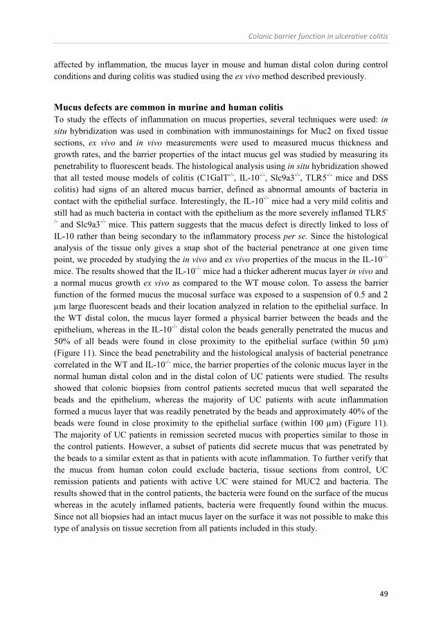
Colonic barrier function in ulcerative colitis
49
affected by inflammation, the mucus layer in mouse and human distal colon during control conditions and during colitis was studied using the ex vivo method described previously.
Mucus defects are common in murine and human colitis To study the effects of inflammation on mucus properties, several techniques were used: in situ hybridization was used in combination with immunostainings for Muc2 on fixed tissue sections, ex vivo and in vivo measurements were used to measured mucus thickness and growth rates, and the barrier properties of the intact mucus gel was studied by measuring its penetrability to fluorescent beads. The histological analysis using in situ hybridization showed that all tested mouse models of colitis (C1GalT-/-, IL-10-/-, Slc9a3-/-, TLR5-/- mice and DSS colitis) had signs of an altered mucus barrier, defined as abnormal amounts of bacteria in contact with the epithelial surface. Interestingly, the IL-10-/- mice had a very mild colitis and still had as much bacteria in contact with the epithelium as the more severely inflamed TLR5-
/- and Slc9a3-/- mice. This pattern suggests that the mucus defect is directly linked to loss of IL-10 rather than being secondary to the inflammatory process per se. Since the histological analysis of the tissue only gives a snap shot of the bacterial penetrance at one given time point, we proceded by studying the in vivo and ex vivo properties of the mucus in the IL-10-/- mice. The results showed that the IL-10-/- mice had a thicker adherent mucus layer in vivo and a normal mucus growth ex vivo as compared to the WT mouse colon. To assess the barrier function of the formed mucus the mucosal surface was exposed to a suspension of 0.5 and 2 µm large fluorescent beads and their location analyzed in relation to the epithelial surface. In the WT distal colon, the mucus layer formed a physical barrier between the beads and the epithelium, whereas in the IL-10-/- distal colon the beads generally penetrated the mucus and 50% of all beads were found in close proximity to the epithelial surface (within 50 µm) (Figure 11). Since the bead penetrability and the histological analysis of bacterial penetrance correlated in the WT and IL-10-/- mice, the barrier properties of the colonic mucus layer in the normal human distal colon and in the distal colon of UC patients were studied. The results showed that colonic biopsies from control patients secreted mucus that well separated the beads and the epithelium, whereas the majority of UC patients with acute inflammation formed a mucus layer that was readily penetrated by the beads and approximately 40% of the beads were found in close proximity to the epithelial surface (within 100 µm) (Figure 11). The majority of UC patients in remission secreted mucus with properties similar to those in the control patients. However, a subset of patients did secrete mucus that was penetrated by the beads to a similar extent as that in patients with acute inflammation. To further verify that the mucus from human colon could exclude bacteria, tissue sections from control, UC remission patients and patients with active UC were stained for MUC2 and bacteria. The results showed that in the control patients, the bacteria were found on the surface of the mucus whereas in the acutely inflamed patients, bacteria were frequently found within the mucus. Since not all biopsies had an intact mucus layer on the surface it was not possible to make this type of analysis on tissue secretion from all patients included in this study.
Jenny Gustafsson
50
Figure 11: Effect of inflammation on the penetrability of the colonic mucus layer to 2µm large green fluorescent beads. A: WT and IL-10-/- mouse distal colon. B: Human sigmoid colon of a control patient and an UC patient with acute inflammation. The epithelial surface is stained blue.
Comment: From this part of the work, it can be concluded that an altered mucus barrier is common in both murine and human colitis and independent of the underlying genetic component responsible for causing the disease. This observation highlights an important aspect of the colonic barrier namely that the different functions such as ion transport, the mucus layer and the immune system are integrated and interference with one system may change the delicate balance and break the barrier. The observation of increased mucus penetrability in the IL-10-/- mice despite a very minor inflammatory response points to a direct effect of the altered immune homeostasis on goblet cell function, alternatively interference with systems that regulate mucus properties. The normal mucus growth and even thicker adherent mucus layer observed in the IL-10-/- mice can be interpreted as more voluminous mucus with an altered structure. In the IL-10-/- mice, the immune homeostasis is driven towards a Th1 and Th17 response with high expression of IFN-γ and IL-17 and low numbers of Tregs. IFN-γ has been shown to induce mucus secretion in rat colon, but whether this is related to the mucus penetrability defect is not known (Songhet et al., 2011).
In the UC group, the vast majority of patients with active inflammation secreted mucus that was penetrated by the fluorescent beads and histological evaluation of tissue sections confirmed that the colonic mucus contained bacteria. Thus, our results support the hypothesis that one of the disease causing factors in UC may be an altered mucus layer that allows bacteria to reach the epithelium and start an immune response. At this stage, we have no solid information regarding which mechanisms that are responsible for these altered mucus properties. However, as mentioned previously, patients with active UC have been shown to have reduced electrogenic ion transport, an altered cytokine profile and shorter glycans on the MUC2 mucin which all are expected to affect mucus properties. The genetic influence in UC is probably multifactorial and it seems likely that patients have more than one defect. Related to the relation between the degree of inflammation and mucus penetrability, it is worth mentioning that patients with mild inflammation (Mayo 1) often had the same degree of increased mucus penetrability as patients with more severe inflammation (Mayo 2-3),
Colonic barrier function in ulcerative colitis
51
suggesting that it is not only the inflammatory activity per se that affects mucus properties. Furthermore, a subset of patients in remission showed a similar degree of mucus penetrability as patients with acute disease.
Taken together, we have presented a proof of concept that alterations in the mucus layer is a common denominator for both human and murine colitis, and we propose that a failing mucus barrier will expose the epithelium to increased amounts of bacteria that can further trigger the mucosal immune reaction and thereby aggravate the disease course.
Jenny Gustafsson
52
GENERAL DISCUSSION
In the present thesis work, techniques were developed that enabled monitoring of epithelial anion secretion and mucus properties in human colonic biopsies, and tissue specimens from mouse small and large intestine. These techniques were then used to dissect the interactions between epithelial transport and mucus properties in normal and inflamed tissue. The results show that epithelial transport plays an important role in regulation of the intestinal mucus in both cystic fibrosis and ulcerative colitis. Furthermore, this work highlights that species, segmental and spatial differences in epithelial functions are something that has to be taken into account in studies of mucosal defense.
Regulation of mucus secretion and mucus properties - lessons from cystic fibrosis In the ileum, CFTR mediated secretion was shown to regulate several aspects of mucus properties including mucus growth, adhesion and penetrability. All these effects could be related to the local bicarbonate concentrations during secretion. Furthermore, the finding that EDTA was able to reverse the adherent mucus phenotype in CF ileum supports the hypothesis that one of the mechanisms by which bicarbonate affects mucus properties is by chelating calcium during the secretory process.
In the colon, where the mucus layer is already adherent, and the lumen is colonized by massive amounts of bacteria, loss of CFTR function does not cause any obvious pathology. This is slightly surprising since activation of the CFTR channel results in a prominent secretory response in both the proximal and distal colon (Nobles et al., 1991; Mall et al., 1999; Gawenis et al., 2010). Our results showed that baseline mucus growth was CFTR independent whereas carbachol induced mucus growth was CFTR dependent. This pattern was seen in spite of the fact that the carbachol-induced capacitance response was intact in the CF colon, that is, CF colon had a seemingly normal rate of carbachol induced mucin granule exocytosis. These results suggest that baseline mucus secretion in the colon relies on bicarbonate secretion from another source than the CFTR. However, during secretagogue induced mucus secretion that primarily targets the goblet cells in the colonic crypts additional opening of the CFTR seems to be essential to fully expand the secreted mucus.
Carbachol induced mucus exocytosis was independent of a functional CFTR channel, but dependent on NKCC1 and K+ channels. The discrepancy between the results in the CF colon and WT colon treated with bumetanide (which both reduced the membrane current response by 90%) could be interpreted to mean that the inhibitory effect of bumetanide on the capacitance response is via its effect on the goblet cell. In the rat jejunum, the goblet cells have been shown to express three times more NKCC1 compared to the neighboring enterocytes (Jakab et al., 2011). Furthermore, asthma induced goblet cell hyperplasia is associated with goblet cell specific up-regulation of NKCC1 (Dolganov et al., 2001; Jakab et al., 2011). Together these results suggest that this transporter is important for goblet cell function; however how it is involved in regulation of mucus secretion is not known. The association between potassium and mucus secretion has been recognized from the extreme cases of mucus producing villus adenomas which can secrete up to 2 liters of mucus per 24 hours and almost completely deplete the plasma pool of potassium (Rowe, 1964).
Colonic barrier function in ulcerative colitis
53
Although many questions regarding the role of ion transport in regulation of mucus properties in the colon remains, this work has provided new insights into how CFTR mediated transport and especially bicarbonate secretion affects mucus properties in the intestine. Direct clinical applications of these findings would be to use inhalation of bicarbonate as a treatment in CF. Although our results are from the small intestine one can hypothesize that similar mechanism are responsible for mucus accumulation in the airways. One major problem with this approach is bicarbonate induced mucus expansion in the airways. A combination treatment with either a substance that reduces mucus release, or a substance that helps to transport the mucus in the proximal direction might prove to be useful.
Epithelial function in the normal human colon It is well known that the absorptive properties of the colonic epithelium differ between the proximal and distal colon, however less is known about segmental differences in the epithelial defense systems such as anion and mucus secretion. With the increasing bacterial density and intraluminal pressure towards the distal end one could assume that the colonic epithelium has adapted to handle this efficiently. Our results from the proximal and distal human colon show that the reactivity to both Ca2+ and cAMP mediated anion secretion differed between the two segments and mucus exocytosis was also more rapid in the proximal than distal colon. The secretory pattern was characterized by a dominating Ca2+ response in the proximal colon and a dominating cAMP response in the distal colon. The physiological meaning of these segmental differences is not known, however it is possible that they are related to the luminal conditions in the respective segment. The luminal content of the proximal colon is loose and watery and a high capacity to produce sustained fluid secretion via cAMP mediated pathways might therefore not be needed. In the distal colon on the other hand, where the luminal content is dense fluid secretion may prove to be useful to flush the epithelium. Regarding the segmental differences in the Ca2+ response, the fast response in the proximal colon would allow the tissue to respond rapidly with large quantities of mucus. In the distal colon the slower mucus response and the lower magnitude of the anion response might results in more dense mucus compared to the proximal colon. Dense mucus in the distal colon will help to protect the tissue from the risk of barrier breakage caused by the high intraluminal pressure and the high numbers of bacteria.
Epithelial function in UC Active UC is known to be associated with loss of absorptive functions due to down-regulation or decreased activity of most transporters that participate in the absorptive process (Greig et al., 2004; Yeruva et al., 2010). This loss of absorptive function has been suggested to be the main mechanism behind diarrhea in acute colitis. So far, there is no evidence for increased anion secretion being involved in the pathogenesis of diarrhea, although the inflamed mucosa does produces large amounts of secretagogues such as prostaglandins (Rampton et al., 1980). One explanation to this discrepancy could be the reduced activity of the Na+/K+-ATPase that is required for electrogenic anion secretion.
From the results in mouse small and large intestine, it is clear that epithelial transport affects mucus properties and it is likely that the altered ionic milieu during acute colitis affects
Jenny Gustafsson
54
the mucus layer. Our results from mouse and human colon showed that a penetrable mucus layer is common in both experimental and human colitis. Thus, both an impaired electrogenic transport and mucus defects are common features of colitis that may be independent of the primary cause of disease. During remission, most patients secreted mucus with normal penetrability which correlated with normal baseline transport and a normal response to both Ca2+ and cAMP mediated secretion in the distal colon. In the proximal colon baseline values were normal but the responsiveness to induced secretion was shifted towards a distal phenotype with increased cAMP mediated secretion and decreased Ca2+ mediated secretion. This would result in an ability to secrete more fluid in response to the same stimuli, which might be beneficial if the epithelium is exposed to an increased amount of antigens. Similarly, the decreased Ca2+-response could be interpreted as a way to reduce mucus expansion and thereby generating denser mucus that further impede the bacterial exposure.
By understanding the mechanisms that are responsible for inducing the increased penetrability of the mucus layer during colitis, one can hopefully in the future use this knowledge to improve the mucus barrier and thereby prevent exacerbations. Since the mucus defect was a general phenomenon during colitis a strengthened mucus layer might be beneficial for a large patient group.
In summary, we show that epithelial secretion of ions and mucus is an integrated process that has been adapted to form mucus with properties suitable for the demands of the respective sites. Alterations in the secretory process can cause severe disease like in the case of cystic fibrosis, but the results from this work show that the mucus phenotype is reversible simply by exposure to high concentrations of bicarbonate. In the colon, where the protective properties of the mucus is based on a dense impermeable layer, problems occur when the penetrability of the mucus increases, allowing bacteria to pass though and access the epithelium. We showed that an altered mucus barrier is a common denominator of acute colitis, but resolution of the inflammation appears to restore the mucus barrier is the majority of patients. The normal mucus barrier during remission was paralleled with restored ion transport in the distal colon, suggesting that effective prophylactic treatment not only induces mucosal healing but it also restores epithelial function in this segment.
Colonic barrier function in ulcerative colitis
55
CONCLUDING REMARKS
The central topic of this thesis is how epithelial transport affects mucus properties and how this in turn affects the regulation of the colonic barrier. In the literature there is increasing evidence that the inner colonic mucus layer is of crucial importance for the colonic barrier and that anion secretion regulates mucus properties. However, there are few studies in human colon and the current knowledge regarding the interaction between anion and mucus secretion is limited. In this thesis we aimed to fill some of these knowledge gaps by developing novel methods suitable for studies of anion and mucus secretion in human colonic biopsies and in mouse intestinal explants, and to apply these methods on central questions regarding regulation of epithelial secretions. The methodological development consist of an ex vivo method for studies of mucus properties in human colon and mouse small and large intestine, and the use of epithelial capacitance to measure mucin granule secretion in intact intestinal tissues from human and mouse colon. The main conclusions can be summarized as follows:
I. Both cAMP and Ca2+ dependent anion secretion in the colon show marked species and segmental dependent differences
II. Ion transport via NKCC1 and K+ channels is essential for secretagogue induced mucus secretion in the colon.
III. CFTR mediated transport plays a pivotal role in the regulation of mucus properties in mouse ileum.
IV. CFTR mediated transport is not required for baseline mucus growth in the colon,
but is essential for secretagogue induced mucus growth.
V. Patients with UC in remission have an altered responsiveness to secretagogues in the proximal colon despite a macroscopically normal mucosa.
VI. Increased mucus penetrability in colon is a common denominator of both murine
and human colitis. In summary, this work shows that epithelial transport plays a crucial role for the formation of a normal mucus layer in both the small and large intestine. In UC patients, mucus and anion secretion was normal or even up-regulated during remission, whereas during acute colitis the mucus barrier becomes severely impaired. Damage to the mucus barrier may start a vicious circle with increasing antigen load, increased inflammation and further damage to the mucus layer eventually resulting in full scale colitis. Interference with the early mucolytic effects of inflammation may be a target for prevention of this detrimental sequence of events.
Jenny Gustafsson
56
FUTURE PERSPECTIVES
Although the results from this thesis shed some light on the role of epithelial transport in the regulation of the intestinal mucus layer and its implication on the colonic defense, there are several new questions that need to be addressed.
When it comes to unraveling the role of epithelial secretion (ion and mucus) in the pathogenesis of UC, we are still at the phenotype level regarding mucus properties and anion secretion. Our results have given a proof of concept that both ion transport and mucus properties contribute to colonic defense but we do not understand the relative importance of basal and secretagogues induced mucus secretion. We also do not know the exact mechanism by which inflammation affects the mucus layer during colitis or how the goblet cell is regulated by the immune system. Furthermore, the pathophysiological role of the increased reactivity of cAMP-mediated anion secretion in the proximal colon UC patients in remission needs to be followed up to determine how this phenomenon affects the colonic barrier. Finally, to fully understand the role of the colonic mucus layer in mucosal defense one has to determine how the penetrability of the mucus is regulated and the mechanisms behind the increased penetrability in colitis.
Colonic barrier function in ulcerative colitis
57
POPULÄRVETENSKAPLIG SAMMANFATTNING
I vår tjocktarm lever det fler bakterier än det finns celler i kroppen. Dessa bakterier förser oss med näring och vitaminer och skyddar oss mot sjukdomsalstrande bakterier. Trots att dessa bakterier behövs för den normala tarmfuktionen så måste de hållas borta från blodbanan. För att klara av detta har kroppen utveckat ett skyddssystem den så kallade tarmbarriären som kan delas upp i tre delar: ett tjockt slemlager, ett enkelt cellager (ytepitelet) och slemhinnans immunsystem. Målet med tarmbarriären är att bakterierna ska ha så lite kontakt som möjligt med immunsystemet. Slemmet som frisätts från tarmväggen lägger sig som ett skyddande lager mellan bakterierna och ytepitelet och förhindrar på så sätt att bakterierna tar sig in i vävnaden och vidare in i blodbanan. Om för mycket kontakt uppstår mellan bakterierna och tarmväggen aktiveras tarmens immunsystem vilket leder till inflammtion som vid den kroniska inflammatoriska tarmsjukdomen ulcerös kolit. Även om man idag vet att slemlagret spelar en viktig roll i tarmbarriären så vet man inte vilka faktorer som reglerar slemmets skyddande egenskaper. Det man vet är att jonkanalen CFTR som driver vätskebildning i tarmen även påverkar slemmets egenskaper eftersom patienter som saknar en fungerande CFTR kanal har tjockt segt slem i lungorna och tarmen.
Målet med den här avhandligen var att försöka förstå hur transport via CFTR kanalen påverkar slemlagrets skyddande egenskaper i tarmen och hur dessa system i sin tur påverkas av inflammation.
För att kunna studera tarmbarriären i människa utvecklade vi två metoder för mätning av ytepitelets vätske och slemproduktion och slemlagrets egenskaper på vävnadsbitar (biopsier) som tas i samband med koloskopi. Dessa två metoder användes sedan för att studera vilka mekanismer som påverakde slembildningen och hur den i sin tur påverkades av en ökad och minskad vätskeproduktion och vid inflammation.
De viktigaste resultaten som presenteras i den här avhandlingen är att de slembildande cellerna i tjocktarmen verkar vara försedda med sitt eget transportmaskineri som gör att de kan fortsätta att arbeta även om de övriga cellerna inte producerar vätska. Undantaget är i situationer där det krävs en snabb kraftig slembildning. Då måste även de övriga epitelcellerna bidra med vätsketransport för att man ska få en snabb tillväxt av slemlagret.
Patienter med ulcerös kolit i lungs skede hade en normal slemproduktion, både gällande mängd och kvalitet men de hade en ökad förmåga att bilda vätska som kan skölja bort bakterier. Vid akut inflammation (kolit) påverkas framförallt kvalitén på slemmet som gör det genomsläppligt för bakterier.
Sammanfattningsvis så visar de här resultaten att patienter med inaktiv sjukdom har en välfungerande barriär som även skulle kunna vara mer effektiv än hos de friska patienterna på grund av den ökade vätskeproduktionen. Vid utveckling av ett skov påverkas dock kvaliteten på det skyddande slemmet vilket ökar genomsläppligheten för bakterier. Detta kan i sin tur ge upphov till en ond cirkel av mer och mer kraftfull aktivering av immunförsvaret. Om man kunde identifiera de exakta mekanismerna bakom inflammationens effekter på slembarriären och behandla dessa med läkemedel skulle detta kunna förhindra utvecklingen av nya skov.
Jenny Gustafsson
58
ADDITIONAL BIBLIOGRAPHY
1. Ambort D, Johansson MEV, Gustafsson JK, Nilsson HE, Ermund A, Johansson BR, Koeck P, Hebert H, Hansson GC. Calcium and pH-dependent Packing and Release of the gel-forming MUC2 Mucin. Proc Natl Acad Sci U S A. 2012 Mar 26
2. Gustafsson JK, Sjövall H, Hansson GC. Ex vivo measurements of mucus secretion by colon explants. Methods Mol Biol. 2012;842:237-43.
3. Johansson ME, Ambort D, Pelaseyed T, Schütte A, Gustafsson JK, Ermund A,
Subramani DB, Holmén-Larsson JM, Thomsson KA, Bergström JH, van der Post S, Rodriguez-Piñeiro AM, Sjövall H, Bäckström M, Hansson GC. Composition and functional role of the mucus layers in the intestine. Cell Mol Life Sci. 2011 Sep 25
4. Gustafsson JK and Greenwood-Van Meerveld B. Amygdala activation by
corticosterone alters visceral and somatic pain in cycling female rats. Am J Physiol Gastrointest Liver Physiol. 2011 Jun;300(6):G1080-5
5. Johansson ME, Gustafsson JK, Sjöberg KE, Petersson J, Holm L, Sjövall H, Hansson
GC. Bacteria penetrate the inner mucus layer before inflammation in the dextran sulfate colitis mode. PLoS One. 2010 Aug 18;5(8):e12238
Colonic barrier function in ulcerative colitis
59
ACKNOWLEDGMENTS
This thesis would not be what it is today without the contribution of a large number of people that I would like to take the opportunity to thank: Henrik thank you for introducing me to the world of integrated physiology. You have managed very well to transfer your enthusiasm for understanding the “bigger picture”. I am very grateful for your constant encouragement, enthusiasm and confidence in me during these years. Gunnar thank you for making me part of your group. Other people aim high but you reach for the stars! If it wasn’t for you this thesis would have been so very different and I am truly grateful for your support and enthusiasm. My co-supervisors Sara and Lena thank you for your help and support during these years. There are not many students that have four supervisors, but I say the more the merrier. The mucin biology group with past and present members: Thank you all for making this time what it has been. Tina for being a good friend that cares for everyone. Karin for steering things around and pushing your papers over the invisible line. André for always being kind and helpful. You should convince Nora that Sweden is the place to be. Daniel for once in a while showing that well-behaved person that is hidden inside of you. Thaher and Joakim soon it is your turn, good luck and thanks for these years. Anna for always being ready to discuss all kind of strange subjects. Frida and Jessica for your funny sarcastic sides. Ana I can teach you Swedish if you teach me how to become that organized. Lisbeth thank you for all the help over the years, I truly appreciate it. Robert your devotion to your work is incredible, but I suggest that you try to find a girlfriend that does not live in a Petri dish… Dan for your wise comments. Karolina it’s great to have you back in the group. Noreen for always being so close to laughter, don’t think that there has been a single day when I haven’t heard you laughing. Catharina for keeping track on everything, it is really nice to have you in the group. Elisabeth, for being such a positive person. Malin B and Richard for nice discussions in the lunch room. The newest members of the group, Hedvig, Ida and Lisa it has been nice to get to know you during these past months. A special thanks to Malin for being a good friend that appreciates all the right things in life (I think you know what I’m thinking of). We might have to make a schedule for at least one random discussion a week, I miss that. The members of Sara, Niclas and Susann’s groups for making this a great place to work at. Diarmuid and Catherine thanks for dinners, BBQs and other things that makes life outside work more fun. Sarah and Harvey I really appreciate that you took the time to read through this thesis. Stefan thanks for nice discussion and lunches over the years Maria I’ve known you almost since day one here in Gothenburg. Thank you for being a great friend and for having such a good taste in music. The Knife is still on my top 10 list. Maybe we should go to Café Cello one day to relive 2005. Bengt, Yvonne and Kanita at the EM facility thank you for introducing me to the world of electron microscopy and for your kindness.
Jenny Gustafsson
60
All the patients that volunteered for the different studies and the staff at the Sahlgrenska University hospital. Leo, Gerda, Sanne, Roy and Pien + 1, thank you for welcoming me into your family. Johan, en bättre bror kan man inte önska sig. Lycka till med din egen avhandlig. Min underbara föräldrar Ulla och Olle som alltid ställer upp och finns där när man behöver er. Tack för allt! Farmor Elsie för att du är så omtänksam och stolt över mig. Den här boken är till dig. And Sjoerd what would these years have been without you? You have been amazing during these last months, helping out in all kind of ways by making figures, cover pictures, reading, formatting, etc. I am so happy and grateful to have you in my life. “No one will ever love you more than I do”. Ik hou van je.
This work was supported by the Swedish Research Council (no. 7461, 21027, 08288), The Swedish Cancer Foundation, The Knut and Alice Wallenberg Foundation, Inga-Britt and Arne Lundberg Foundation, Sahlgren's University Hospital (LUA-ALF, ALFGBG-138841), Wilhelm and Martina Lundgren’s Foundation, Torsten och Ragnar Söderbergs Stiftelser, The Swedish Foundation for Strategic Research - The Mucus-Bacteria-Colitis Center (MBC) of the Innate Immunity Program and Assar Gabrielsson foundation.

Colonic barrier function in ulcerative colitis
61
REFERENCES
(1993). Correlation between Genotype and Phenotype in Patients with Cystic Fibrosis. New England Journal of Medicine 329, 1308-1313.
Alzamora R, O'Mahony F & Harvey BJ. (2011). Estrogen inhibits chloride secretion caused by cholera and
Escherichia coli enterotoxins in female rat distal colon. Steroids 76, 867-876. Amann RI, Binder BJ, Olson RJ, Chisholm SW, Devereux R & Stahl DA. (1990). Combination of 16S rRNA-
targeted oligonucleotide probes with flow cytometry for analyzing mixed microbial populations. Appl Environ Microbiol 56, 1919-1925.
Amasheh S, Barmeyer C, Koch CS, Tavalali S, Mankertz J, Epple HJ, Gehring MM, Florian P, Kroesen AJ,
Zeitz M, Fromm M & Schulzke JD. (2004). Cytokine-dependent transcriptional down-regulation of epithelial sodium channel in ulcerative colitis. Gastroenterology 126, 1711-1720.
Ambort D, Johansson ME, Gustafsson JK, Nilsson HE, Ermund A, Johansson BR, Koeck PJ, Hebert H &
Hansson GC. (2012). Calcium and pH-dependent packing and release of the gel-forming MUC2 mucin. Proc Natl Acad Sci U S A.
Amulic B, Cazalet C, Hayes GL, Metzler KD & Zychlinsky A. (2011). Neutrophil Function: From Mechanisms
to Disease. Annu Rev Immunol. Atuma C, Strugala V, Allen A & Holm L. (2001). The adherent gastrointestinal mucus gel layer: thickness and
physical state in vivo. Am J Physiol Gastrointest Liver Physiol 280, G922-929. Bachmann O, Reichelt D, Tuo B, Manns MP & Seidler U. (2006). Carbachol increases Na+-HCO3- cotransport
activity in murine colonic crypts in a M3-, Ca2+/calmodulin-, and PKC-dependent manner. Am J Physiol Gastrointest Liver Physiol 291, G650-657.
Bachmann O & Seidler U. (2011). News from the end of the gut--how the highly segmental pattern of colonic
HCO transport relates to absorptive function and mucosal integrity. Biol Pharm Bull 34, 794-802. Bachmann O, Wuchner K, Rossmann H, Leipziger J, Osikowska B, Colledge WH, Ratcliff R, Evans MJ, Gregor
M & Seidler U. (2003). Expression and regulation of the Na+-K+-2Cl- cotransporter NKCC1 in the normal and CFTR-deficient murine colon. J Physiol 549, 525-536.
Barker N, van Es JH, Kuipers J, Kujala P, van den Born M, Cozijnsen M, Haegebarth A, Korving J, Begthel H,
Peters PJ & Clevers H. (2007). Identification of stem cells in small intestine and colon by marker gene Lgr5. Nature 449, 1003-1007.
Barreiro-de Acosta M, Alvarez Castro A, Souto R, Iglesias M, Lorenzo A & Dominguez-Munoz JE. (2011).
Emigration to western industrialized countries: A risk factor for developing inflammatory bowel disease. J Crohns Colitis 5, 566-569.
Bekkali N, de Jonge HR, van den Wijngaard RM, van der Steeg AF, Bijlsma PB, Taminiau JA, Desjeux JF &
Benninga MA. (2011). The role of rectal chloride secretion in childhood constipation. Neurogastroenterol Motil 23, 1007-1012.
Bergstrom KS, Kissoon-Singh V, Gibson DL, Ma C, Montero M, Sham HP, Ryz N, Huang T, Velcich A, Finlay
BB, Chadee K & Vallance BA. (2010). Muc2 protects against lethal infectious colitis by disassociating pathogenic and commensal bacteria from the colonic mucosa. PLoS Pathog 6, e1000902.
Bertrand CA, Durand DM, Saidel GM, Laboisse C & Hopfer U. (1998). System for dynamic measurements of
membrane capacitance in intact epithelial monolayers. Biophys J 75, 2743-2756. Bertrand CA & Frizzell RA. (2003). The role of regulated CFTR trafficking in epithelial secretion. Am J Physiol
Cell Physiol 285, C1-18.
Jenny Gustafsson
62
Bertrand CA, Laboisse CL & Hopfer U. (1999). Purinergic and cholinergic agonists induce exocytosis from the same granule pool in HT29-Cl.16E monolayers. Am J Physiol 276, C907-914.
Bertrand PP, Kunze WA, Bornstein JC, Furness JB & Smith ML. (1997). Analysis of the responses of myenteric
neurons in the small intestine to chemical stimulation of the mucosa. Am J Physiol 273, G422-435. Binder HJ & Rawlins CL. (1973). Electrolyte transport across isolated large intestinal mucosa. Am J Physiol
225, 1232-1239. Boige N, Amiranoff B, Munck A & Laburthe M. (1984). Forskolin stimulates adenylate cyclase in human
colonic crypts: interaction with VIP. Eur J Pharmacol 101, 111-117. Boman HG. (2003). Antibacterial peptides: basic facts and emerging concepts. J Intern Med 254, 197-215. Bonoff KM. (1939). Water Absorption from the Colon and Its Relation to Motility. Cal West Med 51, 154-156. Boucher RC, Stutts MJ, Knowles MR, Cantley L & Gatzy JT. (1986). Na+ transport in cystic fibrosis respiratory
epithelia. Abnormal basal rate and response to adenylate cyclase activation. J Clin Invest 78, 1245-1252.
Brandtzaeg P, Farstad IN, Johansen FE, Morton HC, Norderhaug IN & Yamanaka T. (1999). The B-cell system
of human mucosae and exocrine glands. Immunol Rev 171, 45-87. Braun T, Voland P, Kunz L, Prinz C & Gratzl M. (2007). Enterochromaffin cells of the human gut: sensors for
spices and odorants. Gastroenterology 132, 1890-1901. Burgoyne RD & Morgan A. (2007). Membrane trafficking: three steps to fusion. Curr Biol 17, R255-258. Caprilli R, Frieri G, Latella G, Vernia P & Santoro ML. (1986). Faecal excretion of bicarbonate in ulcerative
colitis. Digestion 35, 136-142. Chang WW & Leblond CP. (1971). Renewal of the epithelium in the descending colon of the mouse. I. Presence
of three cell populations: vacuolated-columnar, mucous and argentaffin. Am J Anat 131, 73-99. Chen JH, Stoltz DA, Karp PH, Ernst SE, Pezzulo AA, Moninger TO, Rector MV, Reznikov LR, Launspach JL,
Chaloner K, Zabner J & Welsh MJ. (2010). Loss of anion transport without increased sodium absorption characterizes newborn porcine cystic fibrosis airway epithelia. Cell 143, 911-923.
Cheng H & Leblond CP. (1974). Origin, differentiation and renewal of the four main epithelial cell types in the
mouse small intestine. V. Unitarian Theory of the origin of the four epithelial cell types. Am J Anat 141, 537-561.
Cheng SH, Rich DP, Marshall J, Gregory RJ, Welsh MJ & Smith AE. (1991). Phosphorylation of the R domain
by cAMP-dependent protein kinase regulates the CFTR chloride channel. Cell 66, 1027-1036. Choi JY, Joo NS, Krouse ME, Wu JV, Robbins RC, Ianowski JP, Hanrahan JW & Wine JJ. (2007). Synergistic
airway gland mucus secretion in response to vasoactive intestinal peptide and carbachol is lost in cystic fibrosis. J Clin Invest 117, 3118-3127.
Christofi FL, Wunderlich J, Yu JG, Wang YZ, Xue J, Guzman J, Javed N & Cooke H. (2004). Mechanically
evoked reflex electrogenic chloride secretion in rat distal colon is triggered by endogenous nucleotides acting at P2Y1, P2Y2, and P2Y4 receptors. J Comp Neurol 469, 16-36.
Clauss W, Schafer H, Horch I & Hornicke H. (1985). Segmental differences in electrical properties and Na-
transport of rabbit caecum, proximal and distal colon in vitro. Pflugers Arch 403, 278-282. Collins D, Hogan AM, Skelly MM, Baird AW & Winter DC. (2009). Cyclic AMP-mediated chloride secretion is
induced by prostaglandin F2alpha in human isolated colon. Br J Pharmacol 158, 1771-1776. Cooke HJ. (1998). "Enteric Tears": Chloride Secretion and Its Neural Regulation. News Physiol Sci 13, 269-274.
Colonic barrier function in ulcerative colitis
63
Cooke HJ. (2000). Neurotransmitters in neuronal reflexes regulating intestinal secretion. Ann N Y Acad Sci 915,
77-80. Coombes JL, Robinson NJ, Maloy KJ, Uhlig HH & Powrie F. (2005). Regulatory T cells and intestinal
homeostasis. Immunol Rev 204, 184-194. Costa M, Furness JB, Cuello AC, Verhofstad AA, Steinbusch HW & Elde RP. (1982). Neurons with 5-
hydroxytryptamine-like immunoreactivity in the enteric nervous system: their visualization and reactions to drug treatment. Neuroscience 7, 351-363.
Dagher PC, Rho JI & Charney AN. (1993). Mechanism of bicarbonate secretion in rat (Rattus rattus) colon.
Comp Biochem Physiol Comp Physiol 105, 43-48. Davidson NJ, Leach MW, Fort MM, Thompson-Snipes L, Kuhn R, Muller W, Berg DJ & Rennick DM. (1996).
T helper cell 1-type CD4+ T cells, but not B cells, mediate colitis in interleukin 10-deficient mice. J Exp Med 184, 241-251.
Davis PB. (2006). Cystic fibrosis since 1938. Am J Respir Crit Care Med 173, 475-482. Debongnie JC & Phillips SF. (1978). Capacity of the human colon to absorb fluid. Gastroenterology 74, 698-
703. del Castillo JR & Burguillos L. (2005). Pathways for K+ efflux in isolated surface and crypt colonic cells.
Activation by calcium. J Membr Biol 205, 37-47. Dharmsathaphorn K & Pandol SJ. (1986). Mechanism of chloride secretion induced by carbachol in a colonic
epithelial cell line. J Clin Invest 77, 348-354. Dianda L, Hanby AM, Wright NA, Sebesteny A, Hayday AC & Owen MJ. (1997). T cell receptor-alpha beta-
deficient mice fail to develop colitis in the absence of a microbial environment. Am J Pathol 150, 91-97.
Diener M & Rummel W. (1990). Distension-induced secretion in the rat colon: mediation by prostaglandins and
submucosal neurons. Eur J Pharmacol 178, 47-57. Dolganov GM, Woodruff PG, Novikov AA, Zhang Y, Ferrando RE, Szubin R & Fahy JV. (2001). A novel
method of gene transcript profiling in airway biopsy homogenates reveals increased expression of a Na+-K+-Cl- cotransporter (NKCC1) in asthmatic subjects. Genome Res 11, 1473-1483.
Feldman GM, Berman SF & Stephenson RL. (1988). Bicarbonate secretion in rat distal colon in vitro: a
measurement technique. Am J Physiol 254, C383-390. Forstner G. (1995). Signal transduction, packaging and secretion of mucins. Annu Rev Physiol 57, 585-605. Foxx-Orenstein AE, Kuemmerle JF & Grider JR. (1996). Distinct 5-HT receptors mediate the peristaltic reflex
induced by mucosal stimuli in human and guinea pig intestine. Gastroenterology 111, 1281-1290. Fu J, Wei B, Wen T, Johansson ME, Liu X, Bradford E, Thomsson KA, McGee S, Mansour L, Tong M,
McDaniel JM, Sferra TJ, Turner JR, Chen H, Hansson GC, Braun J & Xia L. (2011). Loss of intestinal core 1-derived O-glycans causes spontaneous colitis in mice. J Clin Invest 121, 1657-1666.
Furness JB. (2008). The enteric nervous system: normal functions and enteric neuropathies. Neurogastroenterol
Motil 20 Suppl 1, 32-38. Fuss IJ, Neurath M, Boirivant M, Klein JS, de la Motte C, Strong SA, Fiocchi C & Strober W. (1996). Disparate
CD4+ lamina propria (LP) lymphokine secretion profiles in inflammatory bowel disease. Crohn's disease LP cells manifest increased secretion of IFN-gamma, whereas ulcerative colitis LP cells manifest increased secretion of IL-5. J Immunol 157, 1261-1270.
Jenny Gustafsson
64
Fyderek K, Strus M, Kowalska-Duplaga K, Gosiewski T, Wedrychowicz A, Jedynak-Wasowicz U, Sladek M, Pieczarkowski S, Adamski P, Kochan P & Heczko PB. (2009). Mucosal bacterial microflora and mucus layer thickness in adolescents with inflammatory bowel disease. World J Gastroenterol 15, 5287-5294.
Garcia MA, Yang N & Quinton PM. (2009). Normal mouse intestinal mucus release requires cystic fibrosis
transmembrane regulator-dependent bicarbonate secretion. J Clin Invest 119, 2613-2622. Garty H & Palmer LG. (1997). Epithelial sodium channels: function, structure, and regulation. Physiol Rev 77,
359-396. Gawenis LR, Bradford EM, Alper SL, Prasad V & Shull GE. (2010). AE2 Cl-/HCO3- exchanger is required for
normal cAMP-stimulated anion secretion in murine proximal colon. Am J Physiol Gastrointest Liver Physiol 298, G493-503.
Geibel JP. (2005). Secretion and absorption by colonic crypts. Annu Rev Physiol 67, 471-490. Geibel JP, Singh S, Rajendran VM & Binder HJ. (2000). HCO(3)(-) secretion in the rat colonic crypt is closely
linked to Cl(-) secretion. Gastroenterology 118, 101-107. Gerbe F, van Es JH, Makrini L, Brulin B, Mellitzer G, Robine S, Romagnolo B, Shroyer NF, Bourgaux JF,
Pignodel C, Clevers H & Jay P. (2011). Distinct ATOH1 and Neurog3 requirements define tuft cells as a new secretory cell type in the intestinal epithelium. J Cell Biol 192, 767-780.
Gitter AH, Bendfeldt K, Schulzke JD & Fromm M. (2000). Trans/paracellular, surface/crypt, and
epithelial/subepithelial resistances of mammalian colonic epithelia. Pflugers Arch 439, 477-482. Gitter AH, Fromm M & Schulzke JD. (1998). Impedance analysis for the determination of epithelial and
subepithelial resistance in intestinal tissues. J Biochem Biophys Methods 37, 35-46. Godl K, Johansson ME, Lidell ME, Morgelin M, Karlsson H, Olson FJ, Gum JR, Jr., Kim YS & Hansson GC.
(2002). The N terminus of the MUC2 mucin forms trimers that are held together within a trypsin-resistant core fragment. J Biol Chem 277, 47248-47256.
Goodhart JF. (1913). A Discussion on Alimentary Toxaemia; its Sources, Consequences, and Treatment. Proc R
Soc Med 6, 294-300. Gopel S, Zhang Q, Eliasson L, Ma XS, Galvanovskis J, Kanno T, Salehi A & Rorsman P. (2004). Capacitance
measurements of exocytosis in mouse pancreatic alpha-, beta- and delta-cells within intact islets of Langerhans. J Physiol 556, 711-726.
Goyal RK & Hirano I. (1996). The enteric nervous system. N Engl J Med 334, 1106-1115. Greig ER, Boot-Handford RP, Mani V & Sandle GI. (2004). Decreased expression of apical Na+ channels and
basolateral Na+, K+-ATPase in ulcerative colitis. J Pathol 204, 84-92. Grubb BR & Gabriel SE. (1997). Intestinal physiology and pathology in gene-targeted mouse models of cystic
fibrosis. Am J Physiol 273, G258-266. Gustafsson JK, Ermund A, Johansson ME, Schutte A, Hansson GC & Sjovall H. (2011). An ex vivo method for
studying mucus formation, properties and thickness in human colonic biopsies and mouse small and large intestinal explants. Am J Physiol Gastrointest Liver Physiol.
Halm DR & Halm ST. (2000). Secretagogue response of goblet cells and columnar cells in human colonic
crypts. Am J Physiol Cell Physiol 278, C212-233. Halm DR, Halm ST, DiBona DR, Frizzell RA & Johnson RD. (1995). Selective stimulation of epithelial cells in
colonic crypts: relation to active chloride secretion. Am J Physiol 269, C929-942. Hanauer SB. (2006). Inflammatory bowel disease: epidemiology, pathogenesis, and therapeutic opportunities.
Inflamm Bowel Dis 12 Suppl 1, S3-9.
Colonic barrier function in ulcerative colitis
65
Hardcastle J, Hardcastle PT, Taylor CJ & Goldhill J. (1991). Failure of cholinergic stimulation to induce a
secretory response from the rectal mucosa in cystic fibrosis. Gut 32, 1035-1039. Hawker PC, McKay JS & Turnberg LA. (1980). Electrolyte transport across colonic mucosa from patients with
inflammatory bowel disease. Gastroenterology 79, 508-511. Hecht G. (1999). Innate mechanisms of epithelial host defense: spotlight on intestine. Am J Physiol 277, C351-
358. Heller F, Florian P, Bojarski C, Richter J, Christ M, Hillenbrand B, Mankertz J, Gitter AH, Burgel N, Fromm M,
Zeitz M, Fuss I, Strober W & Schulzke JD. (2005). Interleukin-13 is the key effector Th2 cytokine in ulcerative colitis that affects epithelial tight junctions, apoptosis, and cell restitution. Gastroenterology 129, 550-564.
Hemlin M, Jodal M, Lundgren O, Sjovall H & Stage L. (1988). The importance of the subepithelial resistance
for the electrical properties of the rat jejunum in vitro. Acta Physiol Scand 134, 79-88. Hirota CL & McKay DM. (2006). Cholinergic regulation of epithelial ion transport in the mammalian intestine.
Br J Pharmacol 149, 463-479. Howe KL, Reardon C, Wang A, Nazli A & McKay DM. (2005). Transforming growth factor-beta regulation of
epithelial tight junction proteins enhances barrier function and blocks enterohemorrhagic Escherichia coli O157:H7-induced increased permeability. Am J Pathol 167, 1587-1597.
Hurst AF. (1922). An Address ON THE SINS AND SORROWS OF THE COLON: Delivered before the
Harrogate Medical Society on November 26th, 1921. Br Med J 1, 941-943. Isgar B, Harman M, Kaye MD & Whorwell PJ. (1983). Symptoms of irritable bowel syndrome in ulcerative
colitis in remission. Gut 24, 190-192. Ishioka T, Kuwabara N, Oohashi Y & Wakabayashi K. (1987). Induction of colorectal tumors in rats by sulfated
polysaccharides. Crit Rev Toxicol 17, 215-244. Iwasaki A & Medzhitov R. (2010). Regulation of adaptive immunity by the innate immune system. Science 327,
291-295. Jahn R & Scheller RH. (2006). SNAREs--engines for membrane fusion. Nat Rev Mol Cell Biol 7, 631-643. Jakab RL, Collaco AM & Ameen NA. (2011). Physiological relevance of cell-specific distribution patterns of
CFTR, NKCC1, NBCe1, and NHE3 along the crypt-villus axis in the intestine. Am J Physiol Gastrointest Liver Physiol 300, G82-98.
Johansson ME, Ambort D, Pelaseyed T, Schutte A, Gustafsson JK, Ermund A, Subramani DB, Holmen-Larsson
JM, Thomsson KA, Bergstrom JH, van der Post S, Rodriguez-Pineiro AM, Sjovall H, Backstrom M & Hansson GC. (2011a). Composition and functional role of the mucus layers in the intestine. Cell Mol Life Sci 68, 3635-3641.
Johansson ME, Gustafsson JK, Sjoberg KE, Petersson J, Holm L, Sjovall H & Hansson GC. (2010). Bacteria
penetrate the inner mucus layer before inflammation in the dextran sulfate colitis model. PLoS One 5, e12238.
Johansson ME & Hansson GC. (2011). Microbiology. Keeping bacteria at a distance. Science 334, 182-183. Johansson ME, Larsson JM & Hansson GC. (2011b). The two mucus layers of colon are organized by the MUC2
mucin, whereas the outer layer is a legislator of host-microbial interactions. Proc Natl Acad Sci U S A 108 Suppl 1, 4659-4665.
Jenny Gustafsson
66
Johansson ME, Phillipson M, Petersson J, Velcich A, Holm L & Hansson GC. (2008). The inner of the two Muc2 mucin-dependent mucus layers in colon is devoid of bacteria. Proc Natl Acad Sci U S A 105, 15064-15069.
Johansson ME, Thomsson KA & Hansson GC. (2009). Proteomic analyses of the two mucus layers of the colon
barrier reveal that their main component, the Muc2 mucin, is strongly bound to the Fcgbp protein. J Proteome Res 8, 3549-3557.
Jones LC, Moussa L, Fulcher ML, Zhu Y, Hudson EJ, O'Neal WK, Randell SH, Lazarowski ER, Boucher RC &
Kreda SM. (2012). VAMP8 is a vesicle SNARE that regulates mucin secretion in airway goblet cells. J Physiol 590, 545-562.
Kaji I, Yasuoka Y, Karaki S & Kuwahara A. (2012). Activation of TRPA1 by luminal stimuli induces EP4-
mediated anion secretion in human and rat colon. Am J Physiol Gastrointest Liver Physiol 302, G690-701.
Kalinowski S & Figaszewski Z. (1992). A new system for bilayer lipid membrane capacitance measurements:
method, apparatus and applications. Biochim Biophys Acta 1112, 57-66. Kawamata K, Hayashi H & Suzuki Y. (2006). Chloride-dependent bicarbonate secretion in the mouse large
intestine. Biomed Res 27, 15-21. Keely SJ & Barrett KE. (2000). Regulation of chloride secretion. Novel pathways and messengers. Ann N Y
Acad Sci 915, 67-76. Keely SJ, Stack WA, O'Donoghue DP & Baird AW. (1995). Regulation of ion transport by histamine in human
colon. Eur J Pharmacol 279, 203-209. Kerem B, Rommens JM, Buchanan JA, Markiewicz D, Cox TK, Chakravarti A, Buchwald M & Tsui LC.
(1989). Identification of the cystic fibrosis gene: genetic analysis. Science 245, 1073-1080. Khan KJ, Ullman TA, Ford AC, Abreu MT, Abadir A, Marshall JK, Talley NJ & Moayyedi P. (2011). Antibiotic
therapy in inflammatory bowel disease: a systematic review and meta-analysis. Am J Gastroenterol 106, 661-673.
Khan WI, Abe T, Ishikawa N, Nawa Y & Yoshimura K. (1995). Reduced amount of intestinal mucus by
treatment with anti-CD4 antibody interferes with the spontaneous cure of Nippostrongylus brasiliensis-infection in mice. Parasite Immunol 17, 485-491.
Kirchgessner AL, Tamir H & Gershon MD. (1992). Identification and stimulation by serotonin of intrinsic
sensory neurons of the submucosal plexus of the guinea pig gut: activity-induced expression of Fos immunoreactivity. J Neurosci 12, 235-248.
Kirk KL, Halm DR & Dawson DC. (1980). Active sodium transport by turtle colon via an electrogenic Na-K
exchange pump. Nature 287, 237-239. Kitajima S, Morimoto M, Sagara E, Shimizu C & Ikeda Y. (2001). Dextran sodium sulfate-induced colitis in
germ-free IQI/Jic mice. Exp Anim 50, 387-395. Kobayashi T, Okamoto S, Hisamatsu T, Kamada N, Chinen H, Saito R, Kitazume MT, Nakazawa A, Sugita A,
Koganei K, Isobe K & Hibi T. (2008). IL23 differentially regulates the Th1/Th17 balance in ulcerative colitis and Crohn's disease. Gut 57, 1682-1689.
Koefoed-Johnsen V & Ussing HH. (1958). The nature of the frog skin potential. Acta Physiol Scand 42, 298-
308. Kokubo A, Yasuoka Y, Nishikitani M, Saigenji K & Kawahara K. (2005). Restoration by VIP of the carbachol-
stimulated Cl- secretion in TTX-treated guinea pig distal colon. Jpn J Physiol 55, 317-324.
Colonic barrier function in ulcerative colitis
67
Kondo M, Nakata J, Arai N, Izumo T, Tagaya E, Takeyama K, Tamaoki J & Nagai A. (2012). Niflumic Acid inhibits goblet cell degranulation in a Guinea pig asthma model. Allergol Int 61, 133-142.
Korn T, Bettelli E, Oukka M & Kuchroo VK. (2009). IL-17 and Th17 Cells. Annu Rev Immunol 27, 485-517. Kuhn R, Lohler J, Rennick D, Rajewsky K & Muller W. (1993). Interleukin-10-deficient mice develop chronic
enterocolitis. Cell 75, 263-274. Kunzelmann K & Mall M. (2002). Electrolyte transport in the mammalian colon: mechanisms and implications
for disease. Physiol Rev 82, 245-289. Kuwahara A, Cooke HJ, Carey HV, Mekhjian H, Ellison EC & McGregor B. (1989). Effects of enteric neural
stimulation on chloride transport in human left colon in vitro. Dig Dis Sci 34, 206-213. Larsson JM, Karlsson H, Crespo JG, Johansson ME, Eklund L, Sjovall H & Hansson GC. (2011). Altered O-
glycosylation profile of MUC2 mucin occurs in active ulcerative colitis and is associated with increased inflammation. Inflamm Bowel Dis.
Laubitz D, Larmonier CB, Bai A, Midura-Kiela MT, Lipko MA, Thurston RD, Kiela PR & Ghishan FK. (2008).
Colonic gene expression profile in NHE3-deficient mice: evidence for spontaneous distal colitis. Am J Physiol Gastrointest Liver Physiol 295, G63-G77.
Lepage P, Hasler R, Spehlmann ME, Rehman A, Zvirbliene A, Begun A, Ott S, Kupcinskas L, Dore J, Raedler
A & Schreiber S. (2011). Twin study indicates loss of interaction between microbiota and mucosa of patients with ulcerative colitis. Gastroenterology 141, 227-236.
Lewis JD, Chuai S, Nessel L, Lichtenstein GR, Aberra FN & Ellenberg JH. (2008). Use of the noninvasive
components of the Mayo score to assess clinical response in ulcerative colitis. Inflamm Bowel Dis 14, 1660-1666.
Lidell ME, Johansson ME, Morgelin M, Asker N, Gum JR, Jr., Kim YS & Hansson GC. (2003). The
recombinant C-terminus of the human MUC2 mucin forms dimers in Chinese-hamster ovary cells and heterodimers with full-length MUC2 in LS 174T cells. Biochem J 372, 335-345.
Loo DD & Kaunitz JD. (1989). Ca2+ and cAMP activate K+ channels in the basolateral membrane of crypt cells
isolated from rabbit distal colon. J Membr Biol 110, 19-28. MacDermott RP, Donaldson RM, Jr. & Trier JS. (1974). Glycoprotein synthesis and secretion by mucosal
biopsies of rabbit colon and human rectum. J Clin Invest 54, 545-554. Mack DR. (2011). Probiotics in inflammatory bowel diseases and associated conditions. Nutrients 3, 245-264. Mall M, Bleich M, Kuehr J, Brandis M, Greger R & Kunzelmann K. (1999). CFTR-mediated inhibition of
epithelial Na+ conductance in human colon is defective in cystic fibrosis. Am J Physiol 277, G709-716. Mall M, Wissner A, Seydewitz HH, Kuehr J, Brandis M, Greger R & Kunzelmann K. (2000). Defective
cholinergic Cl(-) secretion and detection of K(+) secretion in rectal biopsies from cystic fibrosis patients. Am J Physiol Gastrointest Liver Physiol 278, G617-624.
Matharu KS, Mizoguchi E, Cotoner CA, Nguyen DD, Mingle B, Iweala OI, McBee ME, Stefka AT, Prioult G,
Haigis KM, Bhan AK, Snapper SB, Murakami H, Schauer DB, Reinecker HC, Mizoguchi A & Nagler CR. (2009). Toll-like receptor 4-mediated regulation of spontaneous Helicobacter-dependent colitis in IL-10-deficient mice. Gastroenterology 137, 1380-1390 e1381-1383.
Matos JE, Sausbier M, Beranek G, Sausbier U, Ruth P & Leipziger J. (2007). Role of cholinergic-activated
KCa1.1 (BK), KCa3.1 (SK4) and KV7.1 (KCNQ1) channels in mouse colonic Cl- secretion. Acta Physiol (Oxf) 189, 251-258.
Jenny Gustafsson
68
Matsui H, Grubb BR, Tarran R, Randell SH, Gatzy JT, Davis CW & Boucher RC. (1998). Evidence for periciliary liquid layer depletion, not abnormal ion composition, in the pathogenesis of cystic fibrosis airways disease. Cell 95, 1005-1015.
McCormick DA, Horton LW & Mee AS. (1990). Mucin depletion in inflammatory bowel disease. J Clin Pathol
43, 143-146. McDole JR, Wheeler LW, McDonald KG, Wang B, Konjufca V, Knoop KA, Newberry RD & Miller MJ.
(2012). Goblet cells deliver luminal antigen to CD103+ dendritic cells in the small intestine. Nature 483, 345-349.
McGilligan VE, Wallace JM, Heavey PM, Ridley DL & Rowland IR. (2007). The effect of nicotine in vitro on
the integrity of tight junctions in Caco-2 cell monolayers. Food Chem Toxicol 45, 1593-1598. McKenzie GJ, Bancroft A, Grencis RK & McKenzie AN. (1998). A distinct role for interleukin-13 in Th2-cell-
mediated immune responses. Curr Biol 8, 339-342. Monteiro RC & Van De Winkel JG. (2003). IgA Fc receptors. Annu Rev Immunol 21, 177-204. Mowat AM & Bain CC. (2011). Mucosal macrophages in intestinal homeostasis and inflammation. J Innate
Immun 3, 550-564. Muchekehu RW & Quinton PM. (2010). A new role for bicarbonate secretion in cervico-uterine mucus release. J
Physiol 588, 2329-2342. Nanda Kumar NS, Singh SK & Rajendran VM. (2010). Mucosal potassium efflux mediated via Kcnn4 channels
provides the driving force for electrogenic anion secretion in colon. American journal of physiology Gastrointestinal and liver physiology 299, G707-714.
Neutra MR, O'Malley LJ & Specian RD. (1982). Regulation of intestinal goblet cell secretion. II. A survey of
potential secretagogues. Am J Physiol 242, G380-387. Neutra MR, Phillips TL & Phillips TE. (1984). Regulation of intestinal goblet cells in situ, in mucosal explants
and in the isolated epithelium. Ciba Found Symp 109, 20-39. Nikolaus S & Schreiber S. (2007). Diagnostics of inflammatory bowel disease. Gastroenterology 133, 1670-
1689. Nobles M, Diener M, Mestres P & Rummel W. (1991). Segmental heterogeneity of the rat colon in the response
to activators of secretion on the cAMP-, the cGMP- and the Ca(2+)-pathway. Acta Physiol Scand 142, 375-386.
Norkina O, Burnett TG & De Lisle RC. (2004). Bacterial overgrowth in the cystic fibrosis transmembrane
conductance regulator null mouse small intestine. Infect Immun 72, 6040-6049. Ohtani K, Ohtsuka Y, Ikuse T, Baba Y, Yamakawa Y, Aoyagi Y, Fujii T, Kudo T, Nagata S & Shimizu T.
(2010). Increased mucosal expression of GATA-3 and STAT-4 in pediatric ulcerative colitis. Pediatr Int 52, 584-589.
Olsen T, Rismo R, Cui G, Goll R, Christiansen I & Florholmen J. (2011). TH1 and TH17 interactions in
untreated inflamed mucosa of inflammatory bowel disease, and their potential to mediate the inflammation. Cytokine 56, 633-640.
Osbak PS, Bindslev N, Poulsen SS, Kaltoft N, Tilotta MC & Hansen MB. (2007). Colonic epithelial ion
transport is not affected in patients with diverticulosis. BMC Gastroenterol 7, 37. Ouellette AJ. (1999). IV. Paneth cell antimicrobial peptides and the biology of the mucosal barrier. Am J Physiol
277, G257-261.
Colonic barrier function in ulcerative colitis
69
Ousingsawat J, Martins JR, Schreiber R, Rock JR, Harfe BD & Kunzelmann K. (2009). Loss of TMEM16A causes a defect in epithelial Ca2+-dependent chloride transport. J Biol Chem 284, 28698-28703.
Ousingsawat J, Mirza M, Tian Y, Roussa E, Schreiber R, Cook DI & Kunzelmann K. (2011). Rotavirus toxin
NSP4 induces diarrhea by activation of TMEM16A and inhibition of Na+ absorption. Pflugers Arch 461, 579-589.
Owen RL, Piazza AJ & Ermak TH. (1991). Ultrastructural and cytoarchitectural features of lymphoreticular
organs in the colon and rectum of adult BALB/c mice. Am J Anat 190, 10-18. Petersson J, Schreiber O, Hansson GC, Gendler SJ, Velcich A, Lundberg JO, Roos S, Holm L & Phillipson M.
(2011). Importance and regulation of the colonic mucus barrier in a mouse model of colitis. Am J Physiol Gastrointest Liver Physiol 300, G327-333.
Phillips TE, Phillips TH & Neutra MR. (1984). Regulation of intestinal goblet cell secretion. IV. Electrical field
stimulation in vitro. Am J Physiol 247, G682-687. Phillipson M, Johansson ME, Henriksnas J, Petersson J, Gendler SJ, Sandler S, Persson AE, Hansson GC &
Holm L. (2008). The gastric mucus layers: constituents and regulation of accumulation. Am J Physiol Gastrointest Liver Physiol 295, G806-812.
Plaisancie P, Barcelo A, Moro F, Claustre J, Chayvialle JA & Cuber JC. (1998). Effects of neurotransmitters, gut
hormones, and inflammatory mediators on mucus discharge in rat colon. Am J Physiol 275, G1073-1084.
Pollard HB, Tack-Goldman K, Pazoles CJ, Creutz CE & Shulman NR. (1977). Evidence for control of serotonin
secretion from human platelets by hydroxyl ion transport and osmotic lysis. Proc Natl Acad Sci U S A 74, 5295-5299.
Portbury AL, Pompolo S, Furness JB, Stebbing MJ, Kunze WA, Bornstein JC & Hughes S. (1995). Cholinergic,
somatostatin-immunoreactive interneurons in the guinea pig intestine: morphology, ultrastructure, connections and projections. J Anat 187 ( Pt 2), 303-321.
Poulsen JH, Fischer H, Illek B & Machen TE. (1994). Bicarbonate conductance and pH regulatory capability of
cystic fibrosis transmembrane conductance regulator. Proc Natl Acad Sci U S A 91, 5340-5344. Pullan RD, Thomas GA, Rhodes M, Newcombe RG, Williams GT, Allen A & Rhodes J. (1994). Thickness of
adherent mucus gel on colonic mucosa in humans and its relevance to colitis. Gut 35, 353-359. Quinton PM. (1999). Physiological basis of cystic fibrosis: a historical perspective. Physiol Rev 79, S3-S22. Quinton PM. (2008). Cystic fibrosis: impaired bicarbonate secretion and mucoviscidosis. Lancet 372, 415-417. Quinton PM. (2010). Birth of mucus. Am J Physiol Lung Cell Mol Physiol 298, L13-14. Rampton DS, Sladen GE & Youlten LJ. (1980). Rectal mucosal prostaglandin E2 release and its relation to
disease activity, electrical potential difference, and treatment in ulcerative colitis. Gut 21, 591-596. Rask-Madsen J & Hjelt K. (1977). Effect of amiloride on electrical activity and electrolyte transport in human
colon. Scand J Gastroenterol 12, 1-6. Reims A, Strandvik B & Sjovall H. (2006). Epithelial electrical resistance as a measure of permeability changes
in pediatric duodenal biopsies. J Pediatr Gastroenterol Nutr 43, 619-623. Rescigno M, Urbano M, Valzasina B, Francolini M, Rotta G, Bonasio R, Granucci F, Kraehenbuhl JP &
Ricciardi-Castagnoli P. (2001). Dendritic cells express tight junction proteins and penetrate gut epithelial monolayers to sample bacteria. Nat Immunol 2, 361-367.
Resta-Lenert S & Barrett KE. (2003). Live probiotics protect intestinal epithelial cells from the effects of
infection with enteroinvasive Escherichia coli (EIEC). Gut 52, 988-997.
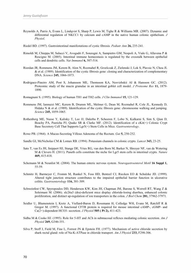
Jenny Gustafsson
70
Reynolds A, Parris A, Evans L, Lindqvist S, Sharp P, Lewis M, Tighe R & Williams MR. (2007). Dynamic and
differential regulation of NKCC1 by calcium and cAMP in the native human colonic epithelium. J Physiol.
Riedel BD. (1997). Gastrointestinal manifestations of cystic fibrosis. Pediatr Ann 26, 235-241. Rimoldi M, Chieppa M, Salucci V, Avogadri F, Sonzogni A, Sampietro GM, Nespoli A, Viale G, Allavena P &
Rescigno M. (2005). Intestinal immune homeostasis is regulated by the crosstalk between epithelial cells and dendritic cells. Nat Immunol 6, 507-514.
Riordan JR, Rommens JM, Kerem B, Alon N, Rozmahel R, Grzelczak Z, Zielenski J, Lok S, Plavsic N, Chou JL
& et al. (1989). Identification of the cystic fibrosis gene: cloning and characterization of complementary DNA. Science 245, 1066-1073.
Rodriguez-Pineiro AM, Post S, Johansson ME, Thomsson KA, Nesvizhskii AI & Hansson GC. (2012).
Proteomic study of the mucin granulae in an intestinal goblet cell model. J Proteome Res 11, 1879-1890.
Romagnani S. (1995). Biology of human TH1 and TH2 cells. J Clin Immunol 15, 121-129. Rommens JM, Iannuzzi MC, Kerem B, Drumm ML, Melmer G, Dean M, Rozmahel R, Cole JL, Kennedy D,
Hidaka N & et al. (1989). Identification of the cystic fibrosis gene: chromosome walking and jumping. Science 245, 1059-1065.
Rothenberg ME, Nusse Y, Kalisky T, Lee JJ, Dalerba P, Scheeren F, Lobo N, Kulkarni S, Sim S, Qian D,
Beachy PA, Pasricha PJ, Quake SR & Clarke MF. (2012). Identification of a cKit(+) Colonic Crypt Base Secretory Cell That Supports Lgr5(+) Stem Cells in Mice. Gastroenterology.
Rowe PB. (1964). A Mucus-Secreting Villous Adenoma of the Rectum. Gut 5, 250-252. Sandle GI, McNicholas CM & Lomax RB. (1994). Potassium channels in colonic crypts. Lancet 343, 23-25. Sato T, van Es JH, Snippert HJ, Stange DE, Vries RG, van den Born M, Barker N, Shroyer NF, van de Wetering
M & Clevers H. (2011). Paneth cells constitute the niche for Lgr5 stem cells in intestinal crypts. Nature 469, 415-418.
Schemann M & Neunlist M. (2004). The human enteric nervous system. Neurogastroenterol Motil 16 Suppl 1,
55-59. Schmitz H, Barmeyer C, Fromm M, Runkel N, Foss HD, Bentzel CJ, Riecken EO & Schulzke JD. (1999).
Altered tight junction structure contributes to the impaired epithelial barrier function in ulcerative colitis. Gastroenterology 116, 301-309.
Schweinfest CW, Spyropoulos DD, Henderson KW, Kim JH, Chapman JM, Barone S, Worrell RT, Wang Z &
Soleimani M. (2006). slc26a3 (dra)-deficient mice display chloride-losing diarrhea, enhanced colonic proliferation, and distinct up-regulation of ion transporters in the colon. J Biol Chem 281, 37962-37971.
Seidler U, Blumenstein I, Kretz A, Viellard-Baron D, Rossmann H, Colledge WH, Evans M, Ratcliff R &
Gregor M. (1997). A functional CFTR protein is required for mouse intestinal cAMP-, cGMP- and Ca(2+)-dependent HCO3- secretion. J Physiol 505 ( Pt 2), 411-423.
Sidhu M & Cooke HJ. (1995). Role for 5-HT and ACh in submucosal reflexes mediating colonic secretion. Am J
Physiol 269, G346-351. Silva P, Stoff J, Field M, Fine L, Forrest JN & Epstein FH. (1977). Mechanism of active chloride secretion by
shark rectal gland: role of Na-K-ATPase in chloride transport. Am J Physiol 233, F298-306.
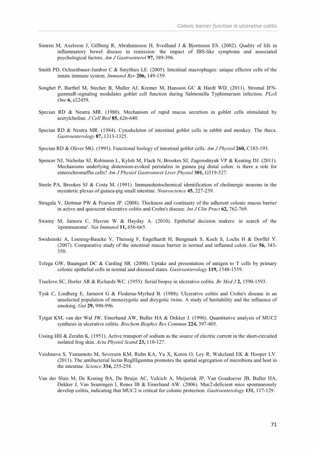
Colonic barrier function in ulcerative colitis
71
Simren M, Axelsson J, Gillberg R, Abrahamsson H, Svedlund J & Bjornsson ES. (2002). Quality of life in inflammatory bowel disease in remission: the impact of IBS-like symptoms and associated psychological factors. Am J Gastroenterol 97, 389-396.
Smith PD, Ochsenbauer-Jambor C & Smythies LE. (2005). Intestinal macrophages: unique effector cells of the
innate immune system. Immunol Rev 206, 149-159. Songhet P, Barthel M, Stecher B, Muller AJ, Kremer M, Hansson GC & Hardt WD. (2011). Stromal IFN-
gammaR-signaling modulates goblet cell function during Salmonella Typhimurium infection. PLoS One 6, e22459.
Specian RD & Neutra MR. (1980). Mechanism of rapid mucus secretion in goblet cells stimulated by
acetylcholine. J Cell Biol 85, 626-640. Specian RD & Neutra MR. (1984). Cytoskeleton of intestinal goblet cells in rabbit and monkey. The theca.
Gastroenterology 87, 1313-1325. Specian RD & Oliver MG. (1991). Functional biology of intestinal goblet cells. Am J Physiol 260, C183-193. Spencer NJ, Nicholas SJ, Robinson L, Kyloh M, Flack N, Brookes SJ, Zagorodnyuk VP & Keating DJ. (2011).
Mechanisms underlying distension-evoked peristalsis in guinea pig distal colon: is there a role for enterochromaffin cells? Am J Physiol Gastrointest Liver Physiol 301, G519-527.
Steele PA, Brookes SJ & Costa M. (1991). Immunohistochemical identification of cholinergic neurons in the
myenteric plexus of guinea-pig small intestine. Neuroscience 45, 227-239. Strugala V, Dettmar PW & Pearson JP. (2008). Thickness and continuity of the adherent colonic mucus barrier
in active and quiescent ulcerative colitis and Crohn's disease. Int J Clin Pract 62, 762-769. Swamy M, Jamora C, Havran W & Hayday A. (2010). Epithelial decision makers: in search of the
'epimmunome'. Nat Immunol 11, 656-665. Swidsinski A, Loening-Baucke V, Theissig F, Engelhardt H, Bengmark S, Koch S, Lochs H & Dorffel Y.
(2007). Comparative study of the intestinal mucus barrier in normal and inflamed colon. Gut 56, 343-350.
Telega GW, Baumgart DC & Carding SR. (2000). Uptake and presentation of antigen to T cells by primary
colonic epithelial cells in normal and diseased states. Gastroenterology 119, 1548-1559. Truelove SC, Horler AR & Richards WC. (1955). Serial biopsy in ulcerative colitis. Br Med J 2, 1590-1593. Tysk C, Lindberg E, Jarnerot G & Floderus-Myrhed B. (1988). Ulcerative colitis and Crohn's disease in an
unselected population of monozygotic and dizygotic twins. A study of heritability and the influence of smoking. Gut 29, 990-996.
Tytgat KM, van der Wal JW, Einerhand AW, Buller HA & Dekker J. (1996). Quantitative analysis of MUC2
synthesis in ulcerative colitis. Biochem Biophys Res Commun 224, 397-405. Ussing HH & Zerahn K. (1951). Active transport of sodium as the source of electric current in the short-circuited
isolated frog skin. Acta Physiol Scand 23, 110-127. Vaishnava S, Yamamoto M, Severson KM, Ruhn KA, Yu X, Koren O, Ley R, Wakeland EK & Hooper LV.
(2011). The antibacterial lectin RegIIIgamma promotes the spatial segregation of microbiota and host in the intestine. Science 334, 255-258.
Van der Sluis M, De Koning BA, De Bruijn AC, Velcich A, Meijerink JP, Van Goudoever JB, Buller HA,
Dekker J, Van Seuningen I, Renes IB & Einerhand AW. (2006). Muc2-deficient mice spontaneously develop colitis, indicating that MUC2 is critical for colonic protection. Gastroenterology 131, 117-129.
Jenny Gustafsson
72
van Doorninck JH, French PJ, Verbeek E, Peters RH, Morreau H, Bijman J & Scholte BJ. (1995). A mouse model for the cystic fibrosis delta F508 mutation. EMBO J 14, 4403-4411.
Velcich A, Yang W, Heyer J, Fragale A, Nicholas C, Viani S, Kucherlapati R, Lipkin M, Yang K & Augenlicht
L. (2002). Colorectal cancer in mice genetically deficient in the mucin Muc2. Science 295, 1726-1729. Vidyasagar S, Rajendran VM & Binder HJ. (2004). Three distinct mechanisms of HCO3- secretion in rat distal
colon. Am J Physiol Cell Physiol 287, C612-621. Vijay-Kumar M, Sanders CJ, Taylor RT, Kumar A, Aitken JD, Sitaraman SV, Neish AS, Uematsu S, Akira S,
Williams IR & Gewirtz AT. (2007). Deletion of TLR5 results in spontaneous colitis in mice. J Clin Invest 117, 3909-3921.
Watt J & Marcus R. (1973). Experimental ulcerative disease of the colon in animals. Gut 14, 506-510. Wehkamp J, Harder J, Weichenthal M, Mueller O, Herrlinger KR, Fellermann K, Schroeder JM & Stange EF.
(2003). Inducible and constitutive beta-defensins are differentially expressed in Crohn's disease and ulcerative colitis. Inflamm Bowel Dis 9, 215-223.
Wehkamp J, Schmid M & Stange EF. (2007). Defensins and other antimicrobial peptides in inflammatory bowel
disease. Curr Opin Gastroenterol 23, 370-378. White H. (1909). A Discussion on "Ulcerative Colitis." Introductroy Address. Proc R Soc Med 2, 79-82. Winslet MC, Allan A, Poxon V, Youngs D & Keighley MR. (1994). Faecal diversion for Crohn's colitis: a
model to study the role of the faecal stream in the inflammatory process. Gut 35, 236-242. Wyllie R. (1999). Gastrointestinal manifestations of cystic fibrosis. Clin Pediatr (Phila) 38, 735-738. Yajima T. (1988). Luminal propionate-induced secretory response in the rat distal colon in vitro. J Physiol 403,
559-575. Yeruva S, Farkas K, Hubricht J, Rode K, Riederer B, Bachmann O, Cinar A, Rakonczay Z, Molnar T, Nagy F,
Wedemeyer J, Manns M, Raddatz D, Musch MW, Chang EB, Hegyi P & Seidler U. (2010). Preserved Na(+)/H(+) exchanger isoform 3 expression and localization, but decreased NHE3 function indicate regulatory sodium transport defect in ulcerative colitis. Inflamm Bowel Dis 16, 1149-1161.
Yu H, Riederer B, Stieger N, Boron WF, Shull GE, Manns MP, Seidler UE & Bachmann O. (2009).
Secretagogue stimulation enhances NBCe1 (electrogenic Na(+)/HCO(3)(-) cotransporter) surface expression in murine colonic crypts. Am J Physiol Gastrointest Liver Physiol 297, G1223-1231.
Yu K, Lujan R, Marmorstein A, Gabriel S & Hartzell HC. (2010). Bestrophin-2 mediates bicarbonate transport
by goblet cells in mouse colon. J Clin Invest 120, 1722-1735. Zachos NC, Tse M & Donowitz M. (2005). Molecular physiology of intestinal Na+/H+ exchange. Annu Rev
Physiol 67, 411-443. Zhou Z, Duerr J, Johannesson B, Schubert SC, Treis D, Harm M, Graeber SY, Dalpke A, Schultz C & Mall MA.
(2011). The ENaC-overexpressing mouse as a model of cystic fibrosis lung disease. J Cyst Fibros 10 Suppl 2, S172-182.
Zhu Y, Ehre C, Abdullah LH, Sheehan JK, Roy M, Evans CM, Dickey BF & Davis CW. (2008). Munc13-2-/-
baseline secretion defect reveals source of oligomeric mucins in mouse airways. J Physiol 586, 1977-1992.