Embed Size (px)
Citation preview

Hereditary DisordersCollie Eye Anomaly
Progressive Retinal AtrophyPatella LuxationElbow Dysplasia
Heart Valve DisordersEntropion and Ectropion

Factors to ConsiderHas a hereditary basis for the condition been
confirmed?What is the incidence?What is the mode of inheritance ? eg
dominant, recessive, sex linked.How can “carriers” of recessive genes be
identified?What is the severity of the disorder?

A sample of teratogenic agents that can damage an embryo/foetus.
Vitamin A excess : eye defects, cleft palateVitamin D and E excess : dental problems,
cleft palate.Vitamin E deficiency : hydrocephalyAntibiotic – chloramphenicol :
cryptorchidismAntibiotic – streptomycin : hearing loss.Aspirin - hydrocephaly, skeletal defectsThis is a short list, and the severity of the
toxicity or the deficiency is not stated.
The Dog’s Eye
The tapetum forms a layer of reflecting tissue on the surface of the choroid

Collie Eye Anomaly

1.The choroid layer is normally darkly pigmented. In choroidal hypoplasia the choroid is very thin, almost transparent, a pale patch is visible at the back of the eye and blood vessels can be seen through the choroid.2. Coloboma (“pits”) are seen on the optic disc.3. Other signs include : a) non-attachment of the retina. b) bleeding within the eye4. Commonest eye disorder in the UK. Rough Collies, Smooth Collies, Border Collies and Shetland Sheepdog.5. Only 6% of cases result in total blindness.6. Autosomal Recessive – but other genes may be responsible for colobomas. - “carrier animals” possible. (chromosome 37)7. Litter screening at 5-12 weeks is essential as the condition may not be identified in the adult. “go normal adults”

Generalised Progressive Retinal Atrophy
Normal : vascular PRA : narrowing of the blood vessels
There is a gradual degeneration of the photoreceptor cells- rods and cones – in the retina. There are two forms of the disorder in which both eyes are affected :
a) rods and cones fail to develop normally and degenerate in the first year of life.
b) rods and cones develop normally and then undergo degeneration, with blindness at 5-7 years old.
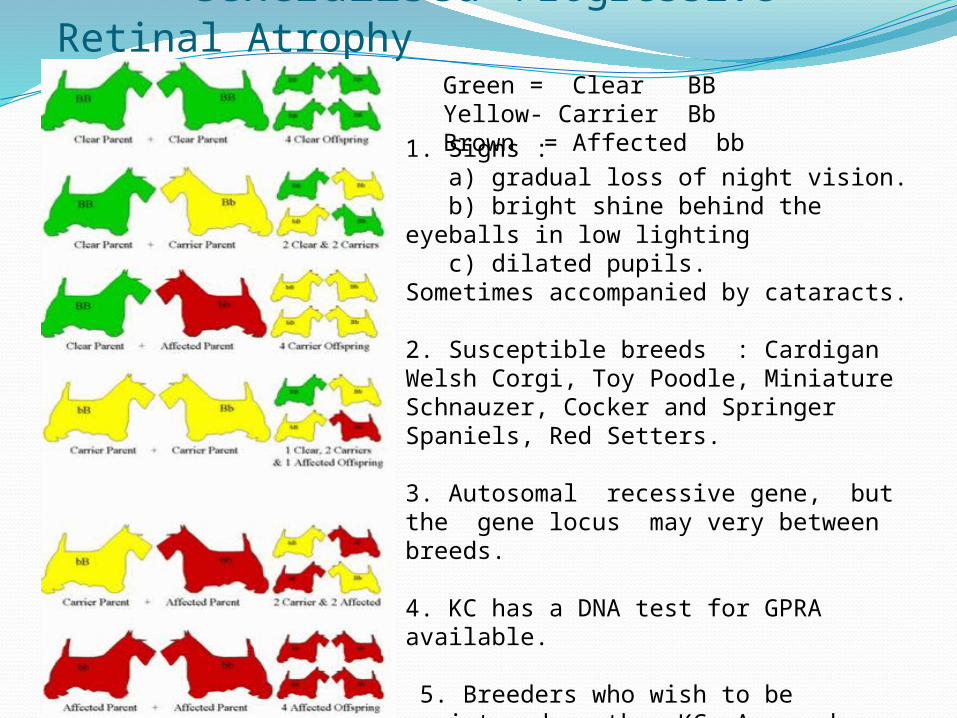
Generalised Progressive Retinal AtrophyGreen = Clear BB Yellow- Carrier BbBrown = Affected bb1. Signs :
a) gradual loss of night vision. b) bright shine behind the eyeballs in low lighting c) dilated pupils.Sometimes accompanied by cataracts.
2. Susceptible breeds : Cardigan Welsh Corgi, Toy Poodle, Miniature Schnauzer, Cocker and Springer Spaniels, Red Setters.
3. Autosomal recessive gene, but the gene locus may very between breeds.
4. KC has a DNA test for GPRA available.
5. Breeders who wish to be registered on the KC Assured Breeders Scheme must have relevant tests for hereditary disorders carried out on potential breeding stock. The results can be sourced on the KC web site.

Patella Luxation1. In the stifle joint the patella prevents
wear and tear to the quadriceps tendon and protects the stifle joint
2. The patella “sits” in the trochlear groove at the distal end of the femur, and slides up and down in the groove when the knee bends and straightens.
3. Small breeds are most at risk, including miniature and toy poodles, yorkies, dachshunds and boston terriers. The basset hound is also susceptible due to the shortening and shape of the femur.
4. In 50% of cases both knees are affected. The patell may displace laterally or to the inside of the knee.5. Signs include a “knock – kneed”
stance. In the long term osteo-arthritis will develop.

Patellar Luxation
1. It is thought that the condition is due to a poor alignment of the complete leg, and that the condition is polygenic – caused by numerous gene pairs. Environmental factors such as overweight and strenuous exercise may play a role.
Elbow Dysplasia

Stages of Elbow Dysplasia

Elbow Dysplasia1 Three conditions that effect one or both of the canine
elbows. The dog may suffer from one or more of the conditions :
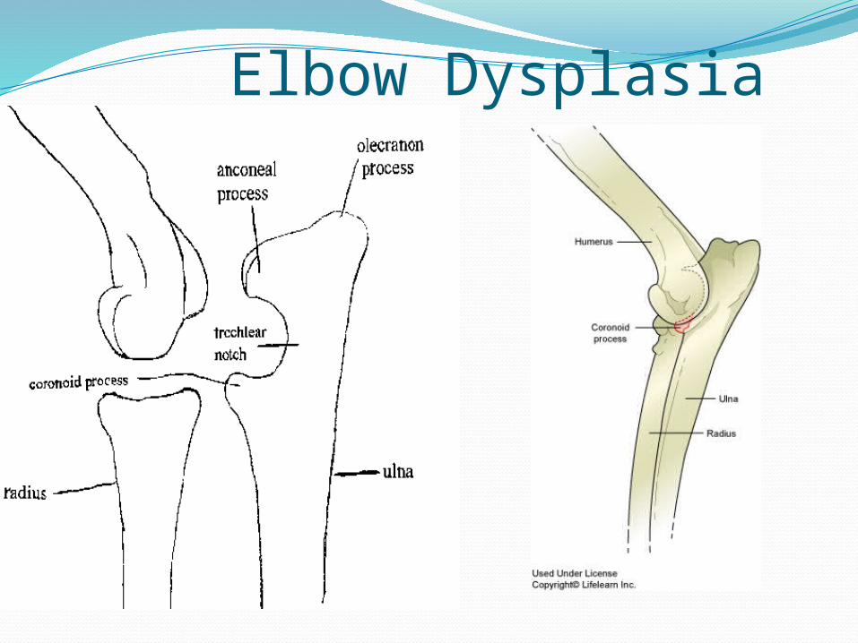
a) Ununited anconeal process (UAC) b) Fragmented coronoid process (FCP) c) Osteochondritus dissecans (OCD)2. Signs a) lameness b) pain on flexing and extending the elbow. c) crepitation (cracking noise) d) osteoarthritis3. Ununited anconeal process (UAC) a) The process forms part of the semi-lunar notch of the ulna
which articulates with the humerus. b) The process develops separately from the other parts of
the ulna and normally unites with the ulna by 20 weeks of age.
c) In this condition the process does not unite with the ulna.

Elbow Dysplasia4. Fragmented coronoid process (FCP) a) The coronoid process also forms part of the semi-
lunar notch which articulates with the humerus. b) The process breaks free from the ulna5. Osteochondritis Dissecans (OCD) a) This occurs when the hyaline cartilage at the end of
a bone separates from the underlying bone and “floats” within the joint capsule.
b) Guide Dogs for the Blind Report (1990) for Labradors and Retrievers. i) high heritability
ii) higher incidence in males than females iii) multifactorial condition : polygenic
with substantial environmental influence eg growth rate, diet, level of exercise

Elbow Dysplasia6. There is a higher risk in medium to large breeds : a) German Shepherd Dog b) Labradors c) Retrievers d) Basset Hounds e) Rottweiler f) Newfoundland7. The BVA/KC Elbow Dysplasia Scheme a) Three X rays of each elbow. Score awarded according
to the presence of UAC, FCP or OCD. b) Each elbow is scored 0 to 3, and the grade given is for
the “worst” elbow. Score of 2 or 3 – Do not breed.

1 FCP2 OCD
1
2
2

Mitral /Bicuspid Valve Disease
Follow the flow of blood through the heart and identify O2 and deO2 blood

An atrio-ventricular valve

Mitral Valve Disease
Chordae tendinae
Valve Flaps (2)
Early sign of a nodule
Severely damaged mitral valve
Papillary Muscle

Mitral Valve Disease – X Ray
The enlarged heart is pushing the trachea dorsally towards the vertebral column. This will cause breathing difficulties (Dyspnoea) and coughing

Mitral Valve Disease1. This is a chronic disease in which there is slow degeneration of the mitral
(bicuspid) valve.2. Initially there is a leakage of blood from the left ventricle back in to the
atrium (Mitral Regurgitation).3. This occurs in cocker spaniels, poodles and terriers at an age of 6-7
years, but is noticed much earlier (3-4 yrs.) in KC/Cav. Spaniels. It appears to be more common in males.
4. The left atrium is “forced” to hold more blood than normal and will begin to enlarge. There will be a backflow of blood towards the lungs. The result is severe pulmonary congestion – excess fluid around the lungs, and this may extend to the abdomen (Ascites).
5. Signs : a) Chronic coughing – enlarged heart pressing on the trachea. b) Exercise intolerance-tires easily
c) Dyspnoea – difficult breathing d) Fainting – lack of O2 to the brain e) Heart Murmur : an extra sound when listening for the “lub – dup” of the valves closing. Measured on a scale of 1 to 6.
6. It is a chronic, progressive disease and will lead to a fainter pulse and increased CRT.
7. Prognosis/Treatment a) Combination of medications that will slow down the disease eg
Diuretics – increase fluid removal and urine output. ACE inhibitors – dilate blood vessels . Pimobendan – improves heart contractions and dilates blood vessels.
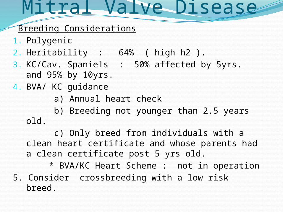
Mitral Valve Disease Breeding Considerations1. Polygenic2. Heritability : 64% ( high h2 ).3. KC/Cav. Spaniels : 50% affected by 5yrs. and
95% by 10yrs.4. BVA/ KC guidance a) Annual heart check b) Breeding not younger than 2.5 years old. c) Only breed from individuals with a clean
heart certificate and whose parents had a clean certificate post 5 yrs old.
* BVA/KC Heart Scheme : not in operation5. Consider crossbreeding with a low risk
breed.

Ectropion and Entropion
Ectropion
EntropionPolygenic Disorders related to the shape of the head and the ligaments around the eye.

Ectropion1. An outward rolling or sagging of the eyelid.
Common in specific dog breeds but rare in cats. a) Basset Hound b) Bloodhounds c) Bull Mastiffs d) St. Bernards e) Newfoudland d) Gordon
Setter2. Signs a) Conjunctivitis b) “Dry Eye” * c) Poor tear
drainage d) Foreign object irritation e) Nictitating
membrane visible3. Treatment : a) Mild Irritation - suitable surface
medications b) Severe Irritation - corrective surgery
* Kerato-conjunctivitis sicca

Entropion1 Eyelid rolls in on itself - one or both eyes, lower
and upper eyelids, but more common in the lower eyelid.
2. Occurs in dogs and cats a) Chow-chow b) Shar-Pei c)
Mastiffs d) Great Dane e) English Bulldog f) Brachycephalic breeds of cat eg Persian cat.3. Signs a) Corneal ulcers. b) Excess tear production. c) Eye discharge and excessive blinking. d) Eye rubbing4. Polygenic – facial skinfolds, brachycephalic
skulls, short muzzles.