Embed Size (px)
Citation preview

er.com/locate/pharmthera
Pharmacology & Therapeutics
Associate editor: P.S. Foster
Clues to asthma pathogenesis from microarray expression studies
Michael S. Rolph *, Mary Sisavanh, Sue M. Liu, Charles R. Mackay
Arthritis and Inflammation Research Program, Garvan Institute for Medical Research, Darlinghurst, Australia
CRC for Asthma, University of Sydney, Camperdown, Australia
Abstract
Asthma is a chronic inflammatory disease characterized by airway hyperresponsiveness (AHR), tissue remodeling, and airflow obstruction.
The pathogenesis of asthma is only partly understood, and there is an urgent need for improved therapeutic strategies for this disease. Microarray
technology has considerable promise as a tool for discovery of novel asthma therapeutic targets, although the field is still in its infancy. A number
of studies have described expression profiles derived from human asthmatic lung tissue, mouse airway tissue, or from key cell types associated
with asthma, but to date relatively few studies have exploited these findings to discover new pathways involved in the pathogenesis of asthma.
Among the genes to have been identified by array studies and validated by further studies are monokine induced by interferon (IFN)-g, fatty acid
binding proteins (FABP), and complement factor 5 (C5). Here we provide examples of microarray approaches to the discovery of new molecules
associated with asthma. We anticipate that these types of analyses will provide considerable insight into asthma pathogenesis and will provide a
wealth of new molecules for downstream analyses such as gene deficient mouse studies, or monoclonal antibody production.
D 2005 Elsevier Inc. All rights reserved.
Keywords: Asthma; Microarray; Mast cell; Bronchial epithelium; Cytokine
Abbreviations: AHR, airway hyperresponsiveness; FABP, fatty acid binding protein; GM-CSF, granulocyte-macrophage colony stimulating factor; HBE, human
bronchial epithelial cells; IFN, interferon; IL, interleukin; MBP, major basic protein; STAT, signal transducer and activator of transcription.
0163-7258/$ - see f
doi:10.1016/j.pharm
* Corresponding
Australia. Tel.: +61
E-mail address:
Contents
. . . . . . 285
. . . . . . 285
. . . . . . 285
. . . . . . 286
. . . . . . 286
. . . . . . 286
. . . . . . 287
. . . . . . 288
. . . . . . 288
. . . . . . 288
. . . . . . 289
. . . . . . 290
. . . . . . 290
. . . . . . 291
. . . . . . 291
1. Introduction . . . . . . . . . . . . . . . . . . . . . . . . . . . . . . . .
1.1. Etiology and pathogenesis of asthma. . . . . . . . . . . . . . . .
1.2. Asthma therapies . . . . . . . . . . . . . . . . . . . . . . . . . .
1.3. Functional genomics . . . . . . . . . . . . . . . . . . . . . . . .
2. Profiling asthmatic tissue . . . . . . . . . . . . . . . . . . . . . . . . .
2.1. Human studies . . . . . . . . . . . . . . . . . . . . . . . . . . .
2.2. Animal models . . . . . . . . . . . . . . . . . . . . . . . . . . .
2.3. Considerations relating to tissue profiling studies . . . . . . . . .
3. Profiling purified and cultured cells . . . . . . . . . . . . . . . . . . . .
3.1. Mast cells . . . . . . . . . . . . . . . . . . . . . . . . . . . . .
3.2. Airway epithelial cells . . . . . . . . . . . . . . . . . . . . . . .
3.3. Airway smooth muscle cells . . . . . . . . . . . . . . . . . . . .
3.4. Th1/Th2 cells . . . . . . . . . . . . . . . . . . . . . . . . . . .
3.5. Eosinophils . . . . . . . . . . . . . . . . . . . . . . . . . . . . .
3.6. Integrating array data from multiple cell types . . . . . . . . . . .
4. Conclusion . . . . . . . . . . . . . . . . . . . . . . . . . . . . . . . .
. . . . . . 292References . . . . . . . . . . . . . . . . . . . . . . . . . . . . . . . . . . . . . . . . . 292
109 (2006) 284 – 294
www.elsevi
ront matter D 2005 Elsevier Inc. All rights reserved.
thera.2005.08.009
author. Arthritis and Inflammation Research Program, Garvan Institute for Medical Research, 384 Victoria Street, Darlinghurst NSW 2010,
2 92958351.
[email protected] (M.S. Rolph).

M.S. Rolph et al. / Pharmacology & Therapeutics 109 (2006) 284–294 285
1. Introduction
Asthma is a chronic inflammatory disease characterized by
airway hyperresponsiveness (AHR), tissue remodeling, and
airflow obstruction (Bochner et al., 1994). The incidence of
asthma in western countries has increased markedly over the
last 20 years, and in countries such as the United States and
Australia it now affects ¨10% of the population (Woolcock et
al., 2001; CDC, 2002). The economic burden of asthma has
also increased, with the annual cost in the United States in 1998
estimated at US$12.7 billion (Weiss & Sullivan, 2001). These
figures indicate that advances in diagnosis, treatment, or
prevention of asthma will lead to major medical and economic
benefits to society.
One of the major advances to come out of the genomic
revolution has been the development of microarray technology.
Using microarrays, it is now possible to profile gene expression
for essentially the entire human genome. In this review, we
describe some of the recent uses of microarray technology for
the study of asthma. Most of these studies are still of a
preliminary nature, but initial data strongly suggests that
microarrays will lead to major advances in our understanding
of asthma etiology and pathogenesis.
1.1. Etiology and pathogenesis of asthma
Asthma is caused by environmental factors in genetically
predisposed individuals. Detailed understanding of the etiology
and underlying pathogenetic mechanisms in asthma is still
lacking, and this is a major impediment to development of
improved diagnostic and therapeutic strategies for this disease.
Atopy is the strongest predisposing factor for the development
of asthma (Lemanske & Busse, 2003), and the majority of
asthma is associated with allergic inflammation characterized
by the presence of mast cells, basophils, eosinophils, mono-
cytes, and Th2 lymphocytes. The allergic inflammatory
response is regulated largely by Th2 cytokines such as
interleukin (IL)-4, IL-5, IL-9, and IL-13, all of which impinge
on the response in a variety of ways. For example, IL-5
regulates differentiation, activation, and survival of eosino-
phils, a key effector cell type in allergic inflammation (Foster et
al., 2001). IL-4 is a highly pleiotropic cytokine, controlling
such events as Th2 cell differentiation, IgE isotype switching,
and expression of vascular cell adhesion molecule-1 on
endothelial cells. IL-13 shares many functions with IL-4 but
in addition is a crucial mediator of airway hyperresponsiveness
(Wynn, 2003).
It is becoming increasingly apparent that allergic inflam-
mation does not account for all the features of asthma. For
example, a number of genetic studies showed a separation in
the inheritance of bronchial hyperresponsiveness and atopy
(Holloway et al., 1999; Davies et al., 2003). Studies examining
the earliest events in the development of asthma have often
demonstrated injury to the lung occurring prior to the
development of asthma, or in the absence of eosinophilic
inflammation (Cokugras et al., 2001; Payne et al., 2003;
Fedorov et al., 2005). Even the role of eosinophils, which have
for long been considered central effector cells in asthmatic
inflammation, is becoming controversial. In vitro and animal
model studies strongly support a role for eosinophils in airway
pathology (Foster et al., 2001). However, clinical trials in
which the levels of circulating and tissue eosinophils were
lowered using anti-IL-5 therapy have questioned a key role for
eosinophils in disease (Leckie et al., 2000).
In recent years, there has been increasing focus on the role
of airway cells and tissue remodeling in the development of
asthma. A number of studies have now shown that airway cells
in asthmatic patients differ from those of non-asthmatics. For
example, smooth muscle cells from asthmatics appear to have
an intrinsically increased rate of proliferation (Johnson et al.,
2001) linked to absence of the C/EBP transcription factor (Roth
et al., 2004). Epithelial cells from asthmatics show increased
susceptibility to rhinovirus infection (Wark et al., 2005) and
oxidant-induced apoptosis (Bucchieri et al., 2002), and release
more IL-8 and granulocyte-macrophage colony stimulating
factor (GM-CSF) when exposed to diesel exhaust particles
(Bayram et al., 1998). In response to these and other findings,
some researchers have suggested that airway inflammation and
remodeling occur as a consequence of increased susceptibility
to injury or impaired healing of the airway epithelium (Davies
et al., 2003).
Asthma is a highly heterogeneous disease, and this presents
an additional challenge for the design of new therapeutics
(Peters, 2003). This heterogeneity is most likely due to a
combination of environmental and genetic factors and
manifests at many levels including etiology, pathogenesis,
prognosis, and response to therapy. To design optimal
therapies and diagnostic tools, it will be necessary to fully
understand the basis of asthma heterogeneity. Gene profiling
technology has the potential to provide important insights in
different forms of asthma, and it has already been used
successfully to probe heterogeneity in other diseases (Alizadeh
et al., 2000).
1.2. Asthma therapies
The most effective asthma therapy at present is treatment
with combination inhalers that contain a corticosteroid and a
long-acting h2-adrenoceptor agonist. Other drugs efficacious
in the treatment of asthma include theophylline, cromolyn,
leukotriene antagonists, and anti-IgE monoclonal antibodies.
The majority of asthma cases can be well controlled using
currently available drugs; however, there is a growing number
of asthmatics whose disease is not optimally controlled,
particularly those with severe asthma. An additional area for
improvement is development of orally active drugs. Unfortu-
nately the pharmaceutical industry has had little success in
developing new therapeutic agents for asthma, not least due to
the complexity of the disease, a failure to fully understand
asthma etiology and pathogenesis, and the lack of suitable
animal models. A large number of targets are currently under
investigation as potential asthma drug targets (Barnes, 2004);
however, few have shown promise so far, and there is a need
for the identification of new drug targets.

M.S. Rolph et al. / Pharmacology & Therapeutics 109 (2006) 284–294286
1.3. Functional genomics
Using microarray technology, it is now relatively straight-
forward to simultaneously assess transcriptional activity of
essentially every gene in an organism’s genome. The develop-
ment of this technology has been an enormous boost to many
areas of medical research—drug target discovery, identification
of novel transcriptional programs, development of molecular
disease classifications, and identification of new prognostic and
diagnostic markers. The myriad applications and experimental
challenges associated with microarray technology have been
widely reviewed elsewhere (Staudt & Brown, 2000; Staudt,
2001; Butte, 2002; Stoughton, 2004) and will not be discussed
here. However, it is worth repeating here an important feature
of microarray-based experiments and the data they generate.
Unlike traditional experimental approaches, a microarray
experiment is almost certain to generate novel data. The real
challenge is to design a microarray experiment to generate data
that can be analyzed in a way that leads to useful and relevant
information. This requires careful consideration of numerous
factors including sample composition (especially when using
tissue samples), methods for data analysis, experimental
variation, and number of replicates required.
In this review, we will discuss the literature that has emerged
over the last few years in which gene profiling has been
employed to address issues relating to the pathogenesis of
asthma. The studies have been separated into 2 broad
categories: first, those studies involving asthmatic tissue
samples, and second, studies involving pure cultures of cell
types implicated in the pathogenesis of asthma. The majority of
asthma gene profiling studies have been aimed at identifying
novel transcriptional programs in cells and tissues that underlie
asthmatic pathology. A large number of these studies are of a
preliminary nature involving the description of novel gene
profiles, and relatively few studies have actually used these
profiles to progress our understanding of asthma pathogenesis.
We expect that in the next few years there will be many
downstream studies, reporting developments arising from
novel transcript profiling data. Microarrays also have consid-
erable potential as diagnostic tools, although to date there have
been very few efforts to apply this technology to asthma
diagnosis.
2. Profiling asthmatic tissue
2.1. Human studies
Our understanding of gene expression in asthma is still at a
very basic level, and gene profiling is likely to be profitable for
almost any aspect of the pathogenesis of this disease. Some
areas of study that we consider to be particularly promising
include analysis of the response to therapy; the molecular
events underlying asthma heterogeneity; and the response of
both airway and inflammatory cells to allergen challenge. The
most obvious way to apply gene-profiling technology to the
study of asthma is to directly analyze gene expression in
human asthmatic tissue samples. As discussed below, this
approach can be very difficult and relatively few such studies
have been reported. In particular, it is difficult to obtain suitable
tissue samples, with biopsies obtained from fiberoptic bron-
choscopy being the main source of tissue. Ongoing therapy for
asthma or other diseases can also be problematic, by having a
confounding effect on gene expression. Nonetheless, a number
of reports have emerged in recent years describing gene
profiles obtained from asthmatic tissue.
To identify genes expressed in airway epithelium in allergic
inflammation, Lilly et al. (2005) used Affymetrix oligonucle-
otide arrays and endobronchial brushing to analyze gene
expression in airway epithelial cells of mild asthmatics before
and after segmental allergen challenge. After statistical analysis
and post hoc data filtering, 141 up-regulated and 8 down-
regulated genes were identified. As well as genes with defined
immune function, a large number of genes involved in tissue
repair and proliferation were induced. A number of these genes
had not previously been associated with asthma, and the data
provide the basis for further mechanistic studies. These authors
also compared their results to the expression profile previously
reported for IL-13-treated airway epithelial cells (Lee et al.,
2001) and found very little overlap in the genes that were
regulated in both studies. This suggests that many factors in
addition to IL-13 are involved in segmental allergen challenge
in vivo and emphasizes the need to integrate in vitro and in
vivo studies.
Laprise et al. (2004) studied gene expression in bronchial
biopsies from healthy controls (n =4) and from asthmatics
(n =4) before and after inhaled corticosteroids (Laprise et al.,
2004). Seventy-nine genes had significantly different expres-
sion levels between the control and asthmatic groups, and 128
genes showed differential expression between pre- and post-
corticosteroid therapy. Corticosteroid therapy reversed ¨25%
of the genes that were regulated in the asthmatic group,
especially genes encoding proteolytic, immune, and extracel-
lular proteins. Although the number of subjects was small, a
number of the genes identified had previously been associated
with asthma. Together with the study by Lilly et al., this work
indicates that bronchial biopsies are suitable tissue source for
gene profiling studies and the preliminary data already point to
some promising candidates for further study.
A slightly different approach was taken by Guajardo et al.
(2005) who studied nasal mucosal cells from healthy children
and those with stable and exacerbated asthma, with the
assumption that gene expression in nasal mucosal cells reflects
gene expression in the respiratory epithelium of the lung. Not
surprisingly, a large number of immune genes were up-
regulated in the exacerbated asthmatic samples. Cilia-related
genes were prominent among the most strongly down-
regulated genes in asthmatics, an unexpected finding suggest-
ing that altered ciliary function may contribute to airway
obstruction in asthma.
Microarrays have considerable potential as tools for
molecular classification and the development of improved
strategies for diagnosis, although to date few studies have
focused on asthma. After screening peripheral blood from
atopic asthmatics, non-atopic asthmatics, and healthy controls,

M.S. Rolph et al. / Pharmacology & Therapeutics 109 (2006) 284–294 287
Brutsche et al. (2002) developed a composite atopy gene
expression (CAGE) score that was superior to total IgE in
differentiating atopic from non-atopic subjects. This study used
low-density arrays and relatively few subjects, but as a proof-
of-principle study, it clearly indicates the potential of this
approach in the development of diagnostic strategies.
2.2. Animal models
A number of gene profiling studies have been undertaken
using animal models of asthma. With the exception of 1 study
using a monkey model of asthma (Zou et al., 2002), all studies
have used the mouse model of acute allergic airway inflam-
mation. Although the usual caveats concerning animal models
apply, these studies have the advantage of obtaining sufficient
quantities of well-defined tissue samples, free of uncontrolled
genetic and environmental heterogeneity.
A particularly elegant use of gene profiling was reported
by Karp et al. (2000) to identify the genetic basis of allergen-
induced airway hyperresponsiveness (AHR) susceptibility in
mouse strains of differing susceptibility. These investigators
had previously used high- and low-responder mouse strains to
identify 2 distinct quantitative trait loci (QTL) on chromo-
some 2 that regulate AHR. Using gene profiling, a panel of
genes differentially expressed between allergen-treated lungs
of high- and low-responder strains was identified. Only one
of these genes, complement factor 5 (C5) was located within
the QTL intervals. SNP-based genotyping identified a
mutation in C5 in high-responder mice that resulted in
deficiency of C5 mRNA and protein, and subsequent
heightened susceptibility to allergen-induced AHR. These
findings may be directly relevant to human disease, because a
recent study has reported on a haplotype within the C5 gene
associated with protection against childhood asthma (Hase-
gawa et. al., 2004).
Marc Rothenberg’s lab has conducted an extensive series of
studies based on gene profiling lung tissue from mouse models
of allergic airway inflammation. Two mouse models were used:
(1) a model based on systemic sensitization with ovalbumin
(OVA), with subsequent aerosol challenge with OVA, and (2)
repeated intranasal challenge with Aspergillus fumigatus
allergen. Although these 2 models vary considerably in terms
of induction (systemic vs. mucosal), their ultimate pathologic
profile is similar, involving tissue eosinophilia, mucus hyper-
secretion, and AHR. Using these 2 models, an asthma gene
expression signature was developed. The expression of 4.7% of
all genes on the chip was regulated following antigen exposure.
A large number of the genes induced at the early time point (3
hr) were associated with pathogen recognition and initiation of
the immune response such as CD14, CD83, IL-1h, and
CXCL1. By 18 hr following allergen exposure, expression of
genes associated with adaptive immunity and tissue remodeling
became more prominent.
Seventeen of the 28 chemokines represented on the chip
were up-regulated, including several not previously associated
with allergic airway inflammation. Surprisingly, expression of
a substantial number of interferon (IFN)-regulated, Th1-
associated chemokines was enhanced, including monokine
induced by IFNg (Mig) and IFNg-inducible protein of 10 kDa
(IP-10) (Fulkerson et al., 2004a, 2004b). Further experiments
indicated that Mig induced a dose-dependent inhibition of
chemokine-induced eosinophil chemotaxis, and neutralization
of Mig enhanced airway eosinophilia in the model of allergic
airway inflammation (Fulkerson et al., 2004a). Thus, the results
provide evidence for a regulatory pathway by which both Th1-
and Th2-associated chemokines regulate eosinophil responses.
This study provides a good example of the utility of gene
profiling for discovery of novel disease-associated genes, in
which array-based discoveries lead to generation of novel
hypotheses that can be tested in biological systems.
A notable feature of the data from mouse model gene
profiles was the strong up-regulation of genes involved in
arginine metabolism, including arginase I, arginase II, and the
cationic amino acid transporter 2 (Zimmermann et al., 2003).
The transcriptional changes correlated with increased lung
arginase activity. Arginase expression was also enhanced in
human asthmatic lung. Previous studies had already implicated
nitric oxide synthase (NOS)-mediated arginine metabolism in
the pathogenesis of asthma (Meurs et al., 2003), and the
findings by Zimmermann now suggest a pivotal and complex
role for arginine metabolism in asthma, involving interplay
between NOS and arginase (Zimmermann et al., 2003; King et
al., 2004a).
These investigators have subsequently mined the array data
set to establish further novel insights into asthma pathogenesis.
In addition to arginase, a number of novel genes potentially
involved in asthma pathogenesis were described and their lung
expression characterized in more detail. This included ADAM8
(King et al., 2004b), small proline rich protein 2 (Zimmermann
et al., 2005), and trefoil factor 2 (Nikolaidis et al., 2003). Most
of this work is descriptive in nature, and its true value will
become apparent when the function of these novel asthma
genes is tested in appropriate models.
An interesting feature of the gene profiling studies by
Rothenberg’s lab was uncovered when they analyzed the effect
of signal transducer and activator of transcription (STAT)-6 on
gene expression in the asthma model. Although STAT6-
mediated responses are central to the development and
manifestations of allergic airway inflammation (Kuperman et
al., 1998, 2002), a substantial number of genes were regulated
in a STAT6-independent manner. In addition, an additional
program of gene expression was induced in STAT6 KO mice
undergoing allergic airway inflammation, a finding that will
need to be considered in any STAT6-based therapeutic
approaches.
Gene profiling has also been used in an attempt to unravel
the differential effects of IL-4 and IL-13 in asthma. Although
IL-4 and IL-13 both act through the same receptor on non-
hematopoietic cells, IL-13 appears to have a more potent effect
than IL-4 on AHR, pulmonary fibrosis, and goblet cell
hyperplasia (Wills-Karp & Chiaramonte, 2003). Gene profiling
was used to test the hypothesis that the enhanced activity of IL-
13 was due to induction of unique genes by this cytokine. A
limited number of genes were identified that were induced by

M.S. Rolph et al. / Pharmacology & Therapeutics 109 (2006) 284–294288
IL-13, but not by IL-4. By analyzing expression of these genes,
it became apparent that IL-4 possesses some counter-regulatory
properties not shared by IL-13 that can suppress STAT6-
mediated responses (Finkelman et al., 2005).
2.3. Considerations relating to tissue profiling studies
There are a number of issues that need to be considered in
designing gene profiling studies of asthmatic lung and biopsy
samples.
2.3.1. Source of tissue
For obvious reasons, large samples of asthmatic human lung
tissue are not readily available for study. The best approach is
to profile bronchial biopsy samples (Laprise et al., 2004; Lilly
et al., 2005), although this has a number of limitations
including the small size of biopsies, the difficulty in ensuring
collection of fully representative samples, and the limited
regions of the lung that are accessible by fiberoptic biopsy
protocols.
2.3.2. Ongoing therapy
Ongoing therapy can potentially have a major effect on gene
expression profiles. Most moderate and severe asthmatics are
taking corticosteroids, which are known to have major
transcriptional effects. It is difficult to fully control for this
issue. The extent of the problem is illustrated by a microarray
study that specifically examined the effect of corticosteroid
treatment on gene expression in asthmatics (Laprise et al.,
2004). This study identified a greater number of genes
regulated by corticosteroids than genes regulated by asthma
itself.
2.3.3. Tissue heterogeneity
For any tissue sample, multiple cell types contribute to the
gene expression profile. This cellular heterogeneity adds
considerable complexity to data interpretation and has the
potential to obscure pathologically important transcripts in
specific cell types. This problem is particularly prominent for
inflammatory diseases, in which a large number of leukocytes
infiltrate the tissue site, making comparisons with uninflamed
control tissue difficult. This has the potential to overwhelm the
data analysis with thousands of differentially regulated genes.
The mouse model studies described above (Section 2.2) were
able to avoid this problem by focusing on time points early
after allergen challenge prior to the major leukocyte influx
(King et al., 2004b). In asthma, different proportions of specific
airway cells such as epithelial cells, smooth muscle cells, and
fibroblasts in healthy and asthmatic subjects contribute to
sampling heterogeneity. Finally, asthmatic changes such as
oedema, inflammation, and mucous gland hypertrophy can
interfere with the depth of sampling and biopsy constituents
(Laprise et al., 2004).
2.3.4. Addressing the challenges of profiling tissue samples
The issues above present significant obstacles to meaningful
functional genomic analysis of asthmatic tissue samples. The
problem of tissue heterogeneity requires that more extensive
validation of the array results be undertaken than in cellular
studies. In particular, immunohistochemistry or in situ hybrid-
ization is necessary to understand the pathologic basis for
differential gene expression across samples.
Laser capture microdissection (LCM) has considerable
potential for overcoming the difficulties associated with tissue
heterogeneity. This technology allows isolation of specific cell
types or even single cells (Luo et al., 1999; Kamme &
Erlander, 2003) and is a particularly powerful strategy in
combination with gene profiling (Kamme & Erlander, 2003;
Peterson et al., 2004; Player et al., 2004). To date there is only
1 report detailing the application of this approach to asthma, in
which LCM was used to define the phenotype of smooth
muscle cells in asthma (Woodruff et al., 2004). The gene
profiling data indicated no difference in gene expression in
SMC from healthy and asthmatic subjects, which supported the
authors’ conclusion that the major defect in asthmatic SMC is
increased proliferation.
An additional approach to tackle tissue heterogeneity is to
integrate gene expression profiles derived from both tissues
and cells. For example, Peterson et al. (2004) used a panel of
expression profiles from purified leukocyte subsets to assist in
characterizing the expression pattern of glomeruli from lupus
nephritis. By doing so, they were able to obtain additional
information about the nature of leukocyte infiltration in the
glomeruli, and how it contributed to specific patterns of gene
expression. This approach has not yet been utilized for asthma
studies.
3. Profiling purified and cultured cells
An alternative approach for identifying new genes that
contribute to the pathogenesis of asthma is to establish gene
expression profiles for pure populations of cell types central
to disease pathogenesis. This is especially valuable when in
vitro models can be established that mimic aspects of the
pathogenesis of asthma, for example, mast cells activated
by cross-linking Fc(R1 or comparison of Th1 and Th2
cells.
3.1. Mast cells
Mast cells are key effector cells during the immune response
to pathogens, but their effector functions are also responsible
for many of the symptoms associated with allergic diseases
such as asthma. Mast cell products, such as histamine and
tryptase, which are stored within preformed cytoplasmic
granules, are rapidly released following IgE-dependent activa-
tion and can be readily detected in bronchial alveolar lavage
samples from asthmatic patients (Wenzel et al., 1988; Broide et
al., 1990, 1991). Despite their important role in disease, mast
cells are relatively understudied because they are difficult to
obtain in high numbers. Microarray technology has made a
major contribution to mast cell research in recent years, by
identifying novel transcriptional programs underlying mast cell
function.

M.S. Rolph et al. / Pharmacology & Therapeutics 109 (2006) 284–294 289
Cross-linking of high affinity IgE receptor (Fc(R1) on mast
cells by multivalent antigens is the main mode of mast cell
activation during an asthmatic response. Early microarray
studies conducted on in vitro cultured mast cells have therefore
focused on profiling gene expression following IgE-dependent
activation (Nakajima et al., 2001, 2002; Sayama et al., 2002).
Sayama et al. (2002) observed up-regulated expression of 18
cytokines and 13 chemokines, which highlights the importance
of this cell type in mediating inflammation. In particular, the
authors noted the expression and up-regulation of IL-11, which
had not been previously described in mast cells (Sayama et al.,
2002). IL-11 has been implicated in airway inflammation and
remodeling and was originally thought to be produced by
eosinophils as IL-11 expression colocalized with major basic
protein (MBP) expression in the subepithelial layer of
asthmatic airways (Minshall et al., 2000). Although MBP
was originally considered a highly eosinophil-specific product,
microarray data have now revealed that mast cells also express
high levels of MBP (Nakajima et al., 2001) necessitating a
reinterpretation of the IL-11 immunostaining data.
During an asthmatic response, a mast cell residing in the
lung will be exposed not only to allergens but also to cytokines
such as IL-4, IL-5, IL-9, and IL-13. Comparing the effects of
Th2 cytokines on mast cell activation via IgE receptor cross-
linking is useful for understanding the complex interactions
between mast cells and factors present in their immediate
cellular environment during an asthmatic response. Lora et al.
(2003) showed that priming with IL-4, IL-5, and IL-9 modified
gene expression following Fc(R1 cross-linking (Lora et al.,
2003). For example, IL-4 favored the induction of genes
involved in cell cycle arrest, consistent with previous reports
that IL-4 stimulated mast cell apoptosis in vitro (Yeatman et al.,
2000). These findings are useful for understanding mast cell
homeostasis, as disease states are commonly associated with
increased mast cell presence.
There is also growing evidence to suggest that mast cells are
involved in fibrosis, a common outcome of long-term
inflammation. To identify genes involved in this pathway, 2
independent studies used microarrays to identify growth factor
expression in mast cells (Okumura et al., 2005; Wang et al.,
2005). Both studies found that activated mast cells express and
secrete the growth factor amphiregulin. Amphiregulin is a
member of the epidermal growth factor family (EGF) and has
been shown to (1) bind EGF receptor (Johnson et al., 1993); (2)
promote fibroblast, epidermal keratinocyte, and tumor cell
growth; and (3) induce a phenotype similar to inflammatory
psoriasis when overexpressed in mice (Cook et al., 1997).
Recombinant amphiregulin stimulated fibroblast proliferation,
which correlated with increased expression of c-fos (Okumura
et al., 2005), and also induced expression of mucin genes,
MUC2 and MUC5AC, in a human pulmonary mucoepider-
moid carcinoma cell line (Wang et al., 2005). These results
suggest that mast cell-derived amphiregulin can directly
influence fibrotic events and goblet cell hyperplasia, which
are common causes of morbidity related to long-term asthma.
Cell surface receptors associated with promoting T-cell, B-
cell, and dendritic cell interactions, such as CD40L, CD82,
SLAM, CD83, 4-1BB ligand, and OX40 ligand, have also been
identified on mast cell microarrays (Sayama et al., 2002;
Okumura et al., 2003; Kashiwakura et al., 2004). Interestingly,
T-cell costimulation by OX40L has been shown to promote the
differentiation of naı̈ve T-cells into Th2 cells (Ohshima et al.,
1998). Further microarray analysis of mast cell subtypes
showed that OX40L is preferentially expressed on cultured
tonsillar mast cells but is also expressed at low levels on
activated lung and peripheral blood-derived mast cells (Kashi-
wakura et al., 2004). The authors also observed that the
enhanced T-cell proliferation observed during co-culture with
mast cells could be blocked by an anti-OX40L antibody.
Tonsillar mast cells are also more commonly found in close
association with T-cells, suggesting that there is cross-talk
occurring between the 2 cell types.
3.2. Airway epithelial cells
The airway epithelium has a multi-faceted role in asthma,
acting not just as a physical barrier, but also as an active
participant in key processes such as tissue remodeling and the
inflammatory response. The central role of the airway
epithelium in asthma has led a number of investigators to
focus on this cell type in array studies designed to identify
novel transcriptional programs underlying disease.
There is considerable evidence that bronchial epithelial cell
responses in asthma are directly regulated by IL-4 and IL-13
(Richter et al., 2001; Kuperman et al., 2002; Cohn et al., 2004).
To characterize such responses, Yuyama et al. (2002) profiled
human bronchial epithelial cells (HBE) from Clonetics
following stimulation with IL-4 or IL-13. The gene expression
profiles induced by IL-4 and IL-13 were highly similar,
consistent with the shared receptor/signaling systems between
IL-4/IL-13. Twelve genes were identified that were consistently
regulated by both IL-4 and IL-13 in multiple cultures. Among
these genes were the cysteine and serine protease inhibitors
squamous cell carcinoma antigen-1 (SCCA1) and SCCA2,
which the authors subsequently found to be increased in the
serum of asthmatic subjects, with a possible role in modulating
activity of house dust mite allergens (Yuyama et al., 2002;
Sakata et al., 2004).
We have also developed gene expression profiles for HBE
stimulated with IL-4 and IL-13, with overall results very
similar to those reported by Yuyama et al. Our studies utilized
higher density arrays and consequently a larger panel of
differentially regulated genes was obtained. One of the genes
most highly up-regulated by IL-4 and IL-13 was the adipocyte
fatty acid binding protein (FABP) aP2. This surprising in vitro
result modeled in vivo allergic inflammation because aP2
expression was increased in airway epithelium of mice
undergoing allergic airway inflammation. Furthermore, using
KO mice we demonstrated a key role for aP2, and the related
FABP mal1, in regulating allergic airway inflammation (Shum
et al., submitted). This finding underscores the value of the
discovery-driven microarray approach, because it would be
very hard to predict Th2 cytokine-regulated expression of aP2
in HBE based on the current literature.
M.S. Rolph et al. / Pharmacology & Therapeutics 109 (2006) 284–294290
An entirely different pattern of gene expression in IL-13
stimulated bronchial epithelial cells was described by Lee et al.
(2001), most likely related to a considerably shorter incubation
time with IL-13 (6 vs. 18 hr). This illustrates an important
caveat in these types of studies, namely, that transcription is a
dynamic process and interpretation of these ‘‘snapshots’’ of
gene expression needs to be made with care.
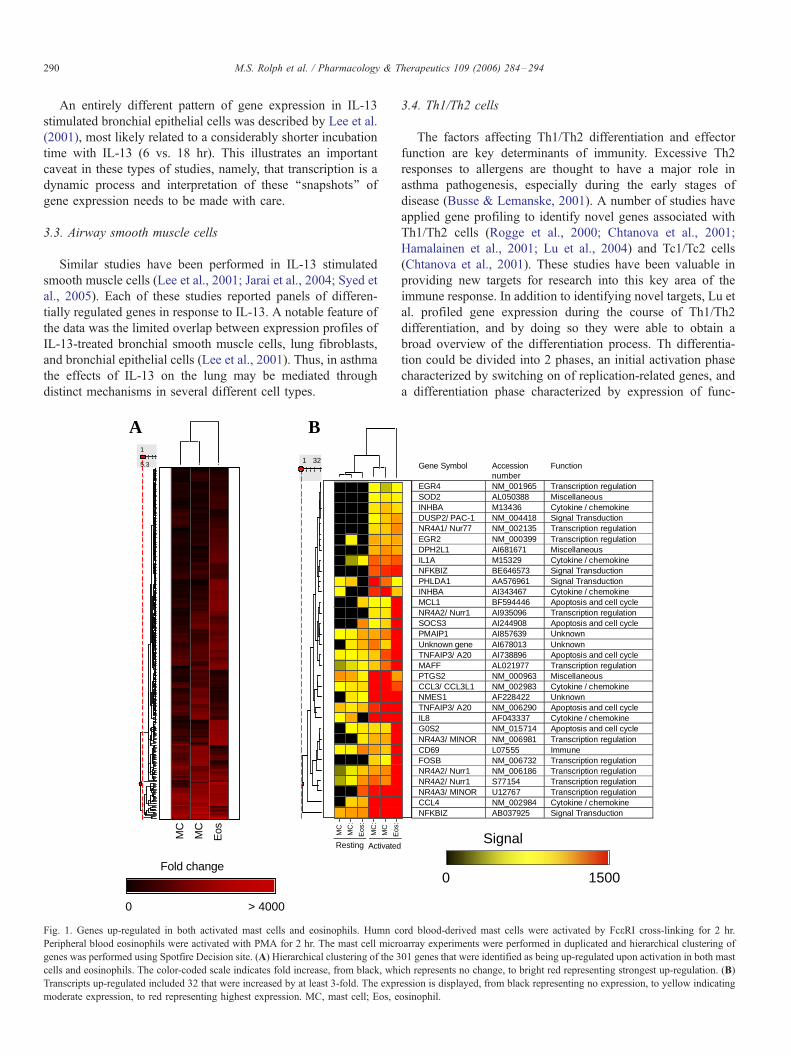
3.3. Airway smooth muscle cells
Similar studies have been performed in IL-13 stimulated
smooth muscle cells (Lee et al., 2001; Jarai et al., 2004; Syed et
al., 2005). Each of these studies reported panels of differen-
tially regulated genes in response to IL-13. A notable feature of
the data was the limited overlap between expression profiles of
IL-13-treated bronchial smooth muscle cells, lung fibroblasts,
and bronchial epithelial cells (Lee et al., 2001). Thus, in asthma
the effects of IL-13 on the lung may be mediated through
distinct mechanisms in several different cell types.
5.3
1
MC
MC
Eos
Fold change
0 > 4000
1 32
MC
MC
:
Eos
:
Resting Activated
MC
:
MC
A B
Fig. 1. Genes up-regulated in both activated mast cells and eosinophils. Humn c
Peripheral blood eosinophils were activated with PMA for 2 hr. The mast cell micr
genes was performed using Spotfire Decision site. (A) Hierarchical clustering of the 3
cells and eosinophils. The color-coded scale indicates fold increase, from black, wh
Transcripts up-regulated included 32 that were increased by at least 3-fold. The expr
moderate expression, to red representing highest expression. MC, mast cell; Eos, e
3.4. Th1/Th2 cells
The factors affecting Th1/Th2 differentiation and effector
function are key determinants of immunity. Excessive Th2
responses to allergens are thought to have a major role in
asthma pathogenesis, especially during the early stages of
disease (Busse & Lemanske, 2001). A number of studies have
applied gene profiling to identify novel genes associated with
Th1/Th2 cells (Rogge et al., 2000; Chtanova et al., 2001;
Hamalainen et al., 2001; Lu et al., 2004) and Tc1/Tc2 cells
(Chtanova et al., 2001). These studies have been valuable in
providing new targets for research into this key area of the
immune response. In addition to identifying novel targets, Lu et
al. profiled gene expression during the course of Th1/Th2
differentiation, and by doing so they were able to obtain a
broad overview of the differentiation process. Th differentia-
tion could be divided into 2 phases, an initial activation phase
characterized by switching on of replication-related genes, and
a differentiation phase characterized by expression of func-
Gene Symbol Accessionnumber
Function
EGR4 NM_001965 Transcription regulationSOD2 AL050388 MiscellaneousINHBA M13436 Cytokine / chemokineDUSP2/ PAC-1 NM_004418 Signal TransductionNR4A1/ Nur77 NM_002135 Transcription regulationEGR2 NM_000399 Transcription regulationDPH2L1 AI681671 MiscellaneousIL1A M15329 Cytokine / chemokineNFKBIZ BE646573 Signal TransductionPHLDA1 AA576961 Signal TransductionINHBA AI343467 Cytokine / chemokineMCL1 BF594446 Apoptosis and cell cycleNR4A2/ Nurr1 AI935096 Transcription regulationSOCS3 AI244908 Apoptosis and cell cyclePMAIP1 AI857639 UnknownUnknown gene AI678013 UnknownTNFAIP3/ A20 AI738896 Apoptosis and cell cycleMAFF AL021977 Transcription regulationPTGS2 NM_000963 MiscellaneousCCL3/ CCL3L1 NM_002983 Cytokine / chemokineNMES1 AF228422 UnknownTNFAIP3/ A20 NM_006290 Apoptosis and cell cycleIL8 AF043337 Cytokine / chemokineG0S2 NM_015714 Apoptosis and cell cycleNR4A3/ MINOR NM_006981 Transcription regulationCD69 L07555 ImmuneFOSB NM_006732 Transcription regulationNR4A2/ Nurr1 NM_006186 Transcription regulationNR4A2/ Nurr1 S77154 Transcription regulationNR4A3/ MINOR U12767 Transcription regulationCCL4 NM_002984 Cytokine / chemokineNFKBIZ AB037925 Signal Transduction
Signal
0 1500
Eos
:
ord blood-derived mast cells were activated by Fc(RI cross-linking for 2 hr.
oarray experiments were performed in duplicated and hierarchical clustering of
01 genes that were identified as being up-regulated upon activation in both mast
ich represents no change, to bright red representing strongest up-regulation. (B)
ession is displayed, from black representing no expression, to yellow indicating
osinophil.
M.S. Rolph et al. / Pharmacology & Therapeutics 109 (2006) 284–294 291
tional gene groups and transcription factors important in Th1/2
biology (Lu et al., 2004). This type of analysis can facilitate
hypothesis generation. For example, based on expression
kinetics during Th1 and Th2 differentiation the authors
speculated that the transcription factor Txk is important during
the early phase of Th1 differentiation.
3.5. Eosinophils
Despite ongoing controversy (Adamko et al., 2003), the
balance of evidence indicates a major effector role for
eosinophils in asthma. Eosinophils readily undergo apoptosis
in the absence of survival factors such as IL-5 and GM-CSF,
and this is a promising area for therapeutic intervention. To
identify pathways involved in eosinophil survival, Temple et al.
(2001) developed gene expression profiles from IL-5 treated
eosinophils. This identified 80 genes, only a minority of which
were likely to be directly involved in apoptosis. To narrow this
list down further, the results were compared with expression
profiles from an IL-5-dependent human erythroleukemic cell
line following IL-5 withdrawal. Four genes were coordinately
regulated in both cellular systems, Pim-1, DSP-5, CD24, and
SLP-76, each of which has already been implicated in
apoptosis in other cellular systems. This illustrates a theme to
be developed further in Section 3.6, that incorporating multiple
distinct cellular models can add considerable power to an array
experiment and its interpretation. The study by Temple et al.
was subsequently extended by an independent group who
showed that GM-CSF induces a very similar expression profile
to IL-5, and that this experimental system also mimics some of
0
Signal
Nav
ïe T
cel
ls
Na ï
ve B
cel
ls
CD
19+
B c
ells
IgG
AE
mem
. B c
ells
IgM
mem
. B c
ells
TF
H
TC
M
Pla
sma
cel
ls
Na ï
ve B
cel
lsC
D19
+ B
cel
ls
IgG
AE
mem
. B c
ells
IgM
mem
. B c
ells
TF
H
TC
M
Pla
sma
cel
ls
Th2
cel
lsT
h2 c
ells
Th1
cel
ls
Lymphoid homing
Inflammatory
Fig. 2. The expression of all the chemokine receptors represented in Affymetrix U
including CCR1-10, CXCR1-6, CX3CR1, XCR1, as well as putative chemokine r
together base on the patterns of expression. Circles represent expression patterns ass
marks in particular leukocyte types associated with lymph node homing, and ‘‘inflam
such as neutrophils. CD19+ B-cells, peripheral blood B-cells; Mem, memory; TFH,
cells; Act., activated.
the changes that an eosinophil undergoes following migration
into the airways (Bates et al., 2004). A similar study looked at
responses of eosinophils to stem cell factor (SCF). In this
system, about 13% of the genes on an ‘‘inflammation-specific’’
array were regulated by SCF, indicating that this growth factor
has a much greater effect on eosinophil function than was
previously appreciated (Oliveira et al., 2002).
3.6. Integrating array data from multiple cell types
The cellular gene profiling experiments detailed above have
been relatively simple in design and scope, mostly comparing
untreated cells with cells treated to reflect some aspect of
asthma pathology. By increasing the number of experimental
variables, such as cell or tissue type, treatment groups, or time
points, the power (and complexity) of gene profiling can be
markedly enhanced. For example, increasing the number of
time points gives much greater insight into a dynamic process,
as illustrated above by studies into Th1/Th2 differentiation (Lu
et al., 2004).
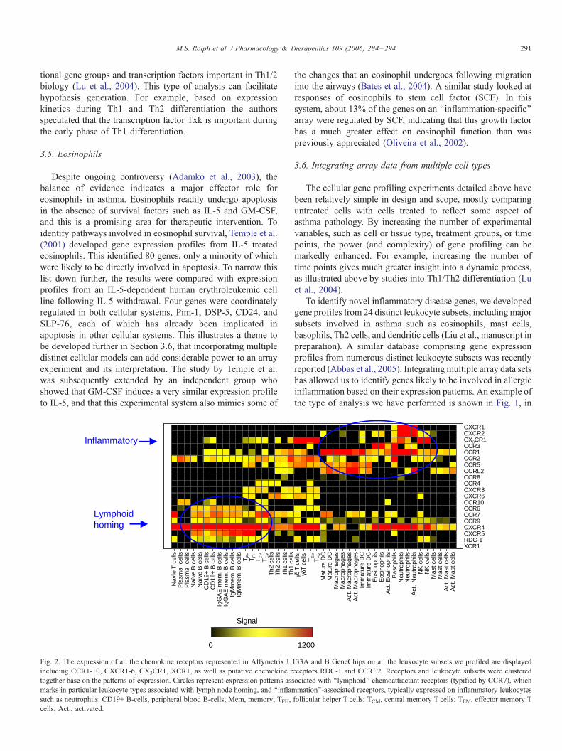
To identify novel inflammatory disease genes, we developed
gene profiles from 24 distinct leukocyte subsets, includingmajor
subsets involved in asthma such as eosinophils, mast cells,
basophils, Th2 cells, and dendritic cells (Liu et al., manuscript in
preparation). A similar database comprising gene expression
profiles from numerous distinct leukocyte subsets was recently
reported (Abbas et al., 2005). Integrating multiple array data sets
has allowed us to identify genes likely to be involved in allergic
inflammation based on their expression patterns. An example of
the type of analysis we have performed is shown in Fig. 1, in
CX3CR1
CCR6
CXCR1CXCR2
CCR1
CXCR4CXCR5
CCR5
CCR3
CCR9
CXCR6
CCR2
CCR7
CCR8CCR4CXCR3
XCR1RDC-1
CCRL2
CCR10
1200
γδ T
cel
ls
TE
M
Mat
ure
DC
Mac
roph
ages
Act
. Mac
roph
ages
Imm
atur
e D
C
Eos
inop
hils
Act
. Eos
inop
hils
Bas
ophi
lsN
eutr
ophi
ls
NK
cel
ls
Mas
t cel
ls
Act
. Mas
t cel
ls
Act
. N
eutr
ophi
ls
Act
. Mas
t cel
ls
Mas
t cel
ls
NK
cel
ls
Neu
trop
hils
Imm
atur
e D
C
Act
. Mac
roph
ages
Mac
roph
ages
Mat
ure
DC
TE
M
γδT
cel
ls
Th1
cel
ls
Eos
inop
hils
133A and B GeneChips on all the leukocyte subsets we profiled are displayed
eceptors RDC-1 and CCRL2. Receptors and leukocyte subsets were clustered
ociated with ‘‘lymphoid’’ chemoattractant receptors (typified by CCR7), which
mation’’-associated receptors, typically expressed on inflammatory leukocytes
follicular helper T cells; TCM, central memory T cells; TEM, effector memory T

M.S. Rolph et al. / Pharmacology & Therapeutics 109 (2006) 284–294292
which genes up-regulated in both mast cells and eosinophils
following activation (mast cells with Fc(R1 cross-linking, and
eosinophils with PMA) were identified. Because mast cells and
eosinophils are the major effector leukocytes in allergic
inflammation, this set of genes is likely to contain a high
proportion of genes involved in allergic effector function. This
type of analysis is made even more powerful by ‘‘subtracting’’
gene profiles from other non-allergic cell types such as
macrophages and neutrophils (Liu et al., manuscript submitted).
An additional strategy that we and others have adopted is to
identify genes whose expression is restricted to a single
leukocyte subset (Liu et al., manuscript submitted) (Nakajima
et al., 2001, 2004; Abbas et al., 2005). In our experience, these
strategies identify numerous genes already implicated in asthma,
but also highlight novel candidates for further study.
Array experiments typically assess the expression of
thousands of genes in a limited number of cell types or tissues.
However, the reverse of this approach—the study of select genes
or molecular families across large numbers of data sets provides
a very effective mechanism to understand the role of genes of
interest in biology.We have a strong interest in certain molecular
families, such as the chemokines and chemokine receptors, and
the TNF superfamily. Fig. 2 shows a heat map representation of
all the chemoattractant receptors in human leukocytes. This type
of analysis allowed us tomake associations between the different
receptors. For instance, 1 poorly characterized receptor, RDC-1,
showed an expression pattern resembling that of the so-called
‘‘lymphoid’’ or ‘‘constitutive’’ homing receptors such as CCR7
or CXCR4. ‘‘Inflammatory’’ receptors such as CXCR1 and
CCR3 are expressed on cell types associated with inflammation,
such as neutrophils and eosinophils. With the public availability
of large data sets for various cell types or biological paradigms,
we believe that interrogation of individual genes or gene families
will become increasingly common.
Large array data sets can be mined for specific types of
targets. For example, Nakajima et al. screened a gene expression
library comprising 10 different cell types to identify cell surface
receptors and ion channels (i.e., ‘‘druggable’’ targets) expressed
on specific granulocyte subsets (Nakajima et al., 2004).
The analysis of gene profiles obtained from tissue samples
can be enhanced by combining the data with profiles obtained
from specific cell types. This strategy has not been employed in
asthma-related studies, but its value has been clearly demon-
strated in other systems. For example, combining gene profiling
data from lupus nephritis glomeruli and individual leukocyte
subsets greatly facilitated interpretation of the glomerular
clustering data (Peterson et al., 2004). Along similar lines, gene
profiles of normal leukocyte subsets have been invaluable in
characterizing gene expression profiles of lymphoid malignan-
cies by providing molecular insights into the cell of origin and
underlying pathogenetic mechanisms (Shaffer et al., 2002).
4. Conclusion
Gene profiling using microarray technology has almost
unlimited potential for discovery-based research and molecular
diagnosis. The power of this approach is also one of its great
challenges because the size of the data sets requires careful and
complex analysis to extract biologically meaningful data. In
most cases, an array experiment is not an end in itself, but a
starting point for hypothesis generation and further experimen-
tation. This is the current status of the application of
microarrays to asthma research. Gene profiles have been
generated, but with few exceptions, they have not yet been
used to make significant advances in our understanding of
asthma. We anticipate numerous advances in asthma research
in the coming years based on initial microarray studies.
Acknowledgements
We thank Tatyana Chtanova, Rebecca Newton, Kim Good,
Sabine Zimmer, Melinda Frost and Stuart Tangye for providing
microarray data for generation of Figs. 1 and 2.
References
Abbas, A. R., Baldwin, D., Ma, Y., Ouyang, W., Gurney, A., Martin, F.,
et al. (2005). Immune response in silico (IRIS): immune-specific genes
identified from a compendium of microarray expression data. Genes
Immun, 6, 319–331.
Adamko, D., Odemuyiwa, S. O., & Moqbel, R. (2003). The eosinophil as a
therapeutic target in asthma: beginning of the end, or end of the beginning?
Curr Opin Pharmacol, 3, 227–232.
Alizadeh, A. A., Eisen, M. B., Davis, R. E., Ma, C., Lossos, I. S., Rosenwald,
A., et al. (2000). Distinct types of diffuse large B-cell lymphoma identified
by gene expression profiling. Nature, 403, 503–511.
Barnes, P. J. (2004). New drugs for asthma. Nat Rev Drug Discov, 3, 831–844.
Bates, M. E., Liu, L. Y., Esnault, S., Stout, B. A., Fonkem, E., Kung, V., et al.
(2004). Expression of interleukin-5- and granulocyte macrophage-colony-
stimulating factor-responsive genes in blood and airway eosinophils. Am J
Respir Cell Mol Biol, 30, 736–743.
Bayram, H., Devalia, J. L., Sapsford, R. J., Ohtoshi, T., Miyabara, Y., Sagai,
M., et al. (1998). The effect of diesel exhaust particles on cell function and
release of inflammatory mediators from human bronchial epithelial cells in
vitro. Am J Respir Cell Mol Biol, 18, 441–448.
Bochner, B. S., Undem, B. J., & Lichtenstein, L. M. (1994). Immunological
aspects of allergic asthma. Annu Rev Immunol, 12, 295–335.
Broide, D. H., Eisman, S., Ramsdell, J. W., Ferguson, P., Schwartz, L. B., &
Wasserman, S. I. (1990). Airway levels of mast cell-derived mediators in
exercise-induced asthma. Am Rev Respir Dis, 141, 563–568.
Broide, D. H., Gleich, G. J., Cuomo, A. J., Coburn, D. A., Federman, E. C.,
Schwartz, L. B., et al. (1991). Evidence of ongoing mast cell and eosinophil
degranulation in symptomatic asthma airway. J Allergy Clin Immunol, 88,
637–648.
Brutsche, M. H., Joos, L., Carlen Brutsche, I. E., Bissinger, R., Tamm, M.,
Custovic, A., et al. (2002). Array-based diagnostic gene-expression score
for atopy and asthma. J Allergy Clin Immunol, 109, 271–273.
Bucchieri, F., Puddicombe, S. M., Lordan, J. L., Richter, A., Buchanan, D.,
Wilson, S. J., et al. (2002). Asthmatic bronchial epithelium is more
susceptible to oxidant-induced apoptosis. Am J Respir Cell Mol Biol, 27,
179–185.
Busse, W. W., & Lemanske Jr., R. F. (2001). Asthma. N Engl J Med, 344,
350–362.
Butte, A. (2002). The use and analysis of microarray data. Nat Rev Drug
Discov, 1, 951–960.
CDC (2002, March 29). Surveillance for asthma—United States, 1980–1999.
CDC Surveillance Summaries. Morb Mort Wkly Rep, vol. 51.
Chtanova, T., Kemp, R. A., Sutherland, A. P., Ronchese, F., & Mackay, C.
R. (2001). Gene microarrays reveal extensive differential gene expression
in both CD4(+) and CD8(+) type 1 and type 2 T cells. J Immunol, 167,
3057–3063.

M.S. Rolph et al. / Pharmacology & Therapeutics 109 (2006) 284–294 293
Cohn, L., Elias, J. A., & Chupp, G. L. (2004). Asthma: mechanisms of disease
persistence and progression. Annu Rev Immunol, 22, 789–815.
Cokugras, H., Akcakaya, N., Seckin, Camcioglu, Y., Sarimurat, N., & Aksoy, F.
(2001). Ultrastructural examination of bronchial biopsy specimens from
children with moderate asthma. Thorax, 56, 25–29.
Cook, P. W., Piepkorn, M., Clegg, C. H., Plowman, G. D., DeMay, J. M.,
Brown, J. R., et al. (1997). Transgenic expression of the human
amphiregulin gene induces a psoriasis-like phenotype. J Clin Invest, 100,
2286–2294.
Davies, D. E., Wicks, J., Powell, R. M., Puddicombe, S. M., & Holgate, S. T.
(2003). Airway remodeling in asthma: new insights. J Allergy Clin
Immunol, 111, 215–225.
Fedorov, I. A., Wilson, S. J., Davies, D. E., & Holgate, S. T. (2005).
Epithelial stress and structural remodelling in childhood asthma. Thorax,
60, 389–394.
Finkelman, F. D., Yang, M., Perkins, C., Schleifer, K., Sproles, A., Santeliz, J.,
et al. (2005). Suppressive effect of IL-4 on IL-13-induced genes in mouse
lung. J Immunol, 174, 4630–4638.
Foster, P. S., Mould, A. W., Yang, M., Mackenzie, J., Mattes, J., Hogan, S. P.,
et al. (2001). Elemental signals regulating eosinophil accumulation in the
lung. Immunol Rev, 179, 173–181.
Fulkerson, P. C., Zimmermann, N., Brandt, E. B., Muntel, E. E., Doepker,
M. P., Kavanaugh, J. L., et al. (2004a). Negative regulation of eosinophil
recruitment to the lung by the chemokine monokine induced by IFN-
gamma (Mig, CXCL9). Proc Natl Acad Sci U S A, 101, 1987–1992.
Fulkerson, P. C., Zimmermann, N., Hassman, L. M., Finkelman, F. D., &
Rothenberg, M. E. (2004b). Pulmonary chemokine expression is
coordinately regulated by STAT1, STAT6, and IFN-gamma. J Immunol,
173, 7565–7574.
Guajardo, J. R., Schleifer, K. W., Daines, M. O., Ruddy, R. M., Aronow, B. J.,
Wills-Karp, M., et al. (2005). Altered gene expression profiles in nasal
respiratory epithelium reflect stable versus acute childhood asthma. J
Allergy Clin Immunol, 115, 243–251.
Hamalainen, H., Zhou, H., Chou, W., Hashizume, H., Heller, R., & Lahesmaa,
R. (2001). Distinct gene expression profiles of human type 1 and type 2 T
helper cells. Genome Biol, 2.
Hasegawa, K., Tamari, M., Shao, C., Shimizu, M., Takahashi, N., Mao, X.Q.,
et al. (2004). Variations in the C3, C3a receptor, and C5 genes affect
susceptibility to bronchial asthma. Hum Genet, 115, 295–301.
Holloway, J. W., Beghe, B., & Holgate, S. T. (1999). The genetic basis of atopic
asthma. Clin Exp Allergy, 29, 1023–1032.
Jarai, G., Sukkar, M., Garrett, S., Duroudier, N., Westwick, J., Adcock, I., et al.
(2004). Effects of interleukin-1beta, interleukin-13 and transforming growth
factor-beta on gene expression in human airway smooth muscle using gene
microarrays. Eur J Pharmacol, 497, 255–265.
Johnson, G. R., Kannan, B., Shoyab, M., & Stromberg, K. (1993).
Amphiregulin induces tyrosine phosphorylation of the epidermal growth
factor receptor and p185erbB2. Evidence that amphiregulin acts exclusively
through the epidermal growth factor receptor at the surface of human
epithelial cells. J Biol Chem, 268, 2924–2931.
Johnson, P. R., Roth, M., Tamm, M., Hughes, M., Ge, Q., King, G., et al.
(2001). Airway smooth muscle cell proliferation is increased in asthma. Am
J Respir Crit Care Med, 164, 474–477.
Kamme, F., & Erlander, M. G. (2003). Global gene expression analysis of
single cells. Curr Opin Drug Discov Dev, 6, 231–236.
Karp, C. L., Grupe, A., Schadt, E., Ewart, S. L., Keane-Moore, M., Cuomo,
P. J., et al. (2000). Identification of complement factor 5 as a susceptibility
locus for experimental allergic asthma. Nat Immunol, 1, 221–226.
Kashiwakura, J., Yokoi, H., Saito, H., & Okayama, Y. (2004). T cell proliferation
by direct cross-talk between OX40 ligand on human mast cells and OX40 on
human T cells: comparison of gene expression profiles between human
tonsillar and lung-cultured mast cells. J Immunol, 173, 5247–5257.
King, N. E., Rothenberg, M. E., & Zimmermann, N. (2004a). Arginine in
asthma and lung inflammation. J Nutr, 134, 2830S–2836S.
King, N. E., Zimmermann, N., Pope, S. M., Fulkerson, P. C., Nikolaidis, N. M.,
Mishra, A., et al. (2004b). Expression and regulation of a disintegrin and
metalloproteinase (ADAM) 8 in experimental asthma. Am J Respir Cell Mol
Biol, 31, 257–265.
Kuperman, D., Schofield, B., Wills-Karp, M., & Grusby, M. J. (1998). Signal
transducer and activator of transcription factor 6 (Stat6)-deficient mice are
protected from antigen-induced airway hyperresponsiveness and mucus
production. J Exp Med, 187, 939–948.
Kuperman, D. A., Huang, X., Koth, L. L., Chang, G. H., Dolganov, G. M., Zhu,
Z., et al. (2002). Direct effects of interleukin-13 on epithelial cells cause
airway hyperreactivity and mucus overproduction in asthma. Nat Med, 8,
885–889.
Laprise, C., Sladek, R., Ponton, A., Bernier, M. C., Hudson, T. J., & Laviolette,
M. (2004). Functional classes of bronchial mucosa genes that are
differentially expressed in asthma. BMC Genomics, 5, 21.
Leckie, M. J., ten Brinke, A., Khan, J., Diamant, Z., O’Connor, B. J., Walls,
C. M., et al. (2000). Effects of an interleukin-5 blocking monoclonal
antibody on eosinophils, airway hyper-responsiveness, and the late
asthmatic response. Lancet, 356, 2144–2148.
Lee, J. H., Kaminski, N., Dolganov, G., Grunig, G., Koth, L., Solomon, C.,
et al. (2001). Interleukin-13 induces dramatically different transcriptional
programs in three human airway cell types. Am J Respir Cell Mol Biol,
25, 474–485.
Lemanske, R. F., & Busse, W. W. (2003). Asthma. J Allergy Clin Immunol, 111,
S502–S519.
Lilly, C. M., Tateno, H., Oguma, T., Israel, E., & Sonna, L. A. (2005). Effects of
allergen challenge on airway epithelial cell gene expression. Am J Respir
Cell Mol Biol, 171, 579–586.
Lora, J. M., Al-Garawi, A., Pickard, M. D., Price, K. S., Bagga, S., Sicoli, J.,
et al. (2003). FcepsilonRI-dependent gene expression in human mast cells
is differentially controlled by T helper type 2 cytokines. J Allergy Clin
Immunol, 112, 1119–1126.
Lu, B., Zagouras, P., Fischer, J. E., Lu, J., Li, B., & Flavell, R. A. (2004).
Kinetic analysis of genomewide gene expression reveals molecule
circuitries that control T cell activation and Th1/2 differentiation. Proc
Natl Acad Sci U S A, 101, 3023–3028.
Luo, L., Salunga, R. C., Guo, H., Bittner, A., Joy, K. C., Galindo, J. E., et al.
(1999). Gene expression profiles of laser-captured adjacent neuronal
subtypes. Nat Med, 5, 117–122.
Meurs, H., Maarsingh, H., & Zaagsma, J. (2003). Arginase and asthma: novel
insights into nitric oxide homeostasis and airway hyperresponsiveness.
Trends Pharmacol Sci, 24, 450–455.
Minshall, E., Chakir, J., Laviolette, M., Molet, S., Zhu, Z., Olivenstein, R., et al.
(2000). IL-11 expression is increased in severe asthma: association with
epithelial cells and eosinophils. J Allergy Clin Immunol, 105, 232–238.
Nakajima, T., Matsumoto, K., Suto, H., Tanaka, K., Ebisawa, M., Tomita, H.,
et al. (2001). Gene expression screening of human mast cells and
eosinophils using high-density oligonucleotide probe arrays: abundant
expression of major basic protein in mast cells. Blood, 98, 1127–1134.
Nakajima, T., Inagaki, N., Tanaka, H., Tanaka, A., Yoshikawa, M.,
Tamari, M., et al. (2002). Marked increase in CC chemokine gene
expression in both human and mouse mast cell transcriptomes following
Fcepsilon receptor I cross-linking: an interspecies comparison. Blood,
100, 3861–3868.
Nakajima, T., Iikura, M., Okayama, Y., Matsumoto, K., Uchiyama, C.,
Shirakawa, T., et al. (2004). Identification of granulocyte subtype-selective
receptors and ion channels by using a high-density oligonucleotide probe
array. J Allergy Clin Immunol, 113, 528–535.
Nikolaidis, N. M., Zimmermann, N., King, N. E., Mishra, A., Pope, S. M.,
Finkelman, F. D., et al. (2003). Trefoil factor-2 is an allergen-induced gene
regulated by Th2 cytokines and STAT6 in the lung. Am J Respir Cell Mol
Biol, 29, 458–464.
Ohshima, Y., Yang, L. P., Uchiyama, T., Tanaka, Y., Baum, P., Sergerie, M.,
et al. (1998). OX40 costimulation enhances interleukin-4 (IL-4) expres-
sion at priming and promotes the differentiation of naive human CD4(+)
T cells into high IL-4-producing effectors. Blood, 92, 3338–3345.
Okumura, S., Kashiwakura, J., Tomita, H., Matsumoto, K., Nakajima, T., Saito,
H., et al. (2003). Identification of specific gene expression profiles in
human mast cells mediated by Toll-like receptor 4 and FcepsilonRI. Blood,
102, 2547–2554.
Okumura, S., Sagara, H., Fukuda, T., Saito, H., & Okayama, Y. (2005).
FcepsilonRI-mediated amphiregulin production by human mast cells

M.S. Rolph et al. / Pharmacology & Therapeutics 109 (2006) 284–294294
increases mucin gene expression in epithelial cells. J Allergy Clin Immunol,
115, 272–279.
Oliveira, S. H., Taub, D. D., Nagel, J., Smith, R., Hogaboam, C. M., Berlin,
A., et al. (2002). Stem cell factor induces eosinophil activation and
degranulation: mediator release and gene array analysis. Blood, 100,
4291–4297.
Payne, D. N., Rogers, A. V., Adelroth, E., Bandi, V., Guntupalli, K. K.,
Bush, A., et al. (2003). Early thickening of the reticular basement
membrane in children with difficult asthma. Am J Respir Crit Care Med,
167, 78–82.
Peters, S. P. (2003). Heterogeneity in the pathology and treatment of asthma.
Am J Med, 115(Suppl 3A), 49S–54S.
Peterson, K. S., Huang, J. F., Zhu, J., D’Agati, V., Liu, X., Miller, N., et al.
(2004). Characterization of heterogeneity in the molecular pathogenesis of
lupus nephritis from transcriptional profiles of laser-captured glomeruli. J
Clin Invest, 113, 1722–1733.
Player, A., Barrett, J. C., & Kawasaki, E. S. (2004). Laser capture
microdissection, microarrays and the precise definition of a cancer cell.
Expert Rev Mol Diagn, 4, 831–840.
Richter, A., Puddicombe, S. M., Lordan, J. L., Bucchieri, F., Wilson, S. J.,
Djukanovic, R., et al. (2001). The contribution of interleukin (IL)-4 and IL-
13 to the epithelial-mesenchymal trophic unit in asthma. Am J Respir Cell
Mol Biol, 25, 385–391.
Rogge, L., Bianchi, E., Biffi, M., Bono, E., Chang, S. Y., Alexander, H., et al.
(2000). Transcript imaging of the development of human T helper cells
using oligonucleotide arrays. Nat Genet, 25, 96–101.
Roth, M., Johnson, P. R., Borger, P., Bihl, M. P., Rudiger, J. J., King, G. G.,
et al. (2004). Dysfunctional interaction of C/EBPalpha and the glucocor-
ticoid receptor in asthmatic bronchial smooth-muscle cells. N Engl J Med,
351, 560–574.
Sakata, Y., Arima, K., Takai, T., Sakurai, W., Masumoto, K., Yuyama, N., et al.
(2004). The squamous cell carcinoma antigen 2 inhibits the cysteine
proteinase activity of a major mite allergen, Der p 1. J Biol Chem, 279,
5081–5087.
Sayama, K., Diehn, M., Matsuda, K., Lunderius, C., Tsai, M., Tam, S. Y., et al.
(2002). Transcriptional response of human mast cells stimulated via the
Fc(epsilon)RI and identification of mast cells as a source of IL-11. BMC
Immunol, 3, 5.
Shaffer, A. L., Rosenwald, A., & Staudt, L. M. (2002). Lymphoid malignan-
cies: the dark side of B-cell differentiation. Nat Rev Immunol, 2, 920–932.
Staudt, L. M. (2001). Gene expression physiology and pathophysiology of the
immune system. Trends Immunol, 22, 35–40.
Staudt, L. M., & Brown, P. O. (2000). Genomic views of the immune system.
Annu Rev Immunol, 18, 829–859.
Stoughton, R. B. (2004). Applications of DNA microarrays in biology. Annu
Rev Biochem.
Syed, F., Panettieri Jr., R. A., Tliba, O., Huang, C., Li, K., Bracht, M., et al.
(2005). The effect of IL-13 and IL-13R130Q, a naturally occurring IL-13
polymorphism, on the gene expression of human airway smooth muscle
cells. Respir Res, 6, 9.
Temple, R., Allen, E., Fordham, J., Phipps, S., Schneider, H. C., Lindauer, K.,
et al. (2001). Microarray analysis of eosinophils reveals a number of
candidate survival and apoptosis genes. Am J Respir Cell Mol Biol, 25,
425–433.
Wang, S. W., Oh, C. K., Cho, S. H., Hu, G., Martin, R., Demissie-Sanders,
S., et al. (2005). Amphiregulin expression in human mast cells and its
effect on the primary human lung fibroblasts. J Allergy Clin Immunol,
115, 287–294.
Wark, P. A., Johnston, S. L., Bucchieri, F., Powell, R., Puddicombe, S., Laza-
Stanca, V., et al. (2005). Asthmatic bronchial epithelial cells have a
deficient innate immune response to infection with rhinovirus. J Exp Med,
201, 937–947.
Weiss, K. B., & Sullivan, S. D. (2001). The health economics of asthma and
rhinitis: I. Assessing the economic impact. J Allergy Clin Immunol, 107,
3–8.
Wenzel, S. E., Fowler III, A. A., & Schwartz, L. B. (1988). Activation of
pulmonary mast cells by bronchoalveolar allergen challenge. In vivo release
of histamine and tryptase in atopic subjects with and without asthma. Am
Rev Respir Dis, 137, 1002–1008.
Wills-Karp, M., & Chiaramonte, M. (2003). Interleukin-13 in asthma. Curr
Opin Pulm Med, 9, 21–27.
Woodruff, P. G., Dolganov, G. M., Ferrando, R. E., Donnelly, S., Hays, S. R.,
Solberg, O. D., Carter, R., et al. (2004). Hyperplasia of smooth muscle in
mild to moderate asthma without changes in cell size or gene expression.
Am J Respir Crit Care Med, 169, 1001–1006.
Woolcock, A. J., Bastiampillai, S. A., Marks, G. B., & Keena, V. A. (2001).
The burden of asthma in Australia. Med J Aust, 175, 141–145.
Wynn, T. A. (2003). IL-13 effector functions. Annu Rev Immunol, 21,
425–456.
Yeatman II, C. F., Jacobs-Helber, S. M., Mirmonsef, P., Gillespie, S. R.,
Bouton, L. A., Collins, H. A., et al. (2000). Combined stimulation with the
T helper cell type 2 cytokines interleukin (IL)-4 and IL-10 induces mouse
mast cell apoptosis. J Exp Med, 192, 1093–1103.
Yuyama, N., Davies, D. E., Akaiwa, M., Matsui, K., Hamasaki, Y., Suminami,
Y., et al. (2002). Analysis of novel disease-related genes in bronchial
asthma. Cytokine, 19, 287–296.
Zimmermann, N., King, N. E., Laporte, J., Yang, M., Mishra, A., Pope, S. M.,
et al. (2003). Dissection of experimental asthma with DNA microarray
analysis identifies arginase in asthma pathogenesis. J Clin Invest, 111,
1863–1874.
Zimmermann, N., Doepker, M. P., Witte, D. P., Stringer, K. F., Fulkerson, P. C.,
Pope, S. M., et al. (2005). Expression and regulation of small proline-rich
protein 2 in allergic inflammation. Am J Respir Cell Mol Biol, 32, 428–435.
Zou, J., Young, S., Zhu, F., Gheyas, F., Skeans, S., Wan, Y., et al. (2002).
Microarray profile of differentially expressed genes in a monkey model of
allergic asthma. Genome Biol, 3.