Embed Size (px)
Citation preview
Cloning of the Human Interferon-Related Developmental Regulator(IFRD1) Gene Coding for the PC4 Protein, a Member of a Novel
Family of Developmentally Regulated Genes
Pasquale Buanne,*,† Barbara Incerti,† Daniele Guardavaccaro,* Virginia Avvantaggiato,‡Antonio Simeone,‡ and Felice Tirone*,1
*Istituto di Neurobiologia CNR, Viale Carlo Marx 43, 00137 Rome; †Telethon Institute of Genetics and Medicine, Via Olgettina 58,20132 Milan; and ‡Istituto Internazionale di Genetica e Biofisica, Via G. Marconi 10, 80100 Naples, Italy
Received December 1, 1997; accepted February 10, 1998
The rat PC4 gene had been initially isolated as anerve growth factor-inducible sequence in PC12 cells.Although its function remains unknown, recently ithas been shown that PC4 is necessary to muscle dif-ferentiation and that it might have a role in signaltransduction. We report the isolation of the humanhomolog of the rat PC4 gene, renamed here IFRD1(interferon-related developmental regulator 1). Sev-eral human IFRD1 clones were identified by searchingthe EST database using the rat IFRD1 (PC4) cDNA as aquery. An EST clone containing the entire ORF waschosen for sequencing. Human IFRD1 presented a pre-dicted protein product of 453 amino acids, highly con-served (90.2% identity) compared to the rat IFRD1(PC4) protein sequences. The mapping assignment ofhuman IFRD1 to chromosome 7q22–q31 was retrievedfrom the UniGene database maintained at NCBI. Acomparison of human IFRD1 (PC4) protein to data-bases revealed 47% identity to the protein encoded bythe human gene SKMc15, originally isolated from achromosome 3-specific library. Therefore, SKMc15 is agene related to IFRD1, being the second member of anovel family. We analyzed their expression during mu-rine development, and we found that mouse IFRD1appears more expressed in specific differentiatingstructures at midgestation, while mouse SKMc15 ishighly expressed soon after gastrulation and in thehepatic primordium, suggesting an involvement inearly hematopoiesis. © 1998 Academic Press
INTRODUCTION
With the aim of identifying genes involved in theprocess of neuronal differentiation, we have isolated in
recent years several immediate-early genes activatedin the rat cell line PC12 by nerve growth factor (NGF;Tirone and Shooter, 1989; Bradbury et al., 1991). Thiscell line, which is derived from a pheochromocytoma,differentiates into sympathetic neurons in the pres-ence of NGF, thus representing a widely used in vitromodel of the differentiative action of NGF on chromaf-fin cells in vivo (Greene and Tischler, 1976). We foundthat one of the genes isolated, PC4, presented a signif-icant similarity with interferon-g, a molecule knownfor its role in cellular differentiation (Tirone andShooter, 1989). Such similarity was between the car-boxy-terminal half of the PC4 cDNA-deduced proteinand the whole interferon-g protein, suggesting the ex-istence of a functional domain within the PC4 protein.Furthermore, the mouse PC4 homolog (TIS7) was iden-tified as a tetradecanoyl phorbol acetate-induced genein mouse NIH3T3 cells (Varnum et al., 1989). AlthoughPC4 function remains unknown, its expression wasfound to be regulated not only during neuronal differ-entiation in vitro and in vivo (Guardavaccaro et al.,1994; Iacopetti et al., 1996), but also during myoblastdifferentiation. PC4 is in fact expressed in the myo-blast and in the differentiated myotube, with a tran-sient decrease after the onset of differentiation. Thisaspect appears to have an important functional signif-icance in muscle differentiation, as directly demon-strated by studies performed in the myoblast cell lineC2C12. In fact, stable inhibition of PC4 expression,using antisense PC4 cDNA transfection or microinjec-tion of anti-PC4 polyclonal antibodies, leads to impair-ment of myogenin and myosin gene expression as wellas of morphological differentiation (Guardavaccaro etal., 1995). Microinjection data in myoblast pointed tothe necessity of PC4 presence when the stimulus fordifferentiation is delivered, suggesting the involve-ment of PC4 in signal transduction (Guardavaccaro etal., 1995). This latter possibility was also implied bythe observation that a fraction of the PC4 protein,which appears to be cytosolic (Guardavaccaro et al.,
Sequence data from this article have been deposited with theEMBL Data Library under Accession No. Y10313.
1 To whom correspondence should be addressed at the Institute ofNeurobiology, National Research Council, Viale Marx 43, 00137Rome, Italy. Telephone: 39-6-86090290. Fax: 39-6-86090370. E-mail:[email protected].
GENOMICS 51, 233–242 (1998)ARTICLE NO. GE985260
2330888-7543/98 $25.00
Copyright © 1998 by Academic PressAll rights of reproduction in any form reserved.
1994, 1995), translocates transiently to the inner sideof the plasma membrane at the beginning of neuronaldifferentiation by NGF in the PC12 cell line (Guarda-vaccaro et al., 1994). As a whole, this evidence suggeststhat PC4 might play a modulatory role in development,during the transition of a determined cell lineage (e.g.,muscle or neuron) to a committed phenotype.
Here we report the cloning, sequencing, and expres-sion analysis in adult tissues of the human homolog ofPC4. In compliance with the HGMW nomenclaturerules, and with the properties observed so far for theprotein product PC4, the gene has been renamedIFRD1 (interferon-related developmental regulator 1).In this study we analyzed in parallel, starting fromearly stages of development, the expression of IFRD1and of the related gene SKMc15 (Latif et al., 1997),obtaining hypotheses on the functional role of thesegenes of the same family.
MATERIALS AND METHODS
Sequence analysis and computer-assisted search of databases.Automated fluorescent DNA sequencing was performed using Per-kin–Elmer 377 Prism machines with both Dye Terminator and DyePrimer Cycle Sequencing chemistries on double-strand plasmid tem-plates. Computer analysis of the sequences was performed with theWisconsin Package version 8.1–UNIX (August 1995). Similaritysearches were performed using BlastN, BlastP, and FastA algo-rithms against the GenBank (release 101, June 1997), EMBL (re-lease 50.0, June 1997), Pir-Protein (release 52.0, March 1997), andSwiss-Prot (release 34.0, November 1996) databases.
Northern analysis. Similar amounts of mRNA from different hu-man tissues, blotted on a nylon filter, were hybridized with humanIFRD1 cDNA 32P labeled with the hexamer primers procedure (Fein-berg and Vogelstein, 1983). Hybridization was performed at 42°C for18 h in 53 SSPE (3 M NaCl, 0.2 M NaH2PO4, 0.02 M EDTA), 23Denhardt’s solution (1% Ficoll 400, 1% polyvinylpyrrolidone, 1%BSA), 100 mg/ml sheared salmon sperm DNA, 2% SDS, 50% form-amide. The mRNA amounts were controlled by hybridizing the filtersto a b-actin probe.
Mice. C57/B16 mice were mated between 9 and 10 PM Day 0.5postcoitum was assumed to begin at the middle of the day of vaginalplugging. Pregnant female mice were killed by cervical dislocation,and embryos were staged according to Theiler (1989), collected inice-cold PBS under a dissection microscope (SV11; Carl Zeiss, Inc.,Thornwood, NY), and fixed in 4% paraformaldehyde overnight.
In situ hybridization. In situ hybridization was carried out asdescribed (Wilkinson and Green, 1990; Wilkinson, 1992), with minormodifications. Both paraffin-embedded and cryostat sections wereanalyzed. Dissected embryos were prefixed in 0.1 M sodium phos-phate buffer (pH 7.3), 4% paraformaldehyde at 4°C overnight andembedded in Tissue-Tek (Miles Laboratories, Inc., Elkhart, IN).Cryosections (8 mm thick) were transferred onto gelatin/chromium(III) potassium sulfate-subbed and dried at room temperature. Be-fore hybridization, slides were postfixed. To obtain probes, mouseSKMc15 and mouse IFRD1 (the latter previously isolated by Varnumet al., 1989, named TIS7) were isolated respectively from mouseembryo (E13.5–14.5) and adult mouse cDNA libraries. The isolates,identified by searching the EST database using TIS7 and humanSKMc15 as a query, corresponded to mouse EST W29669, i.e., TIS7,and to mouse EST W65790, i.e., mouse SKMc15. Their identity waschecked and confirmed by complete sequencing and by Southernanalysis (not shown). The probes used were antisense and sensestrands spanning the SKMc15 gene from nt 1 to 699 (referred to thesequence obtained) and the IFRD1 (TIS7) gene from nt 748 to 1496(referred to the published sequence of TIS7; Varnum et al., 1989).
Transcription reactions with T7 or T3 polymerase (Riboprobe Kit;Promega Biotechnologies, Madison, WI) were carried out in the pres-ence of [35S]CTP (Amersham Corp., Arlington Heights, IL). Thetemplate was then degraded with RNase-free DNase (Pharmacia),and the labeled RNA was purified through a Sephadex G-50 columnand progressively degraded by random alkaline hydrolysis to im-prove access to RNA in situ. The probes were dissolved at a workingconcentration of 13105 cpm/ml in the hybridization mix; 30 ml of theappropriate probe was added to each slide. Hybridization was carriedout overnight at 55°C. The slides were then washed under stringentconditions (65°C, 23 SSC, 50% formamide) and treated with RNase.Autoradiography was performed with Kodak NT/B2 emulsion. Expo-sure times were between 15 and 25 days.
Negative controls using sense strands were tested on sectionsadjacent to those hybridized with the specific antisense strands todetermine the basal background. These probes never gave a detect-able signal.
RESULTS
Cloning and Sequencing of Human IRFD1
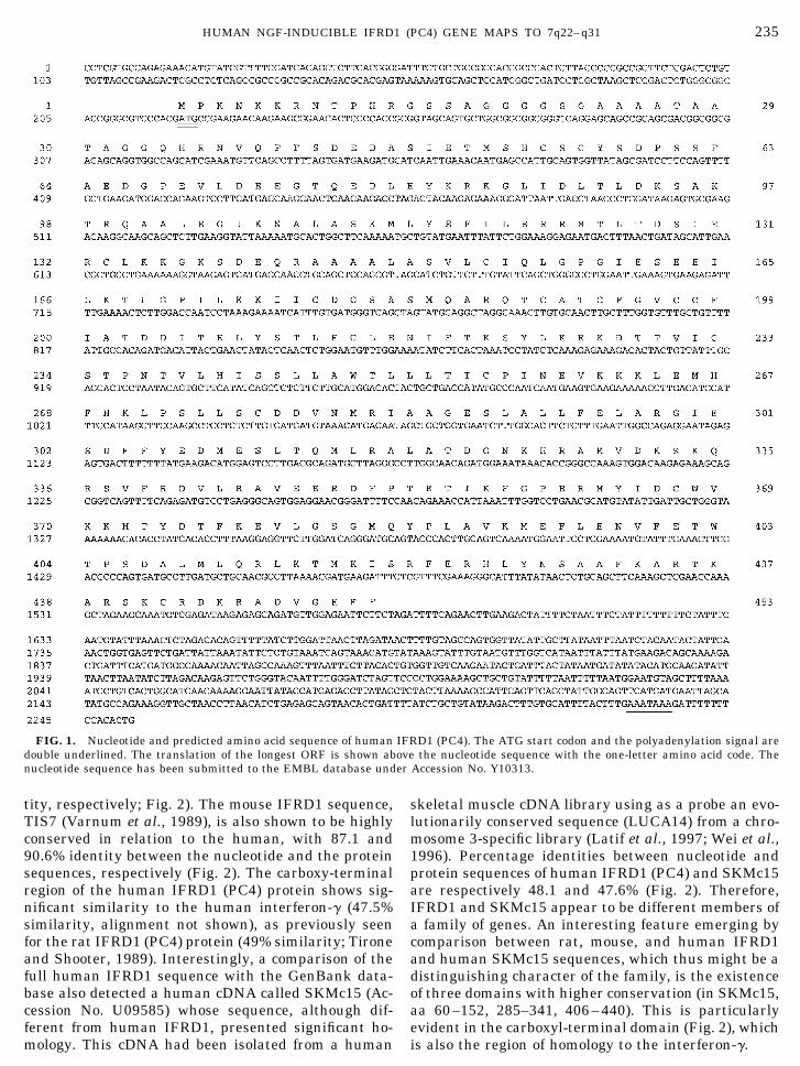
The rat IFRD1 (PC4) cDNA sequence was compared toEST databases (dbEST; Boguski et al., 1993) using theBlastN algorithm (Altschul et al., 1990). We detected 11ESTs having significant homology to the query sequence,from Soares’ infant brain, fetal liver–spleen, and placentacDNA libraries as well as from other human muscle andtestis cDNA libraries (October 1996). Sequence analysiswas then carried out on cDNA clone 52803 (isolated froman infant brain cDNA library), corresponding to a humanEST (H29134) homologous to rat IFRD1 with a P value inthe BlastN output of 3.5310288. Clone 52803 has a cDNAinsert of about 1800 nt, containing the entire coding re-gion. The first in-frame ATG is located 219 nt from the 59end of the clone and fulfills Kozak’s criteria for an initi-ation codon (Kozak, 1984). Comparison of the completesequence of clone 52803 with dbEST indicated the pres-ence of several ESTs belonging to the same transcrip-tional unit and further extending the UTR 39 end of about450 nt. These ESTs were assembled to obtain the com-plete sequence of human IFRD1 cDNA, which has a totallength of 2252 nt, with a 59 UTR of 219 nt and a 39 UTRof 676 nt (Fig. 1). The predicted protein product encodedby the ORF is 453 residues long (Fig. 1) with a calculatedmolecular weight of 50,685. A search for active sites usingthe program Prosite (Bairoch, 1990) showed three poten-tial phosphorylation sites by cAMP-dependent proteinkinase (Thr-9, Thr-127, Thr-373), several potential phos-phorylation sites by casein kinase II, four by proteinkinase C (Ser-95, Thr-353, Thr-373, Thr 416), and one bytyrosine kinase (Tyr-364). Also, potential N-myristoyl-ation sites were detected (residues 13–25 and 105–110),which, however, according to previous experiments per-formed with rat IFRD1 (PC4) cDNA, might be not func-tional (Guardavaccaro et al., 1994).
IRFD1 and SKMc15, Two Membersof a Novel Gene Family
Human and rat IFRD1 (PC4) sequences are highlyhomologous, with a significant identity at both thecDNA and the amino acid levels (86.3 and 90.2% iden-
234 BUANNE ET AL.
tity, respectively; Fig. 2). The mouse IFRD1 sequence,TIS7 (Varnum et al., 1989), is also shown to be highlyconserved in relation to the human, with 87.1 and90.6% identity between the nucleotide and the proteinsequences, respectively (Fig. 2). The carboxy-terminalregion of the human IFRD1 (PC4) protein shows sig-nificant similarity to the human interferon-g (47.5%similarity, alignment not shown), as previously seenfor the rat IFRD1 (PC4) protein (49% similarity; Tironeand Shooter, 1989). Interestingly, a comparison of thefull human IFRD1 sequence with the GenBank data-base also detected a human cDNA called SKMc15 (Ac-cession No. U09585) whose sequence, although dif-ferent from human IFRD1, presented significant ho-mology. This cDNA had been isolated from a human
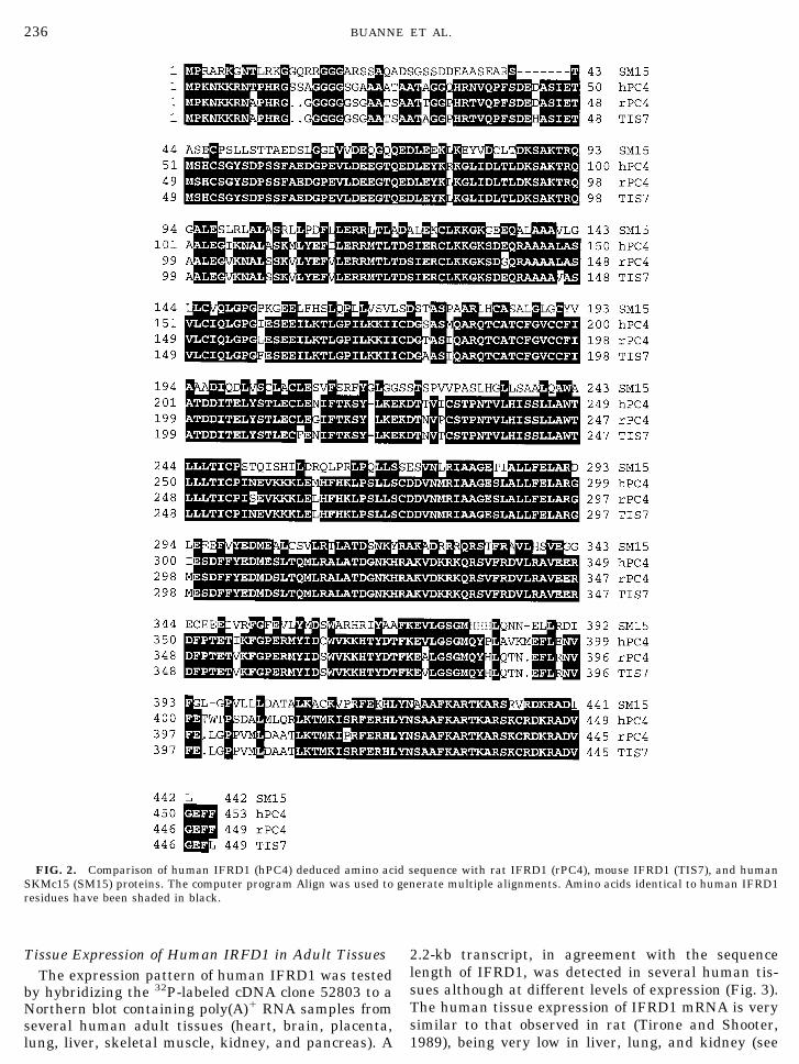
skeletal muscle cDNA library using as a probe an evo-lutionarily conserved sequence (LUCA14) from a chro-mosome 3-specific library (Latif et al., 1997; Wei et al.,1996). Percentage identities between nucleotide andprotein sequences of human IFRD1 (PC4) and SKMc15are respectively 48.1 and 47.6% (Fig. 2). Therefore,IFRD1 and SKMc15 appear to be different members ofa family of genes. An interesting feature emerging bycomparison between rat, mouse, and human IFRD1and human SKMc15 sequences, which thus might be adistinguishing character of the family, is the existenceof three domains with higher conservation (in SKMc15,aa 60–152, 285–341, 406–440). This is particularlyevident in the carboxyl-terminal domain (Fig. 2), whichis also the region of homology to the interferon-g.
FIG. 1. Nucleotide and predicted amino acid sequence of human IFRD1 (PC4). The ATG start codon and the polyadenylation signal aredouble underlined. The translation of the longest ORF is shown above the nucleotide sequence with the one-letter amino acid code. Thenucleotide sequence has been submitted to the EMBL database under Accession No. Y10313.
235HUMAN NGF-INDUCIBLE IFRD1 (PC4) GENE MAPS TO 7q22–q31
Tissue Expression of Human IRFD1 in Adult TissuesThe expression pattern of human IFRD1 was tested
by hybridizing the 32P-labeled cDNA clone 52803 to aNorthern blot containing poly(A)1 RNA samples fromseveral human adult tissues (heart, brain, placenta,lung, liver, skeletal muscle, kidney, and pancreas). A
2.2-kb transcript, in agreement with the sequencelength of IFRD1, was detected in several human tis-sues although at different levels of expression (Fig. 3).The human tissue expression of IFRD1 mRNA is verysimilar to that observed in rat (Tirone and Shooter,1989), being very low in liver, lung, and kidney (see
FIG. 2. Comparison of human IFRD1 (hPC4) deduced amino acid sequence with rat IFRD1 (rPC4), mouse IFRD1 (TIS7), and humanSKMc15 (SM15) proteins. The computer program Align was used to generate multiple alignments. Amino acids identical to human IFRD1residues have been shaded in black.
236 BUANNE ET AL.
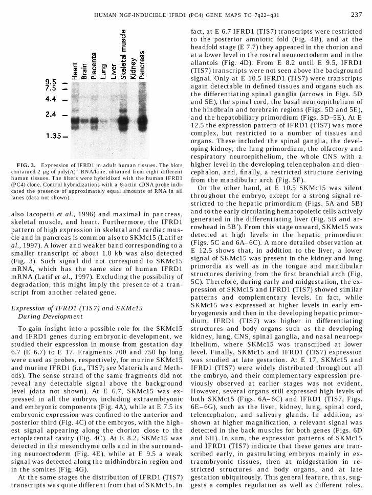
also Iacopetti et al., 1996) and maximal in pancreas,skeletal muscle, and heart. Furthermore, the IFRD1pattern of high expression in skeletal and cardiac mus-cle and in pancreas is common also to SKMc15 (Latif etal., 1997). A lower and weaker band corresponding to asmaller transcript of about 1.8 kb was also detected(Fig. 3). Such signal did not correspond to SKMc15mRNA, which has the same size of human IFRD1mRNA (Latif et al., 1997). Excluding the possibility ofdegradation, this might imply the presence of a tran-script from another related gene.
Expression of IFRD1 (TIS7) and SKMc15During Development
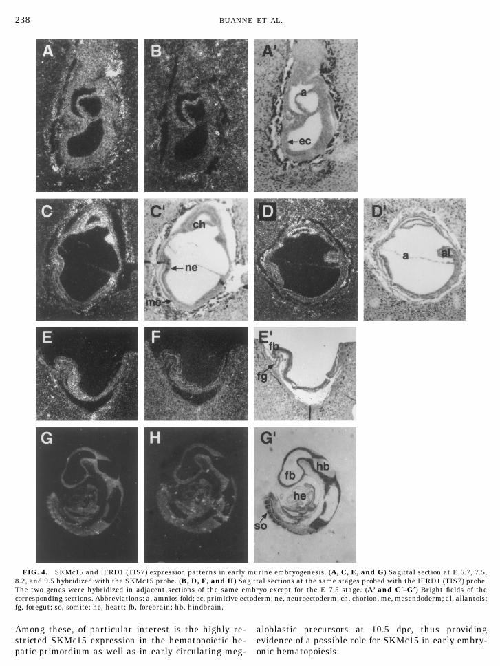
To gain insight into a possible role for the SKMc15and IFRD1 genes during embryonic development, westudied their expression in mouse from gestation day6.7 (E 6.7) to E 17. Fragments 700 and 750 bp longwere used as probes, respectively, for murine SKMc15and murine IFRD1 (i.e., TIS7; see Materials and Meth-ods). The sense strand of the same fragments did notreveal any detectable signal above the backgroundlevel (data not shown). At E 6.7, SKMc15 was ex-pressed in all the embryo, including extraembryonicand embryonic components (Fig. 4A), while at E 7.5 itsembryonic expression was confined to the anterior andposterior third (Fig. 4C) of the embryos, with the high-est signal appearing along the chorion close to theectoplacental cavity (Fig. 4C). At E 8.2, SKMc15 wasdetected in the mesenchyme cells and in the surround-ing neuroectoderm (Fig. 4E), while at E 9.5 a weaksignal was detected along the midhindbrain region andin the somites (Fig. 4G).
At the same stages the distribution of IFRD1 (TIS7)transcripts was quite different from that of SKMc15. In
fact, at E 6.7 IFRD1 (TIS7) transcripts were restrictedto the posterior amniotic fold (Fig. 4B), and at theheadfold stage (E 7.7) they appeared in the chorion andat a lower level in the rostral neuroectoderm and in theallantois (Fig. 4D). From E 8.2 until E 9.5, IFRD1(TIS7) transcripts were not seen above the backgroundsignal. Only at E 10.5 IFRD1 (TIS7) were transcriptsagain detectable in defined tissues and organs such asthe differentiating spinal ganglia (arrows in Figs. 5Dand 5E), the spinal cord, the basal neuroepithelium ofthe hindbrain and forebrain regions (Figs. 5D and 5E),and the hepatobiliary primordium (Figs. 5D–5E). At E12.5 the expression pattern of IFRD1 (TIS7) was morecomplex, but restricted to a number of tissues andorgans. These included the spinal ganglia, the devel-oping kidney, the lung primordium, the olfactory andrespiratory neuroepithelium, the whole CNS with ahigher level in the developing telencephalon and dien-cephalon, and, finally, a restricted structure derivingfrom the mandibular arch (Fig. 5F).
On the other hand, at E 10.5 SKMc15 was silentthroughout the embryo, except for a strong signal re-stricted to the hepatic primordium (Figs. 5A and 5B)and to the early circulating hematopoietic cells activelygenerated in the differentiating liver (Fig. 5B and ar-rowhead in 5B9). From this stage onward, SKMc15 wasdetected at high levels in the hepatic primordium(Figs. 5C and 6A–6C). A more detailed observation atE 12.5 shows that, in addition to the liver, a lowersignal of SKMc15 was present in the kidney and lungprimordia as well as in the tongue and mandibularstructures deriving from the first branchial arch (Fig.5C). Therefore, during early and midgestation, the ex-pression of SKMc15 and IFRD1 (TIS7) showed similarpatterns and complementary levels. In fact, whileSKMc15 was expressed at higher levels in early em-bryogenesis and then in the developing hepatic primor-dium, IFRD1 (TIS7) was higher in differentiatingstructures and body organs such as the developingkidney, lung, CNS, spinal ganglia, and nasal neuroep-ithelium, where SKMc15 was transcribed at lowerlevel. Finally, SKMc15 and IFRD1 (TIS7) expressionwas studied at late gestation. At E 17, SKMc15 andIFRD1 (TIS7) were widely distributed throughout allthe embryo, and their complementary expression pre-viously observed at earlier stages was not evident.However, several organs still expressed high levels ofboth SKMc15 (Figs. 6A–6C) and IFRD1 (TIS7, Figs.6E–6G), such as the liver, kidney, lung, spinal cord,telencephalon, and salivary glands. In addition, asshown at higher magnification, a relevant signal wasdetected in the back muscles for both genes (Figs. 6Dand 6H). In sum, the expression patterns of SKMc15and IFRD1 (TIS7) indicate that these genes are tran-scribed early, in gastrulating embryos mainly in ex-traembryonic tissues, then at midgestation in re-stricted structures and body organs, and at lategestation ubiquitously. This general feature, thus, sug-gests a complex regulation as well as different roles.
FIG. 3. Expression of IFRD1 in adult human tissues. The blotscontained 2 mg of poly(A)1 RNA/lane, obtained from eight differenthuman tissues. The filters were hybridized with the human IFRD1(PC4) clone. Control hybridizations with a b-actin cDNA probe indi-cated the presence of approximately equal amounts of RNA in alllanes (data not shown).
237HUMAN NGF-INDUCIBLE IFRD1 (PC4) GENE MAPS TO 7q22–q31
Among these, of particular interest is the highly re-stricted SKMc15 expression in the hematopoietic he-patic primordium as well as in early circulating meg-
aloblastic precursors at 10.5 dpc, thus providingevidence of a possible role for SKMc15 in early embry-onic hematopoiesis.
FIG. 4. SKMc15 and IFRD1 (TIS7) expression patterns in early murine embryogenesis. (A, C, E, and G) Sagittal section at E 6.7, 7.5,8.2, and 9.5 hybridized with the SKMc15 probe. (B, D, F, and H) Sagittal sections at the same stages probed with the IFRD1 (TIS7) probe.The two genes were hybridized in adjacent sections of the same embryo except for the E 7.5 stage. (A’ and C*–G*) Bright fields of thecorresponding sections. Abbreviations: a, amnios fold; ec, primitive ectoderm; ne, neuroectoderm; ch, chorion, me, mesendoderm; al, allantois;fg, foregut; so, somite; he, heart; fb, forebrain; hb, hindbrain.
238 BUANNE ET AL.
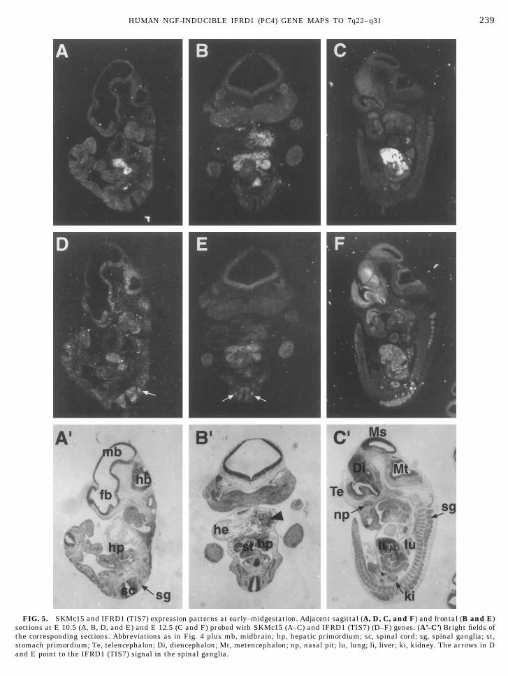
FIG. 5. SKMc15 and IFRD1 (TIS7) expression patterns at early–midgestation. Adjacent sagittal (A, D, C, and F) and frontal (B and E)sections at E 10.5 (A, B, D, and E) and E 12.5 (C and F) probed with SKMc15 (A–C) and IFRD1 (TIS7) (D–F) genes. (A’–C’) Bright fields ofthe corresponding sections. Abbreviations as in Fig. 4 plus mb, midbrain; hp, hepatic primordium; sc, spinal cord; sg, spinal ganglia; st,stomach primordium; Te, telencephalon; Di, diencephalon; Mt, metencephalon; np, nasal pit; lu, lung; li, liver; ki, kidney. The arrows in Dand E point to the IFRD1 (TIS7) signal in the spinal ganglia.
239HUMAN NGF-INDUCIBLE IFRD1 (PC4) GENE MAPS TO 7q22–q31
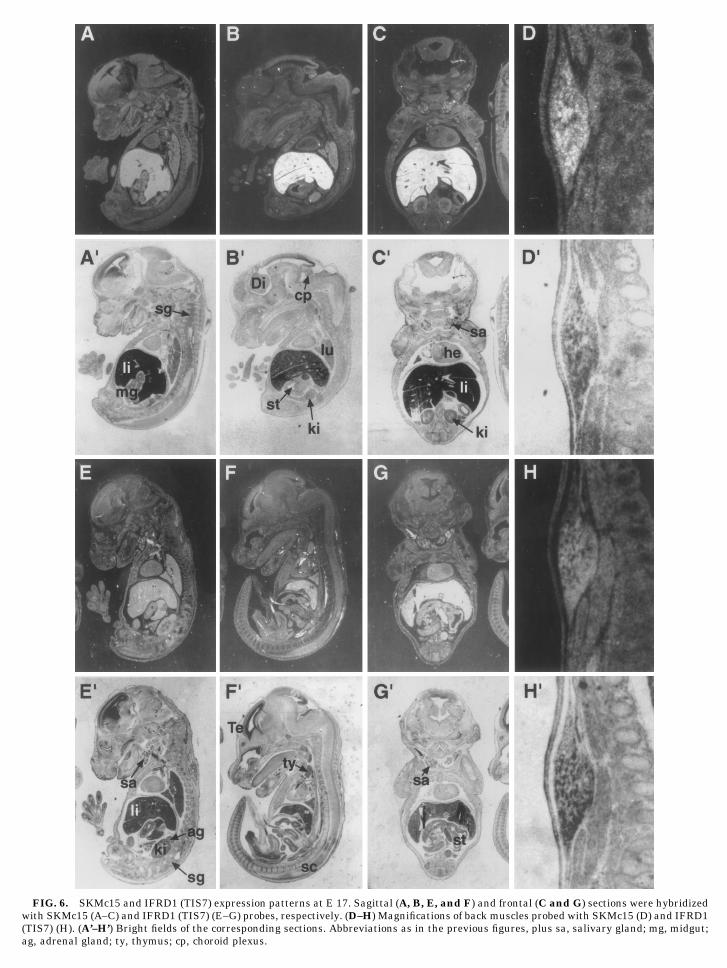
FIG. 6. SKMc15 and IFRD1 (TIS7) expression patterns at E 17. Sagittal (A, B, E, and F) and frontal (C and G) sections were hybridizedwith SKMc15 (A–C) and IFRD1 (TIS7) (E–G) probes, respectively. (D–H) Magnifications of back muscles probed with SKMc15 (D) and IFRD1(TIS7) (H). (A’–H’) Bright fields of the corresponding sections. Abbreviations as in the previous figures, plus sa, salivary gland; mg, midgut;ag, adrenal gland; ty, thymus; cp, choroid plexus.
Chromosomal Localization of PC4
Human EST clone 52803 is one of many EST se-quences homologous to IFRD1 present in the Unigenedatabase, at cluster 7879. STS 32586 (Genethon) andstSG401 (Sanger Centre) have been generated from 39EST sequences belonging to the same cluster. Radia-tion hybrid screening results for these STSs, generatedby both Genethon and the Sanger Centre, indicatedthat the human IFRD1 (PC4) gene maps betweenmarkers D7S523 and D7S486, corresponding to 7q22–q31 (cytogenetic position 1.25–1.39 M, as by Chumakovet al., 1995).
DISCUSSION
The human cDNA whose cloning and sequencing isreported here encodes a protein having 90% amino acididentity to the rat and mouse IFRD1 (PC4 and TIS7)proteins. It can thus be assumed that this cDNA en-codes human IFRD1. Sequence and tissue distributionof IFRD1 appear to be highly conserved between spe-cies, suggesting that the function of IFRD1 is con-served during evolution. The SKMc15 gene, in virtue ofits 47% identity to the IFRD1 sequences, appears to bethe second member of a novel family of genes. Althoughthe molecular function of IFRD1 and SKMc15 is stillunknown, hypotheses on their functional role are sug-gested from our analysis of expression. In the firstplace, both genes have high expression in skeletal andcardiac muscle of the adult human, as seen by North-ern analysis. Furthermore, both genes are expressed inthe embryonic skeletal muscle, attaining an apprecia-ble level at the late gestation period. This fact, in linewith the requirement for IFRD1 (PC4) observed in theprocess of muscle differentiation (Guardavaccaro et al.,1995), suggests that both proteins might share a com-mon function(s), possibly encoded by the three regionsof higher conservation. The same type of suggestioncomes from the observation that during developmentboth genes have qualitatively similar patterns, withearly appearance in the embryo and extraembryonictissues since gastrulation, followed by a common pat-tern of expression in restricted structures at midgesta-tion (such as CNS, kidney, and lung primordia) orubiquitous expression at late gestation. However, be-tween the two genes there are strong and specific dif-ferences in the level of expression, since IFRD1 (TIS7)is expressed much more than SKMc15 in differentiat-ing tissues, such as nervous tissues, kidney, and lung,while SKMc15 is highly expressed in the initial stagesof embryogenesis and in the hepatic primordium.These findings agree with our previous analysis ofIFRD1 (PC4) expression within the CNS, whichshowed that IFRD1 (PC4) was present not only in theventricular zone of the neural tube at the moment ofneuroblast proliferation, but also in the surroundingmantle zone where the differentiated neuron migrates(Iacopetti et al., 1996). Considering all together, it is
plausible to imagine a role for IFRD1 in terminal dif-ferentiation of different cellular districts. This hypoth-esis is consistent with our previous observation of nu-clear translocation of the IFRD1 (PC4) protein duringneuronal differentiation (Guardavaccaro et al., 1994),which suggests a role for IFRD1 (PC4) in this process.Similarly, our present data are very suggestive of aninvolvement of SKMc15 in the early differentiation ofhematic precursors. It is interesting that SKMc15, iso-lated as a putative tumor suppressor gene, has beenexcluded from being a such candidate, based on muta-tional analysis (Latif et al., 1997). Furthermore, wehave observed that IFRD1 does not affect cellular pro-liferation (D.Guardavaccaro, unpublished results). It istherefore likely that SKMc15 and IFRD1 are not im-pinging on cell cycle progression. Certainly furtheranalyses, as well as functional comparisons betweenIFRD1 and SKMc15, might help to assign a functionalrole to this gene family. Concerning the similarity be-tween IFRD1 (PC4) and interferon-g that we have pre-viously observed (Tirone and Shooter, 1989), a compar-ison of interferon-g proteins of different species withrat and human IFRD1 (PC4) still bespeaks commonancestry of these sequences (R. Doolittle, San Diego,pers. commun., December 1996). The interspecies con-servation of the interferon protein, one of the fastestchanging mammalian proteins, is, however, lower thanobserved for IFRD1, indicating different functional as-pects and different positive selection for change. Acomparison of the three-dimensional structures willthus be necessary for a better understanding of theevolutionary relation between IFRD1 and interferon-gproteins. Concerning the chromosomal localization ofIFRD1, which evidently differs from that of SKMc15(assigned to chromosome 3p21; Latif et al., 1997), wecannot at present exclude a functional correlation ofIFRD1 with any of the diseases mapping to the IFRD1locus. Further studies will therefore be necessary toascertain whether IFRD1 is involved in diseases re-lated to development and/or cellular differentiation.
ACKNOWLEDGMENTS
We are grateful to Russel Doolittle for the computer-assisted anal-ysis of the human PC4 protein and to Giuseppe Borsani and AndreaBallabio for critically reading the paper. We thank Sergio Cattadorifor technical assistance. This work was supported by a Telethongrant to F.T. and to A.S. and by an AIRC grant to A.S.; P.B. was therecipient of a fellowship from Telethon. This work was also carriedout under a research contract with NEFAC, Pomezia, Italy, withinthe Neurobiological Systems National Research Plan of the Minis-tero dell’Universita e della Ricerca Scientifica e Tecnologica.
Note added in proof. After submission of the manuscript, a reportappeared indicating that the locus linked to a severe genetic speechand language disorder colocalizes with the chromosomal regionwhere IFRD1/PC4 gene maps, thus making IFRD1/PC4 a potentialcandidate responsible for such disease [Fisher, S. E., Vargha-Khadem, F., Watkins, K. E., Monaco, A. P., and Pembrey, M. E.(1998). Localisation of a gene implicated in a severe speech andlanguage disorder. Nat. Genet. 18: 168–170.]
241HUMAN NGF-INDUCIBLE IFRD1 (PC4) GENE MAPS TO 7q22–q31
REFERENCES
Altschul, S. F., Gish, W., Miller, W., Myers, E. W., and Lipman, D. J.(1990). Basic local alignment search tool. J. Mol. Biol. 215: 403–410.
Bairoch, A. (1990). ‘‘Dictionary of Protein Size and Patterns,’’ Ph.D.thesis, Universite de Geneve.
Boguski, M. S., Lowe, T. M., and Tolstoshev, C. M. (1993). dbEST—Database for ‘‘expressed sequence tags.’’ Nat. Genet. 4: 332–333.
Bradbury, A., Possenti, R., Shooter, E. M., and Tirone, F. (1991).Molecular cloning of PC3, a putatively secreted protein whosemRNA is induced by nerve growth factor and depolarization. Proc.Natl. Acad. Sci. USA 88: 3353–3357.
Chumakov, I. M., Rigault, P., Le Gall, I., Bellanne-Chantelot, C.,Billault, A., Guillou, S., Soularue, P., Guasconi, G., Poullier, E.,Gros, I., et al. (1995). A YAC contig map of the human genome.Nature 377: 175–297.
Doolittle, R. F., and Feng, D. F. (1990). Nearest neighbor procedurefor relating progressively aligned amino acid sequences. MethodsEnzymol. 183: 659–669.
Feinberg, A. P., and Vogelstein, B. (1983). A technique for radiola-beling DNA restriction endonuclease fragments to high specificactivity. Anal. Biochem. 132: 6–13.
Greene, L. A., and Tischler, A.S. (1976). Establishment of a norad-renergic clonal line of rat adrenal pheochromocytoma cells whichrespond to nerve growth factor. Proc. Natl. Acad. Sci. USA 73:2424–2428.
Guardavaccaro, D., Ciotti, M. T., Schafer, B. W., Montagnoli, A., andTirone, F. (1995). Inhibition of differentiation in myoblasts de-prived of the interferon-related protein PC4. Cell Growth Differ. 6:159–169.
Guardavaccaro, D., Montagnoli, A., Ciotti, M. T., Lotti, L., Di Laz-zaro, C., Torrisi, M-R., Gatti, A., and Tirone, F. (1994). Nervegrowth factor regulates the sub-cellular localization of the nervegrowth factor-inducible protein PC4 in PC12 cells. J. Neurosci.Res. 37: 660–674.
Hollenberg, S. M., Cheng, P. F., and Weintraub, H. (1993). Use of aconditional MyoD transcription factor in studies of MyoD trans-activation and muscle determination. Proc. Natl. Acad. Sci. USA90: 8028–8032.
Iacopetti, P., Barsacchi, G., Tirone, F., and Cremisi, F. (1996). Ex-pression of the PC4 gene in the developing rat nervous system.Brain Res. 707: 293–297.
Kozak, M. (1984). Compilation and analysis of sequences upstreamfrom the translational start site in eukaryotic mRNAs. NucleicAcids Res. 12: 857–872.
Latif, F., Duh, F-M., Bader, S., Sekido, Y., Geil, H-L. L., Zbar, B.,Minna, J. D., and Lerman, M. (1997). The human homolog of therodent immediate early response genes, PC4 and Tis7, resides inthe lung cancer tumor suppressor gene region on chromosome3p21. Hum. Genet. 99: 334–341.
Theiler, K. (1989). The house mouse. In ‘‘Atlas of Embryonic Devel-opment,’’ Springer-Verlag, Zurich.
Tirone, F., and Shooter, E. M. (1989). Early gene regulation by nervegrowth factor: Induction of an interferon related gene. Proc. Natl.Acad. Sci. USA 86: 2088–2092.
Varnum, B. C., Lim, R. W., and Herschman, H. R. (1989). Charac-terization of TIS 7, a gene induced in Swiss 3T3 cells by the tumorpromoter tetradecanoyl phorbol acetate. Oncogene 4: 1263–1265.
Wei, M. H., Latif, F., Bader, S., Kashuba, V., Chen, J. Y., Duh, F. M.,Sekido, Y., Lee, C. C., Geil, L., Kuzmin, I., Zabarovsky, E., Klein,G., Zbar, B., Minna, J. D., and Lerman, M. I. (1996). Constructionof a 600-kilobase cosmid clone contig and generation of a transcrip-tional map surrounding the lung cancer tumor suppressor gene(TSG) locus on human chromosome 3p21.3: Progress toward theisolation of a lung cancer TSG. Cancer Res. 56: 1487–1492.
Wilkinson, D. G. (1992). Whole mount in situ hybridization of verte-brate embryos. In ‘‘In situ Hybridization: A Practical Approach,’’IRL Press, Oxford.
Wilkinson, D. G., and Green, J. (1990). In ‘‘Postimplantation MouseEmbryos: A Practical Approach,’’ (D. Rickwood and D. L. Cockroft,Eds.), pp. 155–171, IRL Press, Oxford.
242 BUANNE ET AL.