Embed Size (px)
Citation preview
Proc. Nati. Acad. Sci. USAVol. 84, pp. 7000-7004, October 1987Biochemistry
Cloning and expression of T4 DNA polymerase(DNA replication/expression vector/overproduced protein)
TSUNG-CHUNG LIN, JOHN RUSH, ELEANOR K. SPICER, AND WILLIAM H. KONIGSBERGDepartment of Molecular Biophysics and Biochemistry, Yale University School of Medicine, New Haven, CT 06510
Communicated by Edward A. Adelberg, June 8, 1987
ABSTRACT The structural gene coding for bacteriophageT4 DNA polymerase (gene 43) has been cloned into inducibleplasmid vectors, which provide a source for obtaining largeamounts of this enzyme after induction. The T4 DNA poly-merase produced in this fashion was purified by an innovativethree-step procedure and was fully active.
Studies carried out with bacteriophage T4 have providedmany important insights about the enzymology and mecha-nism of viral DNA replication that will continue to aid in ourunderstanding ofthis process in other organisms (for reviews,see refs. 1-3). One of the advantages of using T4 as a modelsystem for studying DNA replication is that all ofthe proteinsrequired for strand elongation are phage encoded. This hasfacilitated the construction of in vitro systems for primerextension that have many of the characteristics of DNAreplication in vivo, such as fidelity, processivity, and rates ofnucleotide addition (4-6). Although 11 T4-encoded proteinshave been identified as participating in the formation andmovement of DNA replication forks, only 5 of these arenecessary to reconstitute a "core" system in which leading-,but not lagging-, strand synthesis occurs (1, 7). These 5proteins, which form a functional complex at replicationforks, are theDNA polymerase (43P), a single-stranded DNAbinding protein (32P), and the DNA polymerase accessoryproteins (44P/62P and 45P). The genes for 4 of these proteinsare clustered in one region of the T4 phage genome (Fig. 1).Although rapid progress has been made in defining the
function of each of these proteins, certain important prob-lems remain to be solved. These include the location of activesites in proteins with enzymatic activities, the identification
C of interfaces among interacting proteins, and the solution ofthe three-dimensional structure of the replication complex athigh resolution.
In this paper we report cloning of the gene for T4 DNApolymerase (gene 43) and a procedure that has permittedrapid purification of large quantities of the DNA polymerasein a nearly homogenous form, free of contaminating endo-and exonucleases. Since we have also constructed plasmidsthat produce large quantities of the other four proteins of thecore system (ref. 9 and T.-C.L., J.R., and W.H.K., unpub-lished data), our ability to obtain large quantities of T4 DNApolymerase now allows us to reconstruct a DNA replicationcomplex for high-resolution structural studies. This work,together with the extensive genetic and biochemical studiesalready completed (1), should provide the most precisedescription of DNA replication available in any system.
MATERIALS AND METHODSBacterial Strains. Escherichia coli strain 71-18 (K-12,
A[lac, pro], F' lacIQ, ZAM15 pro') was used when propa-gating M13 vectors and when strong repression of the lac or
tac promoters was necessary for constructing certain plas-mids. Strain RR1 [F-, hsdS20 (r-, m-), ara-14, proA2, lac YJ,galK2, rpsL20 (Sm9, xyl-5, mtl-i, supE44, X-] was used formaintaining plasmid pTL43W, and strain HB101 (as RR1except recA13) was used for all other plasmid constructions.
Cloning Vectors. Vectors M13mp8, -mpl9, pUC9, andpUC18 (10) were purchased from Bethesda Research Labo-ratories; plasmid ptacl2 was the kind gift of J. Brosius(Columbia University, New York) (11). Plasmid pGW7,phage A NM761-4 (12, 13), and T4GT7 (14) were the generousgifts of G. Wilson, New England Biolabs.
Cloning Procedures. Restriction endonucleases were pur-chased from New England Biolabs, Boehringer Mannheim,or Bethesda Research Laboratories. The Xho I linker (5'CCTCGAGG 3') was obtained from Pharmacia. The BamHIlinker (5' CCGGATCCGG 3'), Klenow fragment, andpolynucleotide kinase were purchased from Bethesda Re-search Laboratories. Linkers were phosphorylated withpolynucleotide kinase before use.DNA fragments, purified by electrophoresis on agarose
gels, were adsorbed to DEAE membranes (15) and elutedwith buffers containing high concentrations of salt. DEAE-coupled membranes, NA-45, were purchased from Schlei-cher and Schuell and were used according to the instructionsof the manufacturer.
Induction of Gene Expression. Luria-Bertani (LB) mediumsupplemented with ampicillin (100 ,ug/ml) was inoculatedwith frozen stock of E. coli harboring plasmids pTL43W orpTL43Q, grown overnight at 30°C, and then used to inoculatelarger cultures.For analytical experiments (volumes <10 ml), cultures
harboring plasmid pTL43W, grown at 30°C in the mediumdescribed above, were induced by adding an equal volume ofmedium warmed to 54°C, which rapidly brought the temper-ature to 42°C. For larger scale experiments, it was moreconvenient to induce by adding small amounts of boilingmedium with vigorous stirring and monitoring the tempera-ture until it reached 40 or 42°C. Cultures harboring pTL43Qwere induced after growth at 30, 40, or 42°C by adding 3 ,ulof 0.1 M isopropyl ,B-D-thiogalactopyranoside per ml culture.Care was taken to insure that cultures were induced while stillin the logarithmic phase of growth.
Cell Lysis. Cells were lysed according to the procedure ofBurgess and Jendrisak (16), modified as follows. Cells wereresuspended in 50 mM TrisHCI, pH 8.0/2 mM EDTA/0.1mM dithiothreitol/1 mM 2-mercaptoethanol at a density of 50g of cells per liter. Lysozyme was added to 20 ,ug/ml, andphenylmethylsulfonyl fluoride was added to 1 mM. After 30min of vigorous stirring at 25°C, sodium deoxycholate wasadded to 0.05%, and phenylmethylsulfonyl fluoride wasadded again to a final concentration of 1.5 mM. The lysatewas stirred at 25°C for 10 min, placed in ice water, and stirredfor 20 min more. The lysate was then sonicated with aBranson Heat Systems sonicator. Cellular debris was re-moved by centrifugation in a Beckman JA10 rotor at 8000 rpmfor 20 min at 4°C.
7000
The publication costs of this article were defrayed in part by page chargepayment. This article must therefore be hereby marked "advertisement"in accordance with 18 U.S.C. §1734 solely to indicate this fact.

Proc. Natl. Acad. Sci. USA 84 (1987) 7001
g43 regA g62 g44 g45
R H R
26.10 26.68 27.45
HRX A P H
3011
H
32.30
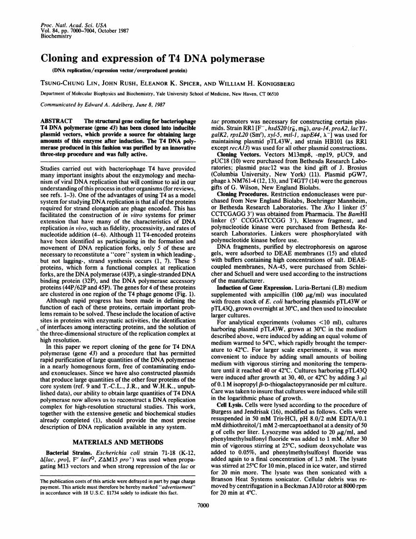
FIG. 1. Restriction map of gene 43 and genes upstream ofgene 43.The promoter (v) and ribosome binding site (v) of gene 43 areindicated. Restriction sites are EcoRI (R), Hindl1 (H), Xho I (X),Ava 1 (A), and Pst I (P). The numbers under certain restriction sitesindicate T4 map units (8).
Enzyme Purification and Enzyme Assays. 43P was purifiedby a slight modification of the method of Nossal (17) for initialenzymatic characterization. DNA polymerase and 3'-to-5'exonuclease activities were assayed according to the methodof Nossal (17), except that at the end of the polymerase assaytritiated deoxynucleotide triphosphate substrates were sep-arated from tritiated product DNA by adsorption to DE 81filters, essentially as described by Brutlag and Kornberg (18).For the rapid purification procedure, single-stranded DNA-cellulose chromatography was performed as described byNossal (17), and affinity chromatography using immobilizedT4 gene 32 protein was performed as described by Formosaet al. (19). NaDodSO4/polyacrylamide gel electrophoresiswas performed according to the method of Laemmli (20).
RESULTSPlasmid Construction Strategy. Gene 43 was cloned as two
separate pieces that were joined together before transfer toexpression vectors. Attempts to clone gene 43 in its entiretyin a single step were not successful, presumably because aputative "lethal" sequence located immediately downstreamof gene 43 interfered with cloning that region of DNA. Allknown restriction sites downstream of gene 43 also turnedout to be downstream of this lethal sequence, so that everyrestriction fragment containing the 3' end of gene 43 alsoincluded this lethal site. This compelled us to revise our initialcloning strategy, which had been to try to clone full-lengthgene 43 in one step, and to clone gene 43 as two fragments thatwere later joined to produce the intact gene.
In addition, the promoter of gene 43 was removed beforejoining the separately cloned segments of gene 43. This wasdone for two reasons. First, the promoter was removed as aprecaution to prevent uncontrolled expression of gene 43,because synthesis of T4 DNA polymerase above a certainlevel is likely to be detrimental to the host cell. Our suspicionthat gene 43 expression would be detrimental to the host wasconfirmed in later experiments, as all attempts to replace thepromoter upstream of intact, reassembled gene 43 failed toproduce stable recombinants. Second, the promoter wasremoved to prevent 43P from repressing its own synthesisfrom expression vectors. Genetic studies have shown that43P regulates the transcription of its gene (21, 22), althoughthe requirements of this autoregulation have not been pre-cisely defined. For these reasons, we decided to remove thegene 43 promoter, leaving its ribosome binding site intact, asan early step in the construction of a full-length gene 43 clone.
Subcloning and Manipulation of the 5' End of Gene 43. The5' region of gene 43 was subcloned from a X-T4 recombinant,X NM761-4 (shown at the top of Fig. 2), originally constructedfrom a partial HindIl digest of cytosine-containing T4 DNAby Wilson and Murray (12, 13). This recombinant phagecontained a 5.6-kilobase-pair (kbp) fragment of T4 DNA(26.68 to 32.30 map units, Fig. 1) and included the codingregions for genes 45, 44, 62, and regA, as well as the largestsegment of gene 43 that had been cloned from T4 DNA as arestriction fragment. The cloning strategy was to transfer arestriction fragment containing gene 43 from X NM761-4 intoa plasmid vector, to remove the genes upstream of gene 43
X761 -4P H P H
N-g43 regA g62 g44 g45- 39kb
Ml 3/KR4C
c-g43
B
N B
Amp (ApUC1
AmpT
AmpXU8D
H
FIG. 2. Separate cloning of the 5' and 3' ends of gene 43 andassembly of intact gene 43. Cloning the 5' end of gene 43 into pUC9(arrow A). A 3.9-kbp Pst I fragment from X NM761-4, containing the5' end of gene 43, was cloned into the Pst I site of pUC9 to give theplasmid p43NR. Removal of the gene 43 promoter and upstream T4genes (arrow B). The gene 43 promoter was removed from p43NR bydigestion with BamHI, followed by a partial Ava I digestion that alsoremoved the 3' end of gene 62 and the regA gene. The 5' overhangmade by Ava I treatment was filled in with Klenow fragment. BamHIlinkers were attached to the resulting blunt ends. After digestion withBamHI, the plasmid was recircularized with DNA ligase to give theplasmid p43N. Construction of the vector pTL18X (arrow C). TheNde I site of pUC18 was changed to an Xho I site. pUC18 wasdigested with Nde I, and the resulting 5' overhang was filled in withKlenow fragment. Xho I linkers were attached. After digestion withXho I, ligation and recircularization gave the plasmid pTL18X.Cloning the 3' end of gene 43 into pTL18X (arrow D). The replicativeform of M13/KR4C was digested with EcoRI, the EcoRI overhangwas filled in with Klenow fragment, and Xho I linkers were attached.The M13 recombinant was then treated with Xho 1 and BamHI, andthe fragment containing the 3' end of gene 43 was isolated. pTL18Xwas also treated with Xho I and BamHI and ligated with the purifiedM13/KR4C fragment to give p43C. Assembly of gene 43 from theseparately cloned 5' and 3' ends (arrow E). p43N and p43C weretreated with BamHl and HindIll, but a partial HindIII digest wasperformed on p43N to prevent cleavage at HindIII sites within gene43. Replacement of the smaller BamHI-HindIII fragment in p43C,which contains sequences already present in the fragment fromp43N, with the BamHI-HindIII (partial) gene 43-containing fragmentfrom p43N gave pTL43, the full-length clone. Restriction sites areAva I (A), BamHI (B), HindIII (H), Nde I (N), and Xho I (X).
along with the gene 43 promoter, and then to reassembleintact gene 43 by adding the separately cloned 3' end.Towards that end, a 3.9-kbp Pst I fragment, containing the 3'end of gene 62, all of regA, and the 5' end of gene 43, wassubcloned into pUC9, generating the plasmid p43NR (Fig. 2,
Biochemistry: Lin et al.
Proc. Natl. Acad. Sci. USA 84 (1987)
arrow A). Further manipulation of p43NR to remove the T4genes upstream of gene 43 and the gene 43 promoter gave theplasmid p43N (Fig. 2, arrow B). It was estimated that thisplasmid contained at least 95% ofgene 43, based on the lengthof the insert and the size of T4 DNA polymerase determinedby NaDodSO4/PAGE. Cloning the 3' end of gene 43 allowedus to deduce the precise size ofgene 43 and showed that p43Nlacked only 21 nucleotides from the 3' end of gene 43.
Cloning the 3' End of Gene 43. In our first attempt to clonethe segment of gene 43, which was not present in X NM761-4,a HindIII-EcoRI fragment was isolated from cytosine-con-taining T4GT7 DNA (14). This fragment spanned T4 mapunits 26.68 to 26.10, a region of the T4 genome that containsthe 3' end of gene 43 (Fig. 1). The HindIII site at map unit26.68 is the site where truncated gene 43 is joined to Xsequences in X NM761-4. Repeated attempts to clone thisHindIII-EcoRI fragment into a variety of vectors, e.g., pUC8and M13mp8, failed, although other T4 DNA fragmentsderived from the same digest were readily cloned. Weassumed that the fragment containing the 3' end of gene 43could not be readily cloned into these vectors because itcontained a nucleotide sequence that interfered with repli-cation of the plasmid or with survival of the E. coli host.Another attempt to clone the 3' end of gene 43 from arestriction fragment was made by digesting the HindIll-EcoRI fragment with Msp Ito produce a 280-bp HindIII-MspI fragment. This smaller fragment also could not be cloned.Thus, it appears that a lethal sequence exists within a 280-bpregion downstream of the HindIII site at 26.68 map units.
Since no other restriction sites were known to existbetween the Msp I and HindIII sites, we separated the 3' endof gene 43 from the lethal sequence by sonicating cytosine-containing T4 DNA. Fragments with an average length of 400bp were used to produce a T4-Ml3mp8 library that wasscreened with a 32P-labeled EcoRI-HindIII restriction frag-ment (map units 27.45 to 26.68) isolated from p43N. DNAsfrom plaques that hybridized with the probe were sequencedto confirm that they contained the probe site. One of thepositive phage isolates, M13/KR4C, contained the probesequence upstream of the HindIII site and 102 bp down-stream from the HindII'site (E.K.S., J.R., C. Fung, J. D.Karam, and W.H.K., unpublished data). The downstreamsequence had a 21-bp open reading frame followed by twotandem in-frame translation termination codons. This 27-bpregion, corresponding to the 3' end of gene 43 with its tandemtermination codons, was cloned into a derivative of pUC18,as described below.Assembly of Gene 43 and Cloning into Expression Vectors.
To join the two portions of gene 43, it was necessary to createa convenient restriction site by inserting a linker downstreamof the 3' end of gene 43. We chose an Xho I linker,anticipating the transfer of gene 43 into the compatible Sal Isite of our overproduction vectors in a later step. Xho Ilinkers were also inserted in pUC18 to form the vectorpTL18X (Fig. 2, arrow C). The 3' end of gene 43 was movedfrom M13/KR4C into pTL18X to produce the plasmid p43C(Fig. 2, arrow D). The 5' end of gene 43 was then transferredfrom p43N to p43C,joining the two segments ofgene 43 at theHindIII site, to give pTL43, containing the entire gene 43(Fig. 2, arrow E).Gene 43, reassembled in pTL43, was then cloned into two
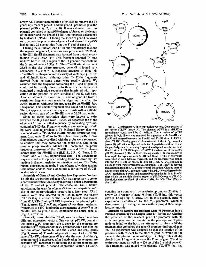
different expression vectors. One vector, pTL9W, containsthe strong X PL promoter, the X gene for the temperaturesensitive c1857 repressor ofthe PL promoter, the X gene for theantitermination protein N, and the X rexA and rexB genes(Fig.' 3, arrow A). Transfer of gene 43 from pTL43 to pTL9Wgave pTL43W, where gene 43 is under the control of the PLpromoter and is expressed after inactivating the temperaturesensitive c1857 repressor by elevating the culture temperature(Fig. 3, arrow B). A second expression vector, pTL19Q,
mp19
M.C.S.
SB
D
g43
FIG. 3. Cloning gene 43 into expression vectors. Construction ofthe vector pTL9W (arrow A). The plasmid pGW7 is a pBR322-Xrecombinant constructed by G. Wilson. The X region of pGW7(shown in bold lines) was removed by digestion with BamHI andEcoRI and inserted between the BamHI and EcoRI sites of pUC9 togive pTL9W. Cloning gene 43 downstream of the X PL promoter(arrow B). pTL43 was digested with Xho I (partial) and BamHI, andthe purified gene 43-containing fragment was ligated into the Sal I andBamHI sites ofpTL9W to give pTL43W. Construction of the vectorpTL19Q (arrow C). The multiple cloning site (M.C.S.) was removedfrom mpl9 by digestion with EcoRI and HindIII. The 5' overhangswere filled in with Klenow fragment, and the fragment was clonedinto the Pvu II site of ptacl2 to give pTL19Q. All P,a-containingplasmids were transformed into E. coli strain 71-18 (lacPQ) to represstranscription from the Ptac promoter until induction. Cloning gene 43downstream of the Ptac promoter (arrow D). pTL43 was digested withXho I (partial) and BamHI and inserted between the Sal I and BamHIsites within the multiple cloning region of pTL19Q to give pTL43Q.Restriction sites are EcoRI (R), BamHI (B), Sal I (S), Xho I (X), andPvu II (P).
contains the strong tac (trp-lac) fusion promoter (23) (Fig. 3,arrow C). Transfer of gene 43 from pTL43 into this vectorgave pTL43Q (Fig. 3, arrow D). In this plasmid, gene 43expression is controlled by the Ptac promoter, which isderepressed by treating cultures with isopropyl 8l-D-thioga-lactopyranoside.
Attempts to Restore the Resident Gene 43 Promoter into aPlasmid Containing Full-Length Gene 43. To find out whetherthe presence of the resident gene 43 promoter with itsstructural gene was detrimental to the propagation of plas-mids or lethal to the host, we attempted to place a 700-bpfragment that contained the gene 43 promoter in front ofgene43. The experiment was designed so that the location of thepromoter with respect to the start of gene 43 would be thesame in the plasmid as in wild-type T4 phage. This 700-bpBamHI-Xho I fragment contains the 3' end ofgene 62 and theentire regA gene as well as -220 bp of the 5' end of gene 43.This fragment was mixed with plasmid pTL43W that had
7002 Biochemistry: Lin et al.

Proc. NatL. Acad. Sci. USA 84 (1987) 7003
been digested with BamHI and Xho I, then ligated success-fully as judged by the banding pattern on agarose gels.Transformation was attempted with this ligation mixture, butno colonies harboring plasmids with the desired constructionwere obtained. To show that technical problems were notresponsible for this negative result, we demonstrated that asimilar-size BamHI-Xho I control fragment, when mixedwith the BamHI-Xho I promoter fragment in a ligationreaction, produced transformants containing only the controlfragment. The presence of regA under PL control in thisconstruction was probably not responsible for the lack ofdesired transformants, since other plasmids containing regAand parts of gene 43 under PL control have been constructed(24). For these reasons, we think the most likely cause for thefailure to obtain plasmids with intact gene 43 and its promoteris that the gene 43 promoter is recognized by the host RNApolymerase and that 43P is produced in sufficient amounts tobe harmful to the cell.
Production and Purification of Large Amounts of Soluble43P. When cells harboring the plasmid pTL43W were in-duced by shifting the culture temperature from 300C to 420C,large amounts of 43P were produced as insoluble aggregates(Fig. 4A). Only high concentrations of protein-denaturingreagents were effective in releasing 43P from the aggregates(data not shown). However, when the induction temperaturewas reduced to 40'C, large amounts of soluble 43P wereproduced (Fig. 4B). When the induction temperature waslowered further, to 380C, very little if any 43P was produced(data not shown). Induction of cells harboring pTL43Q withisopropyl 8-D-thiogalactopyranoside at 40'C and 420Cshowed a similar dependence of 43P solubility on culturetemperature (Fig. 5). However, in contrast to pTL43W,induction ofpTL43Q produced a large quantity of soluble 43Pat temperatures as low as 30TC.A simplified purification procedure allowed rapid process-
ing of large quantities of extracts containing 43P (J.R. andW.H.K., unpublished data). Briefly, 43P was precipitatedfrom the cell lysate with 0.2-0.3% polyethyleneimine andextracted from the polyethyleneimine pellet with 0.5M NaCl.43P was purified further by addition of ammonium sulfate to55% saturation at 40C. Affinity chromatography on single-stranded DNA-cellulose gave a product that was at least 95%pure. Further chromatography using immobilized T4 gene 32protein produced nearly homogenous 43P, free of contami-nating endonuclease and 5'-to-3' exonuclease activities.
A
04k-
15 30 45 60 120 180 min
(n pS p s p S p S p s p s W
1tnU _ _ -_ _ _ _ _
67 kd- - iw
43 kd -
pTL43W PTL43Q- 30° 400 42* 3Q0 4Q0 420 XcPp s S S ps p p scP )
kd
64 -
201-
_4 b
FIG. 5. NaDodSO4/polyacrylamide gel analysis of cell lysatepellets (p) and supernatants (s) 2 hr after induction at 30, 40, or 42°C.Cells harbori~ng pTL43W or pTL43Q were induced. The arrowindicates the position of 43P. kd, kDa; stds, standard molecular sizemarkers.
Characterization of 43P. 43P purified from induced cellsharboring pTL43W and 43P from T4 infected cells wereassayed for DNA polymerase and 3'-to-5' exonuclease ac-tivities (J.R. and W.H.K., unpublished data). We found thatcloned 43P had about twice the specific activity for DNApolymerization as the 43P obtained from T4-infected cellsthat was used as a standard for wild-type levels of enzymaticactivity. The 3'-to-5' exonuclease specific activities of bothproteins were identical.
DISCUSSION
A number of reasons can be advanced to account forsituations where attempts to clone structural genes ha~vefailed. Among the most likely explanations are that the geneproduct may interfere with the viability ofthe host cell or withreplication of the plasmktor that a nucleotide sequence itselfinterferes with propagation of the plasmid. In our attempts toclone gene 43, both of these explanations appeared to bevalid. First, it was necessary to remove the resident promoterof gene 43 and to replace it with one that could be tightlycontrolled. Subsequent attempts to insert the gene 43 pro-
B 15 30 45 60 120
cop s P S P s p s p S94 kd -
k...._67 kd- m% - _ _ _
180 min0
p S :.
W -
l-
43kd-
3Okdr30kd- %
20.1 kd- - 20.i kd
FIG. 4. NaDodSO4/polyacrylamide gel analysis ofpTL43W cell lysate pellets (p) and supernatants (s) at various times after induction at 42°C(A) or at 40°C (B). Two large cultures were made from a culture of cells harboring pTL43W grown at 30°C. The cultures were brought to 40°Cor to 42°C. At the times indicated, aliquots were withdrawn to tubes containing chloramphenicol (at a final concentration of 100 ,ug/ml), storedon ice until all time points were sampled, and then lysed after correcting for differences in cell density. The arrow indicates the position of 43P.kd, kDa; stds, standard molecular size markers.
Biochemistry: Lin et al.

Proc. Natl. Acad. Sci. USA 84 (1987)
moter in the position occupied in wild-type T4 failed to giveany stable recombinants containing the gene 43 promoterinsert. Second, we had to revise our cloning strategy, so thatwe could remove a putative lethal sequence downstreamfrom the 3' end ofgene 43. This lethal sequence resides within280 bp of the gene 43 translation termination codons and maybe a strong early promoter, as indicated by the studies ofKutter and Ruger (8).Our evidence for having obtained clones containing the
correct 3' end of gene 43 is based on three findings: (i) theclones containing the 3' end of gene 43 and the clonescontaining nucleotides upstream from the most distal HindIIIsite in gene 43 share a large, overlapping region of DNAsequence; (ii) we isolated two independent clones containingthe 3' end of gene 43, and the DNA sequences of bothmatched the DNA sequence that was obtained independentlyfor this region of the T4 genome by W. Ruger (personalcommunication), and (iii) carboxypeptidase Y releases thesame amino acid residues from cloned 43P and from 43Pobtained from T4-infected cells.
Cells harboring pTL43W or pTL43Q produce 43P to -10%of the total cellular protein, which is -1000-fold greater thanthe amounts produced by cells infected with wild-type T4phage. This increased level of expression simplifies thepurification scheme enormously. The cloned 43P had thesame specific activity for the 3'-to-5' exonuclease function aswild-type 43P but had twice the specific activity for DNApolymerization. We also found that the cloned 43P had anasparagine at position 214, whereas the 43P from T4-infectedcells contained serine at this position (J.R. and W.H.K.,unpublished data). This alteration may account for thedifference in specific polymerizing activity. After examininga number of T4 DNA polymerases from wild-type strains bypeptide mapping, we found that some had asparagine atposition 214, whereas others had serine at this position (J.R.and W.H.K., unpublished data). For this reason, we believewe have actually cloned one of the forms of gene 43 presentin existing wild-type T4 strains.
It has been observed that some proteins that are normallysoluble form insoluble aggregates when they are produced inlarge amounts in E. coli (25-28). This situation occurs with43P when it is synthesized in cultures at temperatures of420Cusing either pTL43W or pTL43Q. One explanation for theinsolubility of overexpressed 43P at elevated temperatures isthat there is a temperature-dependent step in the normalfolding pathway that becomes rate-limiting at high tempera-tures, so that 43P remains in a partially unfolded state for alonger time than at lower temperatures. This could causeaggregation of partially unfolded 43P through interactionsbetween hydrophobic regions not yet buried in the core of theprotein. By lowering the induction temperature only 20C,most of the 43P produced remained soluble.As described above, we have cloned authentic, full-length
gene 43 and have overproduced and purified a large quantityof enzymatically active 43P. Together with the plasmids wehave constructed to overproduce the other T4 DNA replica-tion core proteins (ref. 9 and T.-C.L., J.R., and W.H.K.,unpublished data), it will now be possible to initiate high-
resolution structural studies on these proteins and theircomplexes.
We thank Iris D. Whitehouse and Karen Rose for their excellenttechnical assistance. This work was supported by GrantsGM12601-20 (to W.H.K.) and GM30191 (to E.K.S.) from the U.S.Public Health Service.
1. Nossal, N. G. & Alberts, B. M. (1983) in Bacteriophage T4,eds. Matthews, C. K., Kutter, E. M., Mosig, G. & Berget,P. B. (Am. Soc. Microbiol., Washington, DC), pp. 71-81.
2. Nossal, N. G. (1983) Annu. Rev. Biochem. 53, 581-615.3. Alberts, B. M. (1983) Cold Spring Harbor Symp. Quant. Biol.
49, 1-12.4. Mace, D. C. & Alberts, B. M. (1984) J. Mol. Biol. 177,
313-327.5. Hibner, U. & Alberts, B. M. (1980) Nature (London) 285,
300-305.6. Sinha, N. K., Morris, C. F. & Alberts, B. M. (1980) J. Biol.
Chem. 255, 4290-4303.7. Nossal, N. G. & Peterlin, B. M. (1979) J. Biol. Chem. 254,
6032-6037.8. Kutter, E. & Ruger, W. (1983) in Bacteriophage T4, eds.
Matthews, C. K., Kutter, E. M., Mosig, G. & Berget, P. B.(Am. Soc. Microbiol., Washington, DC), pp. 277-290.
9. Shamoo, Y., Adari, H., Konigsberg, W. H., Williams, K. R.& Chase, J. (1986) Proc. Natl. Acad. Sci. USA 83, 8844-8848.
10. Yanish-Perron, C., Vieira, J. & Messing, J. (1985) Gene 33,103-119.
11. Amann, E., Brosius, J. & Ptashne, M. (1983) Gene 25,167-178.
12. Wilson, G. G. & Murray, N. E. (1979) J. Mol. Biol. 132,471-491.
13. Hughes, M. B., Yee, A. M. F., Dawson, M. & Karam, J. D.(1987) Genetics 115, 393-403.
14. Kutter, E. & Snyder, L. (1983) in Bacteriophage T4, eds.Matthews, C. K., Kutter, E. M., Mosig, G. & Berget, P. B.(Am. Soc. Microbiol., Washington, DC), pp. 56-57.
15. Dretzen, G., Bellard, M., Sassone-Corsi, P. & Chambon, P.(1981) Anal. Biochem. 112, 295-298.
16. Burgess, R. R. & Jendrisak, J. J. (1975) Biochemistry 14,4634-4638.
17. Nossal, N. G. (1974) in DNA Replication, ed. Wickner, R.(Dekker, New York), pp. 239-255.
18. Brutlag, D. & Kornberg, A. (1972) J. Biol. Chem. 247,241-248.
19. Formosa, T., Burke, R. L. & Alberts, B. M. (1983) Proc. Natl.Acad. Sci. USA 80, 2442-2446.
20. Laemmli, U. (1970) Nature (London) 227, 680-685.21. Russel, M. (1973) J. Mol. Biol. 79, 83-84.22. Miller, R. C., Young, E. T., Epstein, R. H., Krisch, H. M.,
Mattson, T. & Bolle, A. (1981) Virology 110, 98-112.23. DeBoer, H. A., Comstock, L. J. & Vasser, M. (1983) Proc.
Natl. Acad. Sci. USA 80, 21-25.24. Adari, H. Y., Rose, K., Williams, K. R., Konigsberg, W. H.,
Lin, T.-C. & Spicer, E. K. (1985) Proc. Natl. Acad. Sci. USA82, 1901-1905.
25. Gribskov, M. & Burgess, R. R. (1983) Gene 26, 109-118.26. Scheuermann, R. H. & Echols, H. (1984) Proc. Natl. Acad.
Sci. USA 81, 7747-7751.27. Simons, G., Remaut, E., Allet, B., Devos, R. & Fiers, W.
(1984) Gene 28, 55-64.28. Bikel, I., Roberts, T. M., Bladen, M. T., Green, R., Amann,
E. & Livingston, D. M. (1983) Proc. Natl. Acad. Sci. USA 80,906-910.
7004 Biochemistry: Lin et al.