Embed Size (px)
Citation preview
http://ccn.aacnjournals.org CRITICALCARENURSE Vol 27, No. 1, FEBRUARY 2007 39
Kathleen Shaughnessy is the clinical director of cardiothoracic surgery at Abington Memorial Hospital, Abington, Pa.
Corresponding author: Kathleen Shaughnessy, Abington Memorial Hospital, 1200 Old York Road, Abington, PA 19001 (e-mail: [email protected]).
To purchase reprints, contact The InnoVision Group, 101 Columbia, Aliso Viejo, CA 92656. Phone, (800) 899-1712 or (949) 362-2050 (ext 532); fax, (949) 362-2049; e-mail, [email protected].
The patient’s hemodynamic sta-
bility provided the multidisciplinary
team with an opportunity to perform
diagnostic tests to look for a possible
cause for the cardiac arrest. A 2-
dimensional echocardiogram showed
dilatation of the right ventricle and
moderate tricuspid regurgitation
with pulmonary artery systolic pres-
sures of 60 to 65 mm Hg, all abnor-
mal findings in a healthy heart. This
result, coupled with the postpartum
arrest, led to a tentative diagnosis of
pulmonary embolism. A ventilation/
perfusion scan indicated a high
probability of pulmonary embolism,
and a computed tomography scan of
the thorax showed a saddle embolism,
which confirmed the diagnosis.
The definitive diagnosis of pul-
monary embolism proved a great
threat to the patient’s survival, and
an urgent interdisciplinary team
meeting ensued. Physician represen-
tatives from obstetrics, cardiology,
pulmonary medicine, and cardio-
thoracic surgery, as well as a variety
of nursing representatives who
directly provided care for the patient,
played a crucial part in the decision-
making process. The critical care
nurses reported decerebrate postur-
ing of the patient after the cardiac
arrest, which may be indicative of
Kathleen Shaughnessy, MSN, CRNP, CCRN
Whenever a “Code Blue
Maternity” is announced over the
hospital’s loudspeaker, a collective
sigh and silent prayer go up among
the staff. Such an event is especially
traumatic to hospital personnel dur-
ing the holiday season. Our surgical
intensive care and maternity units
are worlds apart, but these worlds
collided one December afternoon
when a 35-year-old patient, whom
we will refer to as Shelby, had a car-
diac arrest shortly after an unevent-
ful cesarean-section delivery at 35
weeks’ gestation. The delivery had
produced a healthy, beautiful baby
girl. Reports of the patient becoming
acutely dyspneic, hypotensive, syn-
copal, and suffering cardiac arrest
quickly filtered through the hallways.
Only moments after the code blue
maternity was called, a group of
emergency personnel responded to
the scene. In conjunction with the
medical professionals already pres-
ent, they acted without hesitation to
save this patient.
Pulseless electrical activity was
quickly identified at the beginning of
the code, and cardiopulmonary resus-
citation was initiated. The patient was
immediately intubated and ventilated
with 100% oxygen. Boluses of intra-
venous epinephrine and atropine were
then given, followed by an infusion of
isotonic sodium chloride solution. The
patient responded to treatment, as evi-
denced by reestablishment of a pulse
and adequate blood pressure.
ClinicalArticle
Author
Massive PulmonaryEmbolism
* This article has been designated for CE credit. A closed-book, multiple-choice examination follows this article, which tests your knowledge of the following objectives:
1. Identify patients at risk for a massive
pulmonary embolus
2. Describe current assessment and diagnostic
strategies for pulmonary embolus
3. Summarize possible medical and surgical
interventions for pulmonary embolus
4. Discuss goals of treatment in the care of
patients with pulmonary embolus
by AACN on May 10, 2018http://ccn.aacnjournals.org/Downloaded from
poor neurological functioning.
Because time was of the essence, a
plan was quickly formulated and put
into action. Surgical embolectomy
was proposed because thrombolytic
therapy was contraindicated in this
patient’s immediate postoperative
state. Measures were quickly taken
by the nurses to ready the patient for
surgery. They also provided support
to the patient’s shocked, desperate
husband and family, who were agree-
able to any treatment that would
offer Shelby a chance for survival.
As the cardiothoracic surgical
nurses worked quickly to prepare
Shelby for surgery, they were acutely
aware that small fragments of throm-
bus could dislodge and travel farther
into the pulmonary arterial tree,
causing pulmonary infarction and
irreversible hypoxia. An intraopera-
tive transesophageal echocardiogram
obtained on the patient’s arrival in
the operating room confirmed a
massive pulmonary embolism. The
procedure was started by placing the
patient on cardiopulmonary bypass
(heart-lung machine) via femoral
cannulation to shunt blood away
from the main pulmonary artery and
provide a bloodless operating field.
The pulmonary artery was incised,
and the incision was extended into
the left and right pulmonary arteries.
A 10-cm-long thrombus was then
manually removed with forceps.
Transesophageal esophagography
after the procedure showed resolu-
tion of the tricuspid regurgitation,
good left ventricular function, and
unobstructed blood flow in the pul-
monary arteries. The patient was
successfully weaned from cardiopul-
monary bypass and transported to
the cardiac surgery intensive care
unit in stable condition. A Gunther
Tulip (Cook, Bloomington, Ind) fil-
ter was placed in the inferior vena
cava to decrease the likelihood of
further embolization. Surgery had
eliminated the primary problem, but
the patient’s full recovery was yet to
be realized.
The cardiothoracic nurses in the
cardiac surgical care unit applied
their skills and expertise to provide
care for this new mother, whose story
had touched them deeply. Many of
the nurses who took care of Shelby
were parents themselves and felt a
personal connection to her, her new
baby, and her family. Although Shelby
was not alert, by postoperative day 2,
her condition had begun to improve.
She demonstrated normal reflexes
and appropriate withdrawal from
painful stimuli, which reassured the
physicians and nurses who were
caring for her.
The nurses relayed the encour-
aging news to her vigilant husband
and family. A room close to the
intensive care unit was obtained for
the family, serving as a place for
them to retreat, shower, and sleep
without having to leave the hospi-
tal. They were encouraged to talk to
Shelby when at the bedside, to touch
her hand, and to participate in her
care whenever possible.
Although the demands of
Shelby’s care were great, the nurses
were acutely aware of how large a
part Shelby’s family played in her
recovery. The staff often inquired
about the whole family, including
Shelby’s newborn and 2 young
daughters, when speaking with her
husband. The family brought in pic-
tures of Shelby’s children to post at
the bedside. These photos warmed
the hearts of all who saw them. While
providing care for Shelby, the nurses
frequently mentioned her children
and their well-being in an effort to
reorient her. Her neurological status
continued to progress, but whether
she would be the Shelby her family
remembered was still uncertain.
Although the responses were
minimal at first, the mention of her
children’s names seemed to reassure
Shelby, quieting her moaning and
thrashing in the bed. This response
provided hope and encouragement
to her family and all who were car-
ing for her. Nursing staff continued
to monitor Shelby’s responses and
overall neurological status, while
caring for her many physical needs.
Shelby was successfully weaned
off of mechanical ventilation on
postoperative day 5; however, she
remained encephalopathic, moaning
and thrashing in the bed. Shelby’s
inability to respond appropriately
greatly disturbed her family, and they
feared a complete neurological recov-
ery would never occur. The nurses
encouraged the family to take it 1 day
at a time, reassuring them that hope
remained for a full recovery.
The nurses reevaluated Shelby’s
overall care at different intervals
throughout her course, and at this
point in her recovery they surmised
that the last thing Shelby remem-
bered was the delivery of her daugh-
ter. It was thought that she might
feel a longing for her baby. In an
attempt to reorient Shelby, the car-
diac surgery and maternity staff
nurses coordinated efforts to bring
mother and baby together at regular
intervals. Shelby became noticeably
calmer as the baby lay next to her.
The nurses observed a dramatic
reduction in Shelby’s heart rate, blood
pressure, and respiratory rate when-
ever she and the baby were united.
40 CRITICALCARENURSE Vol 27, No. 1, FEBRUARY 2007 http://ccn.aacnjournals.org
by AACN on May 10, 2018http://ccn.aacnjournals.org/Downloaded from
42 CRITICALCARENURSE Vol 27, No. 1, FEBRUARY 2007 http://ccn.aacnjournals.org
The union of mother and baby proved
to have a profound effect on Shelby
and to be of considerable therapeu-
tic value, reassuring the family and
staff of a possible full neurological
recovery. Visiting by Shelby’s family
had always been permitted and was
now strongly encouraged.
Shelby regained complete neuro-
logical functioning by postoperative
day 7, much to the relief of her fam-
ily. The dedicated team of medical
professionals, who had worked hard
to see that this family was preserved,
was also relieved. Their efforts dra-
matically improved Shelby’s chance
for a complete recovery. The love of
family and friends and the group of
once-strangers who walked into
Shelby’s life to “just do a good job”
made a difference. Shelby was cared
for and survived this traumatic expe-
rience to be reunited with her new-
born and family.
Shelby was discharged home to
her family on postoperative day 9,
neurologically intact, just 12 days
before Christmas. This patient’s
remarkable recovery was a Christmas
miracle, an answer to the many silent
prayers that helped bring about a
successful outcome.
Description of Pulmonary Embolism
Pulmonary embolism is the lead-
ing cause of maternal death after a live
birth, occurring in 2 out of 100000
live births.1 Pulmonary embolism has
an annual incidence of 60 to 70 per
100000 persons within the general
population.2 Seven world cases of
successful survival after postcesarean
pulmonary embolectomy have been
reported worldwide, and only 2 of
those patients survived without per-
manent neurological damage.1 Vir-
chow’s triad of venous stasis, hyper-
coagulability, and vessel wall damage
triggers a venous thrombus (clot) to
develop in susceptible patients.3 Pul-
monary embolism results when frag-
ments detach from a thrombus, travel
through the venous system, pass
through the right side of the heart,
and lodge in the main branches of
the pulmonary artery. A pulmonary
embolism can also occur as a result
of other fluids or material entering
the vasculature. Deep vein thrombo-
sis, amniotic fluid, air, and iatrogenic
causes are all sources of pulmonary
embolism. A venous thrombus most
commonly originates in the lower
extremities, the pelvis, or the kidneys.
The main symptom of pulmonary
embolism is often a vague complaint
of dyspnea or chest pain, and death
commonly occurs within 1 hour of the
onset of signs and symptoms4 if the
correct diagnosis is not established.
Pulmonary embolism may be
classified as massive, submassive, or
minor, depending on the amount of
pulmonary vasculature affected.
Massive pulmonary embolism is
defined as thrombus occluding more
than 50% of the pulmonary vascula-
ture. Thrombus that occludes the
bifurcation of the pulmonary artery
is termed a saddle embolus.5 When
the embolus lodges in the pulmonary
vasculature, blood flow to the alveoli
beyond the blockage is eliminated.6
The obstruction causes a section of
lung to be ventilated but not perfused,
thus creating intrapulmonary “dead
space.” The blockage acts like a dam,
causing blood pressure to increase in
the vessels upstream. The right ven-
tricle must then generate tremendous
pressure to overcome this obstacle
and maintain forward blood flow
and perfusion to distal organs.
If the right ventricle is unable to
maintain adequate forward blood
flow, right-sided heart failure devel-
ops and leads to hypoxemia, dyspnea,
hypotension, and syncope. In a mas-
sive pulmonary embolism, cardiac
arrest occurs because the left ventri-
cle is unable to maintain adequate
cardiac output.7 Pulseless electrical
activity is the most common rhythm
seen in cardiac arrest. Only 25% of
patients with cardiac arrest survive
the ordeal, and a preoperative cardiac
arrest is the strongest predictor of
postoperative mortality.
Massive pulmonary embolism
with cardiovascular collapse indi-
cates a significant accumulation of
thrombus within the pulmonary
arteries and carries a grim prognosis
if not diagnosed and treated quickly.
Although most patients with massive
pulmonary embolism do not present
in shock, mortality reaches 30% for
those who do.8
Risk FactorsRisk factors for the development
of venous thromboembolism center
on Virchow’s triad of venous stasis,
hypercoagulability, and vessel wall
damage. Advanced age, surgery, and
prolonged immobility from bed rest
or extended confined travel9 are
commonly identified risk factors in
critically ill patients. Anderson et al10
showed that 88% of patients treated
for acute deep venous thrombosis
and/or pulmonary embolism were
more than 40 years of age. Surgical
procedures cause direct damage of
vessel walls and often lead to a period
of postoperative immobilization. Any
abdominal or thoracic surgery lasting
more than 30 minutes increases the
risk of development of venous
thromboembolism.11 The sex of the
by AACN on May 10, 2018http://ccn.aacnjournals.org/Downloaded from
patient is not an independent risk
factor, but many women take oral
contraceptives or other forms of
hormone therapy that contribute to
a hypercoagulable state.3 Pulmonary
embolism is 10 times more likely to
develop in pregnant and postpartum
women than in nonpregnant women.
Smoking, pre-eclampsia, and delivery
by cesarean section are reported to
increase pregnancy-related risk of
venous thromboembolism.12 The
expanding uterus obstructs venous
blood return, posing increased risk
for thrombus formation in the
venous system of the pelvis or lower
extremities. Polycythemia, sickle cell
anemia, and inherited or acquired
thrombophilia, including antithrom-
bin III deficiency, protein C defi-
ciency, protein S deficiency, and
factor V Leiden, also increase the
risk for thromboemboli.13 Obesity,
trauma, burns, cancer, and cardiac
disease are also conditions con-
tributing to the abnormalities that
define Virchow’s triad. Our patient’s
only risk factor for deep venous
thrombosis or pulmonary embolism
was her pregnancy (Table 1).
Signs and Symptoms of Massive Pulmonary Embolism
The “classic” signs and symptoms
of pulmonary embolism, namely
dyspnea, hemoptysis, and chest pain,
occur in fewer than 20% of patients.14
Acute-onset dyspnea and tachypnea
are the most common initial signs
and symptoms, and patients often
report a feeling of apprehension. In
massive pulmonary embolism, 10%
to 15% of patients present with syn-
cope,7 which results from obstruction
of the pulmonary outflow tract by
embolic material. Pressure increases
within the right ventricle, causing
dilatation and dysfunction.15 Dilata-
tion of the right ventricle causes the
tricuspid leaflets to separate, result-
ing in tricuspid regurgitation and
symptoms of right-sided heart failure.
The physical findings include disten-
tion of the jugular vein, increased
central venous pressure, tachycardia,
tachypnea, hypotension, a new mur-
mur due to tricuspid regurgitation,
and decreased oxygen saturation
shown by pulse oximetry (Table 2).
Hypoxemia results from a mismatch
between regional alveolar ventila-
tion and pulmonary blood flow.
Therefore, early recognition and
diagnostic investigation of signs and
symptoms are paramount for avoid-
ance of a catastrophe.
Differential Diagnosis of Chest Pain and Dyspnea
Chest pain and dyspnea are
vague symptoms associated with
many disease processes. Acute
coronary syndrome, aortic dissec-
tion, pneumothorax, acute asthma
or exacerbation of chronic obstruc-
tive pulmonary disease, pneumo-
nia, pericarditis, rib fracture, and
musculoskeletal pain must be
quickly excluded from the list of
possible diagnoses.
Diagnostic FindingsWhen a patient complains of
chest discomfort, an electrocardio-
gram is usually the first test ordered.
This test may eliminate other causes
of chest pain such as myocardial
infarction or aortic dissection.
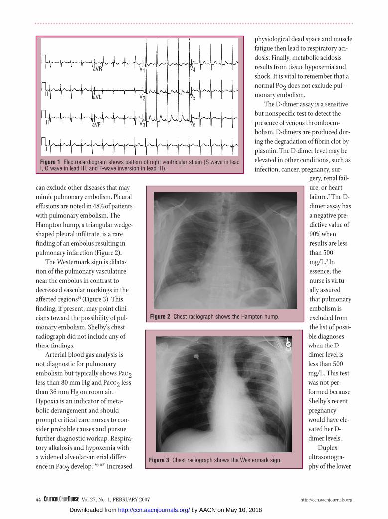
Anterior T-wave inversion is evi-
dent in 85% of cases of pulmonary
embolism.16 This abnormality reflects
inferoposterior ischemia that results
from pressure overload.17(p1902) A pat-
tern of acute right ventricular strain
is highly suggestive of pulmonary
embolism but is present in only 20%
of the cases. This pattern is evidenced
by an S wave in lead I, a Q wave in
lead III, and a T-wave inversion in
lead III (Figure 1). Tall peaked P waves,
tachycardia, new incomplete right
bundle branch block, and right-axis
deviation are the most common
abnormalities noted in patients with
pulmonary embolism. This test was
not performed on Shelby because she
never complained of chest pain. Her
rapid deterioration from acute dysp-
nea to cardiac arrest with pulseless
electrical activity precluded the oppor-
tunity to obtain an electrocardiogram.
Chest radiographs are not diag-
nostic for pulmonary embolism but
http://ccn.aacnjournals.org CRITICALCARENURSE Vol 27, No. 1, FEBRUARY 2007 43
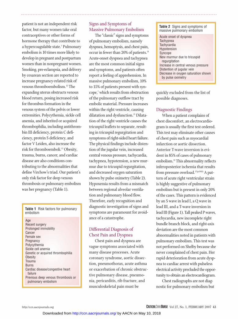
Table 1 Risk factors for pulmonaryembolism
AgeRecent surgeryProlonged immobilityCancerFemale sexPregnancyPolycythemiaSickle cell anemiaGenetic or acquired thrombophiliaObesityTraumaBurnsCardiac disease/congestive heart
failurePrevious deep venous thrombosis or
pulmonary embolism
Table 2 Signs and symptoms ofmassive pulmonary embolism
Acute onset of dyspneaTachypneaTachycardiaHypotensionSyncopeNew murmur due to tricuspid
regurgitationIncrease in central venous pressureDistention of jugular veinDecrease in oxygen saturation shown
by pulse oximetry
by AACN on May 10, 2018http://ccn.aacnjournals.org/Downloaded from
44 CRITICALCARENURSE Vol 27, No. 1, FEBRUARY 2007 http://ccn.aacnjournals.org
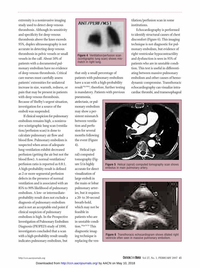
can exclude other diseases that may
mimic pulmonary embolism. Pleural
effusions are noted in 48% of patients
with pulmonary embolism. The
Hampton hump, a triangular wedge-
shaped pleural infiltrate, is a rare
finding of an embolus resulting in
pulmonary infarction (Figure 2).
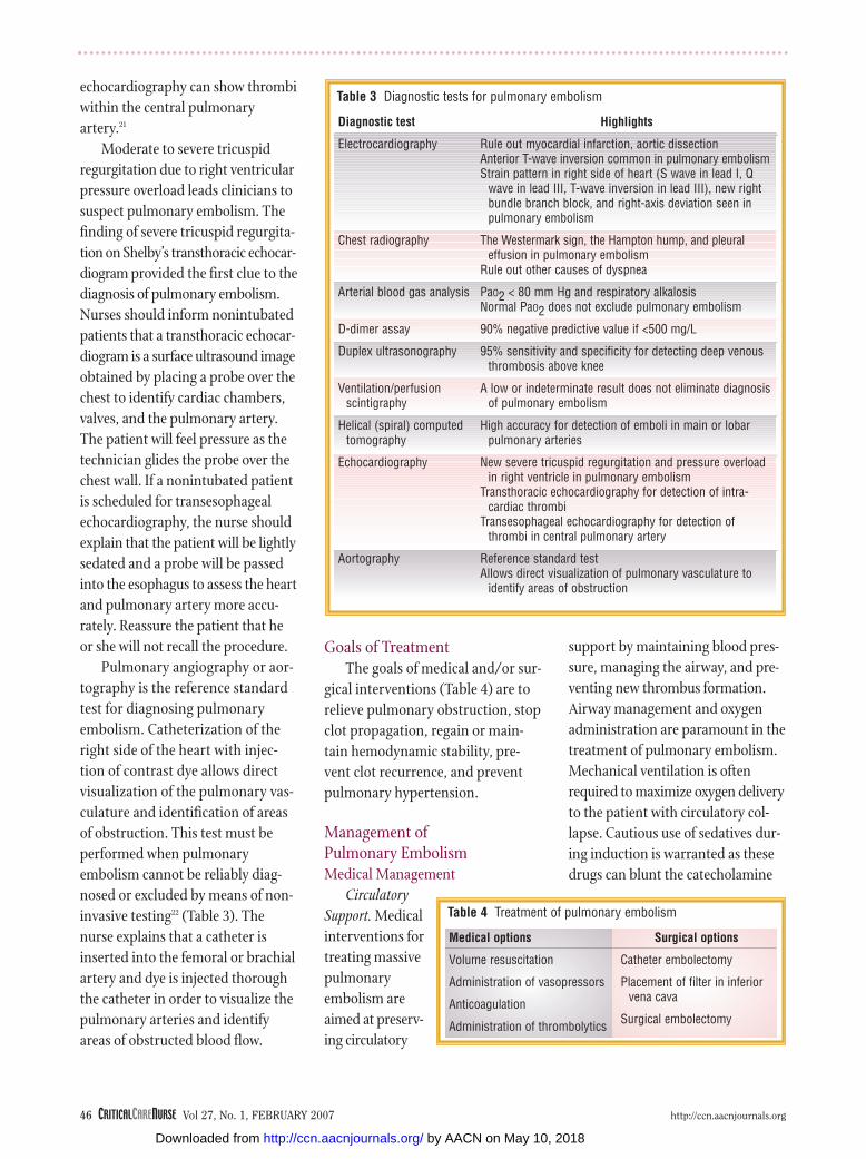
The Westermark sign is dilata-
tion of the pulmonary vasculature
near the embolus in contrast to
decreased vascular markings in the
affected regions14 (Figure 3). This
finding, if present, may point clini-
cians toward the possibility of pul-
monary embolism. Shelby’s chest
radiograph did not include any of
these findings.
Arterial blood gas analysis is
not diagnostic for pulmonary
embolism but typically shows PaO2less than 80 mm Hg and PaCO2 less
than 36 mm Hg on room air.
Hypoxia is an indicator of meta-
bolic derangement and should
prompt critical care nurses to con-
sider probable causes and pursue
further diagnostic workup. Respira-
tory alkalosis and hypoxemia with
a widened alveolar-arterial differ-
ence in PaO2 develop.18(p463) Increased
physiological dead space and muscle
fatigue then lead to respiratory aci-
dosis. Finally, metabolic acidosis
results from tissue hypoxemia and
shock. It is vital to remember that a
normal PO2 does not exclude pul-
monary embolism.
The D-dimer assay is a sensitive
but nonspecific test to detect the
presence of venous thromboem-
bolism. D-dimers are produced dur-
ing the degradation of fibrin clot by
plasmin. The D-dimer level may be
elevated in other conditions, such as
infection, cancer, pregnancy, sur-
gery, renal fail-
ure, or heart
failure.2 The D-
dimer assay has
a negative pre-
dictive value of
90% when
results are less
than 500
mg/L.3 In
essence, the
nurse is virtu-
ally assured
that pulmonary
embolism is
excluded from
the list of possi-
ble diagnoses
when the D-
dimer level is
less than 500
mg/L. This test
was not per-
formed because
Shelby’s recent
pregnancy
would have ele-
vated her D-
dimer levels.
Duplex
ultrasonogra-
phy of the lower
Figure 1 Electrocardiogram shows pattern of right ventricular strain (S wave in leadI, Q wave in lead III, and T-wave inversion in lead III).
I
II
III
II
aVF
aVL
aVR V1
V2
V3 V6
V5
V4
Figure 3 Chest radiograph shows the Westermark sign.
Figure 2 Chest radiograph shows the Hampton hump.
by AACN on May 10, 2018http://ccn.aacnjournals.org/Downloaded from
extremity is a noninvasive imaging
study used to detect deep venous
thrombosis. Although its sensitivity
and specificity for deep venous
thrombosis above the knee exceeds
95%, duplex ultrasonography is not
accurate in detecting deep venous
thrombosis in pelvic vessels or small
vessels in the calf. About 50% of
patients with a documented pul-
monary embolism have no evidence
of deep venous thrombosis. Critical
care nurses must carefully assess
patients’ extremities for unilateral
increase in size, warmth, redness, or
pain that may be present in patients
with deep venous thrombosis.
Because of Shelby’s urgent situation,
investigation for a source of the
emboli was suspended.
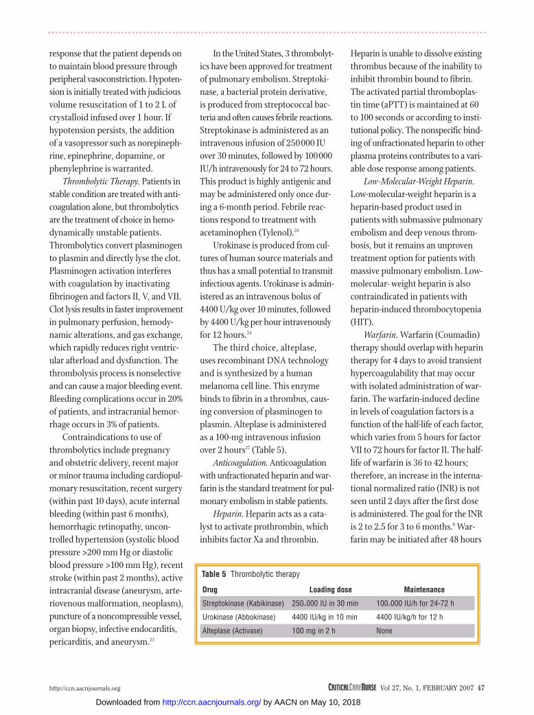
If clinical suspicion for pulmonary
embolism remains high, a noninva-
sive scintigraphic lung scan (ventila-
tion/perfusion scan) is done to
calculate pulmonary air flow and
blood flow. Pulmonary embolism is
suspected when areas of adequate
lung ventilation exhibit decreased
perfusion (getting the air but not the
blood flow). A normal ventilation/
perfusion ratio is reported as 0.8:1.
A high-probability result is defined
as 2 or more segmental perfusion
defects in the presence of normal
ventilation and is associated with an
85% to 90% likelihood of pulmonary
embolism. A low- or intermediate-
probability result does not exclude a
diagnosis of pulmonary embolism
and is not an acceptable end point if
clinical suspicion of pulmonary
embolism is high. In the Prospective
Investigation of Pulmonary Embolism
Diagnosis (PIOPED) study of 1990,
investigators concluded that a scan
with a high-probability result usually
indicates pulmonary embolism, but
that only a small percentage of
patients with pulmonary embolism
have a scan with a high-probability
result19(p1084); therefore, further testing
is mandatory. Patients with previous
pneumonia,
atelectasis, or pul-
monary embolism
may show a per-
sistent mismatch
between ventila-
tion and perfu-
sion for several
months following
the event (Figure
4).
Helical (spi-
ral) computed
tomography (Fig-
ure 5) is highly
accurate for direct
visualization of
large emboli in
the main or lobar
pulmonary arter-
ies, but it requires
a 20- to 30-second
breath-hold,
which may not be
feasible in
patients who are
in unstable condi-
tion.20(pp78-79) This
diagnostic imag-
ing technique is
replacing the ven-
tilation/perfusion scan in some
institutions.
Echocardiography is performed
to identify structural causes of chest
discomfort (Figure 6). This imaging
technique is not diagnostic for pul-
monary embolism, but evidence of
right ventricular hypocontractility
and dysfunction is seen in 95% of
patients who are in unstable condi-
tion. This test is useful in differenti-
ating between massive pulmonary
embolism and other causes of hemo-
dynamic compromise. Transthoracic
echocardiography can visualize intra-
cardiac thrombi, and transesophageal
http://ccn.aacnjournals.org CRITICALCARENURSE Vol 27, No. 1, FEBRUARY 2007 45
Figure 4 Ventilation/perfusion scan(scintigraphic lung scan) shows mis-match in right lung.
Figure 5 Helical (spiral) computed tomography scan showsembolus in main pulmonary artery.
Figure 6 Transthoracic echocardiogram shows dilated rightventricle often seen in massive pulmonary embolism.
by AACN on May 10, 2018http://ccn.aacnjournals.org/Downloaded from
46 CRITICALCARENURSE Vol 27, No. 1, FEBRUARY 2007 http://ccn.aacnjournals.org
echocardiography can show thrombi
within the central pulmonary
artery.21
Moderate to severe tricuspid
regurgitation due to right ventricular
pressure overload leads clinicians to
suspect pulmonary embolism. The
finding of severe tricuspid regurgita-
tion on Shelby’s transthoracic echocar-
diogram provided the first clue to the
diagnosis of pulmonary embolism.
Nurses should inform nonintubated
patients that a transthoracic echocar-
diogram is a surface ultrasound image
obtained by placing a probe over the
chest to identify cardiac chambers,
valves, and the pulmonary artery.
The patient will feel pressure as the
technician glides the probe over the
chest wall. If a nonintubated patient
is scheduled for transesophageal
echocardiography, the nurse should
explain that the patient will be lightly
sedated and a probe will be passed
into the esophagus to assess the heart
and pulmonary artery more accu-
rately. Reassure the patient that he
or she will not recall the procedure.
Pulmonary angiography or aor-
tography is the reference standard
test for diagnosing pulmonary
embolism. Catheterization of the
right side of the heart with injec-
tion of contrast dye allows direct
visualization of the pulmonary vas-
culature and identification of areas
of obstruction. This test must be
performed when pulmonary
embolism cannot be reliably diag-
nosed or excluded by means of non-
invasive testing22 (Table 3). The
nurse explains that a catheter is
inserted into the femoral or brachial
artery and dye is injected thorough
the catheter in order to visualize the
pulmonary arteries and identify
areas of obstructed blood flow.
Goals of TreatmentThe goals of medical and/or sur-
gical interventions (Table 4) are to
relieve pulmonary obstruction, stop
clot propagation, regain or main-
tain hemodynamic stability, pre-
vent clot recurrence, and prevent
pulmonary hypertension.
Management of Pulmonary EmbolismMedical Management
Circulatory
Support. Medical
interventions for
treating massive
pulmonary
embolism are
aimed at preserv-
ing circulatory
support by maintaining blood pres-
sure, managing the airway, and pre-
venting new thrombus formation.
Airway management and oxygen
administration are paramount in the
treatment of pulmonary embolism.
Mechanical ventilation is often
required to maximize oxygen delivery
to the patient with circulatory col-
lapse. Cautious use of sedatives dur-
ing induction is warranted as these
drugs can blunt the catecholamine
Table 3 Diagnostic tests for pulmonary embolism
Diagnostic test
Electrocardiography
Chest radiography
Arterial blood gas analysis
D-dimer assay
Duplex ultrasonography
Ventilation/perfusionscintigraphy
Helical (spiral) computedtomography
Echocardiography
Aortography
Highlights
Rule out myocardial infarction, aortic dissectionAnterior T-wave inversion common in pulmonary embolismStrain pattern in right side of heart (S wave in lead I, Q
wave in lead III, T-wave inversion in lead III), new rightbundle branch block, and right-axis deviation seen inpulmonary embolism
The Westermark sign, the Hampton hump, and pleural effusion in pulmonary embolism
Rule out other causes of dyspnea
PaO2 < 80 mm Hg and respiratory alkalosisNormal PaO2 does not exclude pulmonary embolism
90% negative predictive value if <500 mg/L
95% sensitivity and specificity for detecting deep venousthrombosis above knee
A low or indeterminate result does not eliminate diagnosisof pulmonary embolism
High accuracy for detection of emboli in main or lobarpulmonary arteries
New severe tricuspid regurgitation and pressure overloadin right ventricle in pulmonary embolism
Transthoracic echocardiography for detection of intra-cardiac thrombi
Transesophageal echocardiography for detection ofthrombi in central pulmonary artery
Reference standard testAllows direct visualization of pulmonary vasculature to
identify areas of obstruction
Table 4 Treatment of pulmonary embolism
Medical options
Volume resuscitation
Administration of vasopressors
Anticoagulation
Administration of thrombolytics
Surgical options
Catheter embolectomy
Placement of filter in inferiorvena cava
Surgical embolectomy
by AACN on May 10, 2018http://ccn.aacnjournals.org/Downloaded from
response that the patient depends on
to maintain blood pressure through
peripheral vasoconstriction. Hypoten-
sion is initially treated with judicious
volume resuscitation of 1 to 2 L of
crystalloid infused over 1 hour. If
hypotension persists, the addition
of a vasopressor such as norepineph-
rine, epinephrine, dopamine, or
phenylephrine is warranted.
Thrombolytic Therapy. Patients in
stable condition are treated with anti-
coagulation alone, but thrombolytics
are the treatment of choice in hemo-
dynamically unstable patients.
Thrombolytics convert plasminogen
to plasmin and directly lyse the clot.
Plasminogen activation interferes
with coagulation by inactivating
fibrinogen and factors II, V, and VII.
Clot lysis results in faster improvement
in pulmonary perfusion, hemody-
namic alterations, and gas exchange,
which rapidly reduces right ventric-
ular afterload and dysfunction. The
thrombolysis process is nonselective
and can cause a major bleeding event.
Bleeding complications occur in 20%
of patients, and intracranial hemor-
rhage occurs in 3% of patients.
Contraindications to use of
thrombolytics include pregnancy
and obstetric delivery, recent major
or minor trauma including cardiopul-
monary resuscitation, recent surgery
(within past 10 days), acute internal
bleeding (within past 6 months),
hemorrhagic retinopathy, uncon-
trolled hypertension (systolic blood
pressure >200 mm Hg or diastolic
blood pressure >100 mm Hg), recent
stroke (within past 2 months), active
intracranial disease (aneurysm, arte-
riovenous malformation, neoplasm),
puncture of a noncompressible vessel,
organ biopsy, infective endocarditis,
pericarditis, and aneurysm.23
In the United States, 3 thrombolyt-
ics have been approved for treatment
of pulmonary embolism. Streptoki-
nase, a bacterial protein derivative,
is produced from streptococcal bac-
teria and often causes febrile reactions.
Streptokinase is administered as an
intravenous infusion of 250000 IU
over 30 minutes, followed by 100000
IU/h intravenously for 24 to 72 hours.
This product is highly antigenic and
may be administered only once dur-
ing a 6-month period. Febrile reac-
tions respond to treatment with
acetaminophen (Tylenol).24
Urokinase is produced from cul-
tures of human source materials and
thus has a small potential to transmit
infectious agents. Urokinase is admin-
istered as an intravenous bolus of
4400 U/kg over 10 minutes, followed
by 4400 U/kg per hour intravenously
for 12 hours.24
The third choice, alteplase,
uses recombinant DNA technology
and is synthesized by a human
melanoma cell line. This enzyme
binds to fibrin in a thrombus, caus-
ing conversion of plasminogen to
plasmin. Alteplase is administered
as a 100-mg intravenous infusion
over 2 hours27 (Table 5).
Anticoagulation. Anticoagulation
with unfractionated heparin and war-
farin is the standard treatment for pul-
monary embolism in stable patients.
Heparin. Heparin acts as a cata-
lyst to activate prothrombin, which
inhibits factor Xa and thrombin.
Heparin is unable to dissolve existing
thrombus because of the inability to
inhibit thrombin bound to fibrin.
The activated partial thromboplas-
tin time (aPTT) is maintained at 60
to 100 seconds or according to insti-
tutional policy. The nonspecific bind-
ing of unfractionated heparin to other
plasma proteins contributes to a vari-
able dose response among patients.
Low-Molecular-Weight Heparin.
Low-molecular-weight heparin is a
heparin-based product used in
patients with submassive pulmonary
embolism and deep venous throm-
bosis, but it remains an unproven
treatment option for patients with
massive pulmonary embolism. Low-
molecular- weight heparin is also
contraindicated in patients with
heparin-induced thrombocytopenia
(HIT).
Warfarin. Warfarin (Coumadin)
therapy should overlap with heparin
therapy for 4 days to avoid transient
hypercoagulability that may occur
with isolated administration of war-
farin. The warfarin-induced decline
in levels of coagulation factors is a
function of the half-life of each factor,
which varies from 5 hours for factor
VII to 72 hours for factor II. The half-
life of warfarin is 36 to 42 hours;
therefore, an increase in the interna-
tional normalized ratio (INR) is not
seen until 2 days after the first dose
is administered. The goal for the INR
is 2 to 2.5 for 3 to 6 months.8 War-
farin may be initiated after 48 hours
http://ccn.aacnjournals.org CRITICALCARENURSE Vol 27, No. 1, FEBRUARY 2007 47
Table 5 Thrombolytic therapy
Drug
Streptokinase (Kabikinase)
Urokinase (Abbokinase)
Alteplase (Activase)
Loading dose
250∞000 IU in 30 min
4400 IU/kg in 10 min
100 mg in 2 h
Maintenance
100∞000 IU/h for 24-72 h
4400 IU/kg/h for 12 h
None
by AACN on May 10, 2018http://ccn.aacnjournals.org/Downloaded from
48 CRITICALCARENURSE Vol 27, No. 1, FEBRUARY 2007 http://ccn.aacnjournals.org
of direct thrombin inhibitor therapy
as long as the platelet count is
greater than 100 × 109/L.25 Warfarin
is contraindicated in pregnancy
because it crosses the placenta. War-
farin is safely used in lactating
patients because it is not excreted in
breast milk.
Heparin-Induced Thrombocytopenia
HIT or “heparin allergy” is an
important complication of heparin
therapy and can result in severe
venous and arterial thrombosis.26
This autoimmune disorder occurs in
1% to 3% of patients who receive
heparin-based products. HIT results
from the development of heparin-
associated antibodies. These anti-
bodies induce platelet aggregation in
the presence of heparin. Thrombosis
may occur in both arterial and venous
circulation. All patients who receive
heparin are at risk for HIT, because
no known characteristics of patients
can be used to predict the develop-
ment of HIT.
HIT occurs in 2 forms: HIT-1, also
known as heparin-associated throm-
bocytopenia, is a nonimmune disor-
der that occurs 2 to 3 days after
initiation of heparin therapy. This
disorder may occur in up to 15% of
patients exposed to heparin. The
thrombocytopenia is transient, and
platelet counts rarely decrease to less
than 100 × 109/L. Patients are not at
risk for development of significant
thrombosis. HIT-2 is an immune-
mediated disorder that occurs in 1%
to 3% of patients who receive heparin
and is frequently associated with life-
or limb-threatening thromboembolic
complications (deep venous throm-
bosis, pulmonary embolism, cere-
brovascular accident, myocardial
infarction, extremity ischemia, gan-
grene, and death). This disorder
causes a significant decrease in platelet
count. Thrombocytopenia manifests
5 to 10 days after the start of heparin
therapy, and platelet count decreases
30% to 50%, to less than 100 × 109/L.27
HIT is a clinical diagnosis sup-
ported by confirmatory laboratory
work. The C-serotonin release assay
combines serum from patients with
suspected HIT with serum from
healthy donors and adds this to ther-
apeutic concentrations of heparin. A
positive result detects the C-serotonin
released from the serum of the patient
with suspected HIT.
The treatment for HIT is imme-
diate cessation of use of all heparin-
based products, including
heparin-coated catheters and heparin
flushes. Patients with HIT who require
anticoagulation for deep venous
thrombosis or pulmonary embolism
are treated with a direct thrombin
inhibitor, such as lipirudin or arga-
troban. The direct thrombin inhibitor
is administered until the platelet
count has increased to 100 × 109/L.
Lipirudin is administered as an
intravenous bolus of 0.4 mg/kg fol-
lowed by infusion of 0.15 mg/kg
per hour for an aPTT 1.5 to 2.5
times the time at baseline. Lipirudin
is renally excreted and requires
decreased dosing in patients with
renal insufficiency. Argatroban is
administered as a 2 mg/kg per
minute intravenous infusion, with-
out bolus for an aPTT 1.5 to 3 times
the aPTT at baseline. Argatroban is
metabolized by the liver and may be
useful for a patient with renal
impairment.28(pp2044-2045) Concomitant
warfarin therapy is started once the
platelet count reaches 100 × 109/L.
Argatroban should be continued
until the INR is 4 or greater. This
direct thrombin inhibitor is then dis-
continued and the INR is rechecked
in 4 to 6 hours. If the INR remains at
a therapeutic level (>2), argatroban
does not need to be restarted.
Bivalrudin (Angiomax) is another
direct thrombin inhibitor that is cur-
rently approved by the Food and Drug
Administration for use in patients
undergoing coronary angioplasty
with unstable angina and concomi-
tant aspirin therapy. This drug is used
off-label in some institutions as an
alternative to heparin in patients
with HIT. At this time, however,
there is not enough evidence to sup-
port this indication.
Surgical Management
Catheter Embolectomy. Surgical
interventions are usually reserved for
patients in unstable condition with
contraindications to thrombolytic
therapy. Percutaneous catheter
embolectomy is performed during
pulmonary angiography in an effort to
remove the obstructive embolus. This
procedure may be performed by one
of 2 techniques. The first method is
aspiration embolectomy, in which suc-
tion is applied to remove the embolic
material. The second technique is
mechanical embolectomy, which
involves maceration or fragmentation
of the embolus.2 Mechanical embolec-
tomy has a success rate of 80%, but
there is danger of distal migration of
the pulmonary embolism.
Vena Caval Filters. Percutaneous
filters are placed in the inferior vena
cava to prevent recurrent pulmonary
embolism by preventing clot migra-
tion. These filters are often used for
patients with contraindications to
anticoagulation, those with recur-
rent pulmonary embolism, or those
by AACN on May 10, 2018http://ccn.aacnjournals.org/Downloaded from
who have underlying cardiac or pul-
monary disease in whom a pulmonary
embolism would be fatal. Several fil-
ters are available for use in the United
States, including Greenfield (Boston
Scientific, Natick, Mass), Vena-Tech
LGN or LP (Braun, Bethleham, Pa),
Simon-Nitinol (Bard, Murray Hill,
NJ), Recovery (Bard) bird’s nest, and
Gunther-Tulip (Cook). Complications
of filter placement include thrombo-
sis at the insertion site, filter migra-
tion, erosion through the wall of the
inferior vena cava, or obstruction of
the inferior vena cava during deploy-
ment of the filter.29
Surgical Embolectomy. Surgical
embolectomy involves manual
removal of the thrombus from the
pulmonary artery. This procedure is
usually reserved for patients in unsta-
ble condition who have contraindi-
cations to thrombolytic therapy.29
Extracorporeal membrane oxygena-
tion is a temporary cardiopulmonary
support system used sparingly in the
treatment of patients with circulatory
collapse due to pulmonary embolism.
Extracorporeal membrane oxygena-
tion is used to maintain oxygenation
and tissue perfusion until the throm-
bus can be dissolved or removed.
Nursing CarePreventative measures for deep
venous thrombosis must be used
routinely for any hospitalized patient.
Compression stockings should be
placed on the lower extremities
before the patient gets out of bed.
External pneumatic compression
boots, which intermittently compress
the calf and accelerate deep venous
flow, may be used in bed-bound
patients. Adequate hydration and
early ambulation are encouraged to
prevent deep venous thrombosis.
Daily assessment of extremities for
pain, erythema, and size discrepancy
is vitally important. The Homan sign
(pain on dorsiflexion) is apparent in
only 30% of the cases. The nurse must
recognize risk factors for pulmonary
embolism and vigilantly monitor
patients who are immobilized or
have had their activity restricted for
unexplained tachypnea, tachycardia,
and restlessness. These signs must
not be attributed to anxiety unless a
physical cause has been sought first.
Critically ill or postoperative
patients are at high risk for venous
thromboembolism and should be
assessed for use of prophylactic
heparin or low-molecular-weight
heparin subcutaneously or intra-
venously. If pulmonary embolism is
discovered and the patient is hemo-
dynamically unstable, the nurse will
institute appropriate fluid adminis-
tration and inotropic support to
maintain a systolic blood pressure of
90 mm Hg. If pulmonary hyperten-
sion and right-sided heart failure are
present, the nurse may consider cau-
tious use of a vasodilator such as
nitric oxide. The nurse can provide
short explanations of the diagnostic
tests and invasive procedures to
reassure the patient that this life-
threatening condition is treatable.
Neurological and vascular assess-
ments should be performed hourly
to evaluate organ perfusion. Confu-
sion or agitation may indicate respi-
ratory acidosis (increased PaCO2 and
decreased pH). Weakened pulses
and cool, mottled extremities are
late physical findings that indicate
impending circulatory collapse.
The nurse should suspect pul-
monary embolism as an underlying
cause of syncope and cardiac arrest
with pulseless electrical activity.
Vigilance during anticoagulation or
administration of thrombolytic agents
is warranted to prevent significant
bleeding complications. A decrease
in hemoglobin level and hematocrit
may alert the nurse to occult bleed-
ing in the retroperitoneum or gas-
trointestinal tract. If the nurse
encounters a change in mental sta-
tus or new focal neurological deficits
in a patient receiving thrombolytics,
intracranial hemorrhage must be
eliminated as a possible cause. An
emergent neurosurgical consultation
and an unenhanced computed tomog-
raphy scan of the brain is warranted
in this situation.
Avoidance of unnecessary phle-
botomy, arterial puncture, and other
invasive procedures may reduce the
risk of hemorrhage during throm-
bolytic administration. Significant
hemorrhage requires discontinua-
tion of the thrombolytic agent and
administration of cryoprecipitate or
fresh frozen plasma to reverse the
coagulopathy. Decreasing platelet
counts during heparin administra-
tion may suggest a diagnosis of HIT.
Education of PatientsDuring recovery, the nurses were
instrumental in preparing the patient
for discharge. The nurses instructed
Shelby and her family on the impor-
tance of continued warfarin admin-
istration for 3 to 6 months to prevent
further thrombus development.
Foods high in vitamin K, such as dark
green vegetables and apricots, must
be limited during this time period to
prevent decreased warfarin action.
The nurses informed Shelby that a
therapeutic INR value is between 2
and 3 and that adjustments in the
dosage of warfarin may be needed to
maintain her INR in this range.
http://ccn.aacnjournals.org CRITICALCARENURSE Vol 27, No. 1, FEBRUARY 2007 49
by AACN on May 10, 2018http://ccn.aacnjournals.org/Downloaded from
50 CRITICALCARENURSE Vol 27, No. 1, FEBRUARY 2007 http://ccn.aacnjournals.org
Shelby was advised not to use war-
farin with acetaminophen, nons-
teroidal anti-inflammatory drugs,
amiodarone, or fluoroquinolone
antibiotics because such combina-
tions can quickly elevate the INR.
The nurses reminded Shelby to
stretch every hour during confined
travel and to remain well hydrated
because patients with history of deep
venous thrombosis or pulmonary
embolism are at a greater risk for
subsequent development of venous
thrombosis.15 The nurses encouraged
Shelby to recount her history of deep
venous thrombosis and pulmonary
embolism to future healthcare
providers and to wear a Medic-Alert
bracelet indicating her history of
pulmonary embolism.
SummaryMassive pulmonary embolism in
the setting of syncope and cardiac
arrest is often fatal if not diagnosed
rapidly and correctly. The patient’s
cardiopulmonary status before the
embolic event is a predictor of mor-
bidity and mortality. This postpartum
patient posed unique challenges to
the usual management of pulmonary
embolism. Spiral computed tomog-
raphy could not be used for diagno-
sis because of the patient’s unstable
hemodynamics. Thrombolytic agents
were contraindicated because of the
recent obstetrical delivery and car-
diopulmonary resuscitation. There-
fore, surgical embolectomy and
placement of a filter in the inferior
vena cava were the treatments of
choice. Shelby also received standard
medical treatment, including external
pneumatic compression, heparin,
and warfarin postoperatively.
Two factors were paramount in
the successful outcome of this case.
The first was the rapid diagnosis and
intervention that made survival a
possibility. The second, but perhaps
the most important, factor was the
nursing care of this patient’s entire
family, including her husband, par-
ents, newborn baby, and 2 young
children. The bedside staff facilitated
visits between mother and newborn
in the surgical intensive care unit
and provided expert care, unyielding
support, and encouragement during
this life-threatening event. The
orchestrated interdisciplinary efforts
of many nurses and physicians paved
the way for a modern-day Christmas
miracle for Shelby and her family.
References1. Marty AT, Hilton FL, Spear RK, Greyson B.
Postcesarean pulmonary embolism, sus-tained cardiopulmonary resuscitation,embolectomy, and near death experience.Obstet Gynecol. 2005;106:1153-1155.
2. Owen AR, Gibson MR. Pulmonaryembolism: advances in diagnosis and treat-ment. Care Crit Ill. 2004;20:79-84.
3. Feied C, Handler JA. Pulmonary Embolism.2006. Available at: http://www.emedicine.com/EMERG/topic490.htm. AccessedNovember 15, 2006.
4. Smeltzer SC, Bare BG. Management ofpatients with chest and lower respiratorytract disorders. In: Brunner and Suddarth’sTextbook of Medical-Surgical Nursing.Philadelphia, Pa: Lippincott Williams andWilkins; 2000:471-475.
5. Seifert PC. Cardiac trauma and emergencysurgery. In: Cardiac Surgery PerioperativePatient Care. Philadelphia, Pa: Mosby;2002:559-561.
6. Sole ML, Lamborn ML, Hartshorn JC.Acute respiratory failure. In: Introduction toCritical Care Nursing. 3rd ed. Philadelphia,Pa: WB Saunders; 2001:365-368.
7. Gawlinski A, Hamwi D. Pulmonary disor-ders. In: Acute Care Nurse Practitioner: ClinicalCurriculum and Certification Review. Philadel-phia, Pa: WB Saunders; 1999:90-101.
8. Wan S, Quinlan DJ, Agnelli G, EikelboomJW. Thrombolysis compared with heparinfor the initial treatment of pulmonaryembolism. Circulation. 2004;110:744-749.
9. Scurr JH, Machin SJ, Bailey-King S, MackieIJ, McDonald S, Smith PD. Frequency andprevention of symptom less deep veinthrombosis in long-haul flight: a random-ized trial. Lancet. 2001;357:1485-1489.
10. Anderson FA, Wheeler HB, Goldberg RJ, etal. A population-based perspective of thehospital incidence and case-fatality rates ofdeep vein thrombosis and pulmonaryembolism: the Worcester DVT study. ArchIntern Med. 1991;51:933-935.
11. Anderson FA, Spencer FA. Risk factors forvenous thromboembolism. Circulation.2003;107(suppl):I9-I16.
12. Ros HS, Lichtenstein P, Bellocco R, Peters-son G, Cnattingius S. Pulmonary embolismand stroke in relation to pregnancy: howcan high-risk women be identified? Am JObstet Gynecol. 2002;186:198-203.
13. Fedullo PF, Tapson VF. The evaluation ofsuspected pulmonary embolism. N Engl JMed. 2003;349:1247-1255.
14. Koschel MJ. Pulmonary embolism. Am JNurs. 2004;104:46-50.
15. Cardin T, Martinelli A. Pulmonaryembolism. Crit Care Nurs Q. 2004;27:310-322.
16. Ferrari E, Imbert A, Chevalier T, Mihoubi A,Morand P, Baudouy M. The ECG in pul-monary embolism: predictive value of nega-tive T-waves in precordial leads, 80 casereports. Chest. 1997;111:537-543.
17. Kouchoukos NT, Blacstone EH, Doty DB,Hanley FL, Karp RB, eds. Kirklin/Barratt-Boyes Cardiac Surgery. 3rd ed. Philadelphia,Pa: Churchill-Livingstone; 2003.
18. Fishman AP, Elias JA, Fishman JA, GrippiMA, Kaiser LR, Senior RM. Fishman’s Man-ual of Pulmonary Diseases and Disorders. 3rded. New York, NY: McGraw-Hill; 2002.
19. Cunningham FG, Leveno KJ, Bloom SL,Hauth JC, Gilstrap LC, Wenstrom KD.Williams Obstetrics. 22nd ed. New York, NY:McGraw-Hill; 2005.
20. Todd BA. Acute Care Nurse PractitionerSecrets. St. Louis, Mo: Elsevier Mosby; 2005.
21. Kearon C. Diagnosis of pulmonary embolism.Can Med Assoc J. 2003;168:183-194.
22. Goldhaber SZ. Acute pulmonary embolism:I, epidemiology, pathophysiology, and diag-nosis. Circulation. 2003;108:2726-2729.
23. Goldhaber SZ. Acute pulmonary embolism:II, risk stratification, treatment, and preven-tion. Circulation. 2003;108: 2834-2838.
24. Spratto GR, Woods AL. 2003 Nurse’s DrugHandbook. Clifton Park, NY: Thomson Del-mar Learning; 2003.
25. Sadosty AT. Pulmonary embolism. EmergMed Clin North Am. 2003;21:917-920.
26. Yang JC. Prevention and treatment of deepvein thrombosis and pulmonary embolismin critically ill patients. Crit Care Nurs Q.2005;28:72-79.
27. Barcelona R. Type II heparin-induced throm-bocytopenia: new treatment options. Avail-able at: http://www.clevelandclinicmeded.com/medical_info/pharmacy/septoct2001/thrombocytopenia.htm. Accessed Novem-ber 15, 2006.
28. Lichtman MA, Beutler E, Kipps TJ, Selig-sohn U, Kaushansky K, Prchal J, eds.Williams Hematology. 7th ed. New York, NY:McGraw-Hill; 2006.
29. Dalen JE. Pulmonary embolism: what havewe learned since Virchow? Treatment andprevention. Chest. 2002;122:1801-1817.
by AACN on May 10, 2018http://ccn.aacnjournals.org/Downloaded from
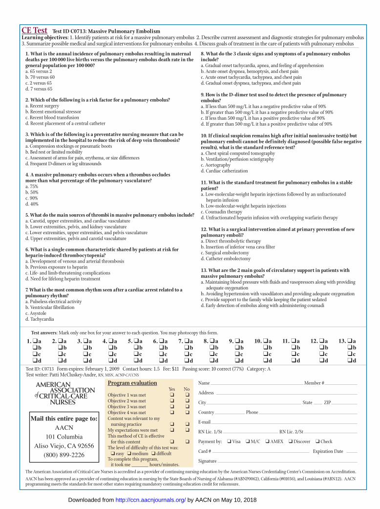
CE Test Test ID C0713: Massive Pulmonary EmbolismLearning objectives: 1. Identify patients at risk for a massive pulmonary embolus 2. Describe current assessment and diagnostic strategies for pulmonary embolus3. Summarize possible medical and surgical interventions for pulmonary embolus 4. Discuss goals of treatment in the care of patients with pulmonary embolus
Program evaluationYes No
Objective 1 was met � �Objective 2 was met � �Objective 3 was met � �Objective 4 was met � �Content was relevant to my
nursing practice � �My expectations were met � �This method of CE is effective
for this content � �The level of difficulty of this test was: � easy � medium � difficult
To complete this program, it took me hours/minutes.
Test answers: Mark only one box for your answer to each question. You may photocopy this form.
1. What is the annual incidence of pulmonary embolus resulting in maternaldeaths per 100 000 live births versus the pulmonary embolus death rate in thegeneral population per 100 000?a. 65 versus 2b. 70 versus 60c. 2 versus 65d. 7 versus 65
2. Which of the following is a risk factor for a pulmonary embolus?a. Recent surgery b. Recent emotional stressor c. Recent blood transfusion d. Recent placement of a central catheter
3. Which is of the following is a preventative nursing measure that can beimplemented in the hospital to reduce the risk of deep vein thrombosis?a. Compression stockings or pneumatic bootsb. Bed rest or limited mobilityc. Assessment of arms for pain, erythema, or size differencesd. Frequent D-dimers or leg ultrasounds
4. A massive pulmonary embolus occurs when a thrombus occludes more than what percentage of the pulmonary vasculature?a. 75%b. 50%c. 90%d. 40%
5. What do the main sources of thrombi in massive pulmonary embolus include?a. Carotid, upper extremities, and cardiac vasculatureb. Lower extremities, pelvis, and kidney vasculaturec. Lower extremities, upper extremities, and pelvis vasculatured. Upper extremities, pelvis and carotid vasculature
6. What is a single common characteristic shared by patients at risk forheparin-induced thrombocytopenia?a. Development of venous and arterial thrombosisb. Previous exposure to heparinc. Life- and limb-threatening complicationsd. Need for lifelong heparin treatment
7. What is the most common rhythm seen after a cardiac arrest related to a pulmonary rhythm?a. Pulseless electrical activityb. Ventricular fibrillationc. Asystoled. Tachycardia
8. What do the 3 classic signs and symptoms of a pulmonary embolusinclude?a. Gradual onset tachycardia, apnea, and feeling of apprehensionb. Acute onset dyspnea, hemoptysis, and chest painc. Acute onset tachycardia, tachypnea, and chest paind. Gradual onset dyspnea, tachypnea, and chest pain
9. How is the D-dimer test used to detect the presence of pulmonary embolus?a. If less than 500 mg/L it has a negative predictive value of 90% b. If greater than 500 mg/L it has a negative predictive value of 90% c. If less than 500 mg/L it has a positive predictive value of 90% d. If greater than 500 mg/L it has a positive predictive value of 90%
10. If clinical suspicion remains high after initial noninvasive test(s) butpulmonary emboli cannot be def initely diagnosed (possible false negativeresults), what is the standard reference test?a. Chest spiral computed tomographyb. Ventilation/perfusion scintigraphyc. Aortography d. Cardiac catherization
11. What is the standard treatment for pulmonary embolus in a stablepatient?a. Low-molecular-weight heparin injections followed by an unfractionated
heparin infusionb. Low-molecular-weight heparin injectionsc. Coumadin therapyd. Unfractionated heparin infusion with overlapping warfarin therapy
12. What is a surgical intervention aimed at primary prevention of newpulmonary emboli?a. Direct thrombolytic therapyb. Insertion of inferior vena cava filter c. Surgical embolectomyd. Catheter embolectomy
13. What are the 2 main goals of circulatory support in patients with massive pulmonary embolus?a. Maintaining blood pressure with fluids and vasopressors along with providing
adequate oxygenationb. Avoiding hypertension with vasodilators and providing adequate oxygenationc. Provide support to the family while keeping the patient sedatedd. Early detection of embolus along with administering coumadi
Mail this entire page to:AACN
101 Columbia
Aliso Viejo, CA 92656
(800) 899-2226
Test ID: C0713 Form expires: February 1, 2009 Contact hours: 1.5 Fee: $11 Passing score: 10 correct (77%) Category: A Test writer: Patti McCluskey-Andre, RN, MSN, ACNP-C/CCNS
9. �a�b�c�d
8. �a�b�c�d
7. �a�b�c�d
6. �a�b�c�d
5. �a�b�c�d
4. �a�b�c�d
3. �a�b�c�d
2. �a�b�c�d
1. �a�b�c�d
12. �a�b�c�d
13. �a�b�c�d
11. �a�b�c�d
10. �a�b�c�d
Name Member #
Address
City State ZIP
Country Phone
RN Lic. 1/St RN Lic. 2/St
Payment by: � Visa � M/C � AMEX � Discover � Check
Card # Expiration Date
Signature
The American Association of Critical-Care Nurses is accredited as a provider of continuing nursing education by the American Nurses Credentialing Center’s Commission on Accreditation.
AACN has been approved as a provider of continuing education in nursing by the State Boards of Nursing of Alabama (#ABNP0062), California (#01036), and Louisiana (#ABN12). AACNprogramming meets the standards for most other states requiring mandatory continuing education credit for relicensure.
by AACN on May 10, 2018http://ccn.aacnjournals.org/Downloaded from
Kathleen ShaughnessyMassive Pulmonary Embolism
http://ccn.aacnjournals.org/Published online Copyright © 2007 by the American Association of Critical-Care Nurses
39-50 27 2007;Crit Care Nurse
http://ccn.aacnjournals.org/cgi/external_ref?link_type=PERMISSIONDIRECTPersonal use only. For copyright permission information:
http://ccn.aacnjournals.org/subscriptions/Subscription Information
http://ccn.aacnjournals.org/misc/ifora.xhtmlInformation for authors
http://www.editorialmanager.com/ccn Submit a manuscript
http://ccn.aacnjournals.org/subscriptions/etoc.xhtmlEmail alerts
362-2049. Copyright ©2016 by AACN. All rights reserved. bimonthly by AACN, 101 Columbia, Aliso Viejo, CA 92656. Telephone: (800) 899-1712, (949) 362-2050, ext. 532. Fax: (949) Critical Care Nurse is an official peer-reviewed journal of the American Association of Critical-Care Nurses (AACN) published
by AACN on May 10, 2018http://ccn.aacnjournals.org/Downloaded from


