Embed Size (px)
Citation preview
Clinical Skin Lesion Diagnosis using Representations Inspired by
Dermatologist Criteria
Jufeng Yang†, Xiaoxiao Sun†, Jie Liang†, Paul L. Rosin‡
†College of Computer and Control Engineering, Nankai University, Tianjin, China‡School of Computer Science and Informatics, Cardiff University, Cardiff, UK
[email protected], {sunxiaoxiaozrt,liang27jie}@163.com, [email protected]
Abstract
The skin is the largest organ in human body. Around
30%-70% of individuals worldwide have skin related health
problems, for whom effective and efficient diagnosis is nec-
essary. Recently, computer aided diagnosis (CAD) systems
have been successfully applied to the recognition of skin
cancers in dermatoscopic images. However, little work has
concentrated on the commonly encountered skin diseases
in clinical images captured by easily-accessed cameras or
mobile phones. Meanwhile, for a CAD system, the repre-
sentations of skin lesions are required to be understandable
for dermatologists so that the predictions are convincing.
To address this problem, we present effective representa-
tions inspired by the accepted dermatological criteria for
diagnosing clinical skin lesions. We demonstrate that the
dermatological criteria are highly correlated with measur-
able visual components. Accordingly, we design six medical
representations considering different criteria for the recog-
nition of skin lesions, and construct a diagnosis system for
clinical skin disease images. Experimental results show that
the proposed medical representations can not only capture
the manifestations of skin lesions effectively, and consis-
tently with the dermatological criteria, but also improve the
prediction performance with respect to the state-of-the-art
methods based on uninterpretable features.
1. Introduction
The skin is directly exposed to the air, which leads to
skin disease being one of the most common human ill-
nesses. It pervades all cultures, occurs at all ages, and af-
fects the health of 30% to 70% of individuals, with even
higher rates for the at-risk subpopulations [21, 22]. A com-
mon technique used by dermatologists for diagnosing skin
diseases is the dermoscope which enables observation of
the latent structures of skin lesions, i.e., a region suffering
from disease, whose effects are otherwise invisible to the
Clinical Images
ABCD
Diagnosing by Dermatologist
Diagnosing by Computer
Dermatologist Criteria Prediction
Computer
Representation
Classifier Prediction
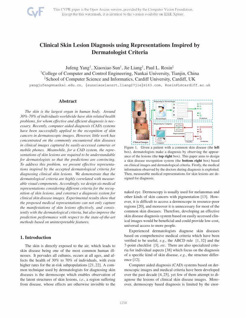
Figure 1. Given a patient with a common skin disease (the left
box), dermatologists make a diagnosis by observing the appear-
ance of the lesions (the top right box). This paper aims to design
a skin disease recognition system (the bottom right box) based
on clinical images and dermatological criteria. Firstly, the medical
information observed by the doctors during diagnosis is exploited.
Then, measurable medical representations for skin lesions are de-
signed for diagnosis.
naked eye. Dermoscopy is usually used for melanomas and
other kinds of skin cancers with pigmentation [13]. How-
ever, it is difficult to access a dermoscope in resource-poor
regions [20], and moreover it is unnecessary for most of the
common skin diseases. Therefore, developing an effective
skin disease diagnosis system based on easily accessed clin-
ical images would be beneficial and could provide low-cost,
universal access to more people.
Experienced dermatologists diagnose skin diseases
based on comprehensive medical criteria which have been
verified to be useful, e.g., the ABCD rule [1, 32] and the
7-point checklist [3], etc. There are also specialized crite-
ria for individual aspects [38] which focus on the diagnosis
of a specific kind of skin disease, e.g., the structure differ-
ence [12].
Computer aided diagnosis (CAD) systems based on der-
moscopic images and medical criteria have been developed
over the past decade [4, 25], yet few of them attempt to di-
agnose the lesions of clinical skin disease images. More-
over, dermoscopy based diagnosis is limited by the enor-
1258
mous expenses and the inconvenience access [20,35], while
experienced dermatologists are able to make effective diag-
nosis for most common diseases by simply observing the
lesions with the naked eye. There are three primary diffi-
culties for recognizing skin disease from a clinical image
captured by easily-accessed devices, e.g., smart-phone, as
compared to dermatoscopic images. (1) The clinical images
are captured with various illumination conditions. (2) The
lesions in clinical images have non-uniform focal lengths
and the size of viewing frame is not consistent. (3) There
are far more categories of diseases in clinical images than
dermoscopic images, since dermoscopy is designed to di-
agnose mainly skin cancers, e.g., melanomas. Despite the
existing difficulties, an automatic diagnosis system for clin-
ical images would be helpful to junior doctors and regular
patients.
There exist several works on diagnosing skin disease by
analyzing digital images [11, 29, 33]. While most of them
employ label information that has been provided by ex-
pert doctors to design the diagnosis systems, they gener-
ally do not consider medical meaning during the diagnosis
process [11, 33]. As a result, the representation they use is
not interpretable which means it is less helpful for convinc-
ing the patients. Meanwhile, unlike other recognition prob-
lems, a diagnosis system needs to be accepted by doctors,
and therefore needs to be consistent with evidence-based
medicine [9].
In this paper, we propose to exploit the dermatologists’
criteria to develop effective and interpretable visual repre-
sentations for lesions in clinical images, of which the main
idea is illustrated in Figure 1. To incorporate the common
criteria into medical representations, we first investigate and
verify the mapping relationship between both concepts. Ac-
cordingly, we design representations for skin lesions relat-
ing to three aspects, i.e., structure, color and shape of the
lesions. First, the structure of each skin disease is rep-
resented by the distribution of the texture. It is measured
mainly by the symmetry property of texture in multiple
spaces. Second, for the representation of color, we pre-
serve only the colors associated with skin lesions for effec-
tive representation from ColorName [39, 40]. We also in-
troduce continuous values for each color to distinguish be-
tween diseases with the same color name but different de-
grees of color, e.g., pityriasis rosea and lichen planus. Third,
to represent the shape of lesions, we consider the peripheral
symmetry and the constrained compactness, etc. We intro-
duce an approximate measurement for the area of the lesion
considering the associated swollen or congested region.
The contributions of this paper are summarized as fol-
lows. Firstly, we verify the measurability of the criteria em-
ployed by the dermatologist, which provides the theoretical
basis to design the diagnosis system for skin disease. Then,
we propose comprehensive medical representations for skin
lesions in clinical images according to the dermatological
criteria. Finally, based on the representations, we develop
a clinically oriented diagnosis system which can identify
skin diseases from clinical skin lesion images. Extensive
experiments on the SD-198 dataset [33] demonstrate the ef-
fectiveness of the proposed diagnosis system.
2. Related Work
We review the previous works from two aspects, i.e., the
diagnosis of skin diseases by dermatologists and the com-
puter aided diagnosis (CAD) system for skin lesions.
2.1. Diagnosing Skin Diseases by Dermatologists
Common skin diseases are primarily diagnosed by ob-
servation. In the diagnosis process, doctors make the diag-
nosis and provide suggestions for treatment according to the
visual information (sometimes along with a questionnaire)
and the empirical criteria. There are few indistinguish-
able diseases which require dermoscopic analysis, biopsy
or histopathological examinations, etc. [17,24]. In the med-
ical community, much research focuses on the diagnosis
of skin disease based on the characteristics of the lesions,
e.g., color [15], shape, etc. For example, color informa-
tion can help with the diagnosis and the recommendations
for treatment of acne vulgaris for patients [34]. In addition,
atopic dermatitis is diagnosed based on the morphological
appearance of the lesions [37]. For most of the pigmented
skin lesions and other diseases, the ABCD criteria [32] and
the 7-point rule [3] are usually employed for diagnosis. As
illustrated in [27], faced with a patient or a clinical image,
the dermatologists analyze the information they observed
from the skin lesions and incorporate the empirical crite-
ria [32] to make the diagnosis. However the diagnosis of
skin lesions by observation is always subjective [30] and
relies on the vast experience of dermatologists. Therefore,
the guidance or suggestions from a decision support sys-
tem can help the dermatologists to make a more objective
and confident diagnosis than that based only on individual
observation and experience. The challenge of designing a
successful CAD system lies on the incorporation of derma-
tological criteria with medical meaning, which is developed
in this paper.
2.2. CAD for Skin Lesions
Earlier research on CAD systems for skin lesions mainly
relates to pigmented skin disease (e.g., melanoma) detec-
tion [8] and classification [16], of which the detected or
predicted results do not satisfactorily improve the diagno-
sis of the clinicians [25]. Recently, with the development
of computer vision and artificial intelligence, there arose
many explorations of using computers to analyze skin le-
sions. [31] discussed the potential benefits of applying dig-
ital imaging to dermatology. [19] proposed an approach to
1259

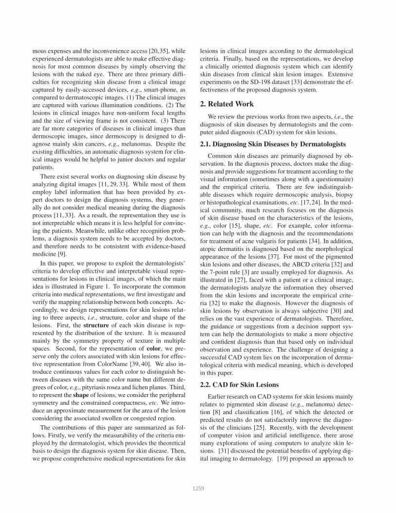
(Asymmetry) (Color) (Different structure)(Border)
(5) (7)
NC=3
(8)
(9)
168~170
185~194
(10)
(11)
(12)
(13)
(14)
(1)
(2)
(3)
(4) (6)
Figure 2. The maps between the criteria employed by dermatologists (top) and the corresponding measurable representations derived by
computer (bottom), i.e., Asymmetry (1-2), Border (3-4), Color (5-10), NC in (8) denotes the number of colors, and the numbers in (10)
denote the gray-values) and Differential structures (11-14). As shown, the dermatological criteria can be transformed into a computationally
tractable representation using specific well-designed features. For example, given (1), dermatologists make diagnoses mainly by observing
the asymmetry property, while the computer generates a binary image including the lesion and the background from which the degree of
asymmetry can easily be calculated by considering both the perimeter and area of the lesion.
recognize the skin tumor profiles, promoting the improve-
ment of dermatological diagnosis via computers. [10] em-
ployed five machine learning methods (e.g., KNN, ANN,
SVM) for classifying the pigmented skin lesions. Subse-
quently, [25] summarized the development of CAD sys-
tems, which also demonstrated the practical benefits of the
diagnosis for clinicians.
Several applications of dermatological diagnosis on
smartphones [18] have been developed, and some clinical
decision support systems (CDSSs) for melanoma diagno-
sis [7] have also been proposed. [9, 26] provided compre-
hensive surveys and evaluations on the aforementioned ap-
plications and systems, which verify that dermatologists
consider the recommendations of CDSSs as important aux-
iliary information for diagnosis. However, the existing sys-
tems diagnose skin diseases mainly using dermoscopic im-
ages, focus on the recognition of the skin cancer [11, 14],
and few of them can handle the common diseases present in
clinical images.
For the common skin diseases, [28, 29] used a dataset
with Question/Answer pairs and image-level labels; the
mean average precision of the classifier trained on the rep-
resentations they designed for skin lesions only achieves
about 25%. [33] collected a dataset of clinical skin disease
images and reported the classification results using a deep
network. The above approaches for analyzing common skin
diseases only employ several existing features developed by
the computer vision community, rather than incorporating
medical meaning for diagnosis. In this paper, we use the ac-
cepted dermatological criteria to design the representations
of skin lesions for effective diagnosis.
3. Criteria of Skin Disease
The typology of skin lesions contains primary and sec-
ondary branches, which can further subdivided into many
subcategories, e.g., viral, pigmented, and keratotic, etc.
These subcategories can also continue to be divided into
various specific skin disease, e.g., herpes simplex, eczema
and callus, etc. Faced with so many kinds of skin dis-
ease, the dermatologist must have acute observation skills
in order to make the preliminary diagnosis of skin lesions.
In practice, there exist clinical criteria used for diagnosis,
which cover the major characteristics of skin lesions and
can help dermatologists to make an accurate diagnosis. In
this work, inspired by dermatological criteria, we aim to
design medical representations used for computer diagnosis
that are consistent with observations from dermatologists.
After investigating the extensive literature relating to skin
lesions, we focus on the original ABCD criteria [1, 32] for
diagnosis as follows.
A: the original Asymmetry property indicates not only the
overall shape of the lesion in clinical images (the le-
sion in Figure 2 (1) is approximately symmetrical), but
also the contour, colors and structures for dermoscopic
images.
B: Border refers to the ill-defined and irregular borders of
the lesion for clinical images (the lesion in Figure 2
(3) has an irregular border). For dermoscopic images,
it means the sharpness of the border.
C: Color variegation means that the colors of skin lesions
for clinical images are not uniform. As shown in Fig-
ure 2 (5, 7), the lesions can either have a single color
or multiple colors. For dermoscopic images, it indi-
cates the presence of the six defined colors, for which
color normalization techniques are required to achieve
adequate recognition due to the illumination changes.
D: Diameter measures the approximate diameter of the
skin lesion for clinical images. In addition, the D cri-
terion represents the Differential structures for dermo-
scopic images. As shown in Figure 2 (11, 13), the le-
sions have different structures which lead to different
diagnoses.
1260

*
Figure 3. The mapping to visual components from dermatological criteria, i.e., ABCD, 7-point, and the two specialized criteria which focus
on a single aspect. A1, D1 and A2, D2 are related to different versions of the ABCD criteria as described in the section on Criteria. “*”
indicates that the criterion corresponds to the physical sensation of patients which cannot be measured in lesion images, e.g., numbness and
itching. The criteria with “Δ” focus on one aspect, e.g., structure or color of the lesions. As shown, the dermatological criteria are highly
correlated with the three visual components.
The ABCD criteria are widely used for diagnosing pig-
mented skin lesions in both clinical images and dermo-
scopic images. Although there are various combinations of
which the meanings are different for clinical images and
dermoscopic images, they contain the same aspects of skin
lesion, i.e., structure, color and shape (asymmetry, bor-
der), etc. The Glasgow 7-point checklist [3] is also a set
of criteria. It contains three major aspects, i.e., change in
size, shape and color, and four minor aspects, i.e., diame-
ter, inflammation, crusting or bleeding and sensory change,
among which inflammation and crusting or bleeding are
considered as properties of the structures (see Figure 2 (11,
13)).
Rather than considering multiple criteria, much research
concentrates on a single aspect of the skin disease, e.g., the
structure difference [12], the color variation [5, 36], the
shape contrast [23], etc. These criteria can guide derma-
tologists to make a decision by observing the specific prop-
erties of skin lesions. Therefore, in this paper, we consider
the comprehensive attributes of the skin diseases by com-
bining the researches concentrated on each criterion and the
original multiple criteria, e.g., ABCD or 7-point, the details
of which can be found in the next section.
4. Proposed Medical Representations
The bottom row of Figure 2 shows the initial measur-
able representations derived by the computer from the cor-
responding clinical images, e.g., Figure 2 (4) is the border
of the lesions in Figure 2 (3). As shown, the characteristics
presented in skin lesions can be distinguished not only by
dermatologists using the naked eye according to the criteria
introduced in the last section, but can also be represented
by the computer. Specifically, the medical representation
of the aforementioned criteria can be generated as follows.
The area, the diameter (D) and the border (B) of lesions
are determined by the number of pixels and the principal
axis of the connected region in computers (see Figure 2 (2,
4)), where Asymmetry (A) can also be calculated by the
geometric information of the connected region. For the cri-
terion of color (C), the computer measures it in different
color spaces (see Figure 2 (6, 8 and 10)), which enables dis-
crimination even for skin diseases with subtle changes of
color, e.g., the tinea in Figure 2 (9). In addition, the clin-
ical manifestations of the skin lesions, e.g., the inflamma-
tion, crusting or bleeding in the 7-point checklist [3], can
be quantitatively determined by the change of texture, color
and structure, on the surface of the skin (see Figure 2 (12,
14)). As shown in Figure 3, the aforementioned criteria are
highly correlated to three visual components, i.e., structure,
color and shape. For example, the Asymmetry property in
the ABCD rules, the “changes in size” and the “changes
in shape” properties in the 7-point checklist can be consid-
ered as the different aspects for the visual component of the
shape. We design representations for each visual compo-
nent based on an interpretable way of which the details are
illustrated as follows.
4.1. Structure Representation
The lesions of different diseases have various epidermal
structures, e.g., scales, lumps, scabs, hemorrhage, which
can be measured by the textures in the clinical images.
Meanwhile, the structure of skin disease has different dis-
tributions, e.g., single and homogeneous lesions or struc-
tureless, which can be measured by the symmetry of the
structure.
4.1.1 Multi-Space Texture of Lesion (MST-L)
While the dermatologist can easily observe the skin le-
sions of a patient, the clinical images of the skin disease
are captured under different illumination conditions which
can affect the calculation of the representations. To rep-
resent the structure of the lesions effectively, we calculate
the texture representation based on different color channels
which reduces the influence of the environment. We pro-
pose the multi-space texture of lesions ��� (�) for each
clinical image � as follows:
��� (�) = [��(�)]��=1, (1)
1261
where ��(�) is the set of the texture features extracted from
the �-th color channels and � denotes the number of spaces.
we employ three channels in our work, i.e., Hue, Saturation
and Brightness, from each of which we extract SIFT fea-
tures for the representation.
4.1.2 Texture Symmetry of Lesion (TS-L)
The asymmetry of structures for skin lesions turns out to
be an effective criterion. Therefore, we propose a represen-
tation based on the texture symmetry of lesions. Firstly, we
divide the lesion region detected by MBD+ [41] into two
parts, i.e., �(�)1 and �(�)2, according to its principal axes.
Then, we extract the texture features for each of the parts.
We finally represent the texture symmetry ��(�) of lesions
in the �-th color channels of the image � as follows:
���(�) = [��(�(�)1), ��(�(�)2), ��(�)]. (2)
Here, we define the symmetry representation ��(�) as:
��(�) = {∣�1�� − �2�� ∣}��=1, (3)
where � is the dimension of the extracted features, �1�� and
�2�� are the �-th entry of ��(�(�)1) and ��(�(�)2), respec-
tively. We measure the texture symmetry in Hue space since
it is scale- and shift-invariant with respect to the light inten-
sity.
4.2. Color Representation
The color of the lesion is a crucial criterion for diagnos-
ing skin diseases. For instance, regular dermatitis appears
red, while stasis dermatitis is always dark-brown. In ad-
dition, due to variations in the illumination, an appropriate
color normalization operation is helpful. In this section, we
design two representations based on the color of the lesions.
4.2.1 Color Name of Lesion (CN-L)
Objects in the real world are colorful, but skin diseases
are restricted to several colors [36]. In computer vision, the
color category is modeled by a mapping function which as-
signs the three channel values of a pixel into a sparse space
spanned by � colors. Inspired by [6], we calculate the
probability vector � = [� (��∣�)]�
�=1 for each color bin in
the � ∗ � ∗ � space (which is sparsely sampled from the
RGB-cube) as follows:
[� (��∣�)]�
�=1 ∝
�∑
�
� (��∣��) �� (∣�� − �∣���) , (4)
where � denotes the original value of the color bin, �� is
� ∗ � ∗ �-value for �, � = 387 is the total number of
the color bins and � is the set of basic colors used in our
work. According to [39] we set the weighting kernel ��
as a Gaussian kernel with � = 5. Based on the inves-
tigation of skin lesions in [5], we set � = 8 and � ={���, ����, ������, ������,�ℎ���, �����, �����, ����} in
this work. The calculation process of � (��∣�) is similar to
that in [39]. Finally, we define the color name of lesions
��(�) as:
��(�) = argmax��
[� (��∣�)]�
�=1 . (5)
4.2.2 Continuous Color Values of Lesion (CCV-L)
Different from the ColorName feature which assigns a
fixed vector for each color name, e.g., for �� = ��� :����� = (1, 0, 0), in this work we assign a continuous value
for each lesion to indicate different degrees of the color. For
each pixel in the image, we define the representation of con-
tinuous color values ��� (�) for each color bin � as:
��� (�) ∝ �(�, �)× �(�), (6)
where �(�, �) indicates the probability of mapping the color
bin � into its nearest color name �. The �(�) is the weight-
ing value of the pixel which is defined as:
�(�) =∑
∣�∣
�(�)�(�), (7)
where �(�) is the frequency of the corresponding color in
the image, �(�) is the color value of � in the ��� space.
4.3. Shape Representation
The shape of skin lesions is an important cue to diag-
nose both the category and the degree of the skin disease.
For example, a nevus lesion can be a circle while a lesion of
psoriasis appears to be scattered. In this paper, we propose
two representations according to the shape of the lesions:
(1) the symmetry of the shape and (2) the constrained com-
pactness of the lesions, both of which are calculated from
the detected lesion regions.
4.3.1 Peripheral Symmetry of Lesion (PS-L)
We first detect the lesion region in each clinical image �
using the MBD+ method. It is then divided into two parts,
i.e., �(�)1 and �(�)2, with approximately equal area and
periphery based on the consistent characteristic of the skin
lesions. Specifically, we find the longest straight line seg-
ment within the lesion, and then partition the region by the
segment’s perpendicular bisector. Finally, we represent the
peripheral symmetry of lesions based on the complementar-
ity of the two parts:
��(�) = � (�(�(�)1), �(�(�)2)), (8)
1262
where �(⋅) denotes the extracted feature of lesions and
� (⋅, ⋅) denotes the concatenation function applied to the two
features.
4.3.2 Adaptive Compactness of Lesion (AC-L)
For a skin disease region, the degree of its approxima-
tion by a circle can be useful for diagnosis, and can be rep-
resented by the criterion of compactness which is defined
as:
Com =4��
� 2, (9)
where � denotes the area and � denotes the perimeter of
the lesion. However, the lesions of skin disease can be
surrounded by swollen or subcutaneous hemorrhage which
may also be detected in clinical images. Note that those re-
gions always appear as lighter colors than those in the cen-
ter of lesions. Therefore, we adaptively calculate the area of
lesions with dynamic weights according to the pixel impor-
tance when measuring the compactness. We define the area
�� of the lesions as follows:
�� =∑
�∈�(�)
�(�∣�, �), (10)
where � denotes the pixel in the lesion �(�). �(�∣�, �) is
the probability of mapping color to a specific color category
in the Color Name feature, which reflects the significance of
the pixel � being the center of the lesion.
5. Experiments
5.1. Setup
Dataset. The SD-198 dataset [33] is the largest publicly
available dataset in this field, containing 198 skin diseases
and 6, 584 clinical images, which are collected by digital
cameras or mobile phones. The images vary in color, ex-
posure, illumination and scale, and include a wide range
of patients with different ages, gender, locations of disease,
colors of skin and different stages of the disease. We use
the standard train/test split provided by SD-198, which has
3, 292 training and 3, 292 testing images.
Evaluated Representations. We compare the proposed
representations with several standard low-level features
from three visual components, i.e., texture (SIFT, HOG,
LBP, BRIEF, SURF, Wavelet and ORB), color (Color His-
togram (CH), Color Name (CN) and ColorSIFT), Border
(GIST, Gabor, Prewitt, Sobel and Canny). Meanwhile, we
visualize the different kinds of designed representation in
Figure 5. We then fuse the best features of each compo-
nent (as determined by experiments) for a comprehensive
combination. Meanwhile, we compare the proposed rep-
resentations against the features derived by state-of-the-art
convolutional neural networks (CNNs), including CaffeNet,
VGGNet and GoogleNet. In the experiments, we also ex-
tract features from the fine-tuned models in order to provide
a fair comparison.
Metrics. In this work, we generate the recognition results
of the contrasted representations using classifiers including
� nearest neighbors (KNN), support vector machine (SVM)
and random forest (RF) (the parameters of classifiers can
be found in supplementary material). We employ classifi-
cation accuracy (ACC) to evaluate the performance of the
tested methods. Considering the recall rate for skin disease
recognition, we also evaluate sensitivity
SE =1
�
�∑
�=1
(�� )�(�� )� + (��)�
, (11)
where � denotes the number of classes, (�� )� and (��)�denotes the number of true positive and false negative esti-
mations for each class, respectively. The SE calculates the
probability of being diagnosed as positive (i.e., the percent-
age of sick people who are correctly identified as having the
condition) [2]. Larger values of both ACC and SE reflect
better diagnostic performance.
5.2. Effectiveness of the Visual Representations
We report the diagnosis performance of different rep-
resentations as well as their combinations in Table 1. As
shown, classifiers with a single low-level representation
(#1∼15) do not perform well. The best features of the low-
level components barely reach about 31% ACC (SURF with
SVM) and 29% SE (CN with RF), mainly because the com-
monly used features can neither reflect the comprehensive
medical information relating to the lesions nor eliminate the
noise in clinical images. Although the integration of com-
ponents represented by low-level features (#16) improves
the recognition performance, the best result is only 48.06%for ACC with RF. However, these results still suggest that
the pathological changes of skin lesions can be captured by
the visual components, which is consistent with Figure 2.
All the proposed representations with the inclusion of
accepted dermatologist criteria improve diagnostic perfor-
mance. For example, CCV-L (#20, which is the extension
of the CN in #9 with same dimensionality) measures the le-
sions by mapping the region to a continuous color space
spanned by eight color categories since the lesions with
same color name yet different color degrees may correspond
to different skin diseases. It achieves an accuracy of 45.32%and a sensitivity of 45.70% with RF, both of which show
around 20% improvement against the original CN feature.
The integration of the components represented by the pro-
posed representations (#23) further improves the diagnostic
performance by more than 10% compared to the integra-
tion of the components represented by low level features. It
1263

Table 1. Diagnosis performance on the SD-198 dataset with different representations and different classifiers, i.e., KNN, SVM and RF. CH
(#8) denotes the Color Histogram feature and CN (#9) denotes the Color Name feature. As shown, the proposed representations (#17∼22)
show better performance than the low-level features (#1∼15). Also, the integration of the components (#23) are more discriminative
against the integration of components represented by low-level features (#16).
Components # Features DimensionKNN SVM RF
ACC SE ACC SE ACC SE
Bas
elin
e
Texture
1 SIFT 21000 20.35 19.17 25.55 24.75 21.42 21.25
2 HOG 12400 19.14 17.85 17.62 14.45 10.54 10.66
3 LBP 23200 15.13 14.80 18.89 14.69 14.61 13.24
4 BRIEF 19200 16.74 15.62 12.21 8.39 15.67 15.03
5 SURF 38400 17.47 16.50 31.17 25.35 27.34 26.52
6 Wavelet 256 15.94 15.52 14.82 12.73 13.37 14.02
7 ORB 19200 20.53 21.44 23.21 22.94 18.86 17.46
Color
8 CH 256 12.33 12.58 4.19 4.41 18.77 16.81
9 CN 21000 20.02 20.10 20.23 21.62 27.64 28.73
10 ColorSIFT 21000 21.29 19.62 22.51 21.43 28.49 27.24
Border
11 GIST 512 21.93 21.52 16.49 17.19 15.01 12.33
12 Gabor 4000 13.67 13.00 10.15 8.62 13.73 12.43
13 Prewitt 900 12.55 13.14 11.91 10.76 11.27 10.87
14 Sobel 10000 12.27 12.03 10.42 10.18 13.46 12.46
15 Canny 10000 15.22 17.16 13.91 14.51 16.46 15.20
Integration 16 1&10&11 2500 47.36 47.23 46.84 47.24 48.06 46.73
Ou
rs
Structure17 MST-L 21000 44.99 45.62 48.06 46.38 43.23 42.73
18 TS-L 21000 47.30 47.80 48.94 47.21 43.92 43.07
Color19 CN-L 21000 42.50 43.24 38.91 39.78 44.59 46.21
20 CCV-L 21000 42.80 43.97 40.13 39.22 45.32 45.70
Shape21 PS-L 10000 30.04 30.47 38.58 38.29 38.94 36.87
22 AC-L 10000 31.50 29.75 39.73 38.92 37.61 35.42
Integration 23 18&20&22 3000 57.62 56.41 56.47 53.15 57.81 56.65
Common Skin Diseases
ACC
Common Skin Diseases
Figure 4. The detailed performance comparison of classification
accuracy (ACC) over six kinds of commonly encountered skin dis-
eases, i.e., (1) Acne Vulgaris, (2) Acute Eczema, (3) Favre Racou-
chot Nail Ridging, (4) Pomade Acne, (5) Syringoma in SD-198
dataset with different representations. The representations in this
figure correspond to the rows in Table 1, e.g., the last column in
each group employ the #23 representation of Table 1.
verifies that the three visual components corresponding to
different medical criteria are complementary to each other,
thus their incorporation provides a convincing diagnosis.
Note that the diagnostic performance of both ACC and SE
in Table 1 varies only slightly when different classifiers
are used, but substantial changes are incurred when em-
ploying different representations, indicating that providing
a comprehensive representation of lesions is a critical fac-
tor for effective diagnosis. Also, the color-based features,
i.e., #8∼10 and #19∼20, achieve better performance on SE
on average than the other features, indicating that lesion
color is particularly discriminative for diagnosis. Further-
more, the RF classifier shows better performance than the
others for color and combined features. This is because the
RF incorporates the hierarchical steps of the diagnosis from
the dependent lesion properties.
We visualize the diagnosis results of 8 representations
for 10 common skin diseases in Figure 4. As shown, single
low-level feature are not sufficient to satisfactorily recog-
nizes common skin diseases, while their combinations per-
form better. When the proposed representations and the
combinations are employed, the diagnosis performance is
further improved, demonstrating that the proposed repre-
sentations capture the characteristics of skin lesions.
5.3. Comparison with the CNN and Doctors
The effectiveness of CNNs has been proven in many
tasks of computer vision due to their powerful feature rep-
resentation, especially for classification tasks. Therefore,
we also compare the proposed representations with deep
features derived from the CaffeNet, VGGNet, GoogleNet
and ResNet, the results of which are reported in Table 2.
As shown, the deep features derived from the fine-tuned
ResNet achieve an ACC of 53.35% and SE of 51.24%,
1264
GT: 5General D: 5Junior D: 5Expert 5Rank1: 5Rank2: 119Rank3: 49
GT: 46General D: 67Junior D: 46Expert 46Rank1: 46Rank2: 39Rank3: 67
GT: 31General D: 68Junior D: 183Expert 55Rank1: 68Rank2: 178Rank3: 31
GT 1General D: --Junior D: 155Expert 1Rank1: 155Rank2: 87Rank3: 1
GT: 15General D: 35Junior D: 35Expert 35Rank1: 35Rank2: 115Rank3: 15
GT : 15General D: 44Junior D: 15Expert 15Rank1: 87Rank2: 124Rank3: 26
GT 92General D: 81Junior D: 81Expert 92Rank1: 92Rank2: 27Rank3: 115
GT: 56General D: 167Junior D: 56Expert 56Rank1: 56Rank2: 176Rank3: 67
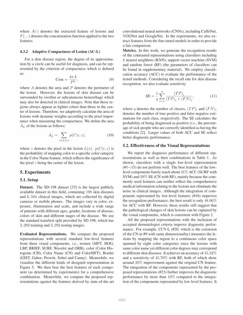
Figure 5. Examples recognized by proposed method and real doctors. Here, ‘GT’ denotes the ground-truth, and each number represents a
class of skin disease. The symbol ‘–’ denotes that the doctor cannot provide a diagnosis for this patient. We report the disease predicted
according to our method, with the top three probabilities in the last three rows. As is shown, the proposed method achieves comparable
performance with the experts, and surpasses the junior doctors for several cases.
Table 2. Comparison to the state-of-the-art deep features derived
by different CNNs and the diagnosis of doctors. There are the clas-
sification performance of the pre-trained CNN models and those
fine-tuned (ft) on SD-198 dataset with the SVM classifier. “Gen-
eral D” is general doctor who does not focus on one specific kind
of disease. “Junior D” is junior dermatologist and “Expert” is an
expert for diagnosing skin lesions.
Method ACC SE
Dee
pfe
ature
s
[33]
CaffeNet 42.31 41.57
CaffeNet + ft 46.69 45.18
VGGNet 37.91 37.25
VGGNet + ft 50.27 48.25
GoogleNet 35.33 35.21
GoogleNet + ft 46.48 45.86
ResNet 48.78 47.62
ResNet + ft 53.35 51.24
Doct
ors General D 49.00 47.50
Junior D 52.00 53.40
Expert 83.29 85.00
Ours 56.47 53.15
which is the state-of-the-art performance. In contrast, the
proposed representations improve ACC and SE with 3.12%and 1.91%, respectively. The lack of a large-scale well-
labeled dataset for this topic limits the application of deep
methods, which cannot learn a distinctive embedding based
on the existing data. In contrast, the proposed medical rep-
resentations which are consistent with the observations of
dermatologists according to empirical criteria do not rely
on such a large-scale dataset. These dermatologist criteria
are employed to distinguish diseases by focusing on certain
manifestations, based on which the designed features are
mapped to a low-rank feature space. Meanwhile, existing
deep models rely on the pre-training process using large-
scale datasets (ImageNet), which have a large semantic gap
regarding the task of skin disease analysis.
In this work, we also report the comparison between the
proposed method and three kind of real doctors, i.e., the
general, junior and the expert. ‘General doctor’ indicates
general doctors with comprehensive knowledge who have
mostly practised in the private clinic and not focus on one
kind of disease. ‘Junior dermatologist’ represents the in-
tern dermatologist. ‘Expert’ denotes senior doctors who
have considerable experience on the skin disease diagnosis.
For each category of doctor we invite two doctors to un-
dertake diagnosis independently followed by a discussion
when they are divergent. Table 2 shows the diagnosis re-
sults of doctor, and the result of our method is better than
the diagnosis of general and junior doctor on SD-198. Note
that all the diagnoses of doctors are made by just observing
the picture, and they will make better diagnoses by touching
the lesion and asking questions. Meanwhile, Figure 5 shows
some examples with the diagnosis results from doctors and
our method. As is shown, our proposed method shows com-
parable performance on diagnosing multiple skin diseases
compared to real doctors.
6. Conclusions
In this paper we address the problem of clinical skin le-
sion diagnosis, which is challenging compared to skin can-
cer recognition on dermatoscopic images. We verify that
the criteria employed by clinicians in the diagnosis process
can be measured by computers. Accordingly, we design
six discriminative and interpretable representations for dis-
tinguishing skin lesions by incorporating the accepted der-
matological criteria. Experiments on a benchmark dataset
demonstrate the proposed representations outperform both
the low-level features and the deep features. Furthermore,
the final performance on clinical images with 198 categories
of skin disease is comparable with dermatologists.
Acknowledgments
This research was supported by NSFC (No.
61620106008, 61572264, 61301238, 61201424), and
the Open Project Program of the National Laboratory of
Pattern Recognition (NLPR).
1265
References
[1] N. R. Abbasi, H. M. Shaw, D. S. Rigel, R. J. Friedman, W. H. Mc-
Carthy, I. Osman, A. W. Kopf, and D. Polsky. Early diagnosis of
cutaneous melanoma: revisiting the ABCD criteria. The Journal of
the American Medical Association, 292(22), 2004.
[2] D. G. Altman and J. M. Bland. Diagnostic tests. 1: Sensitivity and
specificity. British Medical Journal, 308(6943), 1994.
[3] G. Argenziano, G. Fabbrocini, P. Carli, V. De Giorgi, E. Sammarco,
and M. Delfino. Epiluminescence microscopy for the diagnosis of
doubtful melanocytic skin lesions: comparison of the ABCD rule of
dermatoscopy and a new 7-point checklist based on pattern analysis.
Archives of Dermatology, 134(12), 1998.
[4] C. Barata, M. A. Figueiredo, M. E. Celebi, and J. S. Marques. Lo-
cal features applied to dermoscopy images: Bag-of-features versus
sparse coding. In IbPRIA, 2017.
[5] G. S. Barsh. What controls variation in human skin color? PLoS
Biology, 1(1), 2003.
[6] R. Benavente, M. Vanrell, and R. Baldrich. A data set for fuzzy
colour naming. Color Research & Application, 31(1), 2006.
[7] M. Binder, H. Kittler, S. Dreiseitl, H. Ganster, K. Wolff, and H. Pe-
hamberger. Computer-aided epiluminescence microscopy of pig-
mented skin lesions: the value of clinical data for the classification
process. Melanoma Research, 10(6), 2000.
[8] A. P. Dhawan, R. Gordon, and R. M. Rangayyan. Nevoscopy: three-
dimensional computed tomography of nevi and melanomas in situ
by transillumination. IEEE Transactions on Medical Imaging, 3(2),
1984.
[9] S. Dreiseitl and M. Binder. Do physicians value decision support? a
look at the effect of decision support systems on physician opinion.
Artificial Intelligence in Medicine, 33(1), 2005.
[10] S. Dreiseitl, L. Ohno-Machado, H. Kittler, S. Vinterbo, H. Billhardt,
and M. Binder. A comparison of machine learning methods for the
diagnosis of pigmented skin lesions. Journal of Biomedical Infor-
matics, 34(1), 2001.
[11] A. Esteva, B. Kuprel, R. A. Novoa, J. Ko, S. M. Swetter, H. M. Blau,
and S. Thrun. Dermatologist-level classification of skin cancer with
deep neural networks. Nature, 542(7639), 2017.
[12] T. Fitzpatrick, J. Bernhard, T. Cropley, et al. The structure of skin
lesions and fundamentals of diagnosis. Dermatology in General
Medicine, 5, 1999.
[13] A. Gewirtzman, J.-H. Saurat, and R. Braun. An evaluation of der-
moscopy fluids and application techniques. British Journal of Der-
matology, 149(1), 2003.
[14] J. Glaister, A. Wong, and D. A. Clausi. Segmentation of skin le-
sions from digital images using joint statistical texture distinctive-
ness. IEEE Transactions on Biomedical Engineering, 61(4), 2014.
[15] H. M. Gloster and K. Neal. Skin cancer in skin of color. Journal of
the American Academy of Dermatology, 55(5), 2006.
[16] A. Green, N. Martin, G. McKenzie, J. Pfitzner, F. Quintarelli,
B. Thomas, M. O’Rourke, and N. Knight. Computer image analysis
of pigmented skin lesions. Melanoma Research, 1(4), 1991.
[17] T. P. Habif, M. S. Chapman, J. L. Campbell, J. G. Dinulos, and
K. A. Zug. Skin Disease E-Book: Diagnosis and Treatment. Elsevier
Health Sciences, 2011.
[18] A. Hamilton and R. Brady. Medical professional involvement in
smartphone apps in dermatology. British Journal of Dermatology,
167(1), 2012.
[19] H. Handels, T. Roß, J. Kreusch, H. H. Wolff, and S. J. Poeppl. Fea-
ture selection for optimized skin tumor recognition using genetic al-
gorithms. Artificial Intelligence in Medicine, 16(3), 1999.
[20] R. J. Hay and L. C. Fuller. The assessment of dermatological needs in
resource-poor regions. International journal of dermatology, 50(5),
2011.
[21] R. J. Hay, N. E. Johns, H. C. Williams, I. W. Bolliger, R. P. Dellavalle,
D. J. Margolis, R. Marks, L. Naldi, M. A. Weinstock, S. K. Wulf,
et al. The global burden of skin disease in 2010: an analysis of the
prevalence and impact of skin conditions. Journal of Investigative
Dermatology, 134(6), 2014.
[22] M.-L. T. Johnson and J. Roberts. Skin conditions and related need for
medical care among persons 1-74 years, United States, 1971-1974.
Vital & Health Statistics, (212), 1978.
[23] S. Jowett and T. Ryan. Skin disease and handicap: an analysis of the
impact of skin conditions. Social Science & Medicine, 20(4), 1985.
[24] L. S. King. What is disease? Philosophy of Science, 21(3), 1954.
[25] K. Korotkov and R. Garcia. Computerized analysis of pigmented
skin lesions: a review. Artificial Intelligence in Medicine, 56(2),
2012.
[26] I. Maglogiannis and C. N. Doukas. Overview of advanced computer
vision systems for skin lesions characterization. IEEE Transactions
on Information Technology in Biomedicine, 2009.
[27] A. A. Marghoob, L. D. Swindle, C. Z. Moricz, F. A. S. Negron,
B. Slue, A. C. Halpern, and A. W. Kopf. Instruments and new tech-
nologies for the in vivo diagnosis of melanoma. Journal of the Amer-
ican Academy of Dermatology, 49(5), 2003.
[28] O. Razeghi and G. Qiu. 2309 skin conditions and crowd-sourced
high-level knowledge dataset for building a computer aided diagnosis
system. In ISBI, 2014.
[29] O. Razeghi, Q. Zhang, and G. Qiu. Interactive skin condition recog-
nition. In ICME, 2013.
[30] J. Spalding. The doctor with an inherited defect of colour vision:
effect on clinical skills. The British Journal of General Practice,
43(366), 1993.
[31] W. V. Stoecker and R. H. Moss. Digital imaging in dermatology.
Computerized Medical Imaging and Graphics, 16(3), 1992.
[32] W. Stolz, A. Riemann, A. Cognetta, L. Pillet, W. Abmayr,
D. Holzel, P. Bilek, F. Nachbar, and M. Landthaler. ABCD rule
of dermatoscopy-a new practical method for early recognition of
malignant-melanoma. European Journal of Dermatology, 4(7),
1994.
[33] X. Sun, J. Yang, M. Sun, and K. Wang. A benchmark for automatic
visual classification of clinical skin disease images. In ECCV, 2016.
[34] S. C. Taylor, F. Cook-Bolden, Z. Rahman, and D. Strachan. Acne
vulgaris in skin of color. Journal of the American Academy of Der-
matology, 2002.
[35] M. W. Tsang and C. L. Kovarik. Global access to dermatopathology
services: physician survey of availability and needs in sub-saharan
africa. Journal of the American Academy of Dermatology, 63(2),
2010.
[36] Z. J. Twardowski and B. F. Prowant. Classification of normal and
diseased exit sites. Peritoneal Dialysis International, 16(Suppl 3),
1996.
[37] M. Uehara and T. Sawai. A longitudinal study of contact sensitivity
in patients with atopic dermatitis. Archives of Dermatology, 125(3),
1989.
[38] R. P. Usatine and M. Riojas. Diagnosis and management of contact
dermatitis. American Family Physician, 82(3), 2010.
[39] J. Van De Weijer, C. Schmid, and J. Verbeek. Learning color names
from real-world images. In CVPR, 2007.
[40] J. Van De Weijer, C. Schmid, J. Verbeek, and D. Larlus. Learning
color names for real-world applications. IEEE Transactions on Im-
age Processing, 18(7), 2009.
[41] J. Zhang, S. Sclaroff, Z. Lin, X. Shen, B. Price, and R. Mech. Mini-
mum barrier salient object detection at 80 fps. In ICCV, 2015.
1266


![LI ET AL.: SEMI-SUPERVISED SKIN LESION SEGMENTATION …dermoscopy images [14,18]. For example, Jaisakthi et al. [14] proposed a semi-supervised skin lesion segmentation method using](https://img.dokumen.tips/doc/110x75/60658319b2024701434d8eca/li-et-al-semi-supervised-skin-lesion-segmentation-dermoscopy-images-1418-for.jpg)