Embed Size (px)
Citation preview
6/22/05
Send/Handouts 1
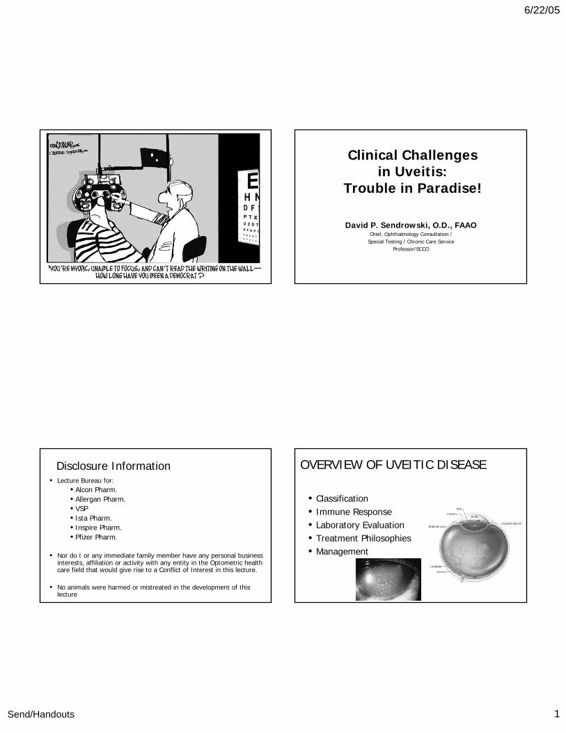
Clinical Challengesin Uveitis:
Trouble in Paradise!
David P. Sendrowski, O.D., FAAOChief, Ophthalmology Consultation /
Special Testing / Chronic Care ServiceProfessor/SCCO
Disclosure Information• Lecture Bureau for:
• Alcon Pharm.• Allergan Pharm.• VSP • Ista Pharm.• Inspire Pharm.• Pfizer Pharm.
• Nor do I or any immediate family member have any personal business interests, affiliation or activity with any entity in the Optometric health care field that would give rise to a Conflict of Interest in this lecture.
• No animals were harmed or mistreated in the development of this lecture
OVERVIEW OF UVEITIC DISEASE
• Classification• Immune Response• Laboratory Evaluation• Treatment Philosophies• Management

6/22/05
Send/Handouts 2
Overview of Immune Response in Uveitis – so simple even a Caveman could understand it!!!!!!
1. Presentation of ocular antigen to T lymphocyte2. Activation and clonal expansion (CD 4 & 8 ^)3. Activated Uveogenic T Cells AND circulating
leukocytes come to eye4. Migration into eye with initial breakdown of
Blood / Ocular barrier.5. Mast cell degranulation and inflammation
amplification.
UVEITIS WORK-UP
• History - vital component– Unilateral or bilateral presentation– Time course– Prior therapy– Systemic diseases - (i.e., Sarcoidosis, Herpes
simplex, Herpes zoster)– STD's - (i.e. AIDS, syphilis, chlamydia)
– Diet - (i.e. Toxoplasmosis)– Pets - (i.e. Toxoplasmosis, Toxocariasis)– IV drug use - (i.e. fungal)– Family history
– Age– Race– Sex– Geographic location (Ohio / Mississippi / Missouri)– Symptoms - (i.e. photophobia, pain, redness,
decreased vision, lacrimation, etc.)

6/22/05
Send/Handouts 3
Best Three Questions to ask and Document in Patient with Uveitis
1. Recent Skin Rashes : Sarcoid, Reiter’s, Bechet’s, Psoriatic Arthritis.
2. Recent Respiratory Problems: Sarcoid, Histo, Coccidiomycosis, TB
3. Recent Joint Pain or Stiffness: Anklyosing Spondylitis, Reiter’s, IBD, Psoriatic Arthritis.
“Suggested” office work up• Physical observation peri-ocular tissues• Pupils• Biomicroscopy
– Endothel. (KPs) type, configuration.– A/C– Iris--look for nodules, PS, AS– Ant. Vitreous
• BIO and Extended Ophthal. (R/o Post. Uveitis)• IOP• Charges (99214 or 99204)
Lab work-up• First time attack- no signs & suggestive Hx.
(none or minimal)• Referral letter to M.D.• If possible - order a few tests if uveitis is
recurrent/severe• Things to ponder:
– 50% labs come up negative– Should be 50% certain of the disease in chair to
direct lab test
Treatment Philosophies

6/22/05
Send/Handouts 4
Steroids• Need a long acting steroid with good anti-
inflammatory properties (Pred. Forte 1%, Lotemax, Durezol – good choices)
• Need to use the steroid aggressively in the first 10 to 14 days (q1h or q2h)– you have no idea what level the inflammation is at.
• If AC cells drop from one grade to a lower grade (3+ 2+), continue with frequency of (q1h or q2h), wait for significant reduction to “taper”
New Steroid
• Never (?) switch steroids if Tx. is working but IOP elevates– uveitis patients are steroid responders too.
• Always evaluate IOP at every visit as well as corneal to R/o HS
• Taper steroid from q1h to q2h when cells have significantly reduced or minimal AC rxn before you start to taper. Inflammation does way more damage than IOP.
Cycloplegia• Good for the first 3 days to reduce photophobia,
PS/PAS, and help shore up blood/ocular barrier• If no synechiae are present D/C after 3 days• If synchiae already there – may need to keep
patient on cyclo. Tx. or if fibrin level is high in AC• If multiple synechiae present - try
atropine/scopolamine/10% Neosynepherine to break synechiae and consider longer duration cycloplegics if synechiae are 180° or greater and IOP is elevating

6/22/05
Send/Handouts 5
Few Items• Steroids cause ptosis (1-2mm) - it goes away after D/C
steroid• Steroids cause mydriasis - it goes away after D/C
steroid• Steroids alters TBUT, corneal rigidity, tear production -
returns to normal after D/C steroid• Pressure usually goes up 10 mmHg for steroid
responder (30% max. with exceptions)• Secondary glaucoma – most handled with topical
therapy, some require surgical intervention• Two types of glaucoma: Pupillary Block Glaucoma
versus secondary steroid induced / AACG (PAS)
Take Home Pearl:
• In an effort to do no harm with a topical steroid, practitioners utilize steroids sparingly causing greater harm to the eye in the long run
Future for biological therapy for uveitis
• Include AB’s, soluble receptors, and cytokines• Anti-TNF alpha– most widely employed*• IntraVitreal Injections (Anti-Inflam. & nano)• Biologics have tremendous potential in the
treatment of ocular inflammation but studies have been limited and side-effects are not completely known.**
• */**Rosenbaum, JT: Current Opinion in Ophthalmology, 2015, 21: 473-477
New and Standard Treatment ModalitiesAntimetabolites
• Azathioprine (Imuran) • Methotrexate (Rheumatrex) • Mycophenolate Mofetil (Cellcept)
• T-Cell Inhibitors• Cyclosporine (Sandimmune, Neoral) • Tacrolimus (Prograf)
• Alkylating Agents• Chlorambucil (Leukeran) • Cyclophophamide (Cytoxan)
• Biologics• Infliximab (Remicade) **• Etanercept (Enbrel) • Interferon (Avonex) • Daclizumab (Zenapax) • Alefacept (Amevive) • Efalizumab (Raptiva)
A protein produced by molecular recombinant DNA technology and designed to have atherapeutic effect on the inflammationbased on current understanding of the disease pathogenesis

6/22/05
Send/Handouts 6
Key Features in Uveitis Treatment• Topical steroids are the mainstay of therapy for
acute iridocyclitis• Systemic corticosteroids are beneficial for
bilateral, non-infectious, endogenous uveitis (avoidance of chronic use due to complications)
• Newer classes of immunomodulatory agents currently are: anti-metabolites, calcineurin inhibitors, alkylating agents and biologics.
• Determination of an immunosuppressive regimen requires a careful assessment of the benefits and risks of therapy
NSAIDS that can be utilized with AU
• Fenoprofen Nalfon• Ketoprofen Oridus• Piroxicam Feldene• Flurbiprofen Ansaid• Ketorolac Toradol• Naproxen Naprosyn• Ibuprofen Motrin, Rufen
Top Causes / Associations of Anterior Uveitis
• Idiopathic (AKA: Undifferentiated)• HLA- B27 related (CRAP)• Sarcoidosis• Herpes Virus (HSV & VZV)• Juvenile Idiopathic arthritis – associated uveitis• Fuch’s Heterochromic Iridocyclitis (FHI)• Posner-Schlossman Syndrome• Syphilis• Masquerade Syndrome
DIFFERENTIAL DIAGNOSIS AND TREATMENT OF UVEITIS

6/22/05
Send/Handouts 7
• Anklyosing Spondylitis– Etiology
• Disease of the axial skeleton• Males (3x) > females (1x)• Ages 20-40• Women– older and more
– Peripheral joint involvement.– Neck pain and breast pain
• Affects 0.1% of Caucasian adults– Lower back pain in morning
lasting 15 min. > 3 months– Other complaints are "pain in
the chest cavity and difficulty with chest expansion"
– anorexia, fever, malaise -systemic signs
– Ocular presentation• Anterior uveitis - usually unilateral• Recurrence in same or other eye• Rapid onset of pain and photophobia• Flare may be heavy or light• Posterior synechiae form quickly• Episodes vary 2-6 weeks

6/22/05
Send/Handouts 8
Fibrin clots or aggregations
Posterior Synechiae
Peripheral Anterior Synechiae
Investigations
• X-ray of sacroiliac joints• (MRI for early enthesopathy if X-rays negative----
enthesopathies are disorders of peripheral ligamentous or muscular attachments)
• ESR / C-reactive protein • RF (-) / ANA (-)• Family Hx of AS• Alkaline phosphotase levels • HLA B27 (+) tissue typing• Vitamin D Levels –Risk of Osteoporosis• Electrocardiogram (??) if heart Dz in the FH

6/22/05
Send/Handouts 9
Ocular / Systemic Therapy
•Topical steroids (q1h to q2h - initially)•Topical cycloplegic agents (QD or BID)•Periocular steroid injections for more severe cases•NSAIDS, COX-2 inhibitors - main stay for systemic
treatment & physical therapy•TNF inhibitors (Etanercept, Ifleximab)
Can’t see. Can’t pee, Can’t Dance with Me
• Reiter's Syndrome (Reactive Arthritis)– Etiology
• Triad (conjunctivitis, arthritis, urethritis) • Anterior Uveitis in 15 to 20%• Males > females (9:1 / 1:1)• Ages 18 - 40 more common
Forms of RS
• Post venereal = conjunctivitis (30-60%) • Arthritis (asymmetric- large weight bearing joints) presenting
with anterior uveitis (15%)• Post dysenteric : Shigella, Yersinia, Salmonella, Campylobacter
– (usually 1 to 4 weeks after the dysentery)• Chlamydia Trachomatis, most common agent causing the
venereal disorder so look for signs of inclusion conjunctivitis or Ureaplasma urealyticum
•Inclusion conjunctivitis is most common presentation post infection (2-4 wks uveitis)
•Superior micropannus of cornea (if chlamydial infection)
•Anterior uveitis arthritic form•Systemic signs with ocular presentation:
keratodermal blennorrhagicum, circinata blanatis (ddx: pustular psoriasis), aphthous stomatitis, rheumatologic signs: plantar fasciitis.
Ocular presentation / Systemic signs

6/22/05
Send/Handouts 10
Keratoderma BlennorrhagicumBalanitis circinataAphthous Ulcers
•Heavy flare and cells (flare may be plasmoid)
•Course is 2-6 weeks•Glaucoma possible after repeated episodes
•X-rays of knees, ankles, feet, heels, Achilles tendon, and sacroiliac area
•Cultures for chlamydia from conjunctiva, urethra
•HLA-B27 (++)•Fecal cultures for post dysenteric•ESR and CBC (leukocytosis with mild anemia ?)
•Creatinine – Elevated
Investigation

6/22/05
Send/Handouts 11
Investigation
•Nail pitting•Palate / tongue ulcers•HLA tissue typing (HLA B27)•RF (-)•ANA (-)
Ocular / Systemic Treatment
•Topical steroids (Q1h to Q2h initially)•Quick removal of steroids may cause return of
inflammation •Topical cycloplegics (QD to BID)•Periocular steroid injections for more severe cases•NSAIDs, second line: methotrexate &
sulfasalazine•Co-management with interest for venereal;
rheumatologist for arthritis is recommended
Systemic therapy (if venereal)
•Oral tetracycline 250mg •Oral doxycycline 100 mg•Oral arithromycin 1000 mg (zithromax)

6/22/05
Send/Handouts 12
– Etiology•Usually African-American (blacks 8-10X > whites)•Also Euro Whites / Japanese patients•Women = men•Multi-systemic disease: hallmark, non-caseating
granulomas (eval. the peri-ocular region)•Accounts for 3-10% of all uveitic cases•50% patients develop ocular sequellae– usually
anterior•Etiology unknown – immunological pathology
Sarcoidosis
•Most commonly seen in the Atlantic Gulf Coast states
•Ages 20-40 (children under age 5) 60 to 70•Disease has anterior and posterior involvement
•Acute / chronic presentation•Systemic symptoms: respiratory (most common symptom) associated with fever, fatigue (27%), dsypnea, weight loss (28%)
•Patients may be asymptomatic at the time of Dx.
•Bilateral iridocyclitis•Dense posterior synechiae•Mild pain and photophobia•Palpebral conjunctival may manifest sarcoid
granulomas (17%)•Lacrimal gland enlargement
Ocular Presentation

6/22/05
Send/Handouts 13
•KPs are extremely large (mutton-fat)•Iris nodules are usually present (20%)•Cataracts and glaucoma are complications (chronic form)
•Posterior segment: (involvement is less frequent)–Vitreous snowballs – located inferiorly and lie on the retinal surface (vitritis)
–"Candle wax drippings" – (en taches de bougie) venule involvement
–Perivenous sheathing–Choroidal lesions (Dalen-Fuch's nodules)

6/22/05
Send/Handouts 14
–Choroidal granuloma (rare)–Chronic cystoid macular edema–Neovascularization of the disc (15%)–Optic disc swelling (40%)
•Skin lesions - erythema nodosum or sarcoid nodules under the skin
•Lungs are frequently affected •Facial nerve palsies are possible as well
•Chest X-ray (hilar adenopathy) Lung is the number one organ affected by the disease
•ACE (+) >67U/L•Serum lysozyme ()•PPD (-)•Blood panel
Investigations
May present as:-Shortness of breath-Chest pain-Persistent dry cough

6/22/05
Send/Handouts 15
•Gallium scan of the head and neck•Biopsy of conjunctiva or skin or lacrimal gland nodule
•ACE and gallium scan will give false negative if patient is taking steroids
•Pulmonary function tests
•Sickle cell disease•Lyme Disease•TB•Idiopathic pars planitis•Histoplasmosis / Coccocidiomycosis
Differential diagnosis
Systemic Treatment1. Spontaneous resolution in 24-36 months (50%)2. Low dose Corticosteroids (20-40 mg/d)3. Cyclosporin therapy – no safe long term approach
with this drug4. Methotrexate--low dose good for long term tx
(azathioprine)5. Chloroquine (Hydrocholoroquine) for pulmonary6. Biologics (Entanercept / Infliximab for refractory
uveitis)
•Topical steroids (q1h to q2h) depends on activity
•Topical cycloplegics (BID or QD)•Periocular steroids if topicals are ineffective (intermediate)
•Oral steroids and histamine H2 blocker if treating intermediate / posterior uveitis, facial nerve palsies, pulmonary problems
•Cyclosporin A - effective in patients intolerant to oral steroids
Ocular Treatment

6/22/05
Send/Handouts 16
•Anti-glaucoma meds (topical and oral) for 2° complications (aqueous suppressants)
•Panretinal photocoagulation for neovascularization
•Patients need to be re-examined in 3-7 days
•Asymptomatic patients seen Q 6 months•Steroid treated patients need to be seen Q 3 months
•Children with Sarcoidosis need to be seen Q 1-3 months
Thank you for taking the course!