Embed Size (px)
Citation preview
CLASSIFICATION OF ANEMIASWhat is anemia, how do you diagnose anemia, and how are the different anemias classified?
DEFINITION OF ANEMIA
In its broadest sense, anemia is a functional inability of the blood to supply the tissue with adequate O2 for proper metabolic function.
Anemia is not a disease, but rather the expression of an underlying disorder or disease.A specific diagnosis is made by:
DEFINITION OF ANEMIAPatient historyPatient physical examSigns and symptoms exhibited by the patient
Hematologic lab findingsIdentification of the cause of anemia
is important so that appropriate therapy is used to treat the anemia.
Anemia is usually associated with decreased levels of hemoglobin and/or a decreased packed cell volume (hematocrit), and/or a decreased RBC count.
DEFINITION OF ANEMIA
Occasionally there is an abnormal hemoglobin with an increased O2 affinity resulting in an anemia with normal or raised hemoglobin levels, hematocrit, or RBC count.
Before making a diagnosis of anemia, one must consider:Age
DEFINITION OF ANEMIA
SexGeographic locationPresence or absence of lung disease
Remember that the bone marrow has the capacity to increase RBC production 5-10 times the normal production.Thus, if all necessary raw products
are available, the RBC life span can decrease to about 18 days before bone marrow compensation is inadequate and anemia develops.
DEFINITION OF ANEMIA
An increased production of RBCs in the bone marrow is seen in the peripheral smear as an increased reticulocyte count since new RBCs are released as reticulocytes.
If the bone marrow production of RBCs remains the same or is decreased with RBCs that have a decreased survival time, anemia will rapidly develop.

DEFINITION OF ANEMIA
There is no mechanism for increasing RBC survival time when there is an inadequate bone marrow response, so anemia will develop rapidly.
In summary, anemia may develop:When RBC loss or destruction exceeds the maximal capacity of bone marrow RBC production or
When bone marrow production is impaired
DEFINITION OF ANEMIA
Various diseases and disorders are associated with decreased hemoglobin levels. These include:Nutritional deficienciesExternal or internal blood lossIncreased destruction of RBCsIneffective or decreased production
of RBCs

DEFINITION OF ANEMIA
Abnormal hemoglobin synthesisBone marrow suppression by toxins,
chemicals, or radiationInfectionBone marrow replacement by
malignant cells
SIGNIFICANCE OF ANEMIA AND COMPENSATORY MECHANISMS
The signs and symptoms of anemia range from slight fatigue to life threatening reactions depending uponRate of onsetSeverityAbility of the body to adapt
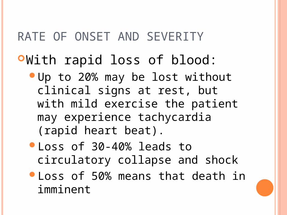
RATE OF ONSET AND SEVERITY
With rapid loss of blood: Up to 20% may be lost without
clinical signs at rest, but with mild exercise the patient may experience tachycardia (rapid heart beat).
Loss of 30-40% leads to circulatory collapse and shock
Loss of 50% means that death in imminent

RATE OF ONSET AND SEVERITY
In slowly developing anemias, a very severe drop in hemoglobin of up to 50% may occur without the threat of shock or death. This is because the body has
adaptive or compensatory mechanisms to allow the organs to function at hemoglobin levels of 50% of normal. These include (in addition to increased bone marrow output):
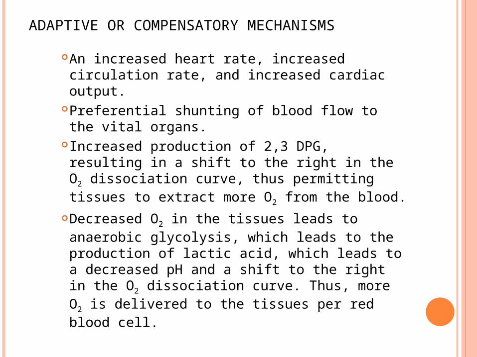
ADAPTIVE OR COMPENSATORY MECHANISMS
An increased heart rate, increased circulation rate, and increased cardiac output.
Preferential shunting of blood flow to the vital organs.
Increased production of 2,3 DPG, resulting in a shift to the right in the O2 dissociation curve, thus permitting tissues to extract more O2 from the blood.
Decreased O2 in the tissues leads to anaerobic glycolysis, which leads to the production of lactic acid, which leads to a decreased pH and a shift to the right in the O2 dissociation curve. Thus, more O2 is delivered to the tissues per red blood cell.
DIAGNOSIS OF ANEMIA
How does one make a clinical diagnosis of anemia?Patient history
Dietary habitsMedication being takenPossible exposure to chemicals and/or toxins
Description and duration of symptoms
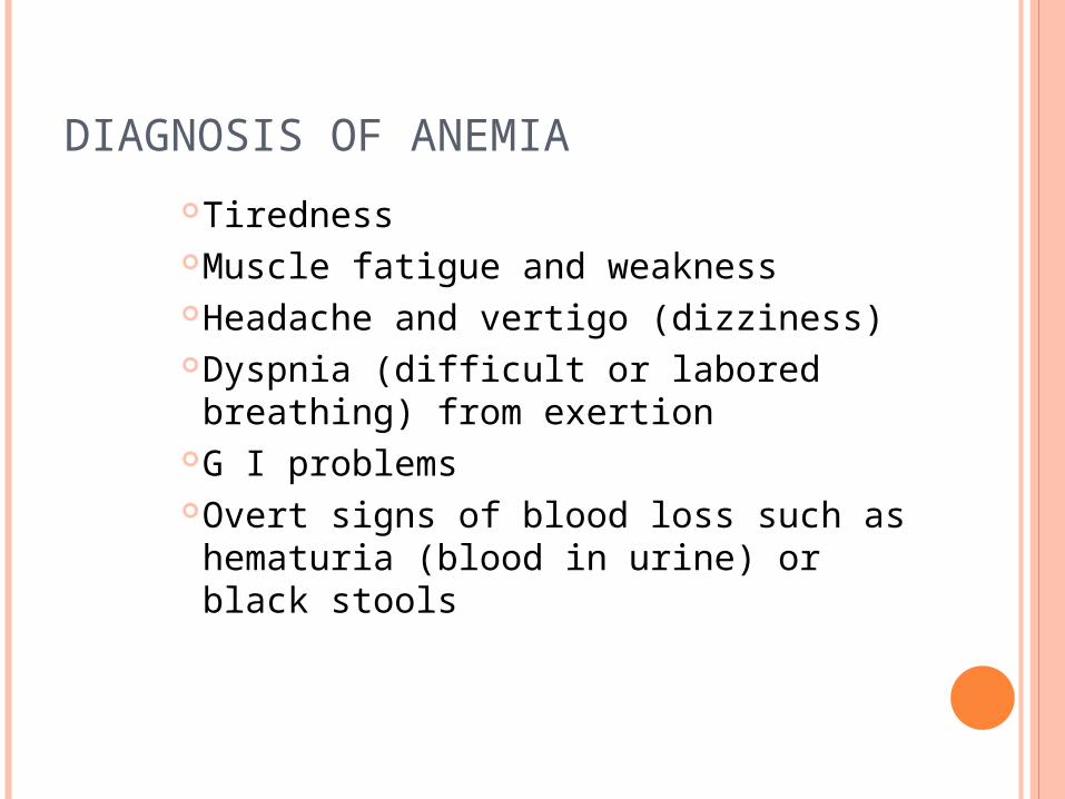
DIAGNOSIS OF ANEMIA
TirednessMuscle fatigue and weaknessHeadache and vertigo (dizziness)Dyspnia (difficult or labored breathing) from exertion
G I problemsOvert signs of blood loss such as hematuria (blood in urine) or black stools
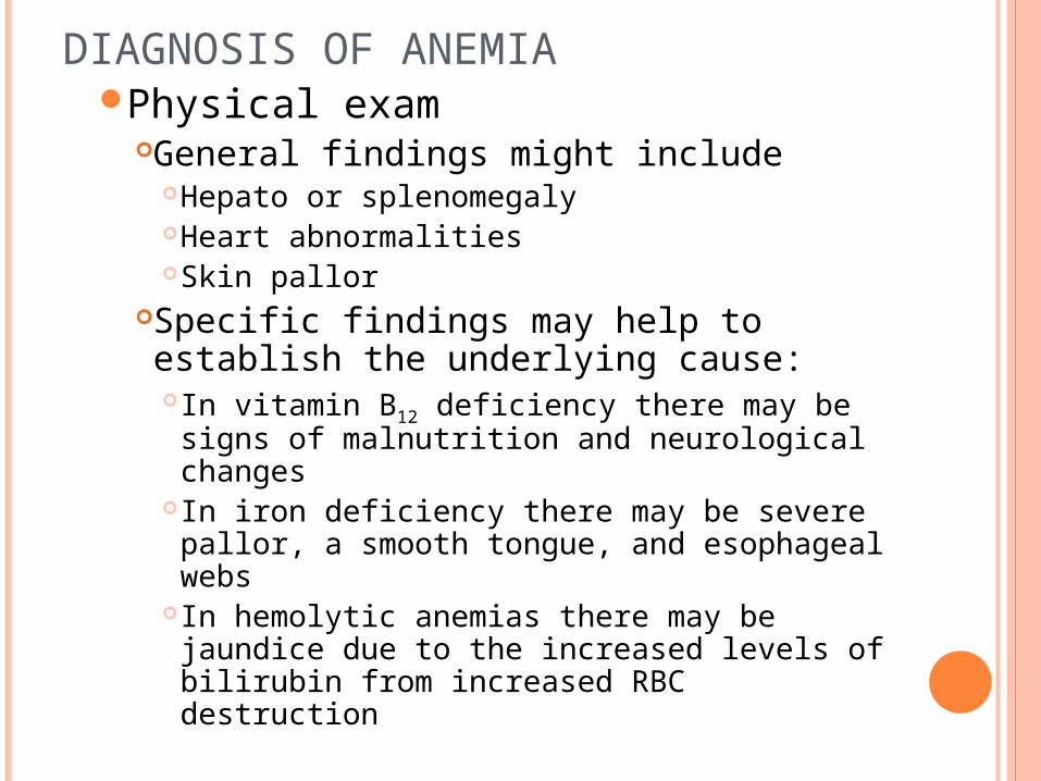
DIAGNOSIS OF ANEMIAPhysical exam
General findings might includeHepato or splenomegalyHeart abnormalitiesSkin pallor
Specific findings may help to establish the underlying cause:In vitamin B12 deficiency there may be signs of malnutrition and neurological changes
In iron deficiency there may be severe pallor, a smooth tongue, and esophageal webs
In hemolytic anemias there may be jaundice due to the increased levels of bilirubin from increased RBC destruction
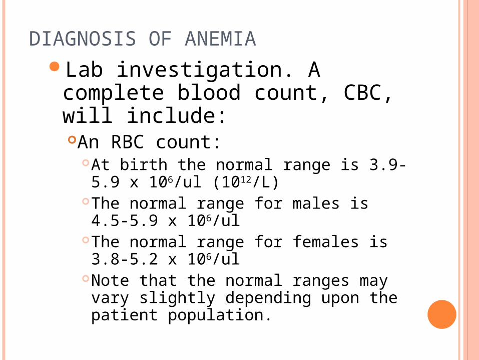
DIAGNOSIS OF ANEMIALab investigation. A complete
blood count, CBC, will include:An RBC count:
At birth the normal range is 3.9-5.9 x 10⁶/ul (1012/L)
The normal range for males is 4.5-5.9 x 10⁶/ul
The normal range for females is 3.8-5.2 x 10⁶/ul
Note that the normal ranges may vary slightly depending upon the patient population.
DIAGNOSIS OF ANEMIA
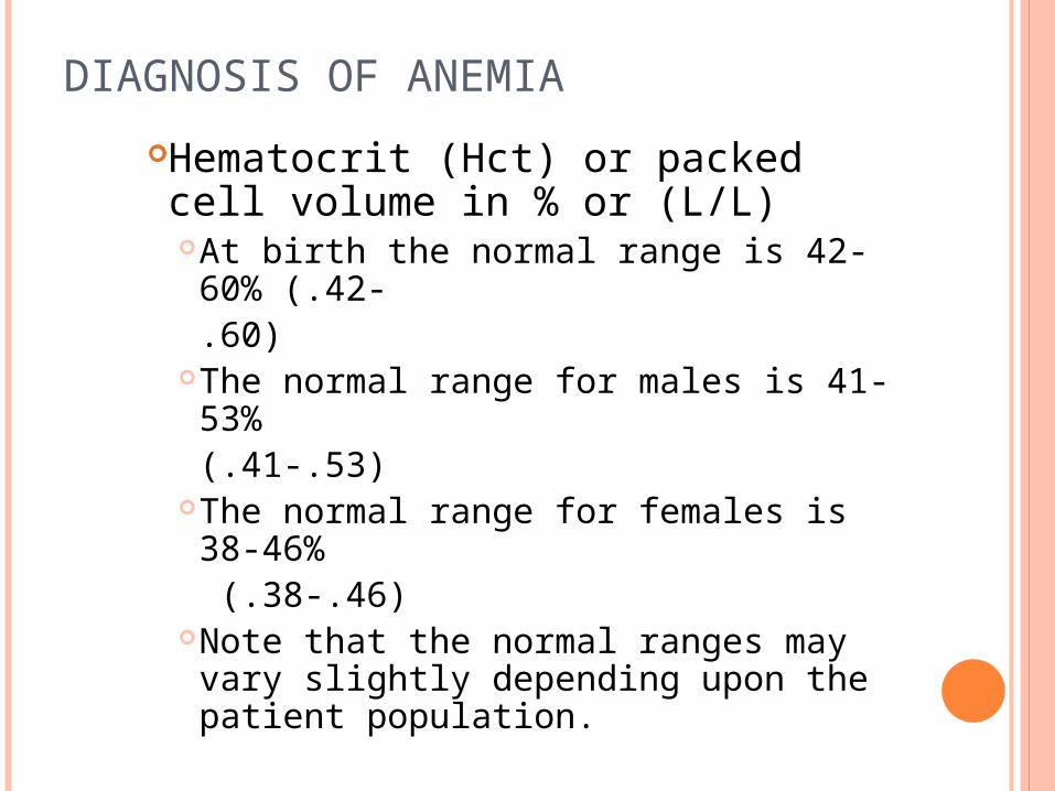
Hematocrit (Hct) or packed cell volume in % or (L/L)At birth the normal range is 42-60% (.42-.60)
The normal range for males is 41-53% (.41-.53)
The normal range for females is 38-46% (.38-.46)
Note that the normal ranges may vary slightly depending upon the patient population.
DIAGNOSIS OF ANEMIAHemoglobin concentration in grams/deciliter - the RBCs are lysed and the hemoglobin is measured spectrophotometricallyAt birth the normal range is 13.5-20 g/dlThe normal range for males is 13.5-17.5 g/dl
The normal range for females is 12-16 g/dlNote that the normal ranges may vary slightly depending upon the patient population.
RBC indices – these utilize results of the RBC count, hematocrit, and hemoglobin to calculate 4 parameters:
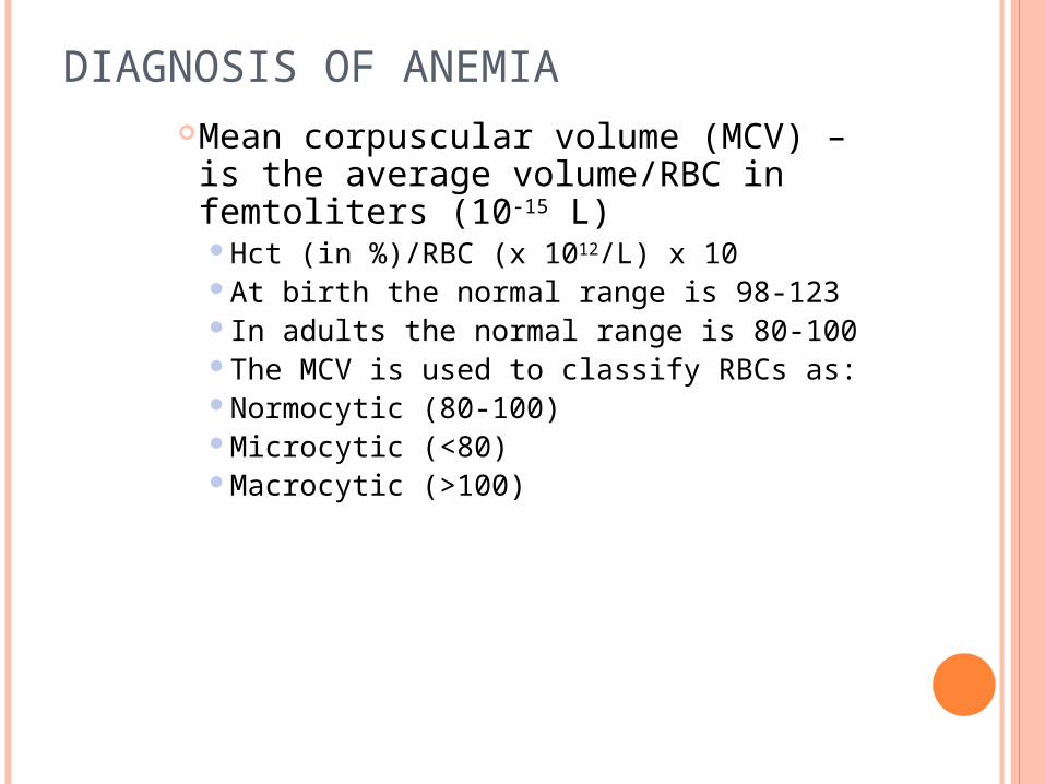
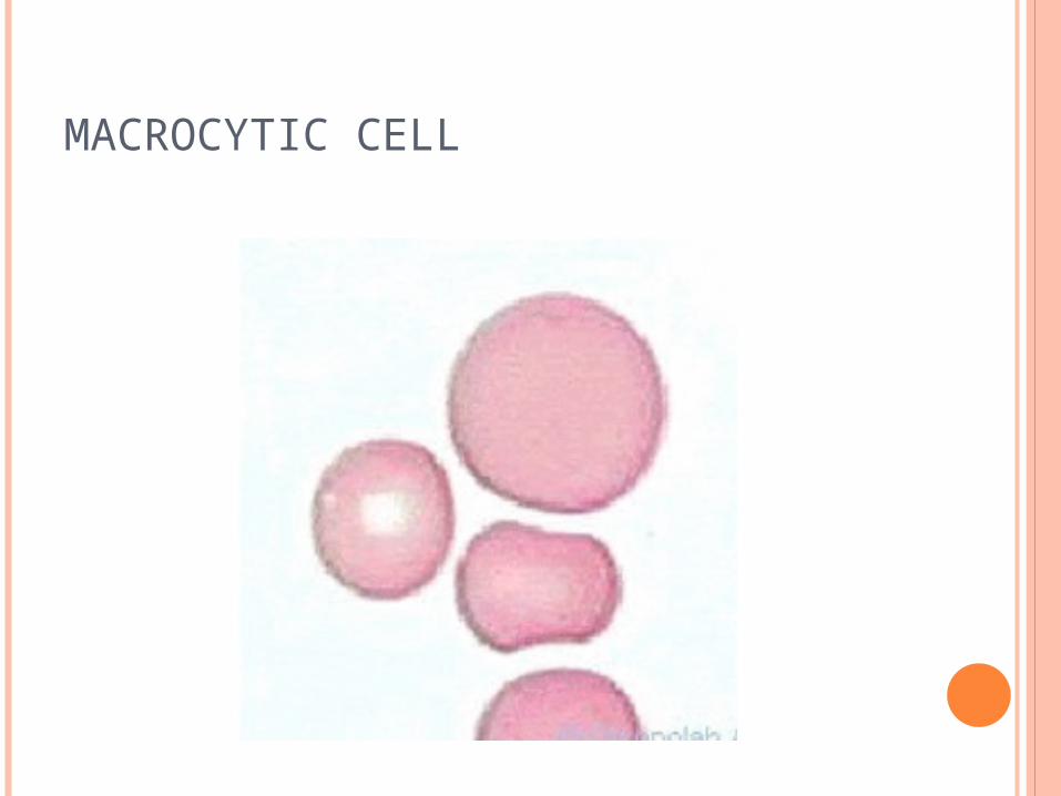
DIAGNOSIS OF ANEMIAMean corpuscular volume (MCV) – is the average volume/RBC in femtoliters (10-15 L)Hct (in %)/RBC (x 1012/L) x 10At birth the normal range is 98-123In adults the normal range is 80-100The MCV is used to classify RBCs as:Normocytic (80-100)Microcytic (<80)Macrocytic (>100)
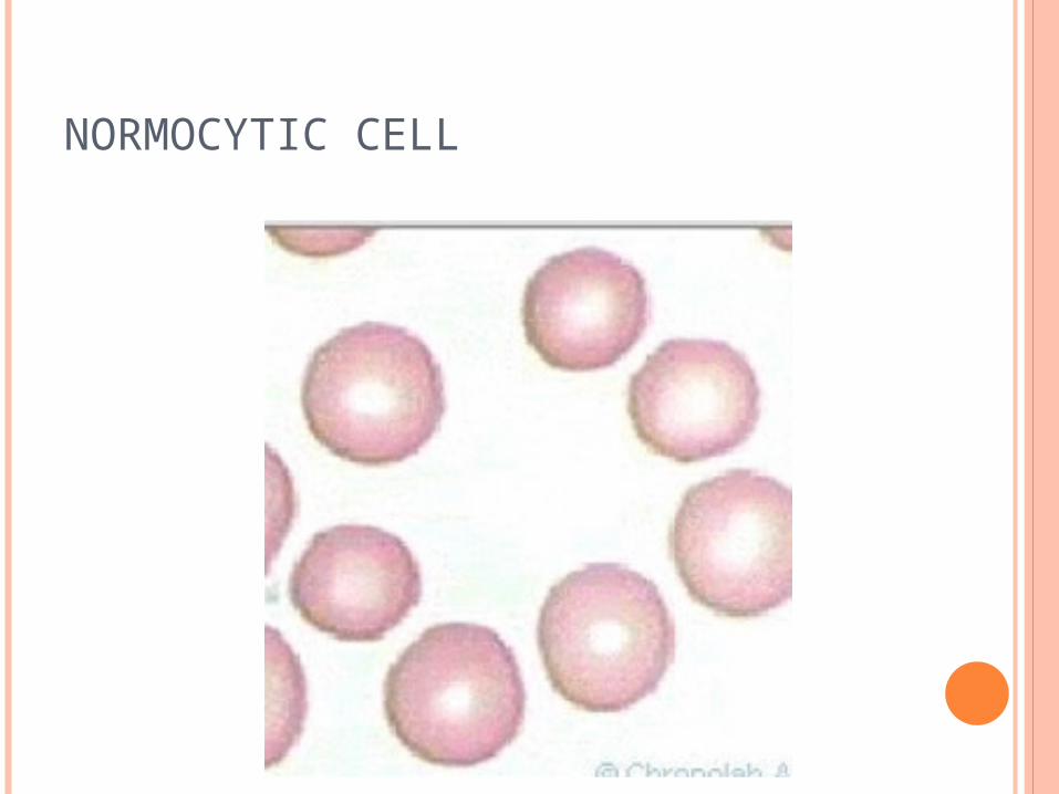
NORMOCYTIC CELL
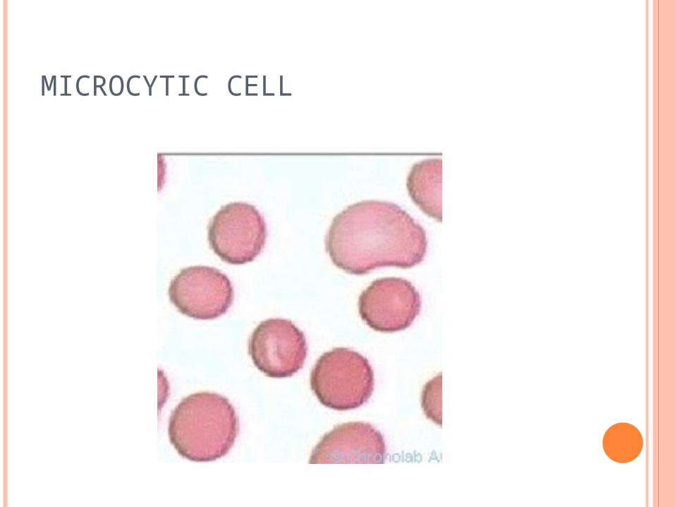
MICROCYTIC CELL
MACROCYTIC CELL
DIAGNOSIS OF ANEMIA
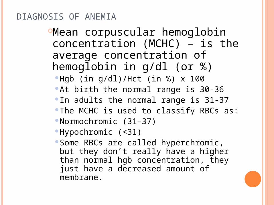
Mean corpuscular hemoglobin concentration (MCHC) – is the average concentration of hemoglobin in g/dl (or %)Hgb (in g/dl)/Hct (in %) x 100 At birth the normal range is 30-36In adults the normal range is 31-37The MCHC is used to classify RBCs as:Normochromic (31-37)Hypochromic (<31)Some RBCs are called hyperchromic, but they don’t really have a higher than normal hgb concentration, they just have a decreased amount of membrane.
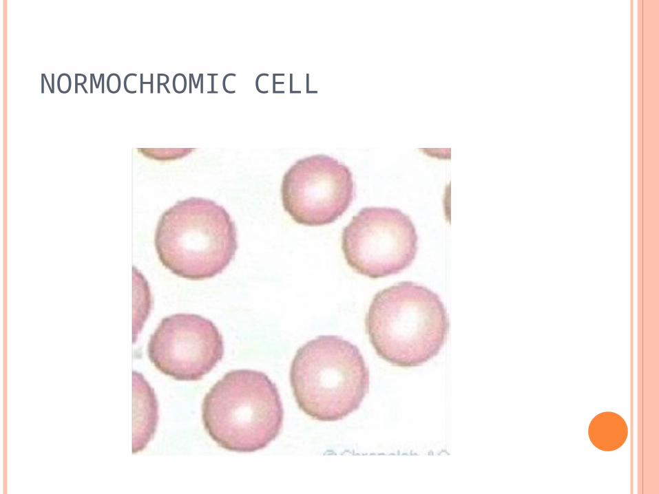
NORMOCHROMIC CELL
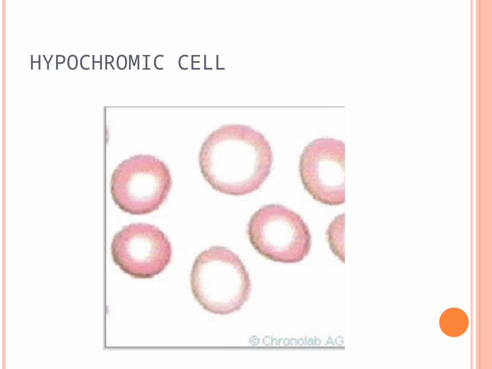
HYPOCHROMIC CELL
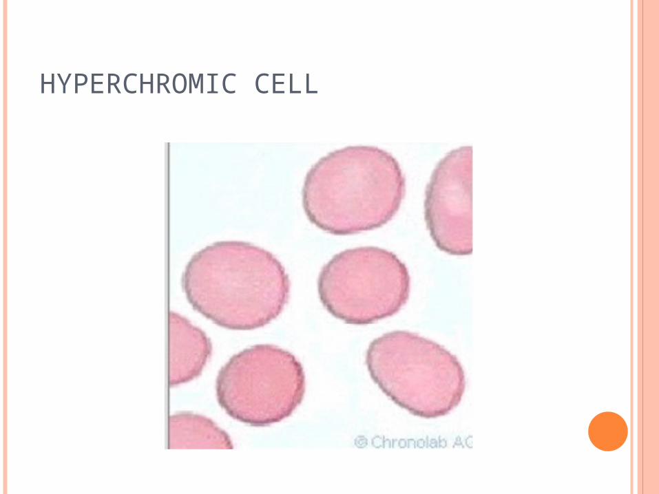
HYPERCHROMIC CELL
DIAGNOSIS OF ANEMIA
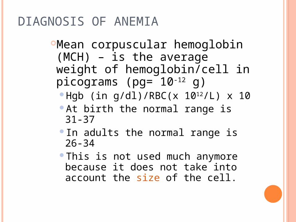
Mean corpuscular hemoglobin (MCH) – is the average weight of hemoglobin/cell in picograms (pg= 10-12 g)Hgb (in g/dl)/RBC(x 1012/L) x 10At birth the normal range is 31-37In adults the normal range is 26-34This is not used much anymore because it does not take into account the size of the cell.

DIAGNOSIS OF ANEMIA
Red cell distribution width (RDW) – is a measurement of the variation in RBC cell sizeStandard deviation/mean MCV x 100The range for normal values is 11.5-14.5%
A value > 14.5 means that there is increased variation in cell size above the normal amount (anisocytosis)
A value < 11.5 means that the RBC population is more uniform in size than normal.
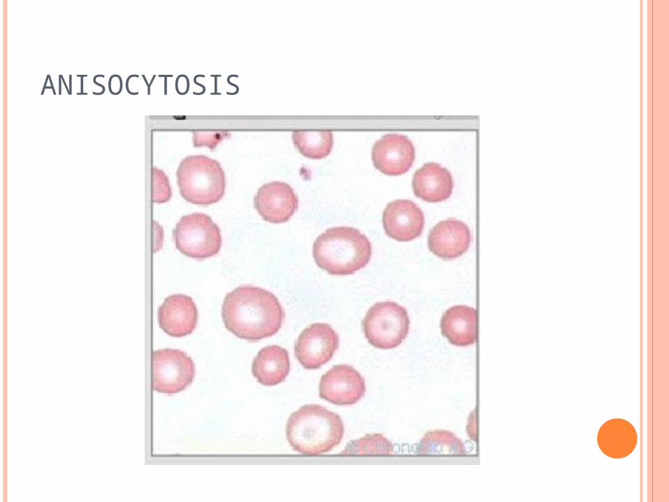
ANISOCYTOSIS
DIAGNOSIS OF ANEMIA
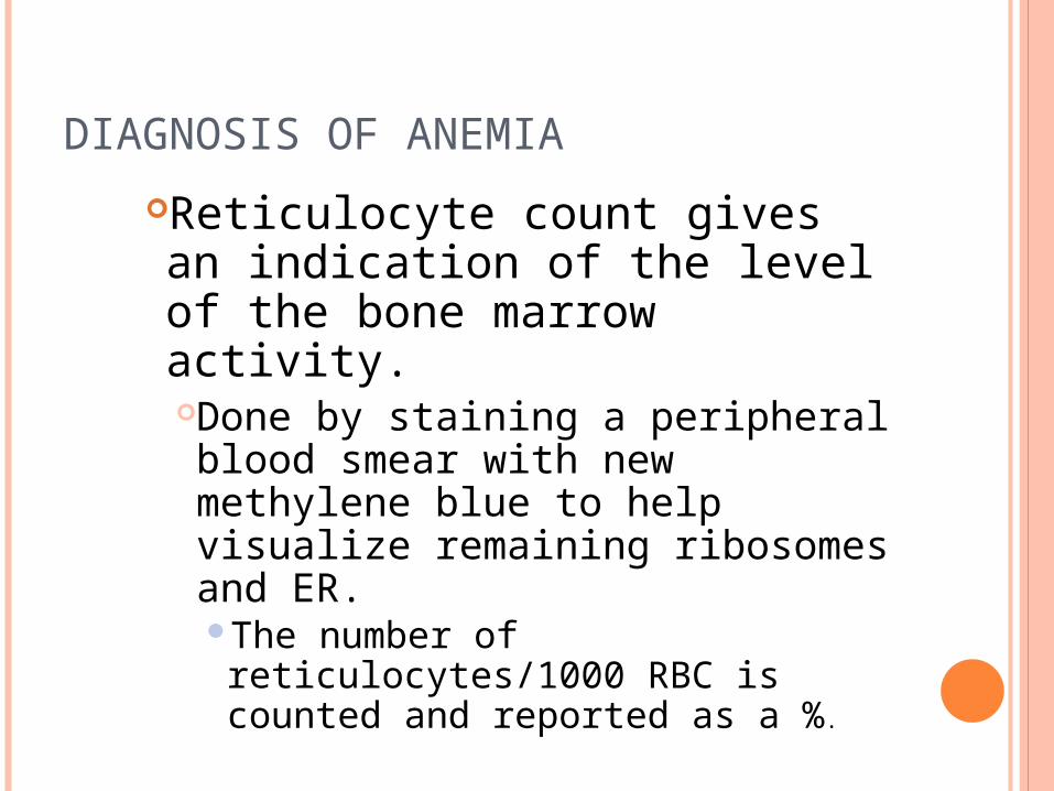
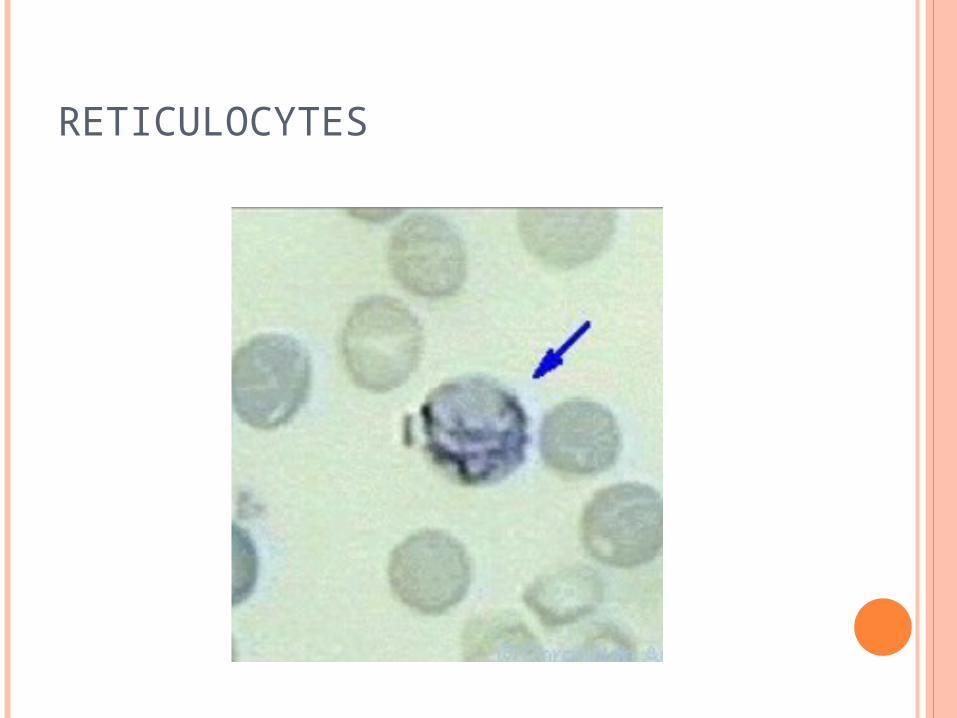
Reticulocyte count gives an indication of the level of the bone marrow activity.Done by staining a peripheral blood smear with new methylene blue to help visualize remaining ribosomes and ER. The number of reticulocytes/1000 RBC is counted and reported as a %.
DIAGNOSIS OF ANEMIA
At birth the normal range is 1.8-8%
The normal range in an adult (i.e. in an individual with no anemia) is .5-1.5%. Note that this % is not normal for anemia where the bone marrow should be working harder and throwing out more reticulocytes per day. In anemia the reticulocyte count should be elevated above the normal values.
RETICULOCYTES
DIAGNOSIS OF ANEMIAThe numbers reported above are only relative values. To get a better indication of what is really going on, a corrected reticulocyte count (patients Hct/.45 (a normal Hct) x the reticulocyte count) or an absolute count (% reticulocytes x RBC count) should be done.
As an anemia gets more severe, younger cells that take longer than 24 hours to mature, are thrown out into the peripheral blood (shift reticulocyte). This may also be corrected for to give the reticulocyte production index (RPI) which is a truer indication of the real bone marrow activity.

DIAGNOSIS OF ANEMIA
Blood smear examination using a Wright’s or Giemsa stain. The smear should be evaluated for the following:Poikilocytosis – describes a variation in the shape of the RBCs. It is normal to have some variation in shape, but some shapes are characteristic of a hematologic disorder or malignancy.
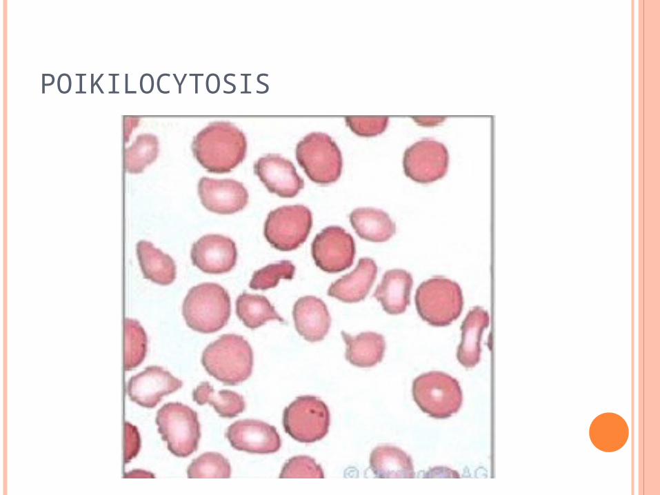
POIKILOCYTOSIS
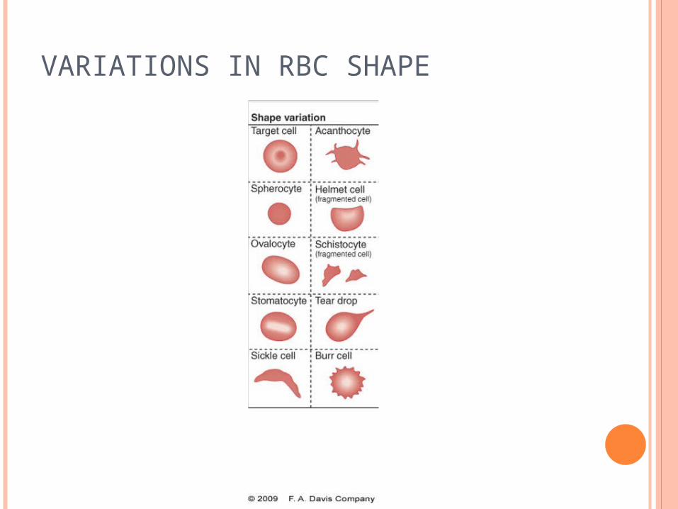
VARIATIONS IN RBC SHAPE
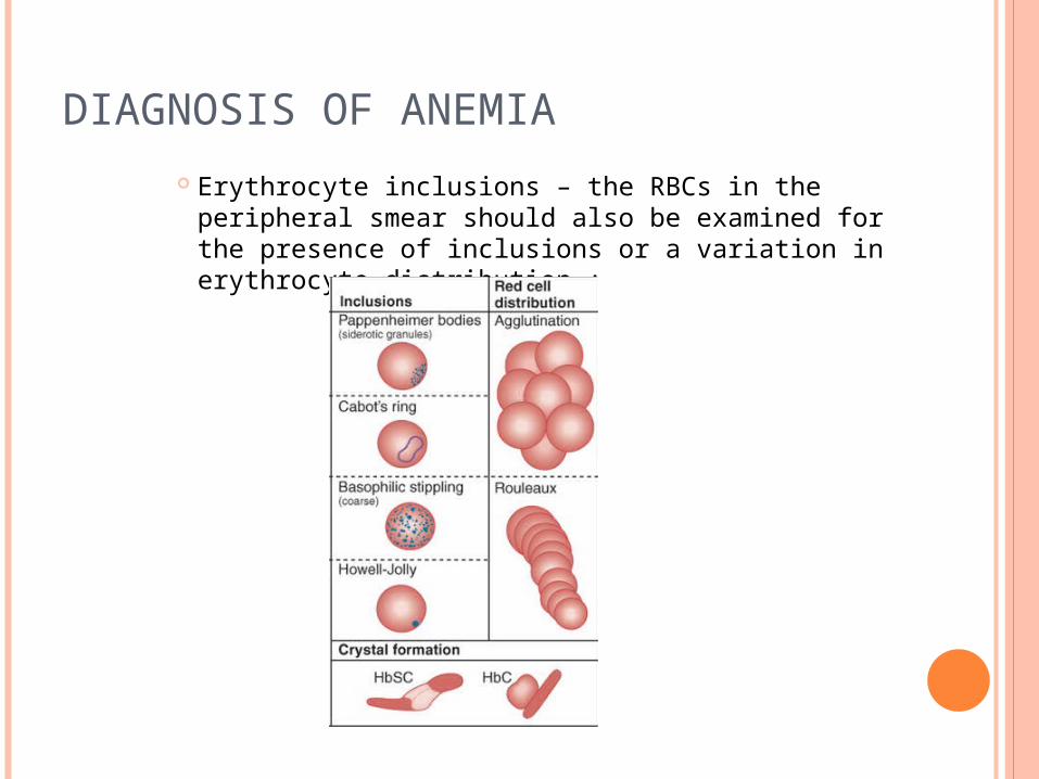
DIAGNOSIS OF ANEMIA
Erythrocyte inclusions – the RBCs in the peripheral smear should also be examined for the presence of inclusions or a variation in erythrocyte distribution :
DIAGNOSIS OF ANEMIA A variation in size should be noted (anisocytosis)
and cells should be classified as Normocytic Microcytic Macrocytic
A variation in hemoglobin concentration (color) should be noted and the cells should be classified as
Normochromic Hypochromic Hyperchromic Polychromasia (pinkish-blue color due to an
increased % of reticulocytes) should be noted. Variation in shape should be noted (poikilocytosis)
and the different shapes found should be indicated Variation in the RBC distribution should be noted
(agglutination or rouleaux formation) Any variations should be classified as slight,
moderate, or marked
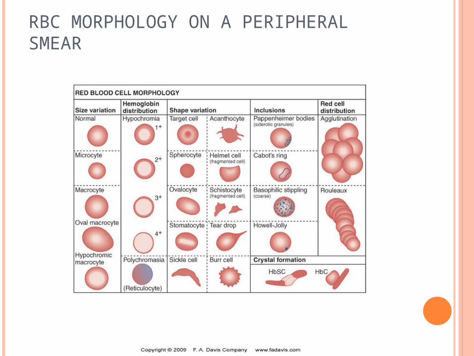
RBC MORPHOLOGY ON A PERIPHERAL SMEAR
DIAGNOSIS OF ANEMIA The peripheral smear should also be examined for
abnormalities in leukocytes or platlets. Some nutritional deficiencies, stem cell disorders, and
bone marrow abnormalities will also effect production, function, and/or morphology of platlets and/or granulocytes.
Finding abnormalities in the leukocytes and/or platlets may provide clues as to the cause of the anemia.
The lab investigation may also include: A bone marrow smear and biopsy
Used only when other tests are not conclusive
DIAGNOSIS OF ANEMIA In a bone marrow sample, the following things
should be noted:Maturation of RBC and WBC seriesRatio of myeloid to erythroid seriesAbundance of iron stores (ringed sideroblasts)Presence or absence of granulomas or tumor cellsRed to yellow ratioPresence of megakaryocytes
Hemoglobin electrophoresis – can be used to identify the presence of an abnormal hemoglobin (called hemoglobinopathies). Different hgbs will move to different regions of the gel and the type of hemoglobin may be identified by its position on the gel after electrophoresis.
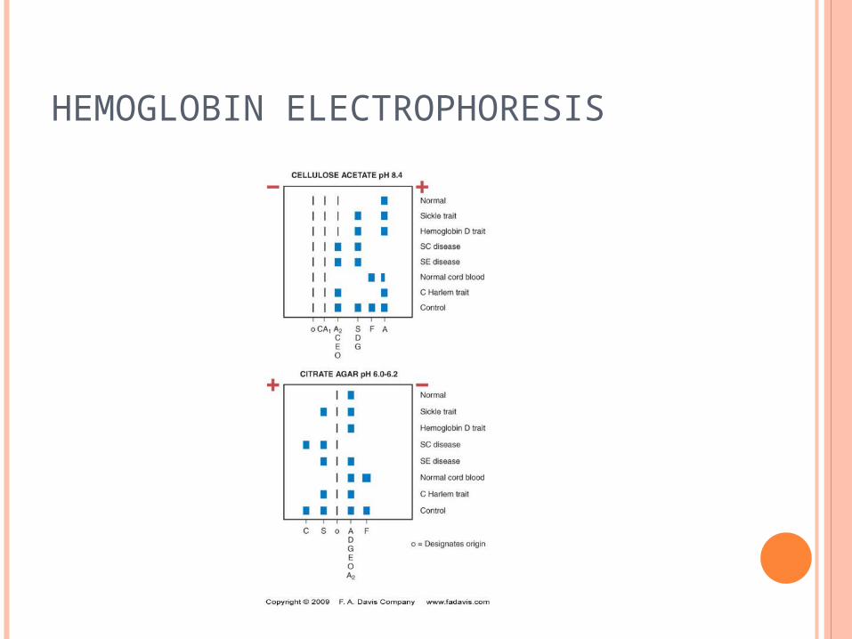
HEMOGLOBIN ELECTROPHORESIS
DIAGNOSIS OF ANEMIA Antiglobulin testing – tests for the presence of
antibody or complement on the surface of the RBC and can be used to support a diagnosis of an autoimmune hemolytic anemia.
Osmotic fragility test – measures the RBC sensitivity to a hypotonic solution of saline. Saline concentrations of 0 to .9% are incubated
with RBCs at room temperature and the percent of hemolysis is measured.
Patients with spherocytes (missing some membrane) have increased osmotic fragility. They have a limited ability take up water in a hypotonic
solution and will, therefore, lyse at a higher sodium concentration than will normal RBCs
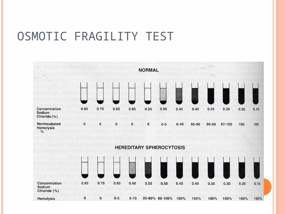
OSMOTIC FRAGILITY TEST
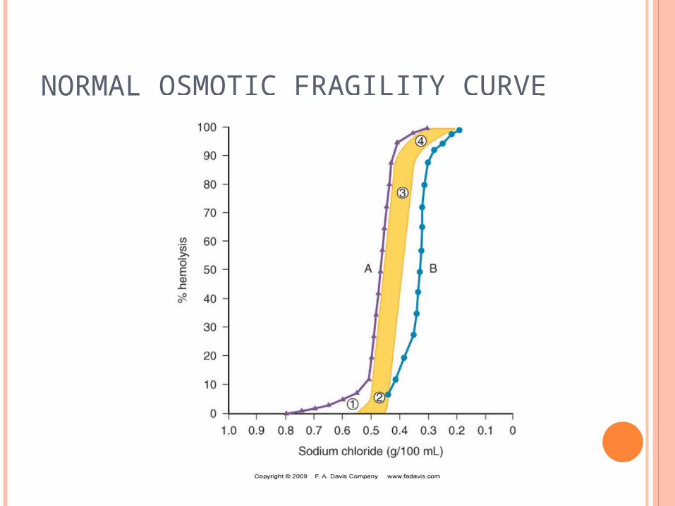
NORMAL OSMOTIC FRAGILITY CURVE
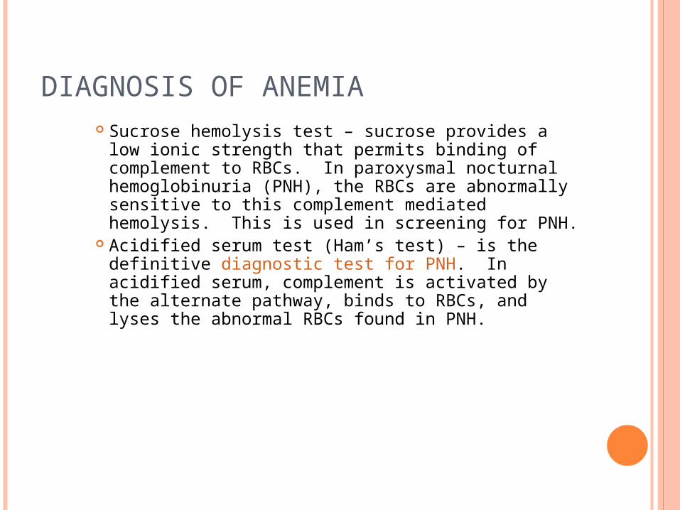
DIAGNOSIS OF ANEMIA Sucrose hemolysis test – sucrose provides a low ionic
strength that permits binding of complement to RBCs. In paroxysmal nocturnal hemoglobinuria (PNH), the RBCs are abnormally sensitive to this complement mediated hemolysis. This is used in screening for PNH.
Acidified serum test (Ham’s test) – is the definitive diagnostic test for PNH. In acidified serum, complement is activated by the alternate pathway, binds to RBCs, and lyses the abnormal RBCs found in PNH.
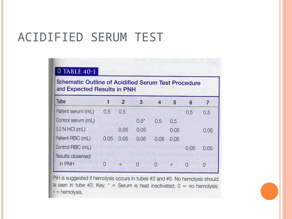
ACIDIFIED SERUM TEST
DIAGNOSIS OF ANEMIA Evaluation of RBC enzymes and metabolic
pathways – enzyme deficiencies in carbohydrate metabolic pathways are usually associated with a hemolytic anemia.
Evaluation of erythropoietin levels – is used to determine if a proper bone marrow response is occurring. Low levels of RBCs could be due to a bone marrow
problem or to a lack of erythropoietin production. Serum iron, iron binding capacity and %
saturation – used to diagnose iron deficiency anemias (more on this later)
Bone marrow cultures – used to determine the viability of stem cells.
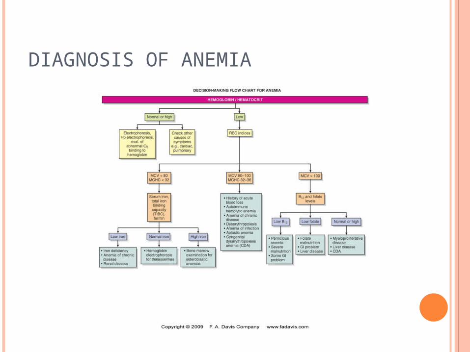
DIAGNOSIS OF ANEMIA
CLASSIFICATION OF ANEMIAS
Anemias may be classified morphologically based on the average size of the cells and the hemoglobin concentration into: Macrocytic Normochromic, normocytic Hypochromic, microcytic
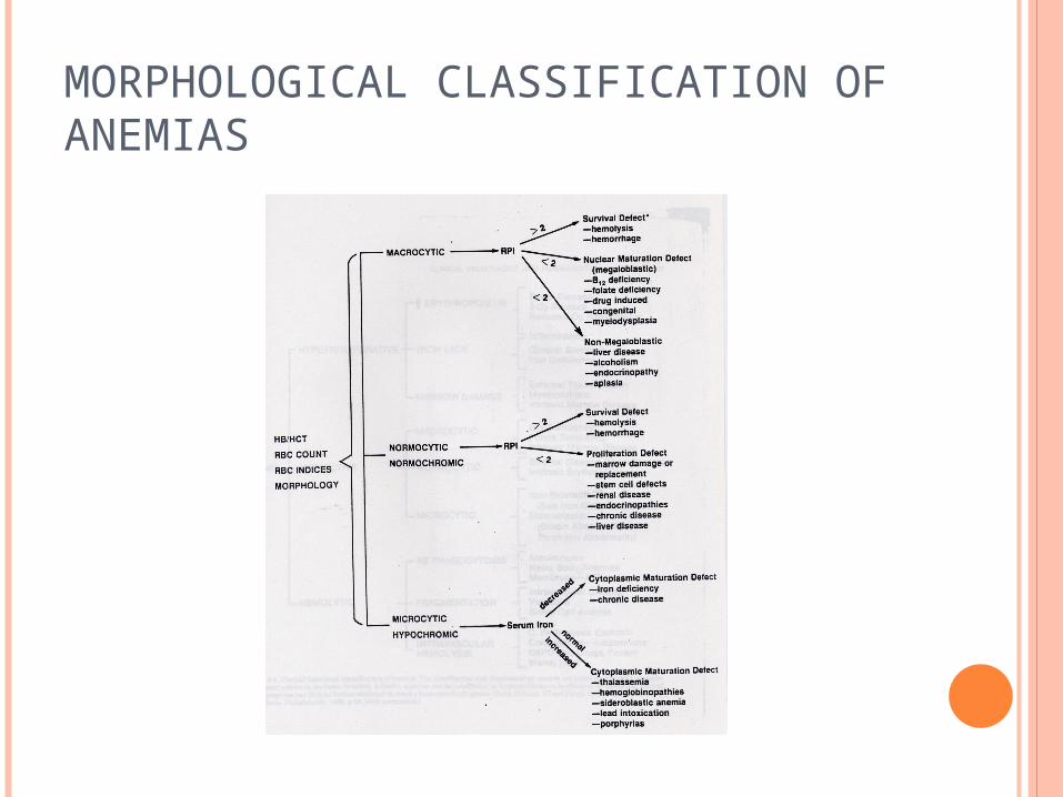
MORPHOLOGICAL CLASSIFICATION OF ANEMIAS
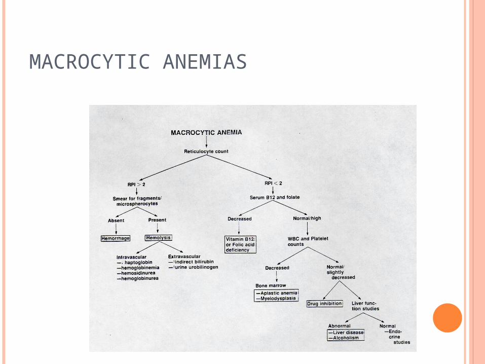
MACROCYTIC ANEMIAS
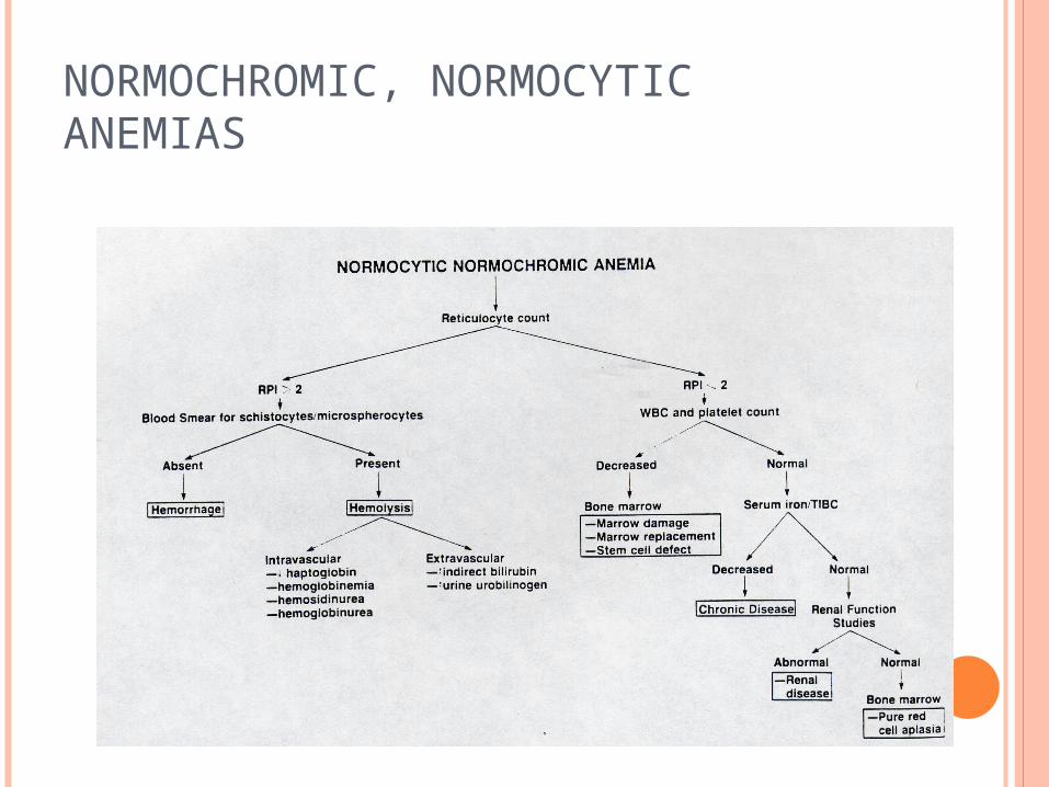
NORMOCHROMIC, NORMOCYTIC ANEMIAS
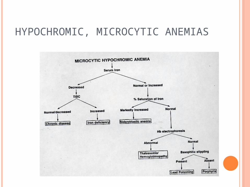
HYPOCHROMIC, MICROCYTIC ANEMIAS
CLASSIFICATION OF ANEMIAS
Anemias may also be classified functionally into: Hypoproliferative (when there is a proliferation
defect) Ineffective (when there is a maturation defect) Hemolytic (when there is a survival defect)
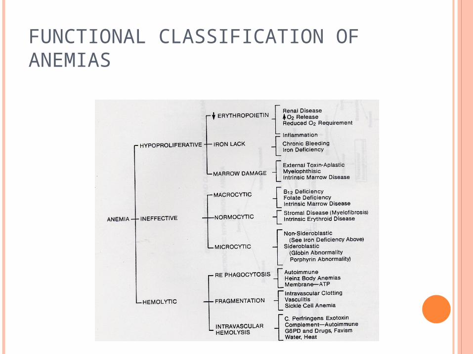
FUNCTIONAL CLASSIFICATION OF ANEMIAS


![Marinov - Anemia and haemorrhagic diatheses 2016 [Eng].ppt - Anemia and... · ANEMIA Time Anemias due to impaired ... Pathway Common Pathway. 4/13/2016 21 ... Marinov - Anemia and](https://img.dokumen.tips/doc/110x75/5d15387088c993e8108c4415/marinov-anemia-and-haemorrhagic-diatheses-2016-eng-anemia-and-anemia.jpg)