Embed Size (px)
Citation preview
1 DOI:10.4158/EP12291.RA © 2013 AACE.
ENDOCRINE PRACTICE Rapid Electronic Article in Press Rapid Electronic Articles in Press are preprinted manuscripts that have been reviewed and accepted for publication, but have yet to be edited, typeset and finalized. This version of the manuscript will be replaced with the final, published version after it has been published in the print edition of the journal. The final, published version may differ from this proof. DOI:10.4158/EP12291.RA © 2013 AACE. Review Article EP12291.RA
CHRONIC KIDNEY DISEASE MINERAL AND BONE DISORDERS (CKD-MBD) WHAT THE ENDOCRINOLOGIST NEEDS TO KNOW
Running title: CKD-MBD and Endocrinologists
Farhad Zangeneh, MD, FACP, FACE¹; Bart L. Clarke, MD, FACE²; Daniel L. Hurley, MD,
FACE²; Nelson B. Watts, MD, FACE³; Paul D. Miller, MD4
From the 1George Washington University School of Medicine, Endocrine, Diabetes & Osteoporosis Clinic (EDOC) Washington, DC, ²Division of Endocrinology, Diabetes, Metabolism and Nutrition. Mayo Clinic, Rochester, Minnesota, 3Director, Mercy Health Osteoporosis and Bone Health Services, Cincinnati, Ohio, and the 4University of Colorado Health Sciences Center, Medical Director, Colorado Center for Bone Research. Address correspondence to Farhad Zangeneh, MD, Endocrinology, Diabetes & Osteoporosis Clinic (EDOC), 46090 Lake Center Plaza, Suite 106, Sterling, VA 20165 E-mail: [email protected]
2 DOI:10.4158/EP12291.RA © 2013 AACE.
ABSTRACT
Objective: Chronic kidney disease mineral and bone disorders (CKD-MBD) are a
spectrum of abnormalities in skeletal hormones, minerals, and bone turnover and mineralization.
This paper focuses on what the endocrinologist should know about the assessment and
management of skeletal and metabolic disorders in CKD-MBD.
Methods: A literature search was reviewed to a) define disturbances of minerals and
hormones in the course of CKD; b) identify the variable radiographic and histomorphometric
changes of CKD-MBD; c) review the association among CKD-MBD, vascular calcification,
cardiovascular disease (CVD), and mortality; and d) clarify issues in CKD-MBD therapy.
Results: Assessment and treatment of CKD-MBD is complicated by progressive changes
in bone minerals and skeletal regulatory hormones as kidney function declines. CKD-MBD is
associated with fracture risk, and studies demonstrate bone mineral density can assess bone loss
and fracture risk in these patients. CKD-MBD treatment continues to evolve. Use of calcium,
phosphate binders, vitamin D, vitamin D receptor analogs, and drugs for osteoporosis and CKD-
MBD treatment are discussed in the context of safety and efficacy for patients with CKD.
Conclusions: The association of CKD with bone disease, vascular calcification, CVD,
and mortality mandates earlier recognition and treatment of CKD-MBD. Osteoporosis as a
distinct entity can be diagnosed and managed in CKD, though assessment of osteoporosis
becomes challenging in late (stage 4-5) CKD. Diabetes is common in early (stage 1-3) CKD. In
addition, 96% of all individuals identified as having CKD have early CKD. The endocrinologist
is uniquely positioned to address and treat both diabetes and many of the metabolic and skeletal
disorders associated with early CKD-MBD, including osteoporosis.
3 DOI:10.4158/EP12291.RA © 2013 AACE.
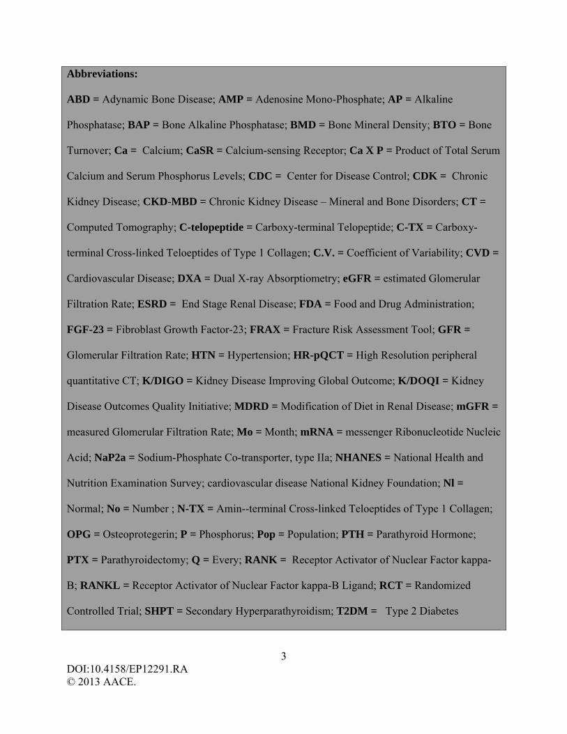
Abbreviations:
ABD = Adynamic Bone Disease; AMP = Adenosine Mono-Phosphate; AP = Alkaline
Phosphatase; BAP = Bone Alkaline Phosphatase; BMD = Bone Mineral Density; BTO = Bone
Turnover; Ca = Calcium; CaSR = Calcium-sensing Receptor; Ca X P = Product of Total Serum
Calcium and Serum Phosphorus Levels; CDC = Center for Disease Control; CDK = Chronic
Kidney Disease; CKD-MBD = Chronic Kidney Disease – Mineral and Bone Disorders; CT =
Computed Tomography; C-telopeptide = Carboxy-terminal Telopeptide; C-TX = Carboxy-
terminal Cross-linked Teloeptides of Type 1 Collagen; C.V. = Coefficient of Variability; CVD =
Cardiovascular Disease; DXA = Dual X-ray Absorptiometry; eGFR = estimated Glomerular
Filtration Rate; ESRD = End Stage Renal Disease; FDA = Food and Drug Administration;
FGF-23 = Fibroblast Growth Factor-23; FRAX = Fracture Risk Assessment Tool; GFR =
Glomerular Filtration Rate; HTN = Hypertension; HR-pQCT = High Resolution peripheral
quantitative CT; K/DIGO = Kidney Disease Improving Global Outcome; K/DOQI = Kidney
Disease Outcomes Quality Initiative; MDRD = Modification of Diet in Renal Disease; mGFR =
measured Glomerular Filtration Rate; Mo = Month; mRNA = messenger Ribonucleotide Nucleic
Acid; NaP2a = Sodium-Phosphate Co-transporter, type IIa; NHANES = National Health and
Nutrition Examination Survey; cardiovascular disease National Kidney Foundation; Nl =
Normal; No = Number ; N-TX = Amin--terminal Cross-linked Teloeptides of Type 1 Collagen;
OPG = Osteoprotegerin; P = Phosphorus; Pop = Population; PTH = Parathyroid Hormone;
PTX = Parathyroidectomy; Q = Every; RANK = Receptor Activator of Nuclear Factor kappa-
B; RANKL = Receptor Activator of Nuclear Factor kappa-B Ligand; RCT = Randomized
Controlled Trial; SHPT = Secondary Hyperparathyroidism; T2DM = Type 2 Diabetes
4 DOI:10.4158/EP12291.RA © 2013 AACE.
Mellitus; U.S. = United States; vCT = volumetric CT; vQCT = volumetric Quantitative CT;
VDRa = Vitamin D Receptor activator; WHO = World Health Organization
INTRODUCTION
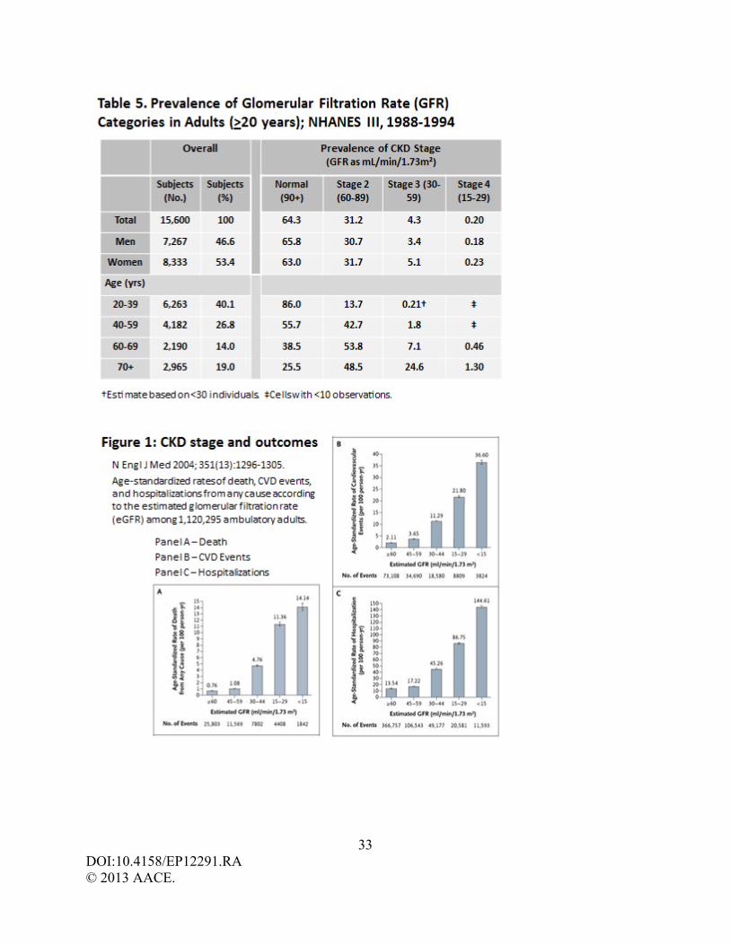
Chronic kidney disease (CKD) is a serious condition associated with increased health-
care expenditures, decreased quality of life, and premature mortality. As reported by the National
Health and Nutrition Examination Survey (NHANES III), CKD affects 11% (19.2 million) of
adult (aged 20 years and older) men and women in the United States (U.S.) (1), and is likely to
further increase with increasing longevity and the increasing incidence of obesity and type 2
diabetes mellitus (T2DM). NHANES is a continuous data survey of health and nutritional status
of U.S. adults, and results are analyzed and released periodically by the Center for Disease
Control (CDC). NHANES has noted a 15.9% increase in the prevalence of CKD between 1988-
1994 and 1999-2004 data bases (cdc.gov). CKD is most common in persons >60 years of age
(39.4% of this population), but also is present in persons aged 40-59 years and 20-39 years
(12.6% and 8.5% of these age groups, respectively). (2) T2DM is the leading cause of CKD in
developed countries (3) and accounts for 45% of all cases of kidney failure (4).
Kidney disease is defined as an abnormality of kidney structure or function with
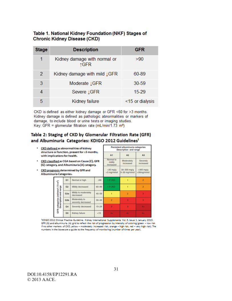
implications for the health of an individual. (5) The National Kidney Foundation (NKF) has
traditionally categorized CKD into five stages based upon the glomerular filtration rate (GFR,
expressed as mL/min/1.73 m2), and commonly reported as mL/min (Table 1). In 2003, CKD
prevalence by stage among the U.S. population was estimated as 3.3% (5.9 million) stage 1,
3.0% (5.3 million) stage 2, 4.3% (7.6 million) stage 3, and 0.2% each for stage 4 and stage 5. (1)
5 DOI:10.4158/EP12291.RA © 2013 AACE.
Kidney Disease Improving Global Outcomes (KDIGO) recently published 2012 clinical practice
guidelines recommend that CKD staging be classified not solely on GFR (G), but also include
cause (C) of injury and albuminuria (A) (Table 2). (5) The inclusion of cause of kidney disease
in staging is fundamentally important to outcome and cause-specific treatment. Albuminuria is a
marker of injury severity and is strongly associated with progression of kidney disease
independent of GFR, and is defined as mildly, moderately, or severely increased. The 2012
KDIGO classification also divides stage 3 CKD into G3a (45-59 mL/min) and G3b (30-44
mL/min) to acknowledge the significant differences in health outcomes and mortality between
these categories (Figure1). (6)
Undiagnosed CKD may lead to the under recognition of associated diseases and lost time
in treating co-morbid diseases at earlier stages of CKD. CKD prevalence is greater among
persons with T2DM (40.2% versus 15.4% without T2DM), cardiovascular disease (CVD) (28.2%
versus 15.4% without CVD), and hypertension (HTN) (24.6% versus 12.5% without HTN). As
reported by Coresh et al., of the 11% of adult Americans with CKD, 96% have stage 1-3 CKD
(GFR >30 mL/min). Thus, a sizeable number of patients with T2DM, CVD, and HTN are at risk
for CKD, and should be identified and screened for co-morbid diseases related to CKD, to
include mineral and bone disease. In a recently reported abstract of 12 million U.S. patients
screened from 2008-2011 by electronic medical records, 44 thousand were found to have T2DM
(mean age 64 years). Of patients with T2DM, 51% had CKD and 22% had stage 3-5 disease,
although 76% of stage 3-5 CKD patients did not have any recorded diagnosis of CKD. (7) The
automatic calculation of estimated GFR (eGFR) on patient laboratory reports will hopefully help
to increase awareness that CKD may be present.
6 DOI:10.4158/EP12291.RA © 2013 AACE.
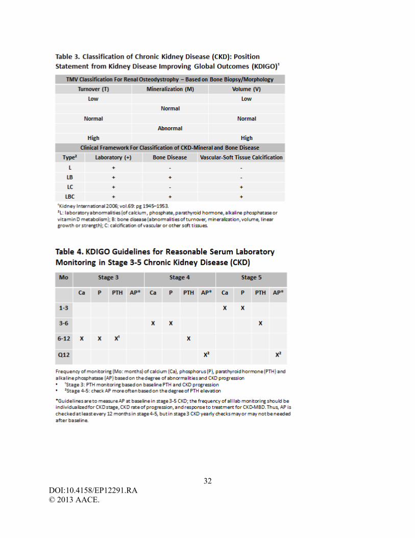
KDIGO sponsored a controversies conference on renal osteodystrophy in 2004 to a)
develop a clear, clinically relevant, and internationally acceptable definition and classification
system, b) develop a consensus for bone biopsy evaluation and classification, and c) evaluate
laboratory and imaging markers for the clinical assessment of patients with CKD. An ideal
classification system for CKD-Mineral and Bone Disorders (CKD-MBD) would allow
categorization of patients based on readily available clinical diagnostic tools and would help
guide treatment. The lack of adequate data and the non-linearity of CKD does not allow for a
classification based on severity or treatment at this time. The proposed KDIGO framework for
classifying CKD-MBD (Table 3) is based on the presence or absence of laboratory abnormalities,
bone disease, and calcification of extraskeletal tissue and is meant to be a descriptive clinical
model. (8) KDIGO recommends that the term renal osteodystrophy be used exclusively to define
altered bone morphology identified by bone biopsy/histomorphometry, and be subsequently
reported as a unified TMV classification system based upon the skeletal parameters of turnover
(T), mineralization (M), and volume (V). Thus, the term CKD-MBD is used to describe a broad
clinical syndrome that develops as a systemic disorder characterized by a constellation of
abnormalities in regulatory hormones and bone mineral, bone turnover and mineralization, and
vascular or soft tissue calcification. CKD-MBD acknowledges the entire spectrum of disease
from early hormonal and mineral disturbances to the late stages of CKD and premature mortality.
Because 96% of all CKD is found in stage 1-3 CKD, and T2DM is common in early CKD,
the endocrinologist is uniquely positioned to address and treat both T2DM and many of the co-
morbid diseases associated with CKD as outlined by the NKF KDOQI guidelines for
management of HTN, dyslipidemia, anemia, CVD, and nutrition (2), and as outlined in the newer
KDIGO guidelines for metabolic bone disease (9). This review will focus on what the
7 DOI:10.4158/EP12291.RA © 2013 AACE.
endocrinologist should know about the development, assessment, and management of CKD-
MBD. For purpose of discussion, where not stated in the text, ‘early CKD’ refers to stage 1-3
disease and ‘late CKD’ refers to stage 4-5 disease. Stage 5 CKD is often referred to as end-stage
renal disease (ESRD). We will not address issues associated with bone disease and kidney
transplantation in ESRD.
Bone Mineral and Regulatory Hormones in CKD-MBD
Disturbances of bone mineral metabolism and regulatory hormones may occur early in
the course of CKD, with perturbations occurring as early as stage 2 CKD, and progressing as
kidney function worsens. (10) CKD is associated with disrupted regulation of fibroblast growth
factor-23 (FGF-23) and the vitamin D–parathyroid hormone (PTH) axis. (11-12) Serum FGF-23
levels rise earlier than and are relatively higher than PTH levels as CKD progresses. FGF-23,
derived from osteocytes, is a phosphaturic hormone that also has multiple tissue effects that
influence bone metabolism. (13) The rise in PTH occurs before serum calcium decreases or
serum phosphate increases significantly, and this eventually leads to secondary
hyperparathyroidism (SHPT). As kidney function declines, a corresponding decrease in vitamin
D receptors (VDR) and calcium-sensing receptors (CaSR) occurs in the parathyroid glands (14)
making them less responsive to the actions of circulating vitamin D and calcium. All these events
worsen SHPT and its potential effect on bone. Many patients with stage 2-4 CKD may go
unrecognized because of a reliance only on serum creatinine to assess renal status, and failure to
more accurately assess renal function using the GFR. The rate of glomerular filtration is
generally regarded as the best overall index of renal function in health and disease. (15) Normal
GFR varies according to age, gender, body size, and race. The measured GFR (mGFR, as
clearance of exogenous filtration markers such as iothalamate) is presently the best direct
8 DOI:10.4158/EP12291.RA © 2013 AACE.
measure of renal function, and the degree of reduction in mGFR correlates with the severity of
structural changes in CKD. Serum measurement of cystatin-C has recently gained attention as a
sensitive, non-creatinine alternative endogenous marker of filtration (16-17) for use when
decisions depend on more accurate knowledge of GFR such as confirming a diagnosis of CKD
or adjusting doses of potentially toxic drugs excreted by the kidney. The 2012 KDIGO
guidelines recommend measuring cystatin C in adults with eGFR 45-59 mL/min who do not
have other makers of kidney damage and if confirmation of CKD is needed for treatment
decisions. However, one should be aware that several factors other than renal function may affect
cystatin-C levels. (18) Serum creatinine levels, like the mGFR, have been shown to fluctuate
throughout the day; this and other determinants (such as measurement inaccuracies for creatinine
and cystatin-C) may account for a substantial portion of the variability in eGFR equations. While
the eGFR can be imprecise, estimating equations adjust for the effect of non-GFR determinants
represented by age, sex, and race. Both the Modification of Diet in Renal Disease (MDRD)
Study equation and the Chronic Kidney Disease Epidemiology Collaboration (CKD-EPI)
equation provide better estimates of GFR than the serum creatinine alone, and the 2012 KDOQI
clinical practice guidelines recommend using the CKD-EPI equation as the most accurate
estimate of kidney function, especially in early CKD.
Stage 3 CKD is often associated with decreased production of 1,25-dihydroxyvitamin D
(calcitriol) (10) in response to a loss of functioning proximal renal tubules and reduced activity
of renal 1-alpha-hydroxylase, leading to parathyroid gland hyperplasia, elevated blood levels of
PTH, and SHPT. The combination of low calcitriol and elevated PTH in CKD is now recognized
as a cause of bone loss (19-21) and a major contributor to bone disease commonly seen in stage 4
CKD and present in almost all patients with stage 5 CKD (22-23). Elevated PTH levels in late
9 DOI:10.4158/EP12291.RA © 2013 AACE.
CKD have also been viewed as a possible contributor to early death (24), although a recent meta-
analysis did not find a significant correlation between PTH levels and mortality (25). Rather, in
4127 patients followed for a median of 60 months, elevated phosphate levels were associated
with increased coronary events and death in patients with stage 2-3 CKD based upon eGFR
(Cockcroft-Gault equation). (26) Increased mortality has been reported with higher serum levels
of both phosphate (25-28) and FGF-23 (29-30) in patients with CKD.
Clinical and experimental evidence supports integrated mechanisms responsible for
SHPT and bone disease in CKD patients. Phosphate retention and hyperphosphatemia directly
stimulate parathyroid gland function. (24) However, perturbations of phosphate retention in stage
2-3 CKD may not be seen by measurement of serum or urine phosphate (10), and possible
explanations are that compensatory increases in FGF-23 (29) and PTH lead to decreased renal
tubular reabsorption of phosphate and increased urinary phosphate excretion. During stages 4-5
CKD, when hyperphosphatemia develops, phosphorus can affect parathyroid function both by
suppressing blood calcium contributing to hypocalcemia, and by acting directly on the
parathyroid glands. Hyperphosphatemia has a direct effect on post-transcriptional increases in
PTH synthesis and secretion (31) and can induce parathyroid hyperplasia independent of low
blood levels of calcium or calcitriol. Phosphate retention interferes with the kidney’s ability to
produce calcitriol, creating a state of vitamin D deficiency and decreased intestinal absorption of
calcium. The calcemic response to PTH infusion is also markedly blunted as early as stage 2
CKD. These abnormalities all contribute to SHPT.
Phosphate homeostasis is primarily regulated by the kidney. Phosphate is filtered by the
renal glomerulus and 80% is then reabsorbed mostly by the proximal nephron’s brush border
membrane type IIa sodium-phosphate co-transporter (NaP2a). PTH increases urinary phosphate
10 DOI:10.4158/EP12291.RA © 2013 AACE.
excretion via cyclic-AMP dependent inhibition of NaP2a expression. However, PTH action does
not account for all of phosphate homeostasis. Recent studies have shown that FGF-23 is involved
in the pathophysiology of CKD (11-12), and plays a major role in various forms of osteomalacia
(32). FGF-23 is a peptide hormone normally secreted by bone osteocytes and osteoblasts in
response to hyperphosphatemia. The rise in FGF-23 mirrors renal phosphate retention and
appears to precede the development of SHPT. Serum FGF-23 levels have been reported to be
persistently increased as early as stage 3 CKD. (29) This FGF-23 rise induces a decline in the
number of intact nephrons and is associated with reduced expression of Klotho, the co-receptor
required for FGF-23 signaling. (33) Patients with CKD have high serum FGF-23 and low Klotho
expression in the kidney and parathyroid glands, raising the concept that they could serve as
biomarkers for progression of disease (29) and response to therapy (34-36). As CKD progresses,
serum phosphate, PTH, and FGF-23 levels continue to increase, while responsiveness to PTH
and FGF-23 decrease. In late stage CKD, abnormally high serum FGF-23 levels can no longer
reduce serum phosphate effectively. Elevated FGF-23 levels are associated with more rapid
CKD progression to ESRD (29), left ventricular hypertrophy (37), and premature mortality (29-
30).
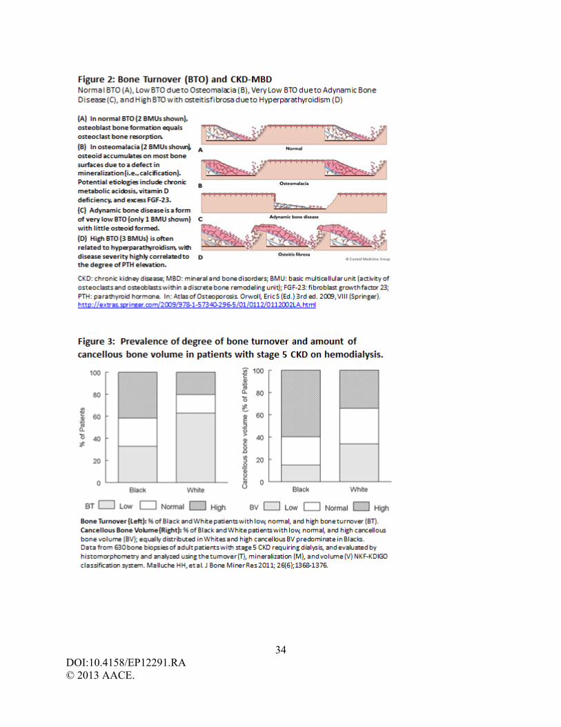
Bone Turnover (BTO) and Mineralization in CKD-MBD
The nature and type of bone disease that develops in CKD-MBD may vary among
patients. Three types of bone disease, as defined by quantitative bone histomorphometry, may be
encountered in patients with CKD: a) increased bone turnover (BTO) and resorption due to
SHPT with or without marrow fibrosis (osteitis fibrosa), b) decreased bone turnover and
formation (termed ‘adynamic bone’), and c) defective bone mineralization (osteomalacia). (38)
Some patients have a mixed pattern. (22-23) In order to clarify the interpretation of bone biopsy
11 DOI:10.4158/EP12291.RA © 2013 AACE.
results in the evaluation of renal osteodystrophy, the TMV classification system allows for three
key histologic descriptors to be reported in any combination (Table 3). Histomorphometry can
thereby help to provide a clinically relevant description of the underlying skeletal pathology
(Figure 2), and assist in guiding therapy.
Factors that cause postmenopausal, idiopathic, or age-related bone loss may contribute to
the skeletal abnormalities of CKD. Inadequate calcium or vitamin D intake or absorption,
hypogonadism, tobacco smoking, glucocorticoid steroid use, immobilization, and poor
nutritional status may by themselves be a cause of bone loss. Osteoporosis, defined by The
National Institutes of Health Consensus Conference on Osteoporosis, is a decrease in the
quantity and/or quality of normally mineralized bone that decreases bone strength and increases
the risk of skeletal fracture. Osteoporosis may coexist with CKD-MBD, and may also be present
years before CKD-MBD becomes evident. In a study of 421 postmenopausal women with
osteoporosis and GFR >50 mL/min, 39% of women had vitamin D deficiency as defined by 25-
hydroxyvitamin D levels <12 ng/dL (<30 nmol/L) and 33% frank SHPT (serum PTH above
normal lab values). (39) In these women, hypovitaminosis D was associated with either an
elevated PTH response and increased BTO or a ‘blunted’ PTH response and low BTO, the latter
theorized to possibly be protective against hypovitaminosis D related bone loss. Lobao et al.
reported on 103 patients with CKD (creatinine clearance 10-78 mL/min) not receiving dialysis,
and found that 50 (48.5%) had low bone mineral density (BMD). (40) In this study, bone loss
was found to be present in patients with both early and late CKD (median creatinine clearance 29
mL/min), with only alkaline phosphatase and PTH levels predictive of low BMD. Bone
histomorphometric analysis on the fifty patients with low BMD revealed adynamic bone disease
in 52.5% and osteomalacia in 42.5%.
12 DOI:10.4158/EP12291.RA © 2013 AACE.
Patients in early stage CKD often have mild SHPT features putting them at risk for bone
loss from high BTO where bone resorption exceeds bone formation. Several studies of bone
histomorphometry in patients with stage 2-4 CKD have shown that most patients have increased
rates of BTO, as defined by increased bone formation and bone resorption rates. (23, 41-42)
There may or may not be a co-existing mineralization defect depending on the degree of
hypocalcemia, vitamin D deficiency, or aluminum deposition in bone. Bone histomorphometry
in 174 patients with GFR 15-50 mL/min (stage 2-4 CKD) reported by Hamdy et al. in 1995
found that 129 patients (74%) had high BTO with osteitis fibrosa, 33 (19%) had osteitis fibrosa
with osteomalacia, and 9 (5%) had low BTO ‘adynamic bone’, while osteomalacia alone (1
patient, 0.6%) and aluminum deposition (2 patients, 1%) were rare. (23) Paired bone biopsies in
62 of these untreated patients showed progression of CKD-MBD in all patients within two years.
These findings are in keeping with the hormone and mineral features of higher PTH, lower
calcitriol, and higher phosphate levels seen in early CKD. Of interest, McCarthy et al. did not
find an association of early CKD and fracture risk in 427 postmenopausal Caucasian women
(median age, 68 years) in a prospective, population-based cohort study followed for up to 25
years (median, 14 years) in Rochester, MN. (43) Although univariant analysis found increased
fracture risk associated with declining renal function, multivariant analysis did not find any
association after adjusting for age, body weight, and BMD. Of note, the baseline creatinine
clearance rate for all patients in this study was 78.7 + 26.6 mL/min, and 44.7 + 12.7 mL/min for
the 20% of subjects in the lowest quintile. While few have examined the longitudinal change in
bone density in relation to BTO in CKD, high BTO from SHPT leads to bone loss (19) due to
bone resorption increased out of proportion to bone formation (38).
13 DOI:10.4158/EP12291.RA © 2013 AACE.
As kidney function deteriorates to late stage CKD, patients are at an increased risk of
fracture. (19-21, 44-49) SHPT can result in markedly increased BTO and even marrow fibrosis
(e.g., activation of precursor mesenchymal cells, which differentiate into fibroblast-like cells and
form fibrous tissue adjacent to bone trabeculae). Osteitis fibrosa is a CKD-MBD manifested by
severe SHPT, excessive BTO, and marrow fibrosis, often with increased osteoid production and
abnormal osteoid mineralization (Figure 2). Coalescence of large multinucleated osteoclasts and
fibrotic marrow may result in ‘brown tumors’ (named for the color of these bone lesions due to
hemosiderin deposits) and appear as lytic or lucent ‘cysts’ via radiographs. Increased osteoid
may be due to increased collagen production that exceeds mineralization, and/or abnormal
mineralization (i.e., osteomalacia). Rapidly deposited, poorly structured, and under-mineralized
osteoid is often referred to as ‘woven bone’, and lacks the lamellar pattern and birefringence seen
histologically in normal bone.
Vitamin D is important for collagen synthesis and maturation, as well as normal
mineralization of osteoid. Factors important in the development of osteomalacia in CKD-MBD
are vitamin D deficiency and/or resistance to calcitriol action. Aluminum deposition in bone can
also lead to a skeletal mineralization defect in CKD. Although osteomalacia can be accurately
diagnosed only by means of a tetracycline double-labeled iliac crest bone biopsy for
histomorphometric analysis, hypovitaminosis D is common in both the general population (50-
51) and in patients with CKD (9-10, 19, 39). The degree of decline in serum levels of 25-
hydroxyvitamin D has been found to be related to biochemical markers of CKD-MBD (directly
with decreases in mGFR, and indirectly with increases in PTH, C-telopeptide, and bone alkaline
phosphatase). (49) Low calcitriol levels are also directly related to the degree of renal
insufficiency (49), and hypovitaminosis D contributes to bone loss through decreased intestinal
14 DOI:10.4158/EP12291.RA © 2013 AACE.
calcium absorption (50, 52), lowered bone formation (52), and increased osteoclastogenesis (53-
54).
Fortunately, stage 4-5 CKD only accounts for 4% of reported CKD in the U.S. (1)
Almost all patients with stage 5 CKD have abnormal bone histology. Cross-sectional studies of
bone histology in dialysis patients reveal different prevalences for types of CKD-MBD. (22-23,
55-56) While high BTO and SHPT is a predominant finding in patients with stage 5 CKD, there
is also a high prevalence of decreased BTO, or ‘adynamic bone disease’ (ABD). (22-23, 56) The
prevalence of ABD has been found to be 30% in patients with stage 4 CKD, and between 15-
60% in patients with stage 5 CKD requiring dialysis. In patients with ESRD requiring
hemodialysis, ABD is more common in Caucasians and less common in African Americans. (22)
ABD is a CKD-MBD disease characterized histologically by low BTO with very little osteoid
accumulation and thin osteoid seams (Figure 2). (57) Both the rate of collagen synthesis by
osteoblasts and the rate of bone matrix mineralization are subnormal. The latter distinguishes
ABD from osteomalacia, where defects in mineralization exceed those in bone formation and
result in a relative osteoid excess and thick osteoid seams. Variability in the prevalence of ABD
reported among studies may be related to differences in excess calcium loading, aluminum
loading, or presence of diabetes. (57-60) Aluminum bone deposition leading to ABD was a much
greater problem in the era of aluminum containing phosphate binders and unrecognized
aluminum contamination in parenterally administered solutions (especially nutrition and
albumin). In theory, ABD is presumed to have an impaired ability to repair skeletal micro-
fractures due to decreased BTO, and thereby result in an increased risk of fracture. However, the
clinical significance of ABD remains to be determined. Barreto et al. identified differences in
bone histology with or without osteoporosis in 98 patients with ESRD treated with hemodialysis.
15 DOI:10.4158/EP12291.RA © 2013 AACE.
(45) In this study, the majority (56%) of patients had ABD with 25% having significant
aluminum bone deposition. Osteoporosis was associated with age, female gender, duration of
amenorrhea, Caucasian ethnicity, and the serum OPG/sRANKL ratio. Neither histologic ABD
findings nor serum levels of PTH, calcium, phosphate, or 25-hydroxyvitamin D were predictive
of osteoporosis, although there was a trend for calcitriol use to be associated with the absence of
osteoporosis (p=0.06).
The mechanisms underlying ABD are not fully known, and it may be seen in late CKD
either before or after initiating dialysis. It has been generally believed and accepted that elevated
PTH levels (2-3 times normal) are necessary to maintain normal rates of bone formation in
patients with stage 4-5 CKD, and thereby prevent ABD from developing. (9, 23, 59-63) Patients
with ABD have lower PTH levels than those with other forms of CKD-MBD (22), and although
over-suppression of parathyroid gland activity from excessive calcium (62) and/or calcitriol or
cinacalcet administration may play a significant role, a similar ‘blunting’ of PTH with low BTO
has been reported in postmenopausal women without CKD. (39) To better understand the
dynamic changes in CKD-MBD following parathyroidectomy (PTX), paired bone biopsies taken
before and after surgery were studied by Yajima et al. in 18 patients with SHPT and stage 5
CKD requiring hemodialysis. (64) PTH levels, markers of BTO, bone osteoclast surfaces, and
marrow fibrosis all decreased markedly 2-4 weeks after PTX. Phosphate (from 5.3 + 1.2 to 2.9 +
1.6 mg/dL) and PTH levels (from 1256.7 + 448.2 to 30.3 + 61.6 pg/mL) decreased significantly
at 2-4 weeks after PTX, and PTH fell below 30 pg/mL in all but two patients. A substantial
increase in osteoid volume and tetracycline label was observed compared with bone biopsies in
both low-PTH and high-PTH stage 5 hemodialysis control groups not having PTX, suggesting
that increased mineralization was taking place. Tetracycline label in PTX subjects was observed
16 DOI:10.4158/EP12291.RA © 2013 AACE.
not only at the mineralization front of trabecular surfaces, but also around the osteocyte lacunar
walls and canaliculi within the basic multicellular units (BMUs). The authors reported an
increase in the number of empty lacunae, a reduction of lacunar volume, and significant decline
in osteocyte number after PTX. Whether or not these acute BMU changes after PTX were in part
due to high doses of calcium and/or vitamin D (1-alpha-hydroxyvitamin D3 as oral alfacalcidol)
administered, and whether or not these bone features persist long-term after PTX is unknown. It
is important to recognize the normal physiology of calcium flux in and out of bone, because
there is a common misconception that bone remodeling is the major mechanism for day-to-day
and minute-to-minute bodily flux of calcium. Osteocytes and bone lining cells, under stimulation
by PTH and other effectors, play a much larger role in determining serum calcium concentrations
than the rate of BTO, both in health and disease. (41)
Malluche et al. studied 630 bone biopsies (obtained from 2003-2008) in patients with
stage 5 CKD requiring hemodialysis for degree of trabecular (cancellous) bone volume, turnover,
and mineralization. (22) Mineralization defects were rare, and present in only 3% of patients. A
total of 62% of Caucasians had predominately low BTO whereas 68% of African Americans had
normal or high BTO (Figure 3). Other racial differences were also evident. Trabecular bone
volume was equally distributed as low, normal, or high in Caucasians whereas in African
Americans trabecular bone volume was high in two-thirds of patients. More than 80% of all
patients with low bone volume had thin trabeculae and low bone formation (i.e., ABD). In
addition, PTH levels varied by race; PTH values 499 + 93, 614 + 100, 805 + 99 for Caucasians
and 172 + 12, 343 + 37, 523 + 37 for African Americans, respectively for low, normal, and high
BTO. Thus, in more current studies in stage 5 CKD-MBD the presence of low bone volume with
low BTO (i.e., ABD) is more frequent than previously appreciated, and defects of mineralization
17 DOI:10.4158/EP12291.RA © 2013 AACE.
(i.e., osteomalacia and aluminum deposition) are rare. This study also confirms that CKD-MBD
differences exist within and between races, and treatment guidelines may therefore not apply
similarly to all patients. The NKF 2002 KDOQI (2) and 2009 KDIGO (9) guidelines accept a
wide range of elevated PTH levels (2-9 times the upper limit of normal) as being optimal in late
stage CKD. In 2008, Barreto and colleagues studied bone histomorphometric change at baseline
and one year to assess the recommended KDOQI PTH range of between 150-300 pg/mL in 97
patients with stage 5 CKD requiring hemodialysis. (65) They found that intact PTH levels <150
pg/mL for identifying low BTO and >300 pg/mL for identifying high BTO had positive
predictive values of 83% and 62%, respectively.
Vascular or Soft Tissue Calcification in CKD-MBD
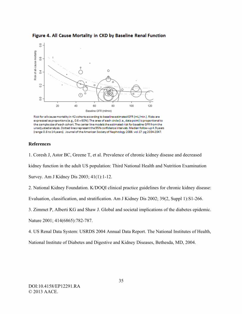
The risk of any cardiovascular event (6), cardiovascular death (66), and death from any
cause (6, 66-67) increases sharply as the eGFR declines below 60 mL/min. (Figures 1 and 4). In
a prospective cohort study of 382 patients with stage 3-5 CKD, the annual mortality rates for
stages 3, 4, and 5 CKD were 3.9%, 6.3%, and 9.2% respectively. (67) However, traditional CVD
risk factors do not entirely account for the elevated mortality in CKD, as seen in a prospective
study of 3879 patients with stage 2-4 CKD followed for a median of 3.5 years. (29)
Hyperphosphatemia, an elevated calcium-phosphate (Ca X P) product (68), and SHPT have all
been linked to increased arterial vascular calcification and/or CVD mortality. As CKD-MBD
progresses, there is a physiologic change for vascular tissue to acquire bone cell characteristics,
with a secondary deposition of calcium in arterial walls and on cardiac valves. One of the
mechanisms of vascular calcification in CKD-MBD is the dedifferentiation of the normal
vasculature, and the acquisition of an osteoblast-like phenotype. In addition, circulating stem
cells originally destined to the vascular bed are recruited to an osteoblast-like phenotype within
18 DOI:10.4158/EP12291.RA © 2013 AACE.
the vasculature. Thus, both bone cell types contribute to arterial vascular calcification in CKD-
MBD.
The correlation of bone turnover, vascular calcification, and mortality in CKD is not well
established. It is of interest that in a study of 2348 healthy postmenopausal women, Schulz et al.
assessed BMD and aortic calcification via lumbar computed tomography (CT) and found
osteoporosis in 70% of subjects, with the degree of aortic calcium accumulation inversely related
to bone BMD and directly related to fracture. (44) This is in keeping with earlier (electronic
beam) coronary CT findings of an association between the degree of coronary calcification and
bone loss. (69) Arterial vascular calcification begins as early as stage 3 CKD and is strongly
linked to bone loss, CVD, and increased mortality. As CKD-MBD progresses, calcium is less
avidly incorporated into the skeleton during bone remodeling. SHPT is associated with bone loss
in patients with CKD (19) and low BMD is a risk factor for mortality in patients with ESRD
needing dialysis (68). Low BTO in CKD may also accelerate CVD if circulating calcium and/or
phosphorus exceed skeletal requirements and an elevated Ca X P product ensues (vascular
calcification may be accelerated if the tissue Ca X P product exceeds 55).
Assessment and Diagnosis in CKD-MBD
Renal Function, Minerals, and Hormones. An assessment of renal function by
measurement of serum creatinine and calculation of the eGFR
(http://www.kidney.org/professionals/kdoqi/gfr_calculator.cfm) should be obtained in any
patient with age >65, diabetes mellitus, osteoporosis, or presence of arterial vascular calcification.
NKF KDIGO guidelines suggest measurement of serum total calcium, phosphorus, 25-
hydroxyvitamin D, PTH, and bone alkaline phosphatase as baseline values if patients are
diagnosed with stage 3 CKD (GFR 30-59 mL/min). As reported by Lobao et al. (40), patients
19 DOI:10.4158/EP12291.RA © 2013 AACE.
with creatine clearance between 10-78 ml/min and significant elevation of alkaline phosphatase
were more likely to have low dual x-ray absorptiometry (DXA) BMD, and ABD or osteomalacia
seen on bone biopsy histomorphometric analysis. KDIGO guidelines also exist for subsequent
laboratory monitoring, but frequency of testing should be individualized as per CKD stage, CKD
rate of progression, and treatment administered for CKD-MBD (Table 4).
FRAX®, Bone Mineral Density, and Spine Radiographs. Patients with CKD-MBD are at
risk of fracture at an earlier age than those without CKD, and use of the World Health
Organization (WHO) Fracture Risk Assessment Tool (FRAX®; http://www.shef.ac.uk/FRAX/)
may underestimate fracture risk. In addition, FRAX® may be unnecessary in the presence of
osteoporosis (defined as BMD T score <-2.5, or prior spine or hip fragility fracture) as these
patients may be candidates for osteoporosis therapy. At present, KDIGO guidelines do not
recommend routine BMD testing in patients with stage 3-5 CKD. However, several studies
demonstrate that DXA BMD is able to assess fracture risk and bone loss over time in patients
with stage 3-4 CKD (70-75) and possibly in ESRD (76), and is also useful in assessing changes
in bone mass following PTX (74). Thus, BMD measurement is indicated in stage 3 CKD (GFR
30-59 mL/min), especially in patients with laboratory or other risk factors for CKD-MBD (40),
and osteoporosis may be diagnosed if the DXA BMD T-score is <-2.5 or if the patient has had a
prior spine or hip fragility fracture. (78-79) Because men and women with stage 4 CKD (GFR
15-29 mL/min) or worse are often >60-70 years of age (Table 5), and both age and renal failure
are strong risk factors for fracture, consideration for BMD measurement also seems warranted in
these patients. However, anterior-posterior (AP) lumbar spine DXA may be falsely elevated if
aortic calcification is present as it cannot be excluded from the AP DXA measurement. Lateral
DXA imaging may be useful in CKD, as it significantly correlated with CT in identifying
20 DOI:10.4158/EP12291.RA © 2013 AACE.
vascular calcification and measuring BMD in 44 men and women with stage 3-4 CKD. (80) It is
important to consider thoracic and lumbar spine radiographs or a vertebral fracture assessment
(VFA) at the time of BMD measurement (especially in patients with significant loss of height,
back pain, or ESRD) to assess for not only prior vertebral fractures but also arterial vascular
calcification, both of which increase fracture risk and may demand more aggressive therapy for
CKD-MBD. (81)
Neither DXA BMD nor volumetric quantitative CT (vQCT) can identify the underlying
bone pathology in CKD-MBD (e.g., SHPT, osteitis fibrosa, ABD, and osteomalacia). The DXA
technique also cannot differentiate cancellous from cortical bone in the spine or elsewhere,
although this limitation does not apply to vQCT scans, and vQCT of cortical lumbar bone has
been shown to be predictive of vertebral fracture risk in ESRD. (82) Rix et al. have identified the
hip and radius as important locations of bone loss in patients with elevated PTH values and GFR
20-80 mL/min (stage 2-4 CKD) (19) corroborating earlier reports of the catabolic effects of
SHPT on cortical bone. The importance of cortical bone loss in CKD is magnified by concerns
regarding its irreversibility and high rates of hip fracture documented in dialysis patients. (47)
Malluche et al. (22) reviewed 630 iliac crest biopsies in patients with ESRD and found that
three-quarters of African Americans had normal cortical thickness but high porosity, whereas
there was approximately the same number of Caucasians who had low or normal cortical
thickness and normal or high porosity. Cortical bone has a lower BTO rate than cancellous bone,
and provides a rigid outer shell to long bones that serves primarily as a structural barrier to
fracture. Cancellous bone consists of an internal trabecular meshwork that provides flexibility
and strength to bone, and also provides a greater surface area that undergoes more rapid BTO
and bone remodeling than cortical bone. The differences between bone loss in cortical and
21 DOI:10.4158/EP12291.RA © 2013 AACE.
cancellous bone, as well as differences in BTO in each of these skeletal compartments, may
indeed have important clinical implications as to fracture risk.
Hip fractures are a significant complication in stage 5 CKD-MBD (46-47, 83), with a
reported incidence up to 17 times greater than seen in the general population (83). In a
retrospective study in 9007 dialysis patients, a U-curve relationship between fracture risk and
PTH levels was detected with fracture risk comparable at all skeletal sites for lowest and highest
PTH levels. (84) Of interest, in dialysis patients with SHPT, following PTX there is a significant
decline in FGF-23 and Ca X P product (36), a reported 32% and 31% decreased fracture rate
respectively for hip and any skeletal fracture (85), and 10-15% lower long-term mortality (24).
Among patients with ESRD requiring hemodialysis, a meta-analysis of 683 patients found lower
DXA BMD measurements related to increased fracture risk. (46) However, in 52 men and
women also with ESRD needing dialysis and followed for one year, high resolution peripheral
quantitative CT (HR-pQCT) scanning of the radius was found to be predictive of non-spine
fractures, whereas neither hip nor spine DXA BMD accurately predicted fractures. (86) One
possible explanation for these findings is that ESRD patients have a selective decline in cortical
(versus trabecular) bone that may not be identified by DXA.
Biochemical Markers (BCM) and Bone Biopsy. In patients with stage 3-5 CKD-MBD,
KDIGO guidelines recommend baseline measurement of serum PTH and bone specific alkaline
phosphatase (BAP) and suggest not to routinely measure bone-derived biochemical markers
(BCM) of collagen degradation (i.e., type I collagen cross-linked telopeptides C-TX and N-TX,
pyridinoline, and deoxypyridinoline). BAP is probably the best readily available BCM for
assessing bone formation in CKD as it is not excreted by the kidney, and increased levels
virtually exclude the presence of ABD. (57) Although BAP elevation in CKD likely reflects
22 DOI:10.4158/EP12291.RA © 2013 AACE.
SHPT, it may also signify recent fracture, hypovitaminosis D, osteomalacia, or (rarely) other
metabolic bone disorders. A significantly elevated PTH also excludes ABD, and marked PTH
elevation (6 times above normal) is indicative of osteitis fibrosa. Normal BAP and normal to
slightly elevated PTH levels in late CKD need to be viewed with caution for possible ABD.
The ‘gold standard’ for the diagnosis and classification of CKD-MBD is the tetracycline
double-labeled bone biopsy. A bone biopsy requires the patient to be referred to a trained
physician-surgeon and medical center that can obtain a proper ‘core’ iliac crest biopsy for
histomorphometric analysis, the latter done at only a few centers in the U.S. In situations where
it is not clear whether high or low BTO disease, or osteomalacia, is present, particularly in late-
stage CKD with normal to mildly elevated PTH, an iliac crest bone biopsy can help distinguish
between CKD-MBD types (Table 3 and Figure 2). (38) Bone biopsy studies have shown
increased prevalence of low BTO during stage 5 CKD in Caucasians, with high BTO common in
African Americans. (22) The prevalence of CKD-MBD has increased in the past few decades
with an increase in both SHPT-related osteitis fibrosa and ABD. (57) The cause of this transition
is not yet clear.
Treatment in CKD-MBD
The development of treatment options that can safely and effectively address serum
phosphorus, PTH levels, bone density, CVD, and mortality in patients with CKD-MBD
continues to evolve. (57, 87)
Calcium. Calcium supplementation in late-stage CKD, particularly when ABD may be
present, remains controversial because it may not improve BMD (88) and may accelerate
vascular calcification and CVD risk by increasing the Ca X P product. The NKF KDOQI and
KDIGO guidelines do not give specific recommendations for calcium supplementation in stage
23 DOI:10.4158/EP12291.RA © 2013 AACE.
4-5 CKD. Calcium can serve as a phosphate binder, although the trend has been away from
calcium use in late CKD-MBD due to other effective phosphorus binders and the concern of
raising the Ca X P product. In a small study of 19 patients with stage 3-4 CKD and consuming a
low phospate diet, administration of calcium acetate lowered elevated serum PTH levels and
urinary phosphate excretion without changes in serum calcium or phosphate. (35) A larger study
of 1188 men (24% African American) having predominately stage 2-4 CKD (8% stage 2, 57%
stage 3, 30% stage 4) reported lower mortality over three years when using a calcium phosphate
binder (median calcium dose 780 mg/day; 507-1014 mg/day at the 25th-75th percentiles). (89)
Spiegel and Brady performed calcium balance studies using 800 mg versus 2000 mg calcium
diets in healthy individuals and patients with stage 3-4 CKD. (90) After 9 days, negative calcium
balance occurred in both groups eating an 800 mg calcium diet, whereas the 2000 mg diet
resulted in positive calcium balance that was modest in healthy persons and marked in those with
CKD. The higher calcium diet significantly decreased PTH and 1,25-dihydroxyvitamin D levels
without change in the serum calcium concentration. Thus, in early CKD, ensuring a modest
(1000-1200 mg) calcium intake appears to be safe and reasonable. In late CKD, some advocate
using 200 mg calcium at each meal as a phosphate binder before the use of other phosphate
binding agents. (91) Others suggest prescribing calcium with caution in late CKD, as randomized
controlled trials (RCTs) in patients with ESRD have shown progression of vascular calcification.
(92) Whether or not calcium supplementation in ESRD leads to increased CVD events is not
well studied.
Phosphate Binders. Dietary phosphate restriction and the use of phosphate binders are
helpful in the treatment of hyperphosphatemia and SHPT with secondary lowering of FGF-23.
(34-35, 89, 91) Lowering serum phosphorus also increases production of calcitriol, which has a
24 DOI:10.4158/EP12291.RA © 2013 AACE.
direct effect on the parathyroid glands to decrease PTH production and secretion. Sevelamer
carbonate and lanthanum carbonate are effective non-calcium phosphate binders, but have been
traditionally used after hyperphosphatemia occurs in stage 4-5 CKD. Sevelamer and calcium
acetate progressively lowered urine phosphate and serum PTH in 40 patients with stage 3 CKD
(creatinine clearance 34.5 mL/min) and SHPT. Importantly, FGF-23 levels were significantly
lowered only by sevelamer, an effect not mediated by (non-significant) changes in serum
phosphorus or 1,25-dihydroxyvitamin D. (35) The addition of lanthanum to a low-phosphate diet
in 18 patients with stage 3 CKD significantly lowered urine phosphorus excretion, tubular
reabsorption of phosphorus, and serum FGF-23 levels without changes in serum phosphorus or
PTH. (34) Although short and long-term studies with use of phosphate binders in stage 3 CKD-
MBD appear promising, long-term prospective clinical trials will be needed to determine if
earlier use of phosphate binders will delay development of late stage CKD-MBD and reduce
mortality. (91)
Vitamin D. Hypovitaminosis D is common in both the general population (49-50) and in
patients with CKD (9-10, 19, 39), and a 25-hydroxyvitamin D level should be measured in all
patients at any stage of CKD. The 1,25-dihydroxyvitamin D assay is inadequate (due to poor
sensitivity and wide C.V.) and should not be used to either assess bodily stores of vitamin D or
monitor vitamin D therapy (either by vitamin D or calcitriol). The 2010 Institutes of Medicine
guidelines recommends a goal 25-hydroxyvitamin D level of 20 ng/mL for the general health of
the population at large, and KDIGO guidelines only suggest that vitamin D deficiency and
insufficiency be corrected in CKD using treatment strategies recommended for the general
population. However, many experts in metabolic bone disease recommend a 25-hydroxyvitamin
D level >30 ng/mL for bone health and treatment of osteoporosis in patients without known
25 DOI:10.4158/EP12291.RA © 2013 AACE.
CKD. (51) In patients with stage 3-4 CKD and SHPT, it is recommended to correct vitamin D
deficiency with cholecalciferol (vitamin D3), and to use calcitriol or vitamin D receptor activator
analogs only if the PTH remains elevated. With the awareness of the importance of vitamin D for
bodily health other than bone and kidney (93), a total 25-hydroxyvitamin D level of 20-30 ng/mL
seems reasonable. However, it is likely best that vitamin D therapy not be used in the presence of
a serum level of phosphate >5.5 mg/dL, PTH >150 pg/mL, or a Ca X P product ≥55. (94)
Vitamin D Receptor Activator Analogs. FGF-23 elevation may inhibit PTH mRNA
activity, osteoblast differentiation, and bone matrix maturation. Thus, FGF-23 elevation may
result in delayed increase in PTH concentrations (and therefore delayed therapy) and suppressed
bone turnover as early as stage 2-3 CKD. Vitamin D receptor activator (VDRa) analog therapy is
expected to mitigate bone loss in CKD-MBD by both suppressing PTH-stimulated bone
resorption and by preventing low BTO due to its stimulatory effect on normal osteoblast
differentiation. (94) There may also be direct effects that inhibit osteoclastogenesis. (47)
Presently, the indication for VDRa analogs in CKD is predicated on lowering elevated serum
phosphate and/or PTH levels. However, the administration of VDRa analogs in early CKD-MBD
with SHPT is often avoided because of concern that over-suppression of PTH will promote low
BTO and development of ABD, as can be seen in late CKD-MBD. (57, 59) In addition, there
may be concerns of adverse effects to include an elevated Ca X P product that may accelerate
CKD progression. However, low dose calcitriol use in stage 5 CKD patients resulted in markedly
less PTH rise than controls, and significant improvement in both spine and hip BMD. (76) RCTs
have assessed the use of VDRa analogs alfacalcidol (23, 70), paricalcitol (95-96), calcitriol (96),
and doxercalciferol (97) in stage 3-4 CKD-MBD compared with placebo. Alfacalcidol reduced
high PTH levels and prevented bone loss at the spine and hip in patients with GFR 20-60
26 DOI:10.4158/EP12291.RA © 2013 AACE.
mL/min (stage 3-4 CKD). (70) This is consistent with an earlier bone histomorphometric study
in patients with GFR 15-50 mL/min that showed alfacalcidol reduced high BTO to more normal
values, did not cause abnormally low BTO or ABD, and improved bone formation in the setting
of pre-treatment ABD. (23) Paricalcitol treatment also significantly reduced elevated PTH levels
in stage 3-4 CKD in 220 patients over 24 weeks (average PTH decline by 42%, and 30%
suppression in 90% of patients) (95) and in 263 patients over 32 weeks (>50% PTH reduction in
62% of patients) (96). In this latter RCT, calcitriol significantly reduced PTH levels >50% in
54% of patients compared with placebo. In these RCTs, serum calcium levels increased for
patients taking calcitriol and alfacalcidol, but neither paricalcitol nor doxercalciferol were
different from controls as to hypercalcemia, hyperphosphatemia, Ca X P product, hypercalciuria,
or adverse events.
Drugs to Treat Osteoporosis. Approved therapies for osteoporosis in the U.S. include
anti-resorptive agents that reduce BTO and an anabolic agent that stimulates bone formation. In
postmenopausal women with osteoporosis, bisphosphonates inhibit osteoclast-mediated bone
resorption and decrease rates of BTO to premenopausal levels. This is usually associated with an
increase in BMD, more at cancellous than cortical bone sites. Bisphosphonates are excreted by
the kidney, and the U.S. Food and Drug Administration (FDA) approved labeling for currently
available bisphosphonates indicates that these agents should not be used to treat osteoporosis in
patients with significantly impaired renal function (GFR <35 mL/min for alendronate and
zoledronate, and <30 mL/min for risedronate and ibandronate). Retrospective reviews and
secondary analysis of previously published RCTs with the anti-resorptive drugs risedronate (71),
alendronate (72), raloxifene (73), and denosumab (75) have reported both increased BMD and
reduced fracture incidence without worsening renal function in patients with early CKD (few
27 DOI:10.4158/EP12291.RA © 2013 AACE.
patients had eGFR <30 mL/min in any of these studies). Because bisphosphonates may increase
the risk of low BTO and ABD as CKD progresses, they are generally not recommended for the
treatment of bone loss in late (stage 4-5) CKD. Denosumab is a human monoclonal antibody to
the receptor activator of nuclear factor-kB ligand (RANKL). Denosumab blocks the binding of
RANKL to RANK and thereby decreases the number and activity of osteoclasts, decreases bone
resorption and BTO, increases BMD, and significantly decreases spine and hip fractures in
postmenopausal women. (98) Denosumab, unlike bisphosphonates, is not metabolized or
excreted by the kidney, and is not retained in bone. (99-100) Jamal et al. studied the effects of
three years denosumab use compared to placebo in postmenopausal women with stage 1-4 CKD
(73 women in stage 4, 2817 in stage 3, 4911 in stage 1-2) and reported increased BMD and
reduced fracture incidence without any adverse mineral or renal effects at any stage of CKD.
(75) Even though denosumab is not contraindicated as osteoporosis therapy in stage 4-5 CKD,
caution should be applied in these patients to insure adequate provision of calcium (up to 1000
mg/day) and vitamin D (up to 800 IU/day) to avoid hypocalcemia. (101) In addition, it is not yet
known if denosumab over suppresses BTO in stage 5 CKD, or changes CKD-MBD histology in
late CKD to cause ABD. We would recommend an iliac crest bone biopsy to assess bone
histology prior to denosumab use in patients with stage 5 CKD. In a 2011 study by Divers and
colleagues of 753 African Americans with T2DM, significant inverse correlations were found
between thoracic and lumbar volumetric BMD and (coronary, carotid, and infrarenal) vascular
calcification, independent of traditional CVD risk factors, supporting the hypothesis that bone
metabolism and vascular calcification are related. (102) However, the potential beneficial effects
of decreasing bone remodeling by any anti-resorptive agent on CDK-MBD vascular calcification
and CVD events would be speculative only and requires further study.
28 DOI:10.4158/EP12291.RA © 2013 AACE.
The biological action of PTH on bone largely depends on pulsatile PTH secretion. This
may explain the risk for ABD in patients receiving active vitamin D via suppression of PTH
release, whereas dialysis patients have a constant exposure to high calcium dialysate levels
which can also suppress PTH. Teriparatide is a 1-34 amino acid human recombinant PTH
hormone approved for treatment of osteoporosis. Teriparatide administration to 485 women with
osteoporosis and stage 2-3 CKD (creatinine clearance 30-79 mL/min) was shown to improve
spine and hip BMD and reduce vertebral and non-vertebral fractures compared with controls.
(74) Transient mild 4-6 hour post-dosing hypercalcemia was observed, but without any long-
term effect on renal function. Teriparatide has not been administered to patients with stage 4-5
CKD. Whether or not daily teriparatide injection therapy might worsen pre-existing SHPT or
possibly improve ABD in patients with CKD-MBD has not been confirmed in clinical trials.
Cinacalcet is a modulator of the calcium sensing receptor (CaSR) and reduces PTH
secretion (and thereby serum calcium) by binding to the CaSR in parathyroid cells. Cinacalcet is
FDA approved in the U.S. for treatment of SHPT due to renal failure. However, the use of
cinacalcet in stage 3-4 CKD has yet to be approved and remains controversial. Only one RCT of
cinacalcet use in stage 3-4 CKD has been published. (103) In that 32 week study of 404 patients,
cinacalcet was found to significantly reduce elevated PTH levels compared with controls (43%
versus 1%). In addition, serum calcium levels were significantly lower (8.9 versus 9.9 mg/dL)
and serum phosphorus levels trended higher (4.5 versus 4.0 mg/dL) with cinacalcet use, but no
adverse mineral or renal events were noted. Because cinacalcet may increase the risk of
hypocalcemia, it is presently generally reserved for patient use in stage 5 CKD, although it has
been safely administered in patients with stage 3-4 CKD who were not candidates for
29 DOI:10.4158/EP12291.RA © 2013 AACE.
parathyroidectomy. (104) Initiating cinacalcet at low dose with gradual titration upward as
needed may reduce the risk of hypocalcemia.
Management of bone loss associated with stage 2-4 CKD-MBD is possible with judicious
selection of the calcium, vitamin D and VDRa analogs, anti-resorptive agents (bisphosphonates
or denosumab), or teriparatide anabolic therapy. In situations where the type of bone disease is
not clinically evident and treatment choice is unclear, a tetracycline-labeled iliac crest bone
biopsy can be used to help determine the presence of high or low BTO or osteomalacia. The
limitation of all of the above noted studies with bisphosphonates, denosumab, teriparatide, and
cinacalcet is that effects of treatment on vascular calcification, bone histomorphometry findings,
and other clinical outcomes were not all included in the various study designs.
CONCLUSION
Early (stage 1-3) CKD comprises the majority (96%) of patients with CKD in the U.S.
Treatment of CKD-MBD is complicated by progressive changes in serum FGF-23, calcium,
phosphate, PTH, and 1,25-dihydroxyvitamin D levels, with changes in bone mass and histology
as kidney function declines. The focus of this article is to briefly review the pathophysiology of
CKD-MBD, and to call for an increased awareness of the NKF KDIGO guidelines and the early
features of CKD-MBD that can be evaluated and treated. In addition, this review discusses
differentiating CKD-MBD from traditional clinical criteria for assessment and treatment of
osteoporosis. Almost 40% of the U.S. population >60 years old have CKD, a population age
group already dealing with a high prevalence of obesity, T2DM, dyslipidemia, HTN, and age-
related bone loss. These diseases impact on the progression of CKD-MBD, and vice versa. The
close association of renal failure with vascular calcification, CVD, and increased mortality
mandates earlier recognition and treatment of CKD-MBD. The endocrinologist is uniquely
30 DOI:10.4158/EP12291.RA © 2013 AACE.
positioned to address and treat not only early CKD-MBD, but also many of the metabolic and
skeletal disorders that accompany CKD-MBD.
31 DOI:10.4158/EP12291.RA © 2013 AACE.
32 DOI:10.4158/EP12291.RA © 2013 AACE.
33 DOI:10.4158/EP12291.RA © 2013 AACE.
34 DOI:10.4158/EP12291.RA © 2013 AACE.
35 DOI:10.4158/EP12291.RA © 2013 AACE.
References
1. Coresh J, Astor BC, Greene T, et al. Prevalence of chronic kidney disease and decreased
kidney function in the adult US population: Third National Health and Nutrition Examination
Survey. Am J Kidney Dis 2003; 41(1):1-12.
2. National Kidney Foundation. K/DOQI clinical practice guidelines for chronic kidney disease:
Evaluation, classification, and stratification. Am J Kidney Dis 2002; 39(2, Suppl 1):S1-266.
3. Zimmet P, Alberti KG and Shaw J. Global and societal implications of the diabetes epidemic.
Nature 2001; 414(6865):782-787.
4. US Renal Data System: USRDS 2004 Annual Data Report. The National Institutes of Health,
National Institute of Diabetes and Digestive and Kidney Diseases, Bethesda, MD, 2004.
36 DOI:10.4158/EP12291.RA © 2013 AACE.
5. Kidney Disease: Improving Global Outcomes (KDIGO) CKD Work Group. KDIGO 2012
Clinical Practice Guideline for the Evaluation and Management of Chronic Kidney Disease.
Kidney inter., Suppl. 2013; 3:1-150.
6. Go AS, Chertow GM, Fan D, et al. Chronic kidney disease and the risks of death,
cardiovascular events, and hospitalization. N Engl J Med 2004; 351(13):1296-1305.
7. Chen S-Y, Lee Y-C, Alas V, et al. Prevalence of undiagnosed chronic kidney disease in
patients with type 2 diabetes mellitus. National Kidney Foundation 2013 Spring Clinical
Meetings. Abstract 179. April 2-6 Orlando FL.
8. Moe S, Drueke T, Cunningham J, et al. Definition, evaluation, and classification of renal
osteodystrophy: A position statement from Kidney Disease: Improving Global Outcomes
(KDIGO). Kidney International 2006; 69:1945-1953.
9. K/DIGO Clinical practice guideline for the diagnosis, evaluation, and treatment of chronic
kidney disease-mineral and bone disorder (CKD-MBD). Kidney Int 2009; 76(Supp 113):S1-
S130.
10. Levin A, Bakris GL, Molitch M, et al. Prevalence of abnormal serum vitamin D, PTH,
calcium, and phosphorus in patients with chronic kidney disease: results of the study to evaluate
early kidney disease. Kidney Int 2007; 71(1):31-38.
11. Fukumoto S and Yamashita T. FGF23 is a hormone-regulating phosphate metabolism –
Unique biological characteristics of FGF23. Bone 2007; 40(5):1190-1195.
12. Danziger J. The bone-renal axis in early chronic kidney disease: an emerging paradigm.
Nephrol Dial Transplant 2008; 23(9):2733-2737.
13. Juppner H, Wolf M and Salusky IB. FGF-23: More than a regulator of renal phosphate
handling? J Bone Miner Res 2010; 25(10):2091-2097.

37 DOI:10.4158/EP12291.RA © 2013 AACE.
14. Goodman WG and Quarles LD. Development and progression of secondary
hyperparathyroidism in chronic kidney disease: lessons from molecular genetics. Kidney Int
2008; 74(3):276-288.
15. Levey AS. Measurement of renal function in chronic renal disease. Kidney Int 1990;
38(1):167-184.
16. Inker LA, Schmid CH, Tighiouart H, et al. Estimating glomerular filtration rate from serum
creatinine and cystatin C. N Engl J Med 2012; 367(1):20-29.
17. Hojs R, Bevc S, Ekart R, et al. Kidney function estimating equations in patients with chronic
kidney disease. Int J Clin Pract 2011; 65(4):458-464.
18. Knight EL, Verhave JC, Spiegelman D, et al. Factors influencing serum cystain C levels
other than renal function and the impact on renal function measurement. Kidney Int 2004;
65(4):1416-1421.
19. Rix M, Andreassen H, Eskildsen P, et al. Bone mineral density and biochemical markers of
bone turnover in patients with predialysis chronic renal failure. Kidney Int 1999; 56(3):1084-
1093.
20. Gal-Moscovici A and Sprague SM. Osteoporosis and chronic kidney disease. Semin Dial
2007; 20(5):423-430.
21. Nickolas TL, Leonard MB and Shane E. Chronic kidney disease and bone fracture: a
growing concern. Kidney Int 2008; 74(6):721-731.
22. Malluche HH, Mawad HW and Monier-Faugere M-C. Renal osteodystrophy in the first
decade of the new millennium: analysis of 630 bone biopsies in black and white patients. J Bone
Miner Res 2011; 26(6):1368-1376.
38 DOI:10.4158/EP12291.RA © 2013 AACE.
23. Hamdy NAT, Kanis JA, Beneton MNC, et al. Effect of alfacalcidol on the natural course of
renal bone disease in mild to moderate renal failure. BMJ 1995; 310(6976):358-363.
24. Kestenbaum B, Andress DL, Schwartz SM, et al. Survival following parathyroidectomy
among United States dialysis patients. Kidney Int 2004; 66(5):2010-2016.
25. Palmer SC, Hayen A, Macaskill P, et al. Serum levels of phosphorus, parathyroid hormone,
and calcium and risks of death and cardiovascular disease in individuals with chronic kidney
disease: a systematic review and meta-analysis. JAMA 2011; 305(11):1119-1127.
26. Tonelli M, Sacks F, Pfeffer M, et al. Relation between serum phosphate level and
cardiovascular event rate in people with coronary disease. Circulation 2005; 112(17):2627-2633.
27. Schwarz S, Trivedi BK, Kalantar-Zadeh K, et al. Association of disorders in mineral
metabolism with progression of chronic kidney disease. Clin J Am Soc Nephrol 2006; 1(4):825-
831.
28. Kestenbaum B, Sampson JN, Rudser KD, et al. Serum phosphate levels and mortality risk
among people with chronic kidney disease. J Am Soc Nephrol 2005; 16(2):520-528.
29. Isakova T, Xie H, Yang W, et al. Fibroblast growth factor 23 and risks of mortality and end-
stage renal disease in patients with chronic kidney disease. JAMA 2011; 305(23);2432-2439.
30. Gutierrez OM, Mannstadt M, Isakova T, et al. Fibroblast growth factor 23 and mortality
among patients undergoing hemodialysis. N Engl J Med 2008; 359(6):584-582.
31. Slatopolsky E, Finch J, Denda M, et al. Phosphorus restriction prevents parathyroid gland
growth: high phosphorus directly stimulates PTH secretion in vitro. J Clin Invest 1996;
97(11):2534-2540.
32. Jan de Beur SM. Tumor-induced osteomalacia. JAMA 2005; 294(10):1260-1267.
39 DOI:10.4158/EP12291.RA © 2013 AACE.
33. John GB, Cheng C-Y and Kuro-o M. Role of Klotho in aging, phosphate metabolism, and
CKD. Am J Kidney Dis 2011; 58(1):127-134.
34. Gonzalez-Parra E, Gonzalez-Casaus ML, Galan A, et al. Lanthanum carbonate reduces
FGF23 in chronic kidney disease stage 3 patients. Nephrol Dial Transplant 2011; 26(8):2567-
2571.
35. Oliveira RB, Cancela ALE, Graciolli FG, et al. Early control of PTH and FGF23 in
normophosphatemic CKD patients: a new target in CKD-MBD therapy? Clin J Am Soc Nephrol
2010; 5(2):286-291.
36. Sato T, Tominaga Y, Ueki T, et al. Total parathyroidectomy reduces elevated circulating
fibroblast growth factor 23 in advanced secondary hyperparathyroidism. Am J Kidney Dis 2004;
44(3):481-487.
37. Gutierrez OM, Januzzi JL, Isakova T, et al. Fibroblast growth factor-23 and left ventricular
hypertrophy in chronic kidney disease. Circulation 2009; 119(19):2545-2552.
38. Parfitt AM. Renal bone disease: a new conceptual framework for the interpretation of bone
histomorphometry. Curr Opin Nephrol Hypertens 2003; 12(4):387-403.
39. Sahota O, Mundey MK, San P, et al. The relationship between vitamin D and parathyroid
hormone: calcium homeostasis, bone turnover, and bone mineral density in postmenopausal
women with established osteoporosis. Bone 2004; 35(1):312-319.
40. Lobao R, Carvalho AB, Cuppari L, et al. High prevalence of low bone mineral density in pre-
dialysis chronic kidney disease patients: Bone histomorphometric analysis. Clinical Nephrology
2004; 62(6):432-439.
41. Parfitt AM. Misconceptions (3): calcium leaves bone only by resorption and enters only by
formation. Bone 2003; 33(3):259-263.
40 DOI:10.4158/EP12291.RA © 2013 AACE.
42. Malluche HH, Ritz E, Lange HP, et al. Bone histology in incipient and advanced renal
failure. Kidney Int 1976; 9(4):355-362.
43. McCarthy JT, Rule AD, Achenbach SJ, et al. Use of renal function measurements for
assessing fracture risk in postmenopausal women. Mayo Clin Proc 2008; 83(11):1231-1239.
44. Schulz E, Arfai K, Liu X, et al. Aortic calcification and the risk of osteoporosis and fractures.
J Clin Endocrinol Metab 2004; 89(9):4246-4253.
45. Barreto FC, Barreto DV, Moyses RMA, et al. Osteoporosis in hemodialysis patients revisited
by bone histomorphometry: a new insight into an old problem. Kidney Int 2006; 69(10):1852-
1857.
46. Jamal SA, Hayden JA and Beyene J. Low bone mineral density and fractures in long-term
hemodialysis patients: a metanalysis. Am J Kidney Dis 2007; 49(5):674-681.
47. Leinau L and Perazella MA. Hip fractures in end-stage renal disease patients: incidence, risk
factors, and prevention. Semin Dial 2006; 19(1):75-79.
48. Chen Q, Kaji H, Iu M-F, et al. Effects of an excess and a deficiency of endogenous
parathyroid hormone on volumetric bone mineral density and bone geometry determined by
peripheral quantitative computed tomography in female subjects. J Clin Endocrinol Metab 2003;
88(10):4655-4658.
49. Urena-Torres P, Metzger M, Haymann JP, et al. Association of kidney function, vitamin D
deficiency, and circulating markers of mineral and bone disorders in CKD. Am J Kidney Dis
2011; 58;(4):544-553.
50. Heaney RP, Dowell S, Hale CA, et al. Calcium absorption varies within the reference range
for serum 25-hydroxyvitamin D. J Am Coll Nutr 2003; 22(2):142-146.
41 DOI:10.4158/EP12291.RA © 2013 AACE.
51. Kennel KA, Drake MT and Hurley DL. Vitamin D deficiency in adults: when to test and how
to treat. Mayo Clin Proc 2010; 85(8):752-758.
52. Panda DK, Miao D, Bolicar I, et al. Inactivation of the 25-hydroxyvitamin D 1α-hydroxylase
and vitamin D receptor demonstrates independent and interdependent effects of calcium and
vitamin D on skeletal and mineral homeostasis. J Biol Chem 2004; 279(16):16754-16766.
53. Takasu H, Sugita A, Uchiyama Y, et al. c-Fos protein as a target of anti-osteoclastogenic
action of vitamin D, and synthesis of new analogs. J Clin Invest 2006; 116(2):528-535.
54. Baldock PA, Thomas GP, Hodge JM, et al. Vitamin D action and regulation of bone
remodeling: suppression of osteoclastogenesis by the mature osteoblast. J Bone Miner Res 2006;
21(10):1618-1626.
55. Ritz E, Malluche H, Krempien B, et al. Pathogenesis of renal osteodystrophy: roles of
phosphate and skeletal resistance to PTH. Adv Exp Med Biol 1978; 103:423-436.
56. Sherrard DJ, Hercz G, Pei Y, et al. The spectrum of bone disease in end-stage renal failure:
an evolving disorder. Kidney Int 1993; 43(2):436-442.
57. Brandenburg VM and Floege J. Adynamic bone disease – bone and beyond. Nephrol Dial
Trans 2008; 3:135-147.
58. Malluche H and Monier-Faugere MC. Risk of adynamic bone disease in dialyzed patients.
Kidney Int Supple 1992; 38:S62-67.
59. Goodman WG, Ramirez JA, Belin TR, et al. Development of adynamic bone in patients with
secondary hyperparathyroidism after intermittent calcitriol therapy. Kidney Int 1994;
46(4):1160-1166.
60. London GM, Marty C, Marchais SJ, et al. Arterial calcifications and bone histomorphometry
in end-stage renal disease. J Am Soc Nephrol 2004; 15(7):1943-1951.
42 DOI:10.4158/EP12291.RA © 2013 AACE.
61. Ferreira A, Frazao JM, Monier-Faugere M-C, et al. Effects of sevelamer hydrochloride and
calcium carbonate on renal osteodystrophy in hemodialysis patients. J Am Soc Nephrol 2008;
19(2):405-412.
62. Haris A, Sherrard DJ and Hercz G. Reversal of adynamic bone disease by lowering of
dialysate calcium. Kidney Int 2006; 70(5):931-937.
63. Spasovski G, Gelev S, Masin-Spasovska J, et al. Improvement of bone and mineral
parameters related to adynamic bone disease by diminishing dialysate calcium. Bone 2007;
41(4):698-703.
64. Yajima A, Inaba M, Tominaga Y, et al. Increased osteocyte death and mineralization inside
bone after parathyroidectomy in patients with secondary hyperparathyroidism. J Bone Miner Res
2010; 25(11):2374-2381.
65. Barreto FC, Barreto DV, Moyses MA, et al. K/DOQI-recommended intact PTH levels do not
prevent low-turnover bone disease in hemodialysis patients. Kidney Int 2008; 73(6):771-777.
66. Tonelli M, Wiebe N, Culleton B, et al. Chronic kidney disease and mortality risk: A
systematic review. J Am Soc Nephrol 2006; 17(7):2034-2047.
67. Landray MJ, Emberson JR, Blackwell L, et al. Prediction of ESRD and death among people
with CKD: The chronic renal impairment in Birmingham (CRIB) prospective cohort study. Am J
Kidney Dis 2010; 56:1082-1094.
68. Taal MW, Roe S, Masud T, et al. Total hip bone mass predicts survival in chronic
hemodialysis patients. Kidney Int 2003; 63(3):1116-1120.
69. Barengolts EI, Berman M, Kukreja SC, et al. Osteoporosis and coronary atherosclerosis in
asymptomatic post-menopausal women. Calcif Tiss Int 1998; 62:209-213.
43 DOI:10.4158/EP12291.RA © 2013 AACE.
70. Rix M, Eskildsen P and Olgaard K. Effects of 18 months of treatment with alfacalcidol on
bone in patients with mild to moderate chronic renal failure. Nephrol Dial Transplant 2004;
19(4):870-876.
71. Miller PD, Roux C, Boonen S, et al. Safety and efficacy of risedronate in patients with age-
related reduced renal function as estimated by the Cockcroft and Gault method: a pooled analysis
of nine clinical trials. J Bone Miner Res 2005; 20(12):2105-2115.
72. Jamal SA, Bauer DC, Ensrud KE, et al. Alendronate treatment in women with normal to
severely impaired renal function: an analysis of the fracture intervention trial. J Bone Miner Res
2007; 22(4):503-508.
73. Ishani A, Blackwell T, Jamal SA, et al. The effect of raloxifene treatment in postmenopausal
women with CKD. J Am Soc Nephrol 2008; 19(7):1430-1438.
74. Miller PD, Schwartz EN, Chen P, et al. Teriparatide in postmenopausal women with
osteoporosis and mild or moderate renal impairment. Osteoporos Int 2007; 18:59-68.
75. Jamal SA, Ljunggren O, Stehman-Breen, et al. Effects of denosumab on fracture and bone
mineral density by level of kidney function. J Bone Miner Res 2011; 26(8):1829-1835.
76. Ruedin P, Rizzoli R, Slosman D, et al. Effects of calcitriol on bone mineral density in
patients with end-stage renal failure. Kidney Int 1994; 45(1):245-252.
77. Yano S, Sugimoto T, Tsukamoto T, et al. Effect of parathyroidectomy on bone mineral
density in hemodialysis patients with secondary hyperparathyroidism: possible usefulness of
preoperative determination of parathyroid hormone level for prediction of bone regain. Horm
Metab Res 2003; 35(4):259-264.
78. Miller PD. Fragility fractures in chronic kidney disease: an opinion-based approach.
Cleveland Clin J Med 2009; 76(12):715-723.
44 DOI:10.4158/EP12291.RA © 2013 AACE.
79. Jamal SA, West SL and Miller PD. Fracture risk assessment in patients with chronic kidney
disease. Ostoporosis Int 2012; 23:1191-1198.
80. Toussaint ND, Lau KK, Strauss BJ, et al. Using vertebral bone densitometry to determine
aortic calcification in patients with chronic kidney disease. Nephrology 2010; 15(5):575-583.
81. Toussaint ND, Elder GJ and Kerr PG. A rational guide to reducing fracture risk in dialysis
patients. Sem Dial 2010; 23(1):43-54.
82. Mares J, Ohlidalovak, Opartna S, et al. Determinates of prevalent vertebral fractures and
progressive bone loss in long-term hemodialysis patients. J Bone Miner Metab 2009; 27(2):217-
223.
83. Coco M and Rush H. Increased incidence of hip fractures in dialysis patients with low serum
parathyroid hormone. Am J Kidney Dis 2000; 36(6):1115–1121.
84. Danese MD, Kim J, Doan QV, et al. PTH and the risks for hip, vertebral, and pelvic fractures
among patients on dialysis. Am J Kidney Dis 2006; 47(1):149-156.
85. Rudser KD, de Boer IH, Dooley A, et al. Fracture risk after parathyroidectomy among
chronic hemodialysis patients. J Am Soc Nephrol 2007; 18(8):2401-2407.
86. Jamal SA, Gilbert J, Gordon C, et al. Cortical pQCT measures are associated with fractures
in dialysis patients. J Bone Miner Res 2006; 21(4):543-548.
87. Gordon PL and Frassetto LA. Management of osteoporosis in CKD stages 3 to 5. Am J
Kidney Dis 2010; 55(5):941-956.
88. Raggi P, James G, Burke SK, et al. Decrease in thoracic vertebral bone attenuation with
calcium-based phosphate binders in hemodialysis. J Bone Miner Res 2005; 20(5):764-772.
89. Kovesdy CP, Kuchmak, Lu JL, et al. Outcomes associated with phosphorus binders in men
with non-dialysis dependent chronic kidney disease. Amer J Kidney Dis 2010; 56(5):842-851.
45 DOI:10.4158/EP12291.RA © 2013 AACE.
90. Spiegel DM and Brady K. Calcium balance in normal individuals and in patients with
chronic kidney disease on low- and high-calcium diets. Kidney International 2012; 81:1116-
1122.
91. Tonelli M, Pannu N, and Manns B. Oral phosphate binders in patients with kidney failure. N
Engl J Med 2010; 362(14):1312-1324.
92. West SL, Swan JD, Jamal SA. Effects of calcium on cardiovascular events in patients with
kidney disease and in a healthy population. Clin J Am Soc Nephrol 2010; 5:S41-S47.
93. Thacher TD and Clarke BL. Vitamin D insufficiency. Mayo Clin Proc 2011; 86(1):50-60.
94. Gal-Moscovici A and Sprague SM. Use of vitamin D in chronic kidney disease patients.
Kidney Int 2010; 78(2):146-151.
95. Coyne D, Acharya M, Qui P, et al. Paricalcitol capsule for the treatment of secondary
hyperparathyroidism in stages 3 and 4 CKD. Am J Kidney Dis 2006; 47(2):263-276.
96. Sprague SM, Llach F, Amdahl M, et al. Paricalcitol versus calcitriol in the treatment of
secondary hyperparathyroidism. Kidney Int 2003; 63(4):1483-1490.
97. Coburn JW, Maung HM, Elangovan L, et al. Doxercalciferol safely suppresses PTH levels in
patients with secondary hyperparathyroidism associated with chronic kidney disease stages 3 and
4. Am J Kidney Dis 2004; 43(5):877-890.
98. Cummings SR, San Martin J, McClung MR, et al. Denosumab for prevention of fractures in
postmenopausal women with osteoporosis. N Engl J Med 2009; 361(8):756-765.
99. Boyle WJ, Simonet WS and Lacey DL. Osteoclast differentiation and activation. Nature
2003; 423(6937):337-342.
100. Delmas PD. Clinical potential of RANKL inhibition for the management of postmenopausal
osteoporosis and other metabolic bone diseases. J Clin Densitom 2008; 11(2):325-338.
46 DOI:10.4158/EP12291.RA © 2013 AACE.
101. Block GA, Bone HG, Fang L, et al. A single-dose study of denosumab in patients with
various degrees of renal impairment. J Bone Miner Res 2012; 27(7):1471-1479.
102. Divers J, Register TC, Langefeld, et al. Relationships between calcified atherosclerotic
plaque and bone mineral density in African Americans with type 2 diabetes. J Bone Miner Res
2011; 26(7):1554-1560.
103. Chonchol M, Locatelli F, Abboud HE, et al. A randomized, double-blind, placebo-
controlled study to assess the efficacy and safety of cinacalcet HCl in participants with CKD not
receiving dialysis. Am J Kid Dis 2009; 53(2):197-207.
104. Forslund T, Koistinen A and Miettinen M. Experience with cinacalcet for secondary
hyperparathyroidism in patients with chronic kidney disease stage III and IV. Clin Med Thera
2009; 1:801-808.