Embed Size (px)
Citation preview

VPM 152 - Winter 2006 CHRONIC INFLAMMATION AND REPAIR
37
CHRONIC INFLAMMATION AND REPAIR
Outcome of the Acute Inflammatory Response
1. Resolution/Termination of the acute inflammatory response a. Inflammatory mediators
i. Mediators have short lives ii. Mediators are degraded after release iii. Mediators are produced in short bursts when stimulus is
present iv. Switch of type of mediator produced
1. anti-inflammatory type a. TGF-•
v. Inhibit production of TNF by macrophages b. Return of normal vascular permeability c. Cessation of leukocyte infiltration and death of neutrophils in tissue d. Removal of excess fluid, WBC’s, foreign material, necrotic debris e. Repair and resolution of inflammation f. Return to normal structure and function
An area of little exudate or tissue damage + reparative stem cells can result in complete resolution.
2. Abscess Formation – see with pyogenic organisms a. Pyogenic Bacteria
i. Arcanobacter pyogenes ii. Streptococcus sp iii. Staphylococcus aureus
3. Fibrosis – Repair by Connective Tissue Replacement a. Substantial tissue destruction b. Occurs when affected tissue can not degenerate c. Occurs when fibrin exudation is abundant
4. Chronic Inflammation a. Persistent stimuli • progression of inflammatory process b. Primary purpose is to contain and remove pathologic agents or
processes. Chronic Inflammation
Definition: Inflammation of prolonged duration (weeks to months to years), in which active inflammation, tissue injury and healing occur at the same time. Characteristics
1. Mononuclear inflammatory cells 2. Tissue destruction 3. Repair
a. Fibroblasts b. Endothelial cell proliferation (angiogenesis)
VPM 152 - Winter 2006 CHRONIC INFLAMMATION AND REPAIR
38
Causes (Clinical Origin) of Chronic Inflammation
1. May progress from acute inflammation a. Acute process can not be resolved
i. Persistence of the agent ii. Interference with normal healing
2. Some injurious agents a. Viral infections b. Persistent microbial infections
i. Mycobacterium ii. Fungi
c. Prolonged exposure to potentially toxic agents d. Autoimmune diseases
3. Can be insidious and slowly progressivve
HISTOLOGIC FEATURES Chronic Mononuclear infiltration (macrophages, lymphocytes, plasma cells) Tissue Destruction Attempts of healing - fibrosis and angiogenesis Acute (often present as well) [chronic-active] Vascular changes Edema, haemorrhage Neutrophilic infiltrate PATHOGENESIS Persistent release of chemical mediators induce 6 Tissue destruction 6 Persistent 8 in blood flow 6 8 vascular permeability 6 Recruitment of inflammatory cells 1E macrophages, lymphocytes, plasma cells 6 Proliferation Parenchymal cells (epithelial) Supportive cells (fibroblasts, capillary endothelial cells)
CELLS Involved in CHRONIC INFLAMMATION Macrophage (m•) “Macrophage - prima dona of chronic inflammation”
Comprised of related cells of bone marrow origin; consisting of monocytes and macrophages, Kupffer cells, sinus histiocytes, alveolar macrophages and microglial cells. This cell makes up the mononuclear phagocyte system (MPS) – formally known as the reticuloendothelial system. Already discussed – see inflammatory cell notes – page 16-17 Function:
1. Filters for particulate matter, microbes and senescent cells 2. Sentinels to specific components of immune system
a. T and B cells b. Recruited to, or immobilized, in inflammatory sites by
lymphocyte products
VPM 152 - Winter 2006 CHRONIC INFLAMMATION AND REPAIR
39
Monocytes - in circulation (t1/2 -24-72 hours) Reserve pool in the bone marrow is not abundant.
Macrophages (in tissue) (t1/2 – 30-60 days [lengthy]) -Emigrate early in acute inflammation (but not as fast as neutrophils) -Within 48 hours predominant cell type
6 "Activated" Macrophages Activators examples: IFN-• (activated T-cells) Bacterial endotoxins Acute inflammatory mediators
Epithelioid cells - specialized macrophages
15-30 micron in diameter Abundant lightly eosinophilic cytoplasm Eccentrically located round to oval nucleus Often contain perinuclear golgi zone -Specialized for secretion Substances that modify the inflammatory reaction - cytokines - Fewer receptors than macrophage - Less phagocytic activity
Multinucleated Giant Cells - formed by the fusion of macrophages
• Types of giant cells o Langhans (nuclei located at periphery) o Foreign body (nuclei scattered throughout cytoplasm)
• Formation o IL-4 o IFN-(
Macrophages at sites of chronic inflammation ** 1. Continued recruitment of monocytes from circulation -Steady expression of adhesion molecules and chemotactic factors C5a, IL-8, PDGF, TGF-ß, chemokines Fragments of collagen, fibronectin, fibrinopeptides 2. Local proliferation of macrophages 3. Immobilization of macrophages at the site Biologically active substances produced by Macrophages can cause: Tissue Injury: Toxic oxygen metabolites Proteases Chemotactic factors (particularly neutrophils) Coagulation factors Arachidonic acid metabolites Nitric oxide

VPM 152 - Winter 2006 CHRONIC INFLAMMATION AND REPAIR
40
Fibrosis: Growth factors (PDGF, FGF, TGFß) Fibrogenic cytokines Angiogenesis factors "Remodelling" collagenases
PLEASE NOTE: Macrophages can produce tissue destruction when inappropriately activated. Explains why tissue destruction is one of the hallmarks of chronic inflammation. This also explains the presence of neutrophils at sites of chronic inflammation
Lymphocytes, Plasma Cells, Eosinophils and Mast Cells
• T and B lymphocytes migrate into inflammatory sites • T lymphocytes have reciprocal relationship to macrophages
o Initially activated by interaction with macrophages § m2 presents “processed” antigen fragments on cell surface
o Activated lymphocytes then produce a variety of mediators § including IFN-( (which activates m2)
o Activated macrophages release cytokines º activate lymphocytes and other cell types § IL-1 TNF
• The result is an inflammatory focus where m2s and T cells persistently stimulate one another until the triggering antigen is removed or some modulating process occurs.
Other Inflammatory Cells
• Eosinophils • Mast Cells • Neutrophils
VPM 152 - Winter 2006 CHRONIC INFLAMMATION AND REPAIR
41
GRANULOMATOUS INFLAMMATION
Definition: A chronic inflammatory reaction which is dominated by macrophages, with epithelioid cells or multinucleated giant cells.
- Granulomatous inflammation is a chronic process - Distinctive pattern of chronic inflammation - Mechanism for dealing with indigestible substances - Principle cells are macrophages and lymphocytes
**Granuloma: Focal area of granulomatous inflammation, consisting of microscopic accumulations of macrophages that are transformed into epithelial-like cells surrounded by a collar of mononuclear leucocytes, principally lymphocytes and occasionally plasma cells. Fibrous connective tissue often surrounds granulomas. The lesions can be small (0.5 – 2 mm), confluent and may become quite large.
Inciting Stimuli/Etio:
Stimuli resistant to phagocyte killing and degradation • Persistence of reaction • Recruitment of macrophages
1. Foreign body granulomas – term used to describe a granuloma which developed as a response to an inert particle or a material which was resistant to digestion/degradation.
a. Foreign body giant cells are associated with this type of reaction.
i. Nuclei are haphazardly arranged b. Inciting agents:
i. Silica ii. Asbestos iii. Lipids – such as mineral oil
2. Plant and Foreign Materials a. Grass awns b. Hair and keratin c. Suture material d. Sponges
3. Bacteria a. Mycobacterium sp b. Actinomyces bovis c. Actinobacillusd lignieresii d. Rhodococcus equi
i. Suppurative response produces a pyogranuloma or pyogranulomatous

VPM 152 - Winter 2006 CHRONIC INFLAMMATION AND REPAIR
42
inflammatory response in some bacteria and fungi – especially prominent in Rhodoccoal infections.
4. Fungi a. Blastomyces dermatitidis b. Coccidioides immitis c. Aspergillus fumigatus
5. Parasites a. Often associated with eosinophilic granulomas
Immune granulomas – Cell Mediated Immunity: 1. Presence of indigestible particles - (can induce CMI) 2. Macrophages engulf material º process/present antigen º T-cell activation º IFN-( macrophages º epithelioid macrophages ú Cytokines IL-2 - activates more T cells Hypersensitivity in Granulomatous Inflammation
Cell mediated hypersensitivity can accelerate the development of granulomatous inflammation, and intensify the reaction.
Gross Lesions
- Diffuse thickening of affected tissue (eg: Johne's disease) - Solid, firm, nodular lesions (eg: Blastomyces dermatitidis) may compress adjacent tissue - May contain organized granulomas with necrotic or suppurative centres - May be difficult to differentiate from other chronic inflammatory reactions. Histologic Lesions
- Dense accumulations of macrophages, epithelioid cells, giant cells and lymphocytes
- Neutrophils and plasma cells are often present - Significant numbers of neutrophils are present in the centre of a granulomatous reaction, the term pyogranulomatous inflammation is often used.
Simple granuloma = organized accumulation of macrophages and epithelioid cells, often rimmed by lymphocytes
Complex granuloma = granuloma with a central area of necrosis 1. Necrosis may lead to calcification / mineralization 2. Necrosis may be due to: -release of oxygen free radicals -and/or lysosomal enzymes -or ischemia

VPM 152 - Winter 2006 CHRONIC INFLAMMATION AND REPAIR
43
REPAIR AND FIBROSIS – HEALING Repair - The process by which lost or necrotic cells are replaced by vital cells. This repair process involves regeneration or fibrosis. Regeneration - Replacement of cells by cells of the identical type. Repair by Fibrosis - Replacement by fibrous connective tissue. Fibrosis is the process resulting in an increase in collagen within tissues. Fibrosis usually results in an increase in connective tissue (fibroblasts and collagen). NOTE: Even as cells and tissues are being injured, events that contain the damage and prepare the surviving cells to replicate are set into motion. Stimuli that induce death in some cells can trigger the activation of replication pathways in others; recruited inflammatory cells not only clean up the necrotic debris but also elaborate mediators that drive the synthesis of new extracellular matrix. Tissue Repair Mechanisms
The repair capability of tissues varies with the organ system in question. Successful repair depends to a large degree on whether the tissues are composed of cells which are labile, stabile or permanent.
Labile cells - - Cells which continue to multiple throughout life to replenish cells lost due to normal turnover
- Continuously dividing and dying -Stem cells have an unlimited capacity to proliferate Progeny may undergo various streams of differentiation - Regeneration from a population of stem cells Epithelial cells: (surface, cuboidal, columnar and transitional) Lymphoid cells Hematopoietic cells - Tissues composed of labile cells regenerate after injury -Provided enough stem cells remain - >1.5% of these cells (normal adult) are in mitoses
Stabile cells (Quiescent) - cells with latent capacity to regenerate - Cells in this category generally have long life spans - Capable of rapid division following tissue damage Epithelial cells of the liver Epithelial cells of the kidney Epithelial cells of the lung Endocrine organs Smooth muscle cells Fibroblasts Endothelial cells
- Requires an intact connective tissue scaffold (basement membrane)
- <1.5% of normal adult cells are in mitosis
VPM 152 - Winter 2006 CHRONIC INFLAMMATION AND REPAIR
44
Permanent cells - (nondividing cells) cells with no regenerative ability
• 0% of cells in mitosis (normal adult) • This is an active area of stem cell research. The goal is to
find stem cells which will result in neurogenesis and myogenesis.
• Cells Neurons Lens epithelium Cardiac muscle cells (?) Skeletal muscle fibres (?)
• Some of these cells regenerate portions of the cell
REPAIR BY CONNECTIVE TISSUE (FIBROSIS)
4 Components 1. Angiogenesis 2. Migration and proliferation of fibroblasts 3. Deposition of ECM 4. Maturation and reorganization of fibrous tissue
Begins within 24 hours of injury Emigration of fibroblasts Induction of fibroblast and endothelial cell proliferation 3-5 days – granulation tissue apparent Granulation tissue defined as, “Highly vascularised tissue that replaces the initial fibrin clot in a wound. Vascularisation is by ingrowth of capillary endothelium from the surrounding vasculature. The tissue is also rich in fibroblasts (that will eventually produce the fibrous tissue) and leucocytes.” Will discuss more in secondary intention wound healing… Angiogenesis - neovascularization
1. Proteolytic degradation of parent vessel basement membrane i. Allow formation of capillary sprouts
2. Migration of endothelial cells to angiogenic stimulus 3. Proliferation of endothelial cells 4. Maturation with organization into capillary tubes (leaky)
i. Recruitment and proliferation of pericytes and smooth muscles cells
- Endothelial progenitor cells arising from bone marrow may play a significant role in angiogenesis.
Neovascularization defined as, “of blood vessels in tissue not normally containing them, or proliferation of blood vessels of a different kind than usual in tissue.
VPM 152 - Winter 2006 CHRONIC INFLAMMATION AND REPAIR
45
Fibrosis Emigration and proliferation of fibroblasts
Growth Factors – Platelet derived growth factor (PDGF), TGF$, fibroblast growth factor (FGF)
From activated endothelium and inflammatory cells (Primarily macrophages)
Deposition of extracellular matrix (ECM) - Fibroblasts synthesize ECM (especially collagen) within 3-5 days
and continues. - Growth Factors
PDGE, FGF, TGF-$, IL-1, TNF Maturation and reorganization of fibrous tissue
Scar Remodeling WOUND HEALING Definition: Process of fibrous replacement and regeneration resulting in restoration of tissue continuity 1. Induction of acute inflammatory response by initial injury 2. Parenchyma cells regenerate (if possible) 3. Migration and proliferation of both parenchymal and fibroblasts 4. Synthesis of extracellular matrix proteins (ECM) 5. Remodelling of parenchymal elements to restores tissue function 6. Remodelling of connective tissue to achieve wound strength Healing by First Intention
Definition: Healing by fibrous adhesion, without suppuration or granulation tissue formation.
Synonym - Primary union Information Epithelial regeneration predominates over fibrosis
Possible with little exudate Requires close apposition of tissues
Example – Surgical Incision – healing Result
Heal with like tissue and little scar formation Process of First Intention Wound Healing 24 hours Neutrophils at the incision margin migrate toward the fibrin clot. Basal epithelial cells at edges of incision increase mitotic activity. 24-48 hours Epithelial cells start to migrate and proliferate. Deposit basement membrane Day 3 – Neutrophils replaced by macrophages.
Collagen fibres are evident at incision margins
VPM 152 - Winter 2006 CHRONIC INFLAMMATION AND REPAIR
46
– vertically oriented and do not bridge the incision • Epithelial cells continue to proliferate.
Day 5 – Neovascularization peaks as granulation tissue fills space. Collagen fibrils become more abundant and begin to bridge the incision.
Week 2 – Continued collagen accumulation and fibroblast proliferation. Leukocyte infiltrate and edema diminish. Blanching occurs as vascular channels regress 4 weeks – Scar formed by fibroblasts and collagen
Few inflammatory cells Tensile strength continues to increase.
Healing by Second Intention Definition: Delayed closure of two granulating surfaces. Occurrence: More extensive injury – more complex reparative process. More granulation tissue More extensive inflammatory component More granulation tissue Factors which can affect Repair (and result in secondary healing)
• Infection, Nutritional, Glucocorticoids, Mechanical factors, Poor perfusion, Foreign bodies
• Type and volume of tissue injured • Location of the injury
Process of Second Intention Wound Healing 24 hours Large tissue defect requires a larger fibrin clot Necrotic debris and exudate production Infiltration of neutrophils Basal epithelial cells at edges of incision increase mitotic activity. 24-48 hours Increased accumulation of neutrophils and macrophages More tissue damage and inflammation Epithelial cells start to migrate and proliferate. Day 3 and continuing onward: Neutrophils remain and macrophages increase in number Active inflammation throughout process
Collagen fibres are evident at incision margins – vertically oriented and do not bridge the incision
• Surface ulcerated, edematous and hyperemic • Epithelial cells continue to proliferate.
Neovascularization and fibrosis continues 6 weeks (or more) (final healing process)
Myofibroblasts result in marked contraction Large thick scar with abundant collagen Thinning of epidermis
VPM 152 - Winter 2006 CHRONIC INFLAMMATION AND REPAIR
47
Wound Strength Immediate (due to sutures) 70% 1 week 10% and increases with time to 70-80% wound strength at 3 months and doesn’t get much better GRANULATION TISSUE • The term granulation tissue derives from its pink, soft, granular appearance
on the surface of wounds. Proliferation of new small blood vessels and fibroblasts are its characteristic histological features.
• Aberrations of cell growth and ECM – exuberant granulation tissue COMPOSITION OF GRANULATION TISSUE
Granulation tissue is composed of four recognizable zones: 1. Zone of necrotic debris and fibrin: superficial area of variable thickness. 2. Zone of macrophages (clean-up) and in-growing capillaries
(angiogenesis, neovascularization). 3. Zone of proliferating capillaries and fibroblasts: budding young blood
vessels grow from the more mature vessels in the deeper zone up into the granulation wound. These vessels grow perpendicular to the surface of the defect
4. Zone of mature fibrous connective tissue: represents the oldest portion of the healing process (a mature collagenous scar tissue has resulted).
MATURATION OF GRANULATION TISSUE
Maturation is the process by which the amount of collagen deposition is increased.
Sequence of events: 1- Bed of granulation tissue matures 2- Deposits of collagen and ground substance (glycosaminoglycan) 3- Granulation bed contracts as it matures 4- Specialization of some fibroblasts into contractile cells FACTORS FAVOURING FIBROSIS (over regeneration with like tissue)
* Severe and prolonged tissue injury * Loss of tissue framework (basement membranes) * Large amounts of exudate * Lack of renewable cell populations
CONSEQUENCES OF FIBROSIS 1.- Loss of functional parenchymal tissue 2.- Alteration of physical properties of tissue Eg1. Skin with scar is more prone to tearing Eg2. Pulmonary fibrosis
6 9 compliance, 9 vital capacity 6 8 work for respiration
VPM 152 - Winter 2006 CHRONIC INFLAMMATION AND REPAIR
48
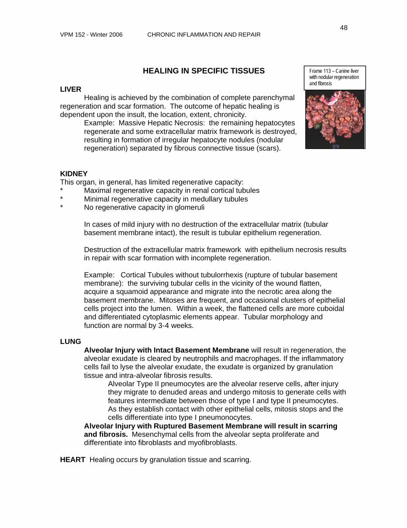
HEALING IN SPECIFIC TISSUES LIVER Healing is achieved by the combination of complete parenchymal regeneration and scar formation. The outcome of hepatic healing is dependent upon the insult, the location, extent, chronicity.
Example: Massive Hepatic Necrosis: the remaining hepatocytes regenerate and some extracellular matrix framework is destroyed, resulting in formation of irregular hepatocyte nodules (nodular regeneration) separated by fibrous connective tissue (scars).
KIDNEY This organ, in general, has limited regenerative capacity: * Maximal regenerative capacity in renal cortical tubules * Minimal regenerative capacity in medullary tubules * No regenerative capacity in glomeruli
In cases of mild injury with no destruction of the extracellular matrix (tubular basement membrane intact), the result is tubular epithelium regeneration. Destruction of the extracellular matrix framework with epithelium necrosis results in repair with scar formation with incomplete regeneration. Example: Cortical Tubules without tubulorrhexis (rupture of tubular basement membrane): the surviving tubular cells in the vicinity of the wound flatten, acquire a squamoid appearance and migrate into the necrotic area along the basement membrane. Mitoses are frequent, and occasional clusters of epithelial cells project into the lumen. Within a week, the flattened cells are more cuboidal and differentiated cytoplasmic elements appear. Tubular morphology and function are normal by 3-4 weeks.
LUNG Alveolar Injury with Intact Basement Membrane will result in regeneration, the alveolar exudate is cleared by neutrophils and macrophages. If the inflammatory cells fail to lyse the alveolar exudate, the exudate is organized by granulation tissue and intra-alveolar fibrosis results.
Alveolar Type II pneumocytes are the alveolar reserve cells, after injury they migrate to denuded areas and undergo mitosis to generate cells with features intermediate between those of type I and type II pneumocytes. As they establish contact with other epithelial cells, mitosis stops and the cells differentiate into type I pneumonocytes.
Alveolar Injury with Ruptured Basement Membrane will result in scarring and fibrosis. Mesenchymal cells from the alveolar septa proliferate and differentiate into fibroblasts and myofibroblasts.
HEART Healing occurs by granulation tissue and scarring.
Frame 113 – Canine liver with nodular regeneration and fibrosis

VPM 152 - Winter 2006 CHRONIC INFLAMMATION AND REPAIR
49
BONE REPAIR Stages in fracture healing 1. Hematoma formation: in the simple traumatic fracture of a long
bone, the broken ends of the bone are misaligned and adjacent soft tissues are torn. Blood vessels are ruptured.
2. Trauma-induced acute inflammation with edema and deposition of fibrin. Monocytes pass into the fracture area and transform into macrophages that play a major role in bone repair.
3. Demolition: macrophages invade and remove fibrin, erythrocytes, debris.
4. Granulation tissue formation (48 hours): invasion of capillary buds and mesenchymal cells derived from periosteum and endosteum, pH low.
5. Soft callus of woven bone and cartilage formation (1 week): mesenchymal osteoblasts differentiate to form bone or cartilage.
6. Woven bone formation from proliferating osteoblasts 7. Lamellar bone formation: woven bone removed by osteoclasts. 8. Remodelling: continued osteoclasts removal and osteoblasts formation; external
callus removed; internal callus hollowed to form bone marrow.
BRAIN C Healing in the brain occurs by proliferation of fibroblasts and glial cells along
vascular networks. When the neuropil is punctured by a sterile instrument, the lesion heals by astrocytic gliosis and fibrosis. The lesion becomes filled with a fibrous core derived from the meninges and perivascular adventitia.
C Astrocytes are stimulated by edema and ischemia, they are less vulnerable to injury than nerve cells. If astrocytes are not destroyed during injury, they form a dendritic network around the wounded neuropil.
C Microglia are migratory, actively phagocytic cells of the neuropil. They engulf lipids and degenerated fragments of dendrites and necrotic neurons.
Bone healing Hematoma formation Frame 13346
Bone healing- Rib fracture Callous formation Frame 3470
Bone healing Hematoma formation and fibrin Frame 13347
Bone healing Intermediate stage Frame 13348
VPM 152 - Winter 2006 CHRONIC INFLAMMATION AND REPAIR
50
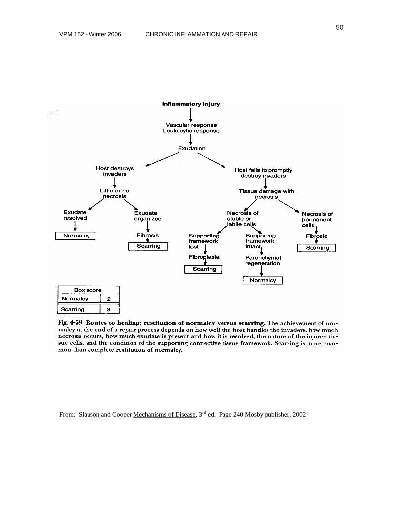
From: Slauson and Cooper Mechanisms of Disease, 3rd ed. Page 240 Mosby publisher, 2002


![Skin Inflammation, [Acute, Suppurative, Chronic, Chronic ... · Skin – Inflammation, [Acute, Suppurative, Chronic, Chronic Active, Granulomatous] presence of mononuclear cells (lymphocytes,](https://img.dokumen.tips/doc/110x75/5f0eb0c97e708231d44075f1/skin-inflammation-acute-suppurative-chronic-chronic-skin-a-inflammation.jpg)