Embed Size (px)
Citation preview
A Prospective Neuroimaging Study of Chemotherapy-Related Cognitive Impairment in
Breast Cancer Patients
Christian Lepage
Thesis submitted to the
Faculty of Graduate and Postdoctoral Studies
in partial fulfillment of the requirements
for the Doctorate in Philosophy degree in clinical psychology.
Psychology
Social Sciences
University of Ottawa
© Christian Lepage, Ottawa, Canada 2016
ii
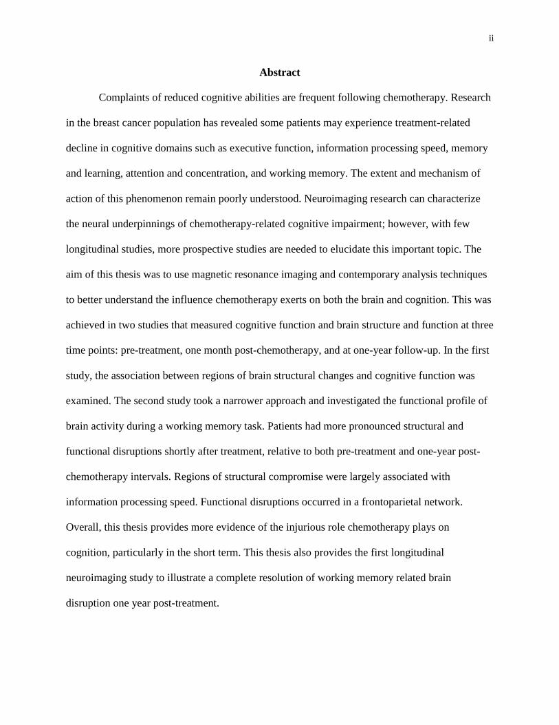
Abstract
Complaints of reduced cognitive abilities are frequent following chemotherapy. Research
in the breast cancer population has revealed some patients may experience treatment-related
decline in cognitive domains such as executive function, information processing speed, memory
and learning, attention and concentration, and working memory. The extent and mechanism of
action of this phenomenon remain poorly understood. Neuroimaging research can characterize
the neural underpinnings of chemotherapy-related cognitive impairment; however, with few
longitudinal studies, more prospective studies are needed to elucidate this important topic. The
aim of this thesis was to use magnetic resonance imaging and contemporary analysis techniques
to better understand the influence chemotherapy exerts on both the brain and cognition. This was
achieved in two studies that measured cognitive function and brain structure and function at three
time points: pre-treatment, one month post-chemotherapy, and at one-year follow-up. In the first
study, the association between regions of brain structural changes and cognitive function was
examined. The second study took a narrower approach and investigated the functional profile of
brain activity during a working memory task. Patients had more pronounced structural and
functional disruptions shortly after treatment, relative to both pre-treatment and one-year post-
chemotherapy intervals. Regions of structural compromise were largely associated with
information processing speed. Functional disruptions occurred in a frontoparietal network.
Overall, this thesis provides more evidence of the injurious role chemotherapy plays on
cognition, particularly in the short term. This thesis also provides the first longitudinal
neuroimaging study to illustrate a complete resolution of working memory related brain
disruption one year post-treatment.
iii
Table of Contents
Abstract ii
List of Tables iv
List of Figures v
Legend vi
Acknowledgements vii
Ethical Standards viii
Statement of Co-Authorship viii
Introduction 1
Chemotherapy 4
Postulated Mechanisms of Impairment 5
Risk Factors 8
Cognition and Chemotherapy 14
Neuroimaging 16
Neuroimaging and CRCI 17
Study Rationale 23
Aims of the Thesis 24
Hypotheses 25
References 27
A Prospective Study of Grey Matter and Cognitive Functioning Alterations in
Chemotherapy-Treated Breast Cancer Patients
44
Abstract 45
Introduction 47
Materials and Methods 50
Results 56
Discussion 64
References 69
Post-chemotherapy recovery of working memory brain activity and functional
connectivity in breast cancer: a prospective fMRI study
78
Abstract 79
Introduction 80
Methods 81
Results 89
Discussion 96
References 102
General Discussion 115
Grey matter volumes and overall cognitive function 115
Working memory and the frontoparietal network 117
Limitations and future directions 119
Conclusion 126
References 128
iv
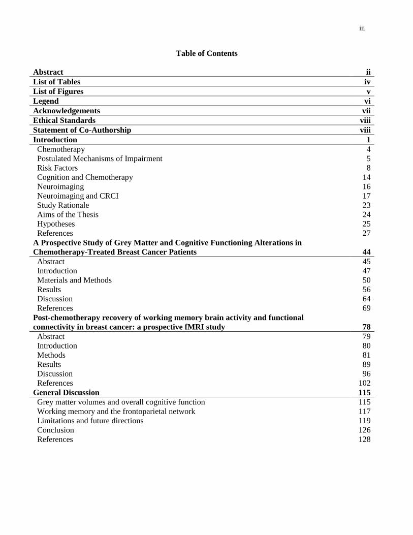
List of Tables
Manuscript 1 Page
Table 1. Demographic and Clinical Characteristics 52
Table 2. Neuropsychological Battery Organized by Cognitive Domain 53
Table 3. Longitudinal Changes in Patient VBM Values 57
Table 4. Patient Within-Group Cognitive Domain Scores ANOVA 60
Table 5. Patient Whole Brain and ROI Grey Matter Volume Correlations with Cognitive Domains 62
Manuscript 2 Page
Table 1. Baseline Demographic and Clinical Characteristics 85
Table 2. Regions of Significantly Greater Working Memory Related Brain Activation in Patients
One Month Post-Chemotherapy Relative to Controls
90
Table 3. Means (Standard Deviations) of Outcome Measure Data 95
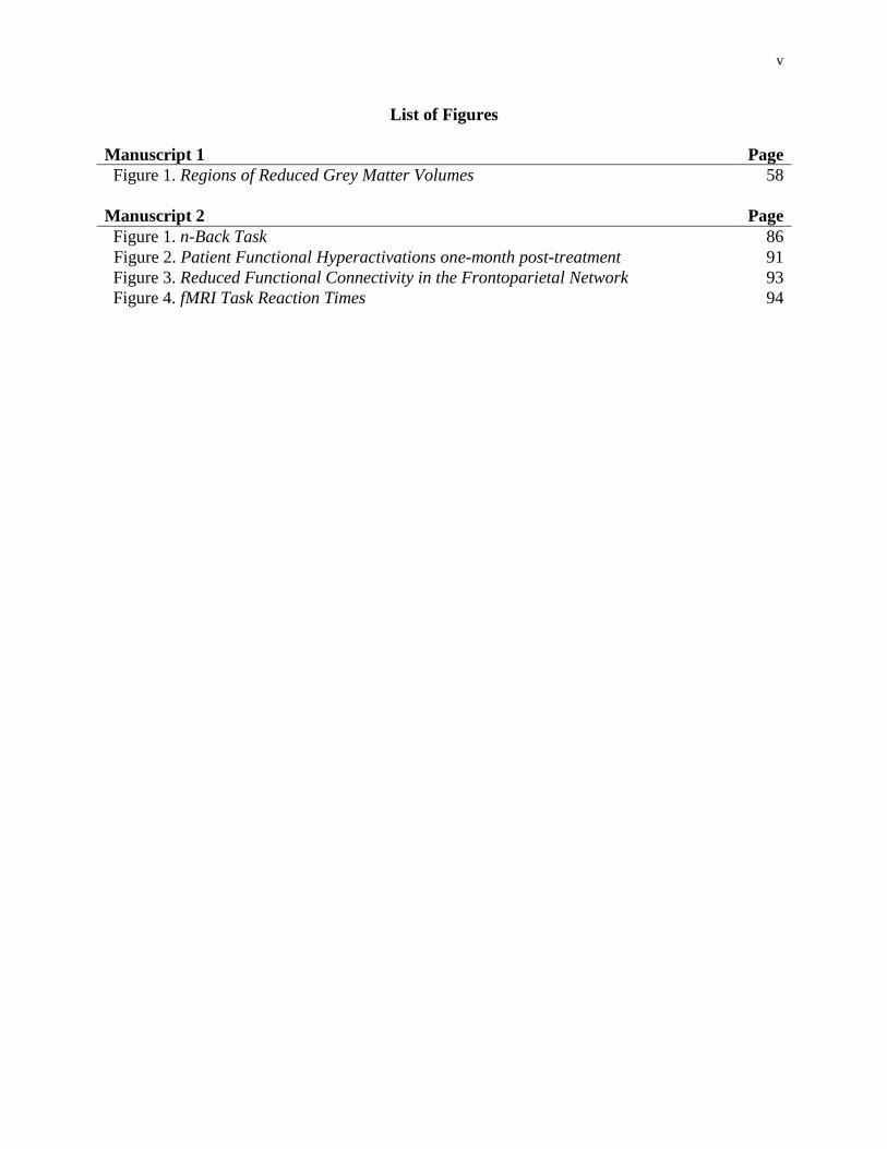
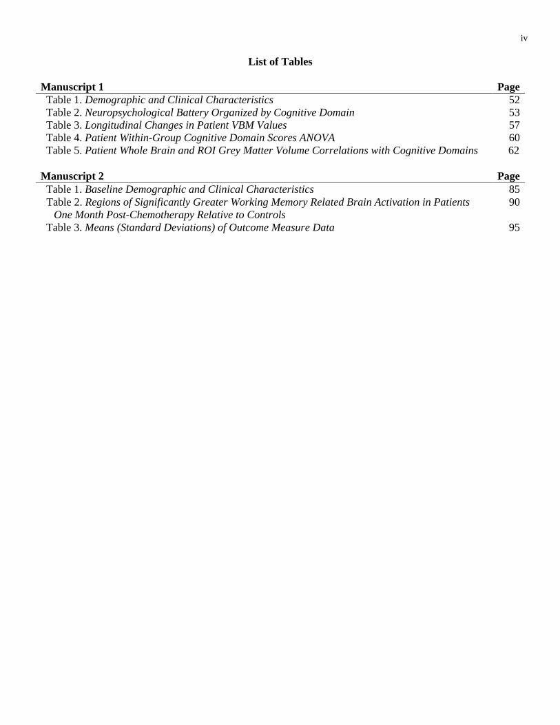
v
List of Figures
Manuscript 1 Page
Figure 1. Regions of Reduced Grey Matter Volumes 58
Manuscript 2 Page
Figure 1. n-Back Task 86
Figure 2. Patient Functional Hyperactivations one-month post-treatment 91
Figure 3. Reduced Functional Connectivity in the Frontoparietal Network 93
Figure 4. fMRI Task Reaction Times 94
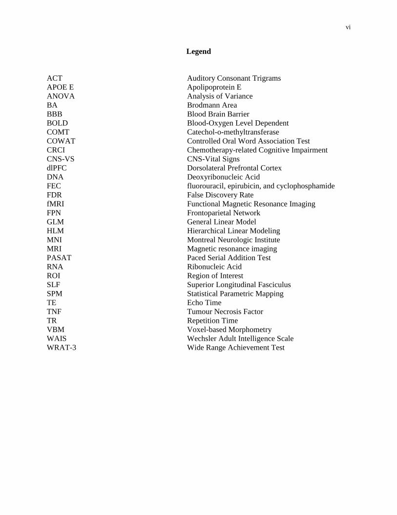
vi
Legend
ACT Auditory Consonant Trigrams
APOE E Apolipoprotein E
ANOVA Analysis of Variance
BA Brodmann Area
BBB Blood Brain Barrier
BOLD Blood-Oxygen Level Dependent
COMT Catechol-o-methyltransferase
COWAT Controlled Oral Word Association Test
CRCI Chemotherapy-related Cognitive Impairment
CNS-VS CNS-Vital Signs
dlPFC Dorsolateral Prefrontal Cortex
DNA Deoxyribonucleic Acid
FEC fluorouracil, epirubicin, and cyclophosphamide
FDR False Discovery Rate
fMRI Functional Magnetic Resonance Imaging
FPN Frontoparietal Network
GLM General Linear Model
HLM Hierarchical Linear Modeling
MNI Montreal Neurologic Institute
MRI Magnetic resonance imaging
PASAT Paced Serial Addition Test
RNA Ribonucleic Acid
ROI Region of Interest
SLF Superior Longitudinal Fasciculus
SPM Statistical Parametric Mapping
TE Echo Time
TNF Tumour Necrosis Factor
TR Repetition Time
VBM Voxel-based Morphometry
WAIS Wechsler Adult Intelligence Scale
WRAT-3 Wide Range Achievement Test
vii
Acknowledgements
There are a number of people who made this project possible through their dedication and
support, for which I am deeply grateful. Thank you, Dr. Andra Smith for giving me this
opportunity and for your inspirational leadership. Your kind-hearted support over the years has
opened more doors for me than I ever thought possible. I am also grateful for the invaluable
support I received by many mentors, colleagues, students, friends, and family throughout this
journey. Thank you, Nancy, Carole, Jeremy, Barb, Rocio, Ola, Selam, Miranda, Angie, Daniel,
Viv, Lyson, and Dan.
I am thankful for the valuable feedback and support of my thesis committee – Drs.
Collins, Taler, Bielajew, and Campbell. I’d also like to thank the staff at the St. Joseph MRI
clinic for their gracious hospitality over the many years.
Thank you to the many women who volunteered for our studies during a critical time in
your lives. I also thank the Canadian Breast Cancer Foundation – Ontario Chapter for funding
the research project. Thank you to the Canadian Institutes of Health Research for your support.
viii
Ethical Standards
A copy of the original approval letter from the Ottawa Hospital Research Ethics Board is
included as Appendix A.
Statement of Co-Authorship
The included manuscripts were crafted with the guidance of Dr. Andra Smith, my thesis
supervisor. As the primary author, I was responsible for the formulation of the research
questions, selection of methods and statistical analyses, data collection, and preparation of the
manuscripts. Dr. Smith provided guidance and assistance in all aspects of this project. Carole
Scherling and Barbara Collins aided in the conceptualization of the overarching research project
from which these studies were drawn, and they provided valuable feedback on the manuscripts
and analyses. Barbara Collins and Joyce MacKenzie oversaw the neuropsychological data
collection. Jeremy Moreau provided assistance with data analysis of the first study. Nancy
Wallis, Emily Barlow-Krelina, Carole Scherling, Miranda Kiyomi Setoguchi, and Zahra Mawani
aided data collection.
1
Introduction
Breast cancer is the most frequently diagnosed cancer among women, afflicting an
estimated 1.67 million women worldwide (Ferlay et al., 2013) and nearly doubling the next-most
common female cancer (Jemal et al., 2011). This trend is mirrored in North America where the
estimated incidence of new cases of breast cancer in 2014 was 232,670 in the United States
(Siegel, Ma, Zou, & Jemal, 2014) and 24,400 in Canada (Canadian Cancer Society, 2014).
Breast cancer survivorship rates have increased over the decades, due in part to the development
of improved treatment options. Since 1986, the five-year relative survival rate for Canadian
breast cancer patients increased from 76% to 88% (Canadian Cancer Society, 2014).
Chemotherapy-based adjuvant therapies supplement principal surgery-based interventions and
have become commonly employed, resulting in greater survivorship among breast cancer
patients (Mariotto et al., 2002).
Subsequent to adjuvant chemotherapy exposure, many breast cancer patients report
cognitive decline (e.g. see (Pullens, De Vries, & Roukema, 2010)), a phenomenon colloquially
referred to as “chemofog” and “chemobrain” (Raffa et al., 2006). Self-perceived deterioration in
mental functioning can adversely impact work and family life for breast cancer survivors
(Boykoff, Moieni, & Subramanian, 2009). Objective evidence of chemotherapy-related cognitive
impairment (CRCI) in breast cancer patients has mounted in the last decades. Varying degrees of
cognitive under-performance are found in many domains, including working memory,
information processing speed, visuospatial ability, attention and concentration, motor
functioning, executive function, and memory (Ahles et al., 2002; Bender et al., 2006; Brezden,
Phillips, Abdolell, Bunston, & Tannock, 2000; Falleti, Sanfilippo, Maruff, Weih, & Phillips,
2005; Fan et al., 2005; Hurria, Rosen, et al., 2006; Jansen, Cooper, Dodd, & Miaskowski, 2011;
2
Kam et al., 2015; Reid-Arndt, Hsieh, & Perry, 2010; van Dam et al., 1998; Wefel, Saleeba,
Buzdar, & Meyers, 2010).
The etiology of CRCI is complex. CRCI is subtle, appears to affect subgroups of patients
only, and is a transient phenomenon for some (Falleti et al., 2005; Stewart, Bielajew, Collins,
Parkinson, & Tomiak, 2006). Despite having the greatest influence on cognitive functioning
during and up to 6 months post treatment (Collins, Mackenzie, Tasca, Scherling, & Smith, 2014;
Jim et al., 2012), chemotherapy is related to impaired cognition decades after treatment (Ahles et
al., 2002; Koppelmans et al., 2012). Furthermore, cognitive dysfunction has been found prior to
the commencement of chemotherapy (Ahles et al., 2008; Jansen et al., 2011; Wefel, Lenzi,
Theriault, Davis, & Meyers, 2004), hinting at other contributing factors to CRCI, including the
cancer itself and emphasizing the need for increased study using other investigative methods.
The neurophysiological correlates of CRCI have been a topic of increased study in recent
years. Prospective neuroimaging studies of brain structure have found abnormalities in both grey
and white matter compartments following chemotherapy (Deprez et al., 2012; McDonald,
Conroy, Ahles, West, & Saykin, 2010; McDonald, Conroy, Smith, West, & Saykin, 2012).
Although some recovery has been noted one year from treatment (McDonald et al., 2010),
retrospective studies have reported brain matter disruption decades after chemotherapy (de
Ruiter et al., 2012; Koppelmans et al., 2012, 2014). Additionally, there is conflicting evidence of
pre-treatment brain structure anomalies, with some support for (McDonald, Conroy, Smith, et al.,
2012) and against (McDonald et al., 2010) baseline differences between breast cancer patients
and control participants before patient adjuvant treatment.
Similarly, functional brain differences have been reported prior to chemotherapy (Askren
et al., 2014; Berman et al., 2014; Cimprich et al., 2010; Scherling, Collins, Mackenzie, Bielajew,

3
& Smith, 2011, 2012). Analogous to the neuropsychological and neuroanatomical literature of
CRCI, evidence suggests that the greatest neurofunctional abnormality is apparent during and
shortly after chemotherapy, relative to baseline and one-year after treatment (McDonald, Conroy,
Ahles, West, & Saykin, 2012). Notably, retrospective studies have reported differences in neural
recruitment during cognitive tasks between breast cancer patients and healthy controls over 10
years after chemotherapy (de Ruiter et al., 2011; Stouten-Kemperman et al., 2015). These mixed
findings further suggest the reduced cognition experienced by breast cancer patients during the
course of chemotherapy may be influenced by disease and other treatment related factors.
In light of the work showing brain structure and function irregularities in breast cancer
patients, there is an impetus to examine the brain networks that support cognitive functioning.
The few studies that have examined functional networks in breast cancer patients reflect the
findings from other neuroimaging modalities, with breast cancer patients showing decreased
overall network efficiency compared to control participants (Bruno, Hosseini, & Kesler, 2012;
Hosseini, Koovakkattu, & Kesler, 2012; Kesler et al., 2013). To date, only one pilot study has
longitudinally examined network integrity in breast cancer patients (Dumas et al., 2013). The
results of that study suggest the default mode and dorsal attention networks are disrupted after
chemotherapy, although the former has displayed some recovery one year after chemotherapy.
Nevertheless, there is a lacuna in the CRCI neuroimaging literature of prospective brain network
integrity investigations.
Thus, the overall aim of this thesis was to use contemporary neuroimaging techniques to
elaborate the current scientific understanding of the neural mechanisms that underlie CRCI. In
order to achieve this goal, this thesis was divided into two studies, each having their own set of
objectives germane to elucidating the influence that chemotherapy exerts on cognition and brain

4
function and structure. The first study used a neuroimaging analysis technique to explore the
course of grey matter volume changes in breast cancer patients and investigate the relationship
between regions of grey matter loss and cognitive function. In the second paper, the integrity of a
working memory network is examined prospectively - the first such study in the CRCI literature.
The two studies are presented in manuscript format, with the structural investigation appearing as
it does in published format, and the functional study appearing as it did during submission to a
peer-reviewed journal. A general discussion follows the articles, summarizes the findings of the
thesis, reviews the implications, acknowledges the limitations of the work, and offers direction
for future research. Before presenting the articles, a general introduction provides the necessary
background information.
Chemotherapy
Chemotherapy drugs used to treat breast cancer can be classified into different groupings,
although some of them have characteristics that overlap two or more categories. Various types of
chemotherapy agents may be used, depending on the stage of cancer and treatment response
(DiPiro, 2009); however, only the most commonly used drugs for early stage breast cancer will
be discussed. Alkylating drugs disrupt the replication and copying processes during the cell
cycle, precluding cancerous cells from repairing damaged DNA (Gerson, Bulgar, Weeks, &
Chabner, 2011). A commonly used alkylating agent in breast cancer is cyclophosphamide
(Morris & Hudis, 2011). Platinum-based antineoplastic drugs are sometimes grouped with
alkylating agents since they destroy cells in a similar fashion. Frequently used platinum-based
drugs in the treatment of breast cancer include cisplatin and carboplatin (Decatris, Sundar, &
O’Byrne, 2004). Antimetabolites supplant the normal building blocks of RNA and DNA during
the chromosomal copying phase of the cell cycle, interfering with cell growth. The
5
antimetabolites used in breast cancer treatment are 5-fluorouracil and methotrexate (Hortobagyi,
2000). Anthracyclines subvert mitosis throughout the cell cycle by disrupting enzymes involved
in DNA replication (Skeel, 2011). Doxorubicin and epirubicin are regularly used anthracyclines
in breast cancer treatment. Finally, the taxanes - docetaxel and paclitaxel - are plant alkaloids
that interfere with microtubules, leading to mitotic arrest and, thus, inhibited cell division (Skeel,
2011). Contemporary regimens employ combinations of chemotherapy drugs to optimize
outcomes for breast cancer patients.
Chemotherapy may be administered preoperatively and after surgical intervention for
breast cancer. It may be delivered in concert with a combination of other adjuvant interventions,
including radiation and hormonal therapies (National Cancer Institute, n.d.). Prior to surgical
removal of the cancerous tissue, neoadjuvant chemotherapy may be dispensed to shrink a tumour
that is currently inoperable or to allow for breast-conserving surgery (Mauri, Pavlidis, &
Ioannidis, 2005). Adjuvant therapies, including chemotherapy, are delivered to destroy cancer
cells or to stop them from dividing (Office on Women’s Health, n.d.). Chemotherapy can be
administered orally and via injection, whether it is given prior to or following surgery. Treatment
occurs for three to six months in weeks-long cycles that vary depending on the drugs used
(American Cancer Society, n.d.).
Postulated mechanisms of impairment
Neurotoxic sequelae have been reported for nearly each category of the chemotherapy
agents (Dropcho, 2004; Scatchard & Lee, 2010; Schagen, Muller, Boogerd, Mellenbergh, & van
Dam, 2006); however, the mechanisms of chemotherapy neurotoxicity remain to be well
characterized. Several processes have been proposed, including direct toxic cellular effects,
6
indirect metabolic abnormalities, inflammatory processes, and vascular influences (Dietrich,
2010).
Blood-brain barrier. The blood-brain barrier (BBB) is generally thought to provide
protection from cytotoxic agents. In fact, it is a major obstacle for the delivery of chemotherapy
agents to brain-based tumours and micrometastatic disease (Deeken & Löscher, 2007). Despite
this, there is some evidence that 5-fluorouracil has a degree of BBB permeability in rodents
(Sakane, Yamashita, Yata, & Sezaki, 1999). The BBB may be susceptible to increased
permeability via the effects of cancer (e.g. micrometastases), radiation treatment, or genetic
variability (Ahles & Saykin, 2007; Wefel, Witgert, & Meyers, 2008). It has been speculated that
a synergistic effect of repetitive exposure to combination of chemotherapy drugs may also
impact the blood-brain barrier in a way that allows lipophilic drugs to penetrate the brain
parenchyma; however, this hypothesis remains to be supported empirically, particularly in
humans (Dietrich, 2010).
Oxidative stress. Oxidative stress is a biochemical condition in which there is an
imbalance between cellular pro-oxidants and antioxidants that leads to irregular cellular
signalling. It has been related to cancer, inflammation, aging, drug action, and drug toxicity
(Sies, 1985). Damage to DNA in neuronal cells from oxidative stress is common (Park et al.,
1998) and may occur via exposure to exogenous toxins (Ozben, 2007). For example, the
anthracycline agent doxorubicin - commonly used to treat breast cancer - is associated with the
induction of oxidative stress (Quiles, Huertas, Battino, Mataix, & Ramírez-Tortosa, 2002). A
recent study by Conroy et al. (2012) found increased oxidative DNA damage in early-stage
breast cancer patients compared to healthy control participants at an average of six years post-
chemotherapy. Notably, the majority (71%) of the regimens included in that study incorporated
7
doxorubicin. Furthermore, the authors found that oxidative DNA damage in the patient group
was associated with reduced grey matter density in temporal and mesencephalic regions.
Similarly, reduced hippocampal volume has been related to increased levels of tumour necrosis
factor (TNF)-alpha in early-stage breast cancer patients (Kesler, Janelsins, Koovakkattu, &
Palesh, 2013). It has been proposed that TNF-alpha, which can pass the BBB and is increased by
doxorubicin, can induce the expression of nitric oxide synthases. In turn, nitric oxide synthases
can then lead to oxidative stress (Chen, Jungsuwadee, Vore, Butterfield, & St Clair, 2007).
Nevertheless, it is not well understood how DNA damage leads to injury in the brain (Ahles &
Saykin, 2007). Oxidative DNA damage has been linked with neurodegenerative disorders that
are characterized by cognitive symptoms (Keller et al., 2005; Wang et al., 2014), suggesting a
relationship between DNA damage and cognitive difficulties.
Proinflammatory cytokine dysregulation. Cytokine is a general term used to describe
small proteins that are extruded from cells and have a distinct impact on intercellular interaction
and communication. Cytokines may interact with host, neighbouring, or remote cells. At times,
they are referred to with function-specific terms: a chemokine is a cytokine with chemotactic
activities, and interleukin is a cytokine made by one leukocyte that acts on other leukocytes. A
cytokine may stimulate the production of another in a remote cell, resulting in cascaded
manufacturing. As part of the immune system, many cell types produce cytokines, including B
cells, T cells, macrophages, mast cells, neutrophils, basophils, and eosinophils (Zhang & An,
2007).
Increased levels of cytokines have been reported in cancer patients treated with standard-
dose chemotherapy (Janelsins et al., 2012; Lotti et al., 2013). Cytokine-induced inflammation
related to chemotherapy has been linked to cognitive diminishment (Janelsins et al., 2012) and
8
self-reported memory complaints (Ganz et al., 2013). As mentioned, increased cytokine levels
(e.g. TNF-alpha) have been related to lower hippocampal volume in breast cancer patients
(Kesler et al., 2013). The authors of that study also reported reduced levels of another cytokine,
interleukin-6, was linked to smaller hippocampal volume, with both cytokines being associated
with memory performance. Using positron-emission tomography, a recent pilot study examined
the relationship between frontal lobe metabolism and inflammatory markers, including C
reactive protein and interleukin-6 (Pomykala et al., 2013). The authors found that at baseline
(which included measures taken post-chemotherapy), the presence of inflammatory markers was
related to left inferior frontal and right inferior lateral metabolic activity; there was no such
relationship for control participants. At one-year follow-up, frontal and medial metabolic activity
remained positively correlated with inflammatory markers in patients; again, there was no link
between these markers and neurometabolic activity in control participants. It has been suggested
that cytokines may be triggered by DNA damage (e.g. caused by chemotherapy drugs), which
could establish a cycle of further DNA damage and cytokine activity, resulting in chronic
inflammation. The inflammation can increase oxidative stress, further compounding this cycle
(Ahles & Saykin, 2007). Overall, the evidence suggests that cytokine deregulation may be
related to CRCI; however, more evidence must accrue before such a relationship can be deemed
definitive.
Risk Factors
CRCI affects a subset of breast cancer patients treated with chemotherapy, suggesting
that the cognitive effects experienced after treatment may be modulated by one or more risk
factors. Some risk factors are supported by evidence of a deleterious influence on cognition
independent of chemotherapy. These may predispose some patients to CRCI. Other risk factors
9
arise from disease and treatment processes that are variable across breast cancer patients.
Potential influences of CRCI that may emerge outside of the processes related to cancer and its
treatment will be discussed first.
Age. Age-related changes in cognition are well-accepted phenomena in the
neuropsychological literature (Salthouse, 2009). The risk of being diagnosed with breast cancer
rises with age, with the highest rates occurring at age 60 and later (Key, Verkasalo, & Banks,
2001). Unsurprisingly, studies of CRCI predominantly include samples whose mean age is above
50 years, necessitating a need to control for age-related changes in cognition. Thus far, it has
been common practice in the CRCI literature to control for age (e.g. see (Scherling & Smith,
2013)). Yet, it has been speculated that chemotherapy may hasten aging and that it can induce a
vulnerability to late-emerging cognitive decline (Schagen & Wefel, 2013). Correspondingly,
cross-sectional studies conducted many years after patient chemotherapy have reported CRCI in
breast cancer patients relative to both healthy and chemotherapy-untreated controls (Ahles et al.,
2002; Koppelmans et al., 2012; Yamada, Denburg, Beglinger, & Schultz, 2010). Although
impairment is reported after age has been controlled for, the cross-sectional nature of these
studies does not preclude the possibility that chemotherapy administration may place women at
greater risk for cognitive decline in the future by hastening age-related changes in cognition.
Age is positively related to executive dysfunction in chemotherapy-treated patients
(Kesler, Kent, & O’Hara, 2011) and with a high degree of memory complaints (Hurria et al.,
2006). Similarly, older age is associated with increased toxicity from chemotherapy exposure
(Hurria et al., 2011; Muss et al., 2007). Thus, it may be that the effects of chemotherapy vary
depending on the age of the patient at the time of administration, such that older women may be
at greater risk of treatment-related cognitive decline than younger ones.
10
Cognitive reserve. In response to increased cognitive demands, there is variability in the
magnitude and efficiency of the associated neural response across individuals. One’s ability to
optimize performance via flexible recruitment of brain networks, proposed to reflect the use of
alternative cognitive strategies, has been labelled cognitive reserve (Stern, 2002). Cognitive
reserve has been postulated to account for the differential outcome among individuals following
brain injury, such that those with a higher degree of it have less functional impairment than
individuals with lower levels (Stern, 2009). Using WRAT-3 (Wilkinson, 1993) reading scores as
a measure of baseline cognitive reserve, Ahles et al. (2010) showed that chemotherapy-treated
breast cancer patients - who were older and had lower pre-treatment cognitive reserve - had
reduced information processing speed compared to control participants. Similarly, pre-treatment
executive network inefficiency is related to breast cancer patients’ complaints of disrupted
cognition and elevated fatigue (Askren et al., 2014).
Educational level is related to cognitive reserve, with higher education acting as a
protective factor against neurodegenerative disorders in old age (Carret et al., 2003). Higher
educational attainment in chemotherapy-treated breast cancer patients is linked to decreased
perseverative errors during executive function tasks (Kesler et al., 2011). Although studies of
cognitive reserve are scarce in the CRCI literature, taken together, current evidence suggests that
it modulates the influence of chemotherapy on cognition.
Genetic factors. Increased susceptibility to brain insult following chemotherapy
administration may be, to an extent, facilitated by genetic variability in genes that manage neural
repair (e.g. see (Ahles & Saykin, 2007)). Apolipoprotein E (APOE) is a component of various
lipoproteins, and its primary role is to transport lipids and cholesterols throughout the body. It is
the major apolipoprotein expressed in the brain, where it has the added functions of mediating
11
synaptogenesis, synaptic plasticity, and neuroinflammation (Chouraki & Seshadri, 2014). The
APOE E4 allele has been identified as a risk factor for the development of Alzheimer’s disease
and cognitive decline in carriers not diagnosed with mild cognitive impairment or Alzheimer’s
disease (Liu, Liu, Kanekiyo, Xu, & Bu, 2013). Although scarce, there is evidence that the APOE
E4 allele in chemotherapy-treated breast cancer patients is related to decreased cognition
compared to non-carriers (Ahles et al., 2003, 2014). However, evidence of neuroimaging
markers of CRCI where APOE E4 status was explored remains equivocal (Ferguson, McDonald,
Saykin, & Ahles, 2007; McDonald, Conroy, Smith, et al., 2012).
Catechol-o-methyltransferase (COMT) is an enzyme that regulates catecholamine
neurotransmitters, such as dopamine, epinephrine, and norepinephrine through deactivation.
COMT enzymes are coded by the COMT gene, which has several alleles, including Val158Met.
This single-nucleotide polymorphism substitutes valine (e.g. Val) with methionine (e.g. Met) at
codon 158 (Lachman et al., 1996). A Val allele of COMT can increase the catabolism of
dopamine by a factor of four, in contrast to that in COMT-Met homozygote carriers. The
Val158Met allele has been linked with dopamine levels in the prefrontal cortex, and a reduced
level of the neurotransmitter in that area may account for the reports of disparate cognition
between COMT-Val and -Met carriers, specifically on tasks of attention and executive function
(Dickinson & Elvevåg, 2009). A retrospective study of CRCI found that COMT-Val carriers in a
breast cancer group with a history of chemotherapy exposure performed more poorly on tests of
overall cognition, complex cognition, attention, verbal fluency, and motor speed, relative to their
COMT-Met carrying counterparts (Small et al., 2011). Although the differences were not
statistically significant after correcting for multiple comparisons, the study suggests that breast
cancer patients who are carriers of COMT-Val may be especially vulnerable cognitively to
12
CRCI. Overall, the study of genetics in CRCI is nascent, and more studies should be conducted
to convincingly determine what role, if any, genes might play in rendering some breast cancer
patients more susceptible than others to cognitive decline following chemotherapy.
Dosage. There is evidence that CRCI may be modulated by the magnitude and duration
of treatment, as well as the number of chemotherapy cycles. An early study of CRCI compared
the effects of high-dose to standard-dose chemotherapy on cognition in breast cancer patients
(van Dam et al., 1998), and found nearly twice the rate of impairment in the former treatment
arm. Additionally, the women who received high-dose chemotherapy were 8.2 times more likely
than chemotherapy-free controls to experience cognitive impairment. A later prospective study
supported those findings, demonstrating that breast cancer patients receiving high-dose
chemotherapy experienced significant cognitive deterioration from pre-treatment baseline to six
months afterwards, relative to healthy controls (Schagen et al., 2006). In contrast, breast cancer
patients who had received either a standard-dose treatment or were chemotherapy-free did not
have a significant decline of cognition relative to healthy controls. Evidence of dose-modulated
CRCI prompted the investigation of cumulative effects of chemotherapy on cognition. In a novel
approach to studying CRCI, Collins et al. (2013) measured the cognitive functioning of breast
cancer patients and controls at pre-treatment baseline, and following patients’ individual
chemotherapy cycles. The authors found that, although the patient group did not significantly
decline on most neuropsychological measures, it did not benefit from repeated testing to the
extent that the healthy control group did. In fact, once practice effects were controlled for, scores
on a composite of overall cognition diminished with successive chemotherapy administrations.
Interestingly, this pattern was observed in a subset of the authors’ larger sample, which consisted
of a group treated with 5-fluorouracil, epirubicin, and cyclophosphamide (FEC) - the regimen
13
that was designated as standard-dose in the aforementioned studies in this section (e.g. (Schagen
et al., 2006; van Dam et al., 1998)). Thus, although women who receive higher doses of
chemotherapy are at increased risk of CRCI, successive administrations of chemotherapy, even
in low-dose regimens, can also render breast cancer patients susceptible to disrupted cognitive
function.
Other adjuvant therapies. The treatment of breast cancer is often multifaceted, with
chemotherapy being one of a number of adjuvant interventions employed concurrently or in
succession to remove the disease. Radiotherapy may be indicated in early stage breast cancer
subsequent to lumpectomy or mastectomy, with an aim to eliminate any cancerous cells that
have lingered or spread to other areas (American Cancer Society, 2015). The cognitively
injurious effects of radiotherapy are well recognized across many cancer populations, including
breast cancer (Jim et al., 2009; Quesnel, Savard, & Ivers, 2009; Shibayama et al., 2014; Small et
al., 2011), with neurological and neuropsychological compromise frequently being the dose-
limiting consideration of this treatment. In fact, radiotherapy is generally not administered
concurrently with chemotherapy due to potential synergistic effects that can exacerbate the
negative consequences associated with these interventions (Bellon & Harris, 2005).
Hormonal therapy is generally indicated for estrogen-positive tumours, due to its ability
to treat hormone-sensitive breast cancer by blocking ovarian function, estrogen production, or
the effects of estrogen (National Cancer Institute, 2012). With most breast cancers being
estrogen-receptor positive (Anderson, Chatterjee, Ershler, & Brawley, 2002), hormone therapy is
commonly administered to breast cancer patients. There are inconsistent findings in the CRCI
literature regarding the cognitive effects of hormonal therapy. Some prospective studies report
that there are no cognitive differences between patients treated with chemotherapy and hormone
14
therapy (Fan et al., 2005; Jenkins et al., 2006), while others have found that the latter was
associated with greater, widespread compromise (Bender et al., 2006; Collins, Mackenzie,
Stewart, Bielajew, & Verma, 2009). Neuroimaging evidence has supported findings in favor of
cognitive disruption following hormone therapy, with one study showing that breast cancer
patients who had taken tamoxifen had smaller hippocampal volumes and diffuse
hypometabolism in frontal brain areas (Eberling, Wu, Tong-Turnbeaugh, & Jagust, 2004). Thus,
within the context of the CRCI literature, disentangling chemotherapy’s contribution to post-
treatment cognitive dysfunction can be confounded by the presence of chemotherapy along with
a combination of radiotherapy and hormone therapy (e.g. see (Tager et al., 2010)).
As evidenced by the array of potential mechanisms of impairment and risk factors, all of
which can vary from one patient to the next, CRCI is a complex phenomenon requiring further
clarification.
Cognition and chemotherapy
Although earlier, cross-sectional studies first raised awareness about CRCI (van Dam et
al., 1998; Wieneke & Dienst, 1995), longitudinal investigations, incorporating a pre-treatment
baseline, importantly showed that breast cancer patients could have clinically significant
reductions in cognitive function over time, despite an absence of cross-sectional between-group
differences, relative to controls (e.g. see (Schagen et al., 2006)). A large number of longitudinal
studies of CRCI have now been conducted, comparing cognitive performance in chemotherapy-
treated patients to both chemotherapy-free and healthy controls (Ahles et al., 2010; Bender et al.,
2006; Collins et al., 2009; Fan et al., 2005; Hermelink et al., 2007; Jenkins et al., 2006; Quesnel
et al., 2009; Tager et al., 2010). Among longitudinal, prospective investigations, CRCI in
patients receiving adjuvant chemotherapy has been found in 12 to 82% of samples, with the
15
cognitive domains of executive function, working memory, information processing speed,
visuospatial ability, attention and concentration, motor functioning, and memory frequently
impacted. The wide range of incidence has been speculated to reflect small sample sizes,
heterogeneous use of neuropsychological assessment batteries and control groups, the presence
of various treatment regimens, and disparate cut-offs used to demarcate impaired from normal
cognitive functioning (Janelsins, Kesler, Ahles, & Morrow, 2014). In spite of this variability, it
has become evident that cognition is adversely impacted in some breast cancer patients following
chemotherapy.
Of the commonly reported cognitive domains affected by chemotherapy, working
memory appears to be the most vulnerable (Bender et al., 2006; Collins et al., 2009; Shilling,
Jenkins, Morris, Deutsch, & Bloomfield, 2005; Stewart et al., 2006). Across a broad range of
cognitive domains, working memory was uniquely lower among chemotherapy-treated patients
compared with hormone-therapy-only controls (Stewart et al., 2008). Interestingly, both groups
scored within normal limits on testing; however, only the chemotherapy-treated group
experienced reliable cognitive deterioration. Although the mechanisms of impairment in CRCI
remain to be understood, animal models suggest that chemotherapy-driven neuroinflammation
may produce a neurotoxic effect on white matter integrity via demyelination (Briones & Woods,
2014). Working memory is related to white matter integrity in many pathways, including the
superior parietal lobule pathway, the medial temporo-frontal pathway, the uncinate fasciculus,
the frontoparietal fasciculus, and the cingulum (Charlton, Barrick, Lawes, Markus, & Morris,
2010). A predilection for white-matter pathways may help explain how chemotherapy exerts its
influence on working memory.
Neuroimaging
16
Neuroimaging methods have been used to better characterize the extent of chemotherapy-
related cognitive dysfunction, and to explore its candidate mechanisms. In the last decade,
evidence has been accumulating that frontal and parietal regions, along with subcortical white
matter structures, may be vulnerable to structural and functional abnormalities following
chemotherapy. This section will provide a brief introduction to some of the non-invasive
neuroimaging techniques used to explore the neurobiological correlates of CRCI, and provide a
review of the findings to date.
Magnetic Resonance Imaging. Magnetic resonance imaging (MRI) is a technology that
permits investigators to directly observe internal anatomy with fine detail by manipulating the
magnetic properties of certain protons. This technology can be used to study the structure and
function of the human brain without exposing patients to radiation. Hydrogen atoms are
abundant in the brain, and possess the nuclear magnetic resonance property - a required
characteristic for MRI. When placed in a uniform external magnetic field, the protons will align
themselves in parallel with the main magnetic field in either a low- or high-energy state. In MRI,
radiofrequency pulses are applied that tip the alignment of the protons. When the radiofrequency
signal is turned off, the spins begin to relax and realign with the main magnetic field. During the
realignment process, detector coils capture energy emitted by protons, and the signal is then
converted to images by a computer. When interpreted by specialized software, data gleaned from
MRI can generate 2- and 3-D images of different tissue types – white matter, grey matter,
cerebrospinal fluid – based on their different rates of relaxation and recovery (Huettel, Song, &
McCarthy, 2009).
Functional magnetic resonance imaging. Functional magnetic resonance imaging
(fMRI) takes advantage of the same physical properties and employs the same scanner as used
17
for structural MRI. Where MRI is used to create high-resolution images of anatomical structures,
fMRI refers to the acquisition of imaging data used to make inferences about the presumed
underlying neuronal activity that drives the changes in MRI signals. Images contain values of the
signals related to neuronal hemodynamic responses. In this way, fMRI can be used as a proxy for
neuronal activity. The metabolic needs of neurons are increased during neural activity, and to
satisfy these demands, an increase in blood flow transports energy to active neurons. This
increase in blood flow replaces deoxyhemoglobin molecules with oxygenated molecules in the
active regions, resulting in a change of local magnetic signal (Huettel et al., 2009). As a result of
this phenomenon - the blood oxygen level dependent (BOLD) effect - fMRI researchers can
study the brain in action.
Neuroimaging and CRCI
In the last decade, neuroimaging studies of CRCI have emerged, generally supporting the
findings from the neuropsychological studies of chemotherapy and cognition. Such studies have
explored the integrity of white and grey matter, functional activations in response to cognitive
tasks, and the efficiency of brain networks. In many cases, abnormal findings in patients exposed
to chemotherapy have been related to decreases in performance on neuropsychological measures.
Another line of evidence comes from the course of brain disruption, which often mirrors the
trajectory of cognitive function. Specifically, from longitudinal neuropsychological and
neuroimaging studies, there is accumulating evidence that disruption is most pronounced during
and shortly after chemotherapy, with some recovery noted within one year after treatment. In the
following sections, findings of CRCI neuroimaging studies of brain structure, function, and
networks will be summarized.
18
Structural studies. Cross-sectional studies have reported white matter tract compromise
in breast cancer patients shortly after chemotherapy (Abraham et al., 2008) and decades after
treatment (de Ruiter et al., 2012; Koppelmans et al., 2014; Stouten-Kemperman et al., 2015).
One of the first prospective studies of white matter integrity found post-treatment irregularities in
the corpus callosum, and frontal and parietal regions that were related to performance in
attention and verbal memory for breast cancer patients relative to chemotherapy-untreated
patients (Deprez et al., 2012). A baseline comparison between these groups revealed no
significant differences in white matter.
Studies of grey matter integrity have been more prominent in the CRCI literature
(Conroy, McDonald, Ahles, West, & Saykin, 2013; Hakamata et al., 2007; Hosseini et al., 2012;
Inagaki et al., 2007; Koppelmans et al., 2014; McDonald et al., 2010; McDonald, Conroy, Smith,
et al., 2012; Scherling, Collins, MacKenzie, et al., 2012), and point to a course of grey matter
alterations that are most pronounced following chemotherapy relative to baseline. With respect
to post-treatment effects, cross-sectional studies have reported long-term grey matter
abnormalities in chemotherapy-treated breast cancer patients up to 9.5 years after treatment (de
Ruiter et al., 2012) and 21 years after chemotherapy (Koppelmans et al., 2012); however, one
cross-sectional study found no CRCI related grey matter attenuation nearly 4 years after
treatment (Inagaki et al., 2007). The first prospective voxel-based morphometry study of the
CRCI population found no pre-chemotherapy structural differences when comparing patients to
controls (McDonald et al., 2010). One month following treatment, the chemotherapy-treated
group displayed broad grey matter alterations that partially recovered one year following
exposure. In light of these mixed findings regarding the course of grey matter following
treatment, Conroy et al. (2013) reported that grey matter density in the right superior and middle
19
frontal gyri was associated with post-chemotherapy interval, and that overall neuropsychological
performance was related to mean grey matter density in those areas.
Thus, although few investigations of white matter have been undertaken in the
chemotherapy and cognition field, those that have been conducted, along with those studying
grey matter, offer similar findings to those from neuropsychological investigations of CRCI.
Specifically, studies of brain structure integrity following chemotherapy suggest some breast
cancer patients are susceptible to its neurotoxic effects, particularly in the short-term following
treatment, with some patients experiencing recovery over time. Further, both grey and white
matter compromise have been observed largely in frontal, parietal, and temporal regions. These
findings support the reported neuropsychological deficits in domains largely subserved by these
regions, including executive function, working memory, information processing speed,
visuospatial ability, attention and concentration, motor functioning, and memory (Wefel et al.,
2008).
Functional Studies. Functional neuroimaging studies have revealed neural activation
differences between chemotherapy-treated breast cancer patients and chemotherapy-unexposed
controls, both cross-sectionally and prospectively. With the exception of a positron-emission
study by Silverman et al. (2007), functional neuroimaging studies of CRCI in breast cancer have
employed fMRI, due in part to its non-invasive ability to provide a high quality, in vivo measure
of neuronal activity with high spatial resolution (Askren et al., 2014; Cimprich et al., 2010;
Conroy et al., 2012, 2013; de Ruiter et al., 2011; Ferguson et al., 2007; S R Kesler et al., 2011;
Kesler, Bennett, Mahaffey, & Spiegel, 2009; López Zunini et al., 2013; McDonald, Conroy,
Ahles, et al., 2012; Saykin et al., 2006; Scherling et al., 2011; Scherling, Collins, Mackenzie, et
al., 2012; Stouten-Kemperman et al., 2015).

20
Functional irregularities between chemotherapy-treated breast cancer patients and
controls have been found during tasks of executive function (de Ruiter et al., 2011; Kesler et al.,
2011) and short-term verbal memory (de Ruiter et al., 2011; Kesler et al., 2009; López Zunini et
al., 2013; Silverman et al., 2007). Both hyper- and hypoactivations have been predominantly
circumscribed to frontal, temporal, and parietal regions. A cross-sectional study by de Ruiter et
al., (2011) found decreased frontal and parietal activation during a planning task and decreased
frontal, temporal, and parietal activation during a paired-association task approximately 10 years
after chemotherapy exposure. These patterns were later related to decreases in white matter
integrity and grey matter volume (de Ruiter et al., 2012). Hyporesponsiveness has also been
reported in frontal regions, including the bilateral superior and middle frontal gyri, during a
verbal declarative memory encoding task (Kesler et al., 2009). Similarly, frontotemporal
hypoactivations have been reported during a verbal recognition task (López Zunini et al., 2013).
In keeping with findings from neuropsychological studies of CRCI that show working
memory to be especially vulnerable to chemotherapy (Stewart et al., 2006), numerous functional
neuroimaging studies of CRCI have focused on the neural underpinnings of this cognitive ability
(Cimprich et al., 2010; Conroy et al., 2012, 2013; Ferguson et al., 2007; McDonald, Conroy,
Ahles, et al., 2012; Saykin et al., 2006; Scherling et al., 2011). The n-back task is a common
paradigm used to engage and assess working memory (Owen, McMillan, Laird, & Bullmore,
2005); it entails sequentially presenting stimuli, such as letters, and requiring the participant to
respond when a stimulus that was presented n times previously is presented again. The n-back
task is popular in investigations of working memory related brain activity due to its robust
recruitment of brain regions associated with working memory, including the dorsolateral
21
prefrontal cortex, lateral premotor cortex, frontal poles, and medial and lateral posterior parietal
cortices (Owen et al., 2005).
In the CRCI literature, the n-back task has been used to demonstrate frontoparietal
working memory related neural activation differences between chemotherapy-treated breast
cancer patients and controls (Conroy et al., 2012, 2013; Ferguson et al., 2007; McDonald,
Conroy, Ahles, et al., 2012; Saykin et al., 2006). Using a two-person monozygotic twin study,
Ferguson et al. (2007) were among the first to show a relationship between chemotherapy
exposure and expansive frontal and parietal hyperactivations during performance of the n-back
task, relative to the activation profile gleaned from the chemotherapy-free twin. A follow-up
prospective study of working memory by McDonald et al. (2012) found that shortly after
treatment, chemotherapy-treated breast cancer patients had attenuated medial and inferior frontal
activations compared to pre-treatment baseline and one-year follow-up. Between groups analyses
in that study revealed baseline hyperactivations in frontal and parietal regions on the part of the
chemotherapy-exposed group that were attenuated one month following treatment, but were
again hyperactive one year after chemotherapy. Despite neural activation differences, patients
and controls appear to perform similarly with respect to reaction times, omissions, and correct
responses (Ferguson et al., 2007; McDonald, Conroy, Ahles, et al., 2012). It has been suggested
that activation differences, coupled with equivocal between-group task performance, are
indicative of a compensatory mechanism whereby cognitive function is preserved in breast
cancer patients, despite changes in neural activation and brain integrity (McDonald, Conroy,
Ahles, et al., 2012; Scherling & Smith, 2013).
Although a large proportion of CRCI neuroimaging studies have investigated working
memory, the relationship between chemotherapy and working memory related neural activity
22
remains unclear. First, only two studies have prospectively investigated working memory neural
signatures (Conroy et al., 2013; McDonald, Conroy, Ahles, et al., 2012). Next, prior to beginning
treatment, breast cancer patients can display activation abnormalities during working memory
tasks (Cimprich et al., 2010; McDonald, Conroy, Ahles, et al., 2012; Scherling et al., 2011). It
has been proposed that pre-treatment working memory dysfunction in newly diagnosed breast
cancer patients can reflect fatigue, stress, and anxiety (Cimprich et al., 2010; Scherling et al.,
2011), both of which can have a detrimental impact on working memory capacity (Chee et al.,
2006; Shackman et al., 2006). Thus, further investigation is required, given the importance of
one’s ability to hold information online, and that this function over others appears to be the most
negatively impacted by chemotherapy.
Brain network studies. In block-design fMRI studies, temporal correlations among brain
regions activated during a task condition can imply neural network associations. The analytical
approach to identify these temporal correlations has been labelled functional connectivity
(Friston & Buchel, 2003). A recent longitudinal pilot study explored functional connectivity in
nine breast cancer survivors as they performed the n-back task (Dumas et al., 2013). Decreased
connectivity in the dorsal attention network was observed one month following chemotherapy;
however, levels returned to baseline one year post-chemotherapy. In contrast, default mode
network connectivity showed persistent decreased connectivity at the one-month and one-year
post-chemotherapy intervals. Disrupted default mode network connectivity at rest has been found
to discriminate chemotherapy-exposed breast cancer patients from their unexposed counterparts
(Kesler et al., 2013), providing further support that dysfunctional networks may contribute to
CRCI. Taken together, the results from the few existing fMRI studies of the breast cancer
23
population underscore the need for increased longitudinal investigations and further elucidation
of the underlying functional correlates of CRCI.
Study Rationale
Neuropsychological sequelae have been documented in broad areas of cognition
following chemotherapy administration, with working memory particularly susceptible to the
injurious effects of chemotherapy (Stewart et al., 2006). Cognitive decline is related to both low
and high dose regimens (Jenkins et al., 2006; van Dam et al., 1998), and has been found to have
a dose-response relationship with chemotherapy (Collins et al., 2013).
Neuroimaging has been used increasingly in the last decade to characterize the
neurobiological underpinnings of CRCI. In addition to altered white matter profiles, widespread
grey matter volume reductions following chemotherapy have been noted, suggesting that the
impact of chemotherapy is non-specific. Compromised grey matter has been found in medial
temporal structures (e.g. hippocampus, parahippocampal gyrus; (Bergouignan et al., 2011;
Inagaki et al., 2007; Kesler et al., 2013)), bilateral frontal regions (Inagaki et al., 2007;
McDonald et al., 2010; McDonald, Conroy, Smith, et al., 2012), cerebellum (de Ruiter et al.,
2012; McDonald et al., 2010), and parieto-occipital areas (de Ruiter et al., 2012; Inagaki et al.,
2007). Overall total brain volume reductions in the absence of focal grey and white matter losses
in long-term breast cancer survivors have been reported (Koppelmans et al., 2012).
Few studies have examined the neuropsychological impact of grey matter loss in breast
cancer patients. Grey matter density in the right anterior frontal cortex in chemotherapy-treated
breast cancer patients has been positively related to time-since-treatment, and overall
neuropsychological performance (Conroy et al., 2012). Subjective executive function
performance has also been related to post-treatment frontal grey matter volume reductions
24
(McDonald, Conroy, Smith, et al., 2012). Thus, although there is converging evidence that both
cognition and brain matter are impacted similarly by chemotherapy, the extant literature on the
topic is scarce. More work is needed to characterize the relationship between grey matter loss
and cognition in chemotherapy-treated breast cancer patients.
In contrast to non-specific structural brain insults following chemotherapy, functional
neuroimaging studies have predominantly revealed aberrant activation signatures in frontal and
parietal regions. Post-treatment, executive function tasks have elicited hypoactivations in the
dorsolateral prefrontal and posterior parietal cortices (de Ruiter et al., 2011; Kesler et al., 2011).
Similarly, engagement in working memory tasks has been associated with hyperactivity in
inferior and broad frontal areas, as well as in the parietal cortex (Ferguson et al., 2007;
McDonald, Conroy, Ahles, et al., 2012). Irregular parietal and frontal activations have also been
found in response to tasks that engaged verbal memory (de Ruiter et al., 2011; Kesler et al.,
2009; López Zunini et al., 2013; Silverman et al., 2007). Given that working memory is the
cognitive domain that is most impacted following chemotherapy, and that it is subserved by
frontoparietal circuitry (Barbey, Koenigs, & Grafman, 2013; Charlton et al., 2010), more work
should be performed to elucidate the influence of chemotherapy on these networks.
Aims of the Thesis
This thesis had two primary purposes. The first was to describe the longitudinal
relationship between grey matter alterations and cognitive function in chemotherapy-treated
breast cancer patients. Although a limited number of CRCI studies have explored the course of
grey matter, they studied the relationship of its attenuation with subjective neuropsychological
functioning (McDonald et al., 2010; McDonald, Conroy, Smith, et al., 2012). Thus, this thesis
attempted to extend the existing CRCI literature by incorporating the use of a robust
25
neuropsychological battery to better characterize CRCI and its relation to grey matter
attenuation.
The second aim of this thesis was to prospectively investigate the neural basis of working
memory, given that this cognitive domain and its brain substrates appear most vulnerable for
breast cancer patients receiving chemotherapy. To accomplish the goals of the thesis, two studies
were conducted, each with pertinent hypotheses.
Hypotheses
Study 1. Given that current voxel-based morphometry studies suggest that grey matter
reductions are most pronounced soon after chemotherapy and partially resolve over time (Conroy
et al., 2013; McDonald et al., 2010), it was hypothesized that breast cancer patients would have
broadly reduced grey matter volumes following chemotherapy, and that some recovery would be
observed one year after treatment. Specific regions hypothesized to display attenuated recovery
included the bilateral prefrontal cortex, medial temporal lobes, and the inferior parietal lobule.
Secondly, since participants of this study are a subset of participants from a larger
neuropsychological study (Collins et al., 2013) in which there was a dose-response decline of
cognitive function, it is hypothesized that areas exhibiting grey matter loss would be related to
cognitive dysfunction. This would be observed, particularly, in executive function and working
memory.
Study 2. With previous research showing pre-treatment working memory related
hyperactivations in breast cancer patients compared to controls (McDonald, Conroy, Ahles, et
al., 2012), it was hypothesized that breast cancer patients would have baseline hyperactivity
bilaterally in the dorsolateral prefrontal cortex, and the superior parietal regions. Further, it was
26
hypothesized that the hyperactivations would be most pronounced and widespread within those
regions shortly following chemotherapy, and partially resolve one year after treatment.
A second aim of this study was to investigate the functional connectivity of the
frontoparietal network, which consists of regions frequently disrupted by chemotherapy,
including the dorsolateral prefrontal cortex, and the superior parietal lobule (Cole et al., 2013). It
was hypothesized that the frontoparietal network would display the greatest disruption shortly
after chemotherapy, relative to baseline and one year post-treatment.
27
References
Abraham, J., Haut, M. W., Moran, M. T., Filburn, S., Lemiuex, S., & Kuwabara, H. (2008).
Adjuvant chemotherapy for breast cancer: effects on cerebral white matter seen in diffusion
tensor imaging. Clinical Breast Cancer, 8(1), 88–91.
Ahles, T. A., Li, Y., McDonald, B. C., Schwartz, G. N., Kaufman, P. A., Tsongalis, G. J., …
Saykin, A. J. (2014). Longitudinal assessment of cognitive changes associated with
adjuvant treatment for breast cancer: the impact of APOE and smoking. Psycho-Oncology,
23(12), 1382–1390.
Ahles, T. A., & Saykin, A. J. (2007). Candidate mechanisms for chemotherapy-induced
cognitive changes. Nature Reviews Cancer, 7(3), 192–201.
Ahles, T. A., Saykin, A. J., Furstenberg, C. T., Cole, B., Mott, L. A., Skalla, K., … Silberfarb, P.
M. (2002). Neuropsychologic impact of standard-dose systemic chemotherapy in long-term
survivors of breast cancer and lymphoma. Journal of Clinical Oncology, 20(2), 485–493.
Ahles, T. A., Saykin, A. J., McDonald, B. C., Furstenberg, C. T., Cole, B. F., Hanscom, B. S., …
Kaufman, P. A. (2008). Cognitive function in breast cancer patients prior to adjuvant
treatment. Breast Cancer Research and Treatment, 110(1), 143–152.
Ahles, T. A., Saykin, A. J., McDonald, B. C., Li, Y., Furstenberg, C. T., Hanscom, B. S., …
Kaufman, P. A. (2010). Longitudinal assessment of cognitive changes associated with
adjuvant treatment for breast cancer: impact of age and cognitive reserve. Journal of
Clinical Oncology, 28(29), 4434–4440.
Ahles, T. A., Saykin, A. J., Noll, W. W., Furstenberg, C. T., Guerin, S., Cole, B., & Mott, L. A.
(2003). The relationship of APOE genotype to neuropsychological performance in long-
term cancer survivors treated with standard dose chemotherapy. Psycho-Oncology, 12(6),
28
612–619.
American Cancer Society. (2015). Radiation therapy for breast cancer. Retrieved July 4, 2015,
from http://www.cancer.org/cancer/breastcancer/detailedguide/breast-cancer-treating-
radiation
American Cancer Society. (n.d.). Chemotherapy for breast cancer. Retrieved June 6, 2015, from
http://www.cancer.org/cancer/breastcancer/detailedguide/breast-cancer-treating-
chemotherapy
Anderson, W. F., Chatterjee, N., Ershler, W. B., & Brawley, O. W. (2002). Estrogen receptor
breast cancer phenotypes in the Surveillance, Epidemiology, and End Results database.
Breast Cancer Research and Treatment, 76(1), 27–36.
Askren, M. K., Jung, M., Berman, M. G., Zhang, M., Therrien, B., Peltier, S., … Cimprich, B.
(2014). Neuromarkers of fatigue and cognitive complaints following chemotherapy for
breast cancer: a prospective fMRI investigation. Breast Cancer Research and Treatment,
147(2), 445–455.
Barbey, A. K., Koenigs, M., & Grafman, J. (2013). Dorsolateral prefrontal contributions to
human working memory. Cortex, 49(5), 1195–1205.
Bellon, J. R., & Harris, J. R. (2005). Chemotherapy and radiation therapy for breast cancer: what
is the optimal sequence? Journal of Clinical Oncology, 23(1), 5–7.
Bender, C. M., Sereika, S. M., Berga, S. L., Vogel, V. G., Brufsky, A. M., Paraska, K. K., &
Ryan, C. M. (2006). Cognitive impairment associated with adjuvant therapy in breast
cancer. Psycho-Oncology, 15(5), 422–430.
Bergouignan, L., Lefranc, J. P., Chupin, M., Morel, N., Spano, J. P., & Fossati, P. (2011). Breast
cancer affects both the hippocampus volume and the episodic autobiographical memory
29
retrieval. PloS One, 6(10), e25349.
Berman, M. G., Askren, M. K., Jung, M., Therrien, B., Peltier, S., Noll, D. C., … Cimprich, B.
(2014). Pretreatment worry and neurocognitive responses in women with breast cancer.
Health Psychology, 33(3), 222–231.
Boykoff, N., Moieni, M., & Subramanian, S. K. (2009). Confronting chemobrain: an in-depth
look at survivors’ reports of impact on work, social networks, and health care response.
Journal of Cancer Survivorship: Research and Practice, 3(4), 223–232.
Brezden, C. B., Phillips, K. A., Abdolell, M., Bunston, T., & Tannock, I. F. (2000). Cognitive
function in breast cancer patients receiving adjuvant chemotherapy. Journal of Clinical
Oncology, 18(14), 2695–2701.
Briones, T. L., & Woods, J. (2014). Dysregulation in myelination mediated by persistent
neuroinflammation: possible mechanisms in chemotherapy-related cognitive impairment.
Brain, Behavior, and Immunity, 35, 23–32.
Bruno, J., Hosseini, S. M. H., & Kesler, S. (2012). Altered resting state functional brain network
topology in chemotherapy-treated breast cancer survivors. Neurobiology of Disease, 48(3),
329–338.
Canadian Cancer Society. (2014). Breast Cancer Statistics. Retrieved February 25, 2015, from
http://www.cancer.ca/en/cancer-information/cancer-type/breast/statistics/?region=on
Carret, N. L., Lafont, S., Letenneur, L., Dartigues, J. F., Mayo, W., & Fabrigoule, C. (2003). The
Effect of Education on Cognitive Performances and Its Implication for the Constitution of
the Cognitive Reserve. Developmental Neuropsychology, 23(3), 317–337.
Charlton, R. A., Barrick, T. R., Lawes, I. N. C., Markus, H. S., & Morris, R. G. (2010). White
matter pathways associated with working memory in normal aging. Cortex, 46(4), 474–489.
30
Chee, M. W. L., Chuah, L. Y. M., Venkatraman, V., Chan, W. Y., Philip, P., & Dinges, D. F.
(2006). Functional imaging of working memory following normal sleep and after 24 and 35
h of sleep deprivation: Correlations of fronto-parietal activation with performance.
NeuroImage, 31(1), 419–428.
Chen, Y., Jungsuwadee, P., Vore, M., Butterfield, D. A., & St Clair, D. K. (2007). Collateral
damage in cancer chemotherapy: oxidative stress in nontargeted tissues. Molecular
Interventions, 7(3), 147–156.
Chouraki, V., & Seshadri, S. (2014). Chapter Five - Genetics of Alzheimer’s Disease. In T.
Friedmann, J. C. Dunlap, & S. F. Goodwin (Eds.), Advances in Genetics (Vol. 87, pp. 245–
294). Academic Press.
Cimprich, B., Reuter-Lorenz, P., Nelson, J., Clark, P. M., Therrien, B., Normolle, D., … Welsh,
R. C. (2010). Prechemotherapy alterations in brain function in women with breast cancer.
Journal of Clinical and Experimental Neuropsychology, 32(3), 324–331.
Cole, M. W., Reynolds, J. R., Power, J. D., Repovs, G., Anticevic, A., & Braver, T. S. (2013).
Multi-task connectivity reveals flexible hubs for adaptive task control. Nature
Neuroscience, 16(9), 1348–1355.
Collins, B., Mackenzie, J., Stewart, A., Bielajew, C., & Verma, S. (2009). Cognitive effects of
chemotherapy in post-menopausal breast cancer patients 1 year after treatment. Psycho-
Oncology, 18(2), 134–143.
Collins, B., MacKenzie, J., Tasca, G. A., Scherling, C., & Smith, A. (2013). Cognitive effects of
chemotherapy in breast cancer patients: a dose–response study. Psycho-Oncology, 22(7),
1517–1527.
Collins, B., Mackenzie, J., Tasca, G. A., Scherling, C., & Smith, A. (2014). Persistent cognitive
31
changes in breast cancer patients 1 year following completion of chemotherapy. Journal of
the International Neuropsychological Society, 20(4), 370–379.
Conroy, S. K., McDonald, B. C., Ahles, T. A., West, J. D., & Saykin, A. J. (2013).
Chemotherapy-induced amenorrhea: a prospective study of brain activation changes and
neurocognitive correlates. Brain Imaging and Behavior, 7(4), 491–500.
Conroy, S. K., McDonald, B. C., Smith, D. J., Moser, L. R., West, J. D., Kamendulis, L. M., …
Saykin, A. J. (2012). Alterations in brain structure and function in breast cancer survivors:
effect of post-chemotherapy interval and relation to oxidative DNA damage. Breast Cancer
Research and Treatment, 137(2), 493–502.
De Ruiter, M. B., Reneman, L., Boogerd, W., Veltman, D. J., Caan, M., Douaud, G., …
Schagen, S. B. (2012). Late effects of high-dose adjuvant chemotherapy on white and gray
matter in breast cancer survivors: converging results from multimodal magnetic resonance
imaging. Human Brain Mapping, 33(12), 2971–2983.
De Ruiter, M. B., Reneman, L., Boogerd, W., Veltman, D. J., van Dam, F. S. A. M., Nederveen,
A. J., … Schagen, S. B. (2011). Cerebral hyporesponsiveness and cognitive impairment 10
years after chemotherapy for breast cancer. Human Brain Mapping, 32(8), 1206–1219.
Decatris, M. P., Sundar, S., & O’Byrne, K. J. (2004). Platinum-based chemotherapy in metastatic
breast cancer: current status. Cancer Treatment Reviews, 30(1), 53–81.
Deeken, J. F., & Löscher, W. (2007). The blood-brain barrier and cancer: transporters, treatment,
and Trojan horses. Clinical Cancer Research, 13(6), 1663–1674.
Deprez, S., Amant, F., Smeets, A., Peeters, R., Leemans, A., Van Hecke, W., … Sunaert, S.
(2012). Longitudinal assessment of chemotherapy-induced structural changes in cerebral
white matter and its correlation with impaired cognitive functioning. Journal of Clinical
32
Oncology, 30(3), 274–281.
Dickinson, D., & Elvevåg, B. (2009). Genes, cognition and brain through a COMT lens.
Neuroscience, 164(1), 72–87.
Dietrich, J. (2010). Chemo fog: chemotherapy associated central nervous system damage. In R.
Raffa & R. Tallarida (Eds.), Advances in Experimental Medicine and Biology (Vol. 678, pp.
77–85). New York: SpringerLink.
DiPiro, C. V. (2009). Breast Cancer. In B. G. Wells, J. T. DiPiro, T. L. Schwinghammer, & C. V.
DiPiro (Eds.), Pharmacotherapy Handbook (7th ed., pp. 679–688). New York: McGraw Hill
Medical.
Dropcho, E. J. (2004). Neurotoxicity of cancer chemotherapy. Seminars in Neurology, 24(4),
419–426.
Dumas, J. A., Makarewicz, J., Schaubhut, G. J., Devins, R., Albert, K., Dittus, K., & Newhouse,
P. A. (2013). Chemotherapy altered brain functional connectivity in women with breast
cancer: a pilot study. Brain Imaging and Behavior, 7(4), 524–532.
Eberling, J. L., Wu, C., Tong-Turnbeaugh, R., & Jagust, W. J. (2004). Estrogen- and tamoxifen-
associated effects on brain structure and function. NeuroImage, 21(1), 364–371.
Falleti, M. G., Sanfilippo, A., Maruff, P., Weih, L., & Phillips, K.-A. (2005). The nature and
severity of cognitive impairment associated with adjuvant chemotherapy in women with
breast cancer: a meta-analysis of the current literature. Brain and Cognition, 59(1), 60–70.
Fan, H. G. M., Houédé-Tchen, N., Yi, Q.L., Chemerynsky, I., Downie, F. P., Sabate, K., &
Tannock, I. F. (2005). Fatigue, menopausal symptoms, and cognitive function in women
after adjuvant chemotherapy for breast cancer: 1- and 2-year follow-up of a prospective
controlled study. Journal of Clinical Oncology, 23(31), 8025–8032.
33
Ferguson, R. J., McDonald, B. C., Saykin, A. J., & Ahles, T. A. (2007). Brain structure and
function differences in monozygotic twins: possible effects of breast cancer chemotherapy.
Journal of Clinical Oncology, 25(25), 3866–3870.
Ferlay, J., Soerjomataram, I., Ervik, M., Dikshit, R., Eser, S., Mathers, C., … Bray, F. (2013).
Cancer Incidence and Mortality Worldwide: IARC CancerBase No. 11. Retrieved February
28, 2015, from http://globocan.iarc.fr/Pages/fact_sheets_cancer.aspx
Friston, K., & Buchel, C. (2003). Functional Connectivity. In R. S. J. Frackowiak, K. J. Friston,
C. D. Frith, R. J. Dolan, C. J. Price, S. Zeki, … W. D. Penny (Eds.). Human Brain Function
(pp. 999–1018). London: Elsevier.
Ganz, P. A., Bower, J. E., Kwan, L., Castellon, S. A., Silverman, D. H. S., Geist, C., … Cole, S.
W. (2013). Does tumor necrosis factor-alpha (TNF-α) play a role in post-chemotherapy
cerebral dysfunction? Brain, Behavior, and Immunity, 30 Suppl, S99–108.
Gerson, S. L., Bulgar, A. D., Weeks, L. D., & Chabner, B. A. (2011). Alkylating Agents. In B.
Chabner & D. L. Longo (Eds.), Cancer Chemotherapy and Biotherapy: Principles and
Practice (5th ed., pp. 268–309). Philadelphia: Lippincott Williams & Wilkins Health.
Hakamata, Y., Matsuoka, Y., Inagaki, M., Nagamine, M., Hara, E., Imoto, S., … Uchitomi, Y.
(2007). Structure of orbitofrontal cortex and its longitudinal course in cancer-related post-
traumatic stress disorder. Neuroscience Research, 59(4), 383–389.
Hermelink, K., Untch, M., Lux, M. P., Kreienberg, R., Beck, T., Bauerfeind, I., & Münzel, K.
(2007). Cognitive function during neoadjuvant chemotherapy for breast cancer: results of a
prospective, multicenter, longitudinal study. Cancer, 109(9), 1905–1913.
Hortobagyi, G. N. (2000). Developments in chemotherapy of breast cancer. Cancer, 88(12
Suppl), 3073–3079.
34
Hosseini, S. M. H., Koovakkattu, D., & Kesler, S. R. (2012). Altered small-world properties of
gray matter networks in breast cancer. BMC Neurology, 48(3), 329–338.
Huettel, S. A., Song, A. W., & McCarthy, G. (2009). Functional Magnetic Resonance Imagaing
(2nd ed.). Massachussetts: Sinauer.
Hurria, A., Goldfarb, S., Rosen, C., Holland, J., Zuckerman, E., Lachs, M. S., … Hudis, C.
(2006). Effect of adjuvant breast cancer chemotherapy on cognitive function from the older
patient’s perspective. Breast Cancer Research and Treatment, 98(3), 343–348.
Hurria, A., Rosen, C., Hudis, C., Zuckerman, E., Panageas, K. S., Lachs, M. S., … Holland, J.
(2006). Cognitive function of older patients receiving adjuvant chemotherapy for breast
cancer: a pilot prospective longitudinal study. Journal of the American Geriatrics Society,
54(6), 925–931.
Hurria, A., Togawa, K., Mohile, S. G., Owusu, C., Klepin, H. D., Gross, C. P., … Tew, W. P.
(2011). Predicting chemotherapy toxicity in older adults with cancer: a prospective
multicenter study. Journal of Clinical Oncology, 29(25), 3457–3465.
Inagaki, M., Yoshikawa, E., Matsuoka, Y., Sugawara, Y., Nakano, T., Akechi, T., … Uchitomi,
Y. (2007). Smaller regional volumes of brain gray and white matter demonstrated in breast
cancer survivors exposed to adjuvant chemotherapy. Cancer, 109(1), 146–156.
Janelsins, M. C., Kesler, S. R., Ahles, T. A., & Morrow, G. R. (2014). Prevalence, mechanisms,
and management of cancer-related cognitive impairment. International Review of
Psychiatry , 26(1), 102–113.
Janelsins, M. C., Mustian, K. M., Palesh, O. G., Mohile, S. G., Peppone, L. J., Sprod, L. K., …
Morrow, G. R. (2012). Differential expression of cytokines in breast cancer patients
receiving different chemotherapies: implications for cognitive impairment research.
35
Supportive Care in Cancer, 20(4), 831–839.
Jansen, C. E., Cooper, B. A., Dodd, M. J., & Miaskowski, C. A. (2011). A prospective
longitudinal study of chemotherapy-induced cognitive changes in breast cancer patients.
Supportive Care in Cancer, 19(10), 1647–1656.
Jemal, A., Bray, F., Center, M. M., Ferlay, J., Ward, E., & Forman, D. (2011). Global cancer
statistics. CA: A Cancer Journal for Clinicians, 61(2), 69–90.
Jenkins, V., Shilling, V., Deutsch, G., Bloomfield, D., Morris, R., Allan, S., … Winstanley, J.
(2006). A 3-year prospective study of the effects of adjuvant treatments on cognition in
women with early stage breast cancer. British Journal of Cancer, 94(6), 828–834.
Jim, H. S. L., Donovan, K. A., Small, B. J., Andrykowski, M. A., Munster, P. N., & Jacobsen, P.
B. (2009). Cognitive functioning in breast cancer survivors: a controlled comparison.
Cancer, 115(8), 1776–1783.
Jim, H. S. L., Phillips, K. M., Chait, S., Faul, L. A., Popa, M. A., Lee, Y.-H., … Small, B. J.
(2012). Meta-analysis of cognitive functioning in breast cancer survivors previously treated
with standard-dose chemotherapy. Journal of Clinical Oncology, 30(29), 3578–3587.
Kam, J. W. Y., Brenner, C. A., Handy, T. C., Boyd, L. A., Liu-Ambrose, T., Lim, H. J., …
Campbell, K. L. (2015). Sustained attention abnormalities in breast cancer survivors with
cognitive deficits post chemotherapy: An electrophysiological study. Clinical
Neurophysiologyogy. http://doi.org/10.1016/j.clinph.2015.03.007
Keller, J. N., Schmitt, F. A., Scheff, S. W., Ding, Q., Chen, Q., Butterfield, D. A., & Markesbery,
W. R. (2005). Evidence of increased oxidative damage in subjects with mild cognitive
impairment. Neurology, 64(7), 1152–1156.
Kesler, S. R., Kent, J. S., & O’Hara, R. (2011). Prefrontal cortex and executive function
36
impairments in primary breast cancer. Archives of Neurology, 68(11), 1447–1453.
Kesler, S. R., Janelsins, M., Koovakkattu, D., Palesh, O., Mustian, K., Morrow, G., & Dhabhar,
F. S. (2013). Reduced hippocampal volume and verbal memory performance associated
with interleukin-6 and tumor necrosis factor-alpha levels in chemotherapy-treated breast
cancerl survivors. Brain, Behavior, and Immunity, 30, S109–S116.
Kesler, S. R., Bennett, F. C., Mahaffey, M. L., & Spiegel, D. (2009). Regional brain activation
during verbal declarative memory in metastatic breast cancer. Clinical Cancer Research,
15(21), 6665–6673.
Kesler, S. R., Wefel, J. S., Hosseini, S. M. H., Cheung, M., Watson, C. L., & Hoeft, F. (2013).
Default mode network connectivity distinguishes chemotherapy-treated breast cancer
survivors from controls. Proceedings of the National Academy of Sciences of the United
States of America, 110(28), 11600–11605.
Key, T. J., Verkasalo, P. K., & Banks, E. (2001). Epidemiology of breast cancer. The Lancet
Oncology, 2(3), 133–140.
Koppelmans, V., Breteler, M. M. B., Boogerd, W., Seynaeve, C., Gundy, C., & Schagen, S. B.
(2012). Neuropsychological performance in survivors of breast cancer more than 20 years
after adjuvant chemotherapy. Journal of Clinical Oncology, 30(10), 1080–1086.
Koppelmans, V., de Groot, M., de Ruiter, M. B., Boogerd, W., Seynaeve, C., Vernooij, M. W.,
… Breteler, M. M. B. (2014). Global and focal white matter integrity in breast cancer
survivors 20 years after adjuvant chemotherapy. Human Brain Mapping, 35(3), 889–899.
Koppelmans, V., de Ruiter, M. B., van der Lijn, F., Boogerd, W., Seynaeve, C., van der Lugt, A.,
… Schagen, S. B. (2012). Global and focal brain volume in long-term breast cancer
survivors exposed to adjuvant chemotherapy. Breast Cancer Research and Treatment,
37
132(3), 1099–1106.
Lachman, H. M., Papolos, D. F., Saito, T., Yu, Y. M., Szumlanski, C. L., & Weinshilboum, R.
M. (1996). Human catechol-O-methyltransferase pharmacogenetics: description of a
functional polymorphism and its potential application to neuropsychiatric disorders.
Pharmacogenetics, 6(3), 243–250.
Liu, C. C., Liu, C.-C., Kanekiyo, T., Xu, H., & Bu, G. (2013). Apolipoprotein E and Alzheimer
disease: risk, mechanisms and therapy. Nature Reviews. Neurology, 9(2), 106–118.
Lotti, F., Jarrar, A. M., Pai, R. K., Hitomi, M., Lathia, J., Mace, A., … Rich, J. N. (2013).
Chemotherapy activates cancer-associated fibroblasts to maintain colorectal cancer-
initiating cells by IL-17A. The Journal of Experimental Medicine, 210(13), 2851–2872.
López Zunini, R. A., Scherling, C., Wallis, N., Collins, B., MacKenzie, J., Bielajew, C., &
Smith, A. M. (2013). Differences in verbal memory retrieval in breast cancer chemotherapy
patients compared to healthy controls: a prospective fMRI study. Brain Imaging and
Behavior, 7(4), 460–477.
Mariotto, A., Feuer, E. J., Harlan, L. C., Wun, L.-M., Johnson, K. A., & Abrams, J. (2002).
Trends in Use of Adjuvant Multi-Agent Chemotherapy and Tamoxifen for Breast Cancer in
the United States: 1975–1999. Journal of the National Cancer Institute, 94(21), 1626–1634.
Mauri, D., Pavlidis, N., & Ioannidis, J. P. A. (2005). Neoadjuvant versus adjuvant systemic
treatment in breast cancer: a meta-analysis. Journal of the National Cancer Institute, 97(3),
188–194.
McDonald, B. C., Conroy, S. K., Ahles, T. A., West, J. D., & Saykin, A. J. (2010). Gray matter
reduction associated with systemic chemotherapy for breast cancer: a prospective MRI
study. Breast Cancer Research and Treatment, 123(3), 819–828.
38
McDonald, B. C., Conroy, S. K., Ahles, T. A., West, J. D., & Saykin, A. J. (2012). Alterations in
brain activation during working memory processing associated with breast cancer and
treatment: a prospective functional magnetic resonance imaging study. Journal of Clinical
Oncology, 30(20), 2500–2508.
McDonald, B. C., Conroy, S. K., Smith, D. J., West, J. D., & Saykin, A. J. (2012). Frontal gray
matter reduction after breast cancer chemotherapy and association with executive
symptoms: a replication and extension study. Brain, Behavior, and Immunity, 30 Suppl,
S117–25.
Morris, P. G., & Hudis, C. A. (2011). Systemic therapy of breast cancer. In Roland T Skeel & S.
N. Khleif (Eds.), Handbook of cancer chemotherapy (pp. 310–319). Philadelphia:
Lippincott Williams & Wilkins.
Muss, H. B., Berry, D. A., Cirrincione, C., Budman, D. R., Henderson, I. C., Citron, M. L., …
Hudis, C. A. (2007). Toxicity of older and younger patients treated with adjuvant
chemotherapy for node-positive breast cancer: the Cancer and Leukemia Group B
Experience. Journal of Clinical Oncology, 25(24), 3699–3704.
National Cancer Institute. (2012). Hormone therapy for breast cancer. Retrieved July 4, 2015,
from http://www.cancer.gov/types/breast/breast-hormone-therapy-fact-sheet
National Cancer Institute. (n.d.). Adjuvant and neoadjuvant therapy for breast cancer. Retrieved
June 6, 2015, from http://www.cancer.gov/types/breast/adjuvant-fact-sheet
Office on Women’s Health. (n.d.). Early-stage breast cancer treatment fact sheet. Retrieved June
6, 2015, from https://www.womenshealth.gov/publications/our-publications/fact-
sheet/early-stage-breast-cancer.html
Owen, A. M., McMillan, K. M., Laird, A. R., & Bullmore, E. (2005). N-back working memory

39
paradigm: a meta-analysis of normative functional neuroimaging studies. Human Brain
Mapping, 25(1), 46–59.
Ozben, T. (2007). Oxidative stress and apoptosis: impact on cancer therapy. Journal of
Pharmaceutical Sciences, 96(9), 2181–2196.
Park, D. S., Morris, E. J., Stefanis, L., Troy, C. M., Shelanski, M. L., Geller, H. M., & Greene, L.
A. (1998). Multiple pathways of neuronal death induced by DNA-damaging agents, NGF
deprivation, and oxidative stress. The Journal of Neuroscience, 18(3), 830–840.
Pomykala, K. L., Ganz, P. A., Bower, J. E., Kwan, L., Castellon, S. A., Mallam, S., …
Silverman, D. H. S. (2013). The association between pro-inflammatory cytokines, regional
cerebral metabolism, and cognitive complaints following adjuvant chemotherapy for breast
cancer. Brain Imaging and Behavior, 7(4), 511–523.
Pullens, M. J. J., De Vries, J., & Roukema, J. A. (2010). Subjective cognitive dysfunction in
breast cancer patients: a systematic review. Psycho-Oncology, 19(11), 1127–1138.
Quesnel, C., Savard, J., & Ivers, H. (2009). Cognitive impairments associated with breast cancer
treatments: results from a longitudinal study. Breast Cancer Research and Treatment,
116(1), 113–123.
Quiles, J. L., Huertas, J. R., Battino, M., Mataix, J., & Ramírez-Tortosa, M. C. (2002).
Antioxidant nutrients and adriamycin toxicity. Toxicology, 180(1), 79–95.
Raffa, R. B., Duong, P. V., Finney, J., Garber, D. A., Lam, L. M., Mathew, S. S., … Jen Weng,
H.-F. (2006). Is “chemo-fog”/“chemo-brain” caused by cancer chemotherapy? Journal of
Clinical Pharmacy and Therapeutics, 31(2), 129–138.
Reid-Arndt, S. A., Hsieh, C., & Perry, M. C. (2010). Neuropsychological functioning and quality
of life during the first year after completing chemotherapy for breast cancer. Psycho-
40
Oncology, 19(5), 535–544.
Sakane, T., Yamashita, S., Yata, N., & Sezaki, H. (1999). Transnasal delivery of 5-fluorouracil
to the brain in the rat. Journal of Drug Targeting, 7(3), 233–240.
Salthouse, T. A. (2009). When does age-related cognitive decline begin? Neurobiology of Aging,
30(4), 507–514.
Saykin, A. J., McDonald, B. C., Ahles, T., Chesnut, L. A., Wang, P. J., Furstenberg, C. T., …
Mamourian, A. C. (2006). Altered brain activation following systemic chemotherapy for
breast cancer: interim analysis from a prospective fMRI study. In Abstract presented at 34th
Annual Meeting of the International Neuropsychological Society.
Scatchard, K., & Lee, S. M. (2010). Neurotoxicity of Chemotherapy. In J. Rees & P. Y. Wen
(Eds.), Neuro-Oncology (pp. 352–371). Philadelphia: Saunders.
Schagen, S. B., Muller, M. J., Boogerd, W., Mellenbergh, G. J., & van Dam, F. S. A. M. (2006).
Change in cognitive function after chemotherapy: a prospective longitudinal study in breast
cancer patients. Journal of the National Cancer Institute, 98(23), 1742–1745.
Schagen, S. B., & Wefel, J. S. (2013). Chemotherapy-related changes in cognitive functioning.
European Journal of Cancer Supplements, 11(2), 225–232.
Scherling, C., Collins, B., Mackenzie, J., Bielajew, C., & Smith, A. (2011). Pre-chemotherapy
differences in visuospatial working memory in breast cancer patients compared to controls:
an FMRI study. Frontiers in Human Neuroscience, 5(122), 1–21.
Scherling, C., Collins, B., Mackenzie, J., Bielajew, C., & Smith, A. (2012). Prechemotherapy
differences in response inhibition in breast cancer patients compared to controls: a
functional magnetic resonance imaging study. Journal of Clinical and Experimental
Neuropsychology, 34(5), 543–560.

41
Scherling, C., Collins, B., MacKenzie, J., Lepage, C., Bielajew, C., & Smith, A. (2012).
Structural brain differences in breast cancer patients compared to matched controls prior to
chemotherapy. International Journal of Biology, 4(2), 3-25.
Scherling, C., & Smith, A. (2013). Opening up the window into “chemobrain”: a neuroimaging
review. Sensors, 13(3), 3169–3203.
Shackman, A. J., Sarinopoulos, I., Maxwell, J. S., Pizzagalli, D. A., Lavric, A., & Davidson, R.
J. (2006). Anxiety selectively disrupts visuospatial working memory. Emotion, 6(1), 40.
Shibayama, O., Yoshiuchi, K., Inagaki, M., Matsuoka, Y., Yoshikawa, E., Sugawara, Y., …
Uchitomi, Y. (2014). Association between adjuvant regional radiotherapy and cognitive
function in breast cancer patients treated with conservation therapy. Cancer Medicine, 3(3),
702–709.
Shilling, V., Jenkins, V., Morris, R., Deutsch, G., & Bloomfield, D. (2005). The effects of
adjuvant chemotherapy on cognition in women with breast cancer—preliminary results of
an observational longitudinal study. Breast, 14(2), 142–150.
Siegel, R., Ma, J., Zou, Z., & Jemal, A. (2014). Cancer statistics, 2014. CA: A Cancer Journal
for Clinicians, 64(1), 9–29.
Sies, H. (1985). Oxidative Stress: Introductory Remarks. In H. Sies (Ed.), Oxidative Stress (pp.
1–7). Orlando: Academic Press.
Silverman, D. H. S., Dy, C. J., Castellon, S. A., Lai, J., Pio, B. S., Abraham, L., … Ganz, P. A.
(2007). Altered frontocortical, cerebellar, and basal ganglia activity in adjuvant-treated
breast cancer survivors 5-10 years after chemotherapy. Breast Cancer Research and
Treatment, 103(3), 303–311.
Skeel, R T. (2011). Classification, use, and toxicity of clinically useful chemothreapy and
42
moleculary targeted therapy. In Roland T Skeel & S. N. Khleif (Eds.), Handbook of cancer
chemotherapy (pp. 693–842). Philadelphia: Lippincott Williams & Wilkins.
Small, B. J., Rawson, K. S., Walsh, E., Jim, H. S. L., Hughes, T. F., Iser, L., … Jacobsen, P. B.
(2011). Catechol-O-methyltransferase genotype modulates cancer treatment-related
cognitive deficits in breast cancer survivors. Cancer, 117(7), 1369–1376.
Stern, Y. (2002). What is cognitive reserve? Theory and research application of the reserve
concept. Journal of the International Neuropsychological Society, 8(3), 448–460.
Stern, Y. (2009). Cognitive reserve. Neuropsychologia, 47(10), 2015–2028.
Stewart, A., Bielajew, C., Collins, B., Parkinson, M., & Tomiak, E. (2006). A meta-analysis of
the neuropsychological effects of adjuvant chemotherapy treatment in women treated for
breast cancer. The Clinical Neuropsychologist, 20(1), 76–89.
Stewart, A., Collins, B., Mackenzie, J., Tomiak, E., Verma, S., & Bielajew, C. (2008). The
cognitive effects of adjuvant chemotherapy in early stage breast cancer: a prospective study.
Psycho-Oncology, 17(2), 122–130.
Stouten-Kemperman, M. M., de Ruiter, M. B., Koppelmans, V., Boogerd, W., Reneman, L., &
Schagen, S. B. (2015). Neurotoxicity in breast cancer survivors ≥10 years post-treatment is
dependent on treatment type. Brain Imaging and Behavior, 9(2), 275–284.
Tager, F. A., McKinley, P. S., Schnabel, F. R., El-Tamer, M., Cheung, Y. K. K., Fang, Y., …
Hershman, D. L. (2010). The cognitive effects of chemotherapy in post-menopausal breast
cancer patients: a controlled longitudinal study. Breast Cancer Research and Treatment,
123(1), 25–34.
Van Dam, F. S., Schagen, S. B., Muller, M. J., Boogerd, W., vd Wall, E., Droogleever Fortuyn,
M. E., & Rodenhuis, S. (1998). Impairment of cognitive function in women receiving
43
adjuvant treatment for high-risk breast cancer: high-dose versus standard-dose
chemotherapy. Journal of the National Cancer Institute, 90(3), 210–218.
Wang, X., Wang, W., Li, L., Perry, G., Lee, H.-G., & Zhu, X. (2014). Oxidative stress and
mitochondrial dysfunction in Alzheimer’s disease. Biochimica et Biophysica Acta, 1842(8),
1240–1247.
Wefel, J. S., Lenzi, R., Theriault, R. L., Davis, R. N., & Meyers, C. A. (2004). The cognitive
sequelae of standard-dose adjuvant chemotherapy in women with breast carcinoma: results
of a prospective, randomized, longitudinal trial. Cancer, 100(11), 2292–2299.
Wefel, J. S., Saleeba, A. K., Buzdar, A. U., & Meyers, C. A. (2010). Acute and late onset
cognitive dysfunction associated with chemotherapy in women with breast cancer. Cancer,
116(14), 3348–3356.
Wefel, J. S., Witgert, M. E., & Meyers, C. A. (2008). Neuropsychological sequelae of non-
central nervous system cancer and cancer therapy. Neuropsychology Review, 18(2), 121–
131.
Wieneke, M. H., & Dienst, E. R. (1995). Neuropsychological assessment of cognitive
functioning following chemotherapy for breast cancer. Psycho-Oncology, 4(1), 61–66.
Wilkinson, G. S. (1993). WRAT-3: wide range achievement test administration manual.
Wilmington, DE: PAR Inc.
Yamada, T. H., Denburg, N. L., Beglinger, L. J., & Schultz, S. K. (2010). Neuropsychological
outcomes of older breast cancer survivors: cognitive features ten or more years after
chemotherapy. The Journal of Neuropsychiatry and Clinical Neurosciences, 22(1), 48–54.
Zhang, J. M., & An, J. (2007). Cytokines, inflammation, and pain. International Anaesthesiology
Clinics, 45(2), 27–37.
44
A Prospective Study of Grey Matter and Cognitive Functioning Alterations in
Chemotherapy-Treated Breast Cancer Patients
Lepage, C., Smith, A. M1, Moreau, J., Barlow-Krelina, E., Wallis, N., Collins, B.,
MacKenzie. J., & Scherling, C.
Published in SpringerPlus (2014), Volume 3:444, pages 1-10. doi: 10.1186/2193-1801-3-444
45
Abstract
Purpose: Subsequent to chemotherapy treatment, breast cancer patients often report a
decline in cognitive functioning that can adversely impact many aspects of their lives. Evidence
has mounted in recent years indicating that a portion of breast cancer survivors who have
undergone chemotherapy display reduced performance on objective measures of cognitive
functioning relative to comparison groups. Neurophysiological support for chemotherapy-related
cognitive impairment has been accumulating due to an increase in neuroimaging studies in this
field; however, longitudinal studies are limited and have not examined the relationship between
structural grey matter alterations and neuropsychological performance. The aim of this study was
to extend the cancer-cognition literature by investigating the association between grey matter
attenuation and objectively measured cognitive functioning in chemotherapy-treated breast
cancer patients.
Methods: Female breast cancer patients (n = 19) underwent magnetic resonance imaging
after surgery but before commencing chemotherapy, one month following treatment, and one
year after treatment completion. Individually matched controls (n = 19) underwent imaging at
similar intervals. All participants underwent a comprehensive neuropsychological battery
comprising four cognitive domains at these same time points. Longitudinal grey matter changes
were investigated using voxel-based morphometry.
Results: One month following chemotherapy, patients had distributed grey matter
volume reductions. One year after treatment, a partial recovery was observed with alterations
persisting predominantly in frontal and temporal regions. This course was not observed in the
healthy comparison group. Processing speed followed a similar trajectory within the patient

46
group, with poorest scores obtained one month following treatment and some improvement
evident one year post-treatment.
Conclusion: This study provides further credence to patient claims of altered cognitive
functioning subsequent to chemotherapy treatment.
47
Patient reports of cognitive changes subsequent to chemotherapy exposure abound in the
breast cancer population. Self-perceived deterioration in mental functioning can adversely
impact work and family life for breast cancer survivors (Boykoff, Moieni, & Subramanian,
2009). Evidence of chemotherapy-related cognitive impairment (CRCI) in breast cancer patients
has mounted in the last several decades, as both retrospective cross-sectional and prospective
longitudinal neuropsychological studies have found varying degrees of cognitive
underperformance in chemotherapy-exposed breast cancer patients (for a review, see (O’Farrell,
MacKenzie, & Collins, 2013)). Executive functioning, processing speed, and memory are
domains frequently identified as vulnerable to chemotherapy exposure in this population (Wefel
& Schagen, 2012). Meta-analyses suggest that CRCI is subtle, may affect a subgroup of patients
only, and that, for some, it is a transient phenomenon (Falleti, Sanfilippo, Maruff, Weih, &
Phillips, 2005; Stewart, Bielajew, Collins, Parkinson, & Tomiak, 2006). CRCI appears to have
the greatest influence on cognitive functioning immediately following treatment to six months
post treatment (Jim et al., 2012). However, some studies have found mild impairment years
beyond treatment (Ahles et al., 2002; Koppelmans et al., 2012), while others have found pre-
chemotherapy impairment in the breast cancer population (Ahles et al., 2007; Wefel, Saleeba,
Buzdar, & Meyers, 2010), hinting at other contributing factors including the disease itself and
highlighting the need for prospective longitudinal study designs.
Neuroimaging studies of chemotherapy-exposed breast cancer patients have started to
elucidate the neural underpinnings of CRCI (for reviews, see (McDonald & Saykin, 2013;
Scherling & Smith, 2013)). Research into the neuroanatomical correlates of CRCI has employed
voxel-based morphometry (VBM) to explore grey matter compromise in the breast cancer
population (Conroy et al., 2012; de Ruiter et al., 2012; Hakamata et al., 2007; Hosseini,
48
Koovakkattu, & Kesler, 2012; Inagaki et al., 2007; Koppelmans et al., 2012; McDonald, Conroy,
Ahles, West, & Saykin, 2010; McDonald, Conroy, Smith, West, & Saykin, 2012; Scherling,
Collins, MacKenzie, et al., 2012; Yoshikawa, Matsuoka, Yamasue, et al., 2005; Yoshikawa,
Matsuoka, Inagaki, et al., 2005). VBM is a technique that enables researchers to make voxel-by-
voxel comparisons of images of segmented brain matter volumes between groups of participants
in an automated and unbiased manner (Ashburner & Friston, 2000; Good et al., 2002). The
breast cancer literature suggests that the course of grey matter loss is similar to the course of
CRCI. An early, retrospective VBM study found prefrontal and temporal grey matter reductions
in a chemotherapy-exposed group four months after exposure; however, these differences were
not present when the same study was conducted on a larger group a mean of 4.2 years since
chemotherapy exposure (Inagaki et al., 2007). Some studies have found grey matter
abnormalities in breast cancer patients at approximately 9.5 years after treatment (de Ruiter et
al., 2012) and 21 years after chemotherapy (Koppelmans et al., 2012), suggesting that a subset of
breast cancer patients exposed to chemotherapy are vulnerable to long-term grey matter deficits
after chemotherapy exposure. The first prospective VBM study to investigate chemotherapy-
related brain matter changes in breast cancer patients found no pre-chemotherapy structural
differences between breast cancer patients and healthy controls while conducting a whole-brain
analysis (McDonald et al., 2010). One month following treatment, the chemotherapy-exposed
group displayed distributed grey matter attenuation that partially recovered one year subsequent
to treatment. That was the first study to demonstrate a pattern of grey matter attenuation
consistent with the course of cognitive impairment reported in neuropsychological studies,
warranting a replication and extension study examining the link between neuropsychological
functioning and grey matter disruption in chemotherapy-treated breast cancer patients.
49
To date, only one VBM study has investigated the relationship between grey matter
volume and the results of a comprehensive neuropsychological assessment (Conroy et al., 2012).
In that retrospective study, grey matter density in the right superior and middle frontal gyri was
positively correlated with post-chemotherapy interval. Furthermore, overall neuropsychological
performance was positively related to mean grey matter density of these regions. In light of those
important findings, and given the cross-sectional design of that study, there exists a need for an
increase in longitudinal studies examining grey matter alterations and their relationship with
neuropsychological functioning.
In the present study, we employed VBM to measure longitudinal differences in whole-
brain grey matter in breast cancer patients exposed to chemotherapy and we examined the
relationship of these grey matter alterations to performance on a comprehensive
neuropsychological battery. The present work extends a preliminary study conducted by our
group that compared pre-chemotherapy volumetric differences between breast cancer patients
and healthy controls (Scherling, Collins, MacKenzie, et al., 2012). Given that current VBM
studies suggest that grey matter reductions are most pronounced soon after chemotherapy and
partially resolve over time (Conroy et al., 2012; McDonald et al., 2010), it was hypothesized that
breast cancer patients would have broadly reduced grey matter volumes following chemotherapy
and that some recovery would be observed one year after treatment. We further hypothesized
that frontotemporal areas exhibiting grey matter loss would be related to cognitive dysfunction,
based on two lines of evidence. First, participants of this study were a subset of participants from
a larger neuropsychological study (Collins, MacKenzie, Tasca, Scherling, & Smith, 2013) that
showed a dose-response decline of cognitive functioning. Secondly, previous studies have
demonstrated grey matter loss in frontotemporal regions and functional studies (de Ruiter et al.,
50
2011; Ferguson, McDonald, Saykin, & Ahles, 2007; Kesler, Bennett, Mahaffey, & Spiegel,
2009; Kesler, Kent, & O’Hara, 2011; Lopez Zunini et al., 2013; McDonald, Conroy, Ahles,
West, & Saykin, 2012) have shown abnormal activations in these areas during executive
functioning and memory tasks.
Material and methods
Participants
Twenty-three early-stage breast cancer patients and 23 healthy controls matched on age,
sex, and education were recruited from the Ottawa Hospital Regional Cancer Centre following
patient surgery to remove the cancer, but before patient chemotherapy commencement. Two
patients withdrew from the study after treatment. At one year post-treatment, one patient
withdrew and another had a recurrence and was excluded from the study. Members of the control
group were recruited either by patient nomination or via print and web-based advertisements.
The final sample for this study consisted of 19 breast cancer patients and 19 healthy controls.
The present sample is a subset of participants from a larger study in which 60 breast cancer
patients and their matched controls underwent longitudinal neuropsychological assessment
(Collins et al., 2013) with a portion (38%) agreeing to further participate in imaging studies. As
part of a larger imaging study, participants performed fMRI tasks related to verbal memory
retrieval, response inhibition, and working memory following the structural scan (López Zunini
et al., 2013; Scherling, Collins, Mackenzie, Bielajew, & Smith, 2011, 2012).
Clinical and demographic characteristics, including chemotherapy regimens, are listed in
Table 1. Inclusion criteria for both groups were: 1) female; 2) no previous history of cancer or
chemotherapy; 3) between 18 and 65 years of age at diagnosis; 4) fluent in English; and, 5)
minimum of grade 8 education. Potential participants were excluded due to the presence of any
51
of the following: 1) metastasis of disease beyond axillary lymph nodes, 2) neo-adjuvant
chemotherapy treatment, 3) serious psychiatric illness, neurological illness, or substance abuse,
4) MRI incompatibilities (e.g. metal implants, claustrophobia). This study was approved by the
Ottawa Hospital Research Ethics Board, and the University of Ottawa Research Ethics Board.
Neuropsychological assessment
Prior to chemotherapy, following each patient’s chemotherapy cycle, and one year after
treatment completion, patients underwent a pencil-and-paper neuropsychological test battery as
well as a computerized cognitive test (CNS-Vital Signs (Gualtieri & Johnson, 2006, 2008)). The
traditional neuropsychological tests (Benedict, 1997; Brandt & Benedict, 2001; Brown, 1958;
Delis, Kaplan, & Kramer, 2001; Fischer, Jak, Kniker, Rudick, & Cutter, 2001; Rao, Leo,
Bernardin, & Unverzagt, 1991; US Army, 1944; Wechsler, 1997), listed in Table 2, were
selected to parallel the cognitive domains covered by the computerized test battery and on the
basis of their previously-observed sensitivity to the effects of cancer treatments (Stewart et al.,
2008), their established reliability and validity (Benedict, 1997; Brandt & Benedict, 2001; Delis
et al., 2001; Lezak, Howieson, Loring, Hannay, & Fischer, 2004; Strauss, Sherman, & Spreen,
2006; Wechsler, 1997), and the recommendations from the International Cognition and Cancer
Task Force (Wefel, Vardy, Ahles, & Schagen, 2011). To mitigate practice effects, raw
neuropsychological patient data were converted to standardized scores based on the means and
standard deviations of the control group. Four domain-specific cognitive summary scores were
computed on rational and empirical grounds: Processing Speed, Working Memory, Verbal
Memory, and Visual Memory. Further elaboration of the assessments and the methodology
employed to create the cognitive domains used in this study is provided elsewhere (Collins et al.,
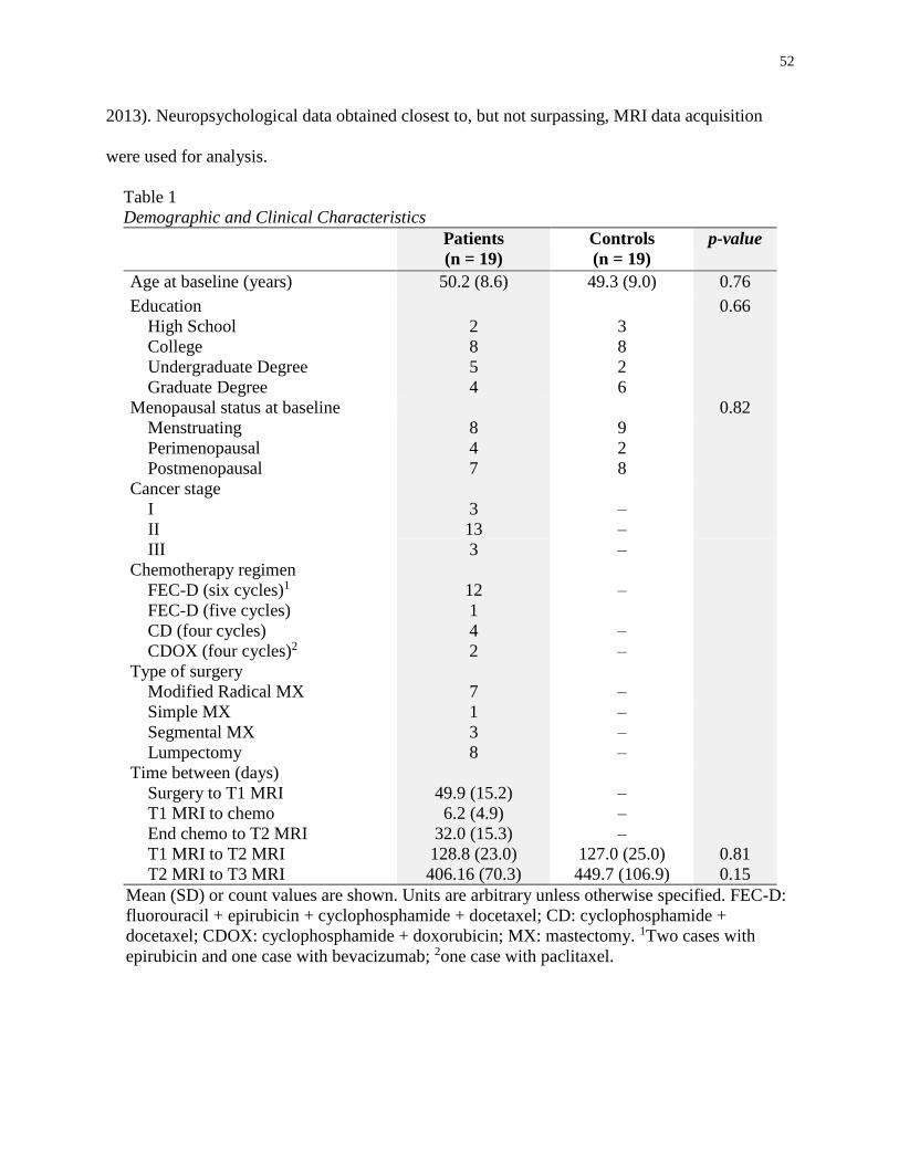
52
2013). Neuropsychological data obtained closest to, but not surpassing, MRI data acquisition
were used for analysis.
Table 1
Demographic and Clinical Characteristics
Patients
(n = 19)
Controls
(n = 19)
p-value
Age at baseline (years) 50.2 (8.6) 49.3 (9.0) 0.76
Education 0.66
High School 2 3
College 8 8
Undergraduate Degree 5 2
Graduate Degree 4 6
Menopausal status at baseline 0.82
Menstruating 8 9
Perimenopausal 4 2
Postmenopausal 7 8
Cancer stage
I 3 –
II 13 –
III 3 –
Chemotherapy regimen
FEC-D (six cycles)1 12 –
FEC-D (five cycles) 1
CD (four cycles) 4 –
CDOX (four cycles)2 2 –
Type of surgery
Modified Radical MX 7 –
Simple MX 1 –
Segmental MX 3 –
Lumpectomy 8 –
Time between (days)
Surgery to T1 MRI 49.9 (15.2) –
T1 MRI to chemo 6.2 (4.9) –
End chemo to T2 MRI 32.0 (15.3) –
T1 MRI to T2 MRI 128.8 (23.0) 127.0 (25.0) 0.81
T2 MRI to T3 MRI 406.16 (70.3) 449.7 (106.9) 0.15
Mean (SD) or count values are shown. Units are arbitrary unless otherwise specified. FEC-D:
fluorouracil + epirubicin + cyclophosphamide + docetaxel; CD: cyclophosphamide +
docetaxel; CDOX: cyclophosphamide + doxorubicin; MX: mastectomy. 1Two cases with
epirubicin and one case with bevacizumab; 2one case with paclitaxel.
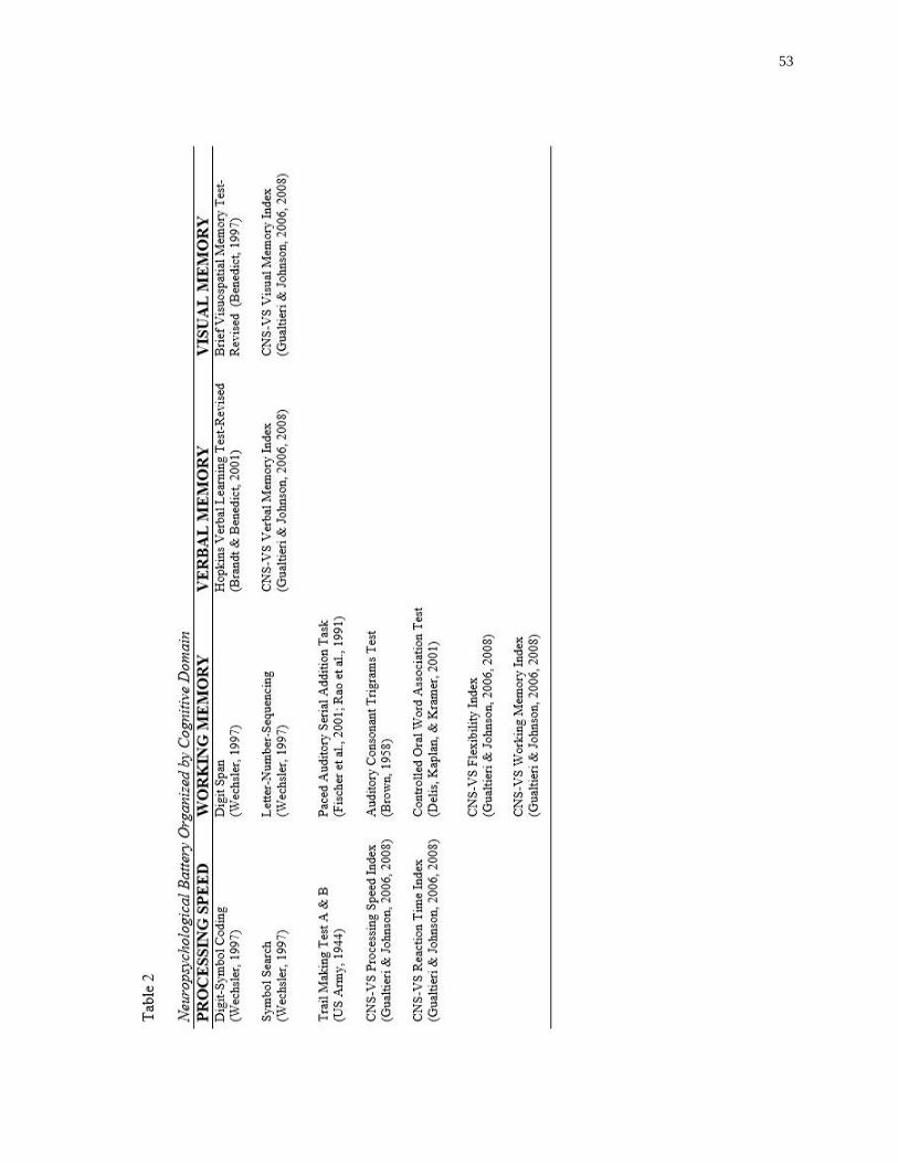
53
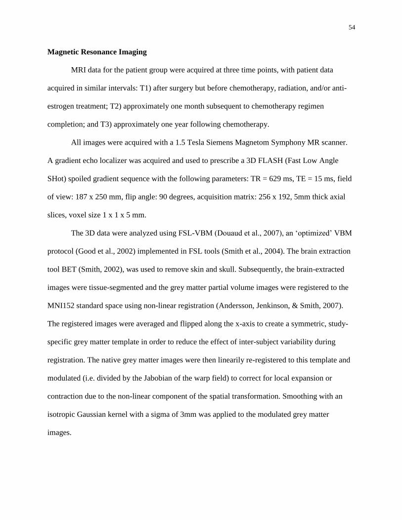
54
Magnetic Resonance Imaging
MRI data for the patient group were acquired at three time points, with patient data
acquired in similar intervals: T1) after surgery but before chemotherapy, radiation, and/or anti-
estrogen treatment; T2) approximately one month subsequent to chemotherapy regimen
completion; and T3) approximately one year following chemotherapy.
All images were acquired with a 1.5 Tesla Siemens Magnetom Symphony MR scanner.
A gradient echo localizer was acquired and used to prescribe a 3D FLASH (Fast Low Angle
SHot) spoiled gradient sequence with the following parameters: TR = 629 ms, TE = 15 ms, field
of view: 187 x 250 mm, flip angle: 90 degrees, acquisition matrix: 256 x 192, 5mm thick axial
slices, voxel size 1 x 1 x 5 mm.
The 3D data were analyzed using FSL-VBM (Douaud et al., 2007), an ‘optimized’ VBM
protocol (Good et al., 2002) implemented in FSL tools (Smith et al., 2004). The brain extraction
tool BET (Smith, 2002), was used to remove skin and skull. Subsequently, the brain-extracted
images were tissue-segmented and the grey matter partial volume images were registered to the
MNI152 standard space using non-linear registration (Andersson, Jenkinson, & Smith, 2007).
The registered images were averaged and flipped along the x-axis to create a symmetric, study-
specific grey matter template in order to reduce the effect of inter-subject variability during
registration. The native grey matter images were then linearily re-registered to this template and
modulated (i.e. divided by the Jabobian of the warp field) to correct for local expansion or
contraction due to the non-linear component of the spatial transformation. Smoothing with an
isotropic Gaussian kernel with a sigma of 3mm was applied to the modulated grey matter
images.

55
Next, within-group voxel-wise threshold-free cluster enhancement-based (Smith &
Nichols, 2008) GLM analyses were conducted using permutation-based non-parametric testing
with 5,000 permutations on whole-brain grey matter volumes. Statistical maps of within-group
comparisons thresholded at p < 0.01 uncorrected for multiple comparisons were used to generate
region of interest (ROI) masks. Uncorrected values were used as a means of selecting ROIs for
further analysis. A composite whole-brain mask covering all regions of significant differences
and 14 masks covering the intersection of these regions and anatomical ROIs were defined with
the AAL atlas tool (Tzourio-Mazoyer et al., 2002) using WFU PickAtlas (Maldjian, Laurienti, &
Burdette, 2004; Maldjian, Laurienti, Kraft, & Burdette, 2003) were created.
Region of Interest Analysis
Using R (R Development Core Team, 2014), ROI data were compared across time points
with Welch’s t-tests and p values were adjusted for multiple comparisons using the Benjamini-
Hochberg procedure (Benjamini & Hochberg, 1995).
Neuropsychological and Demographic Data
Differences across neuropsychological scores were investigated with repeated measures
ANOVA and Tukey pairwise comparisons. The relationship between grey matter volumes and
neuropsychological performance within the patient group was examined with HLM7
(Raudenbush, Bryk, Cheong, & Condon, 2011) using a two-level hierarchical linear model
(HLM; Raudenbush & Bryk, 2002) with time points nested within patients. Distinct HLM
analyses were conducted in order to assess the correlation between each ROI and each cognitive
domain. Welch’s two sample t-tests were used to compare all demographic data, except in the
case of nominal data where Fisher’s exact tests were used.
Results
56
Sample Characteristics
Demographic characteristics are listed in Table 1. The patient group ranged in age from
35 to 64 years and the controls ranged in age from 31 to 61 years. The interval between scanning
sessions did not differ between groups (p > 0.05, Table 1). For patients, T1 neuropsychological
assessments were conducted on average 10.20 days (SD = 8.12) before chemotherapy, T2
neuropsychological assessments were conducted on average 17.75 days (SD = 7.17) after final
chemotherapy exposure, and T3 neuropsychological assessments were completed on average
392.5 days (SD = 46.77) following T2 assessments. After T2 and before T3, 10 patients
commenced hormonal therapy. Similarly, during this interval 13 patients underwent
radiotherapy. Between T2 and T3, all patients that were either menstruating or perimenopausal at
T1 became menopausal; however, this status did not change for controls.
Within-group grey matter changes
Table 3 shows the grey matter volume differences between scans for patients in the
composite whole-brain mask and ROIs. At T2 relative to T1, patients showed a reduction of grey
matter volume in frontal, temporal, parietal, and occipital regions (Figure 1). There were no areas
of increased volume at T2 relative to T1. At one year after chemotherapy relative to T1,
significant grey matter reductions were observed in bilateral frontal and temporal regions and all
other reductions observed from T1 to T2 were no longer significant. Controls did not show any
decrease from T1 to T2; however, they did display an unexpected increase in grey matter volume
in the right amygdala from T1 to T2. At T3 relative to T1, this increase was no longer
significant; yet, there was a significant increase in grey matter volume in the left lingual gyrus.

57

58
Figure 1. Images are presented in radiological view where right is the patient’s left side and left is the
patient’s right side. Coordinates presented in MNI space. a) middle frontal gyrus (36 28 22); b) precuneus
(20 -58 25); c) gyrus rectus (6 48 -20); d) hippocampus (34 -14 -20) e) middle temporal gyrus (-58 -4 -20);
f) anterior cingulate (4 38 5); g) paracentral lobule (10 -36 75); h) medial orbitofrontal gyrus (-32 44 -5); i)
inferior orbitofrontal gyrus (-32 28 -5); j) superior temporal gyrus (-65 2 -5); k) insular cortex (-36 6 0); l)
inferior frontal operculum (-54 10 0); m) anterior cingulate (-10 38 10); n) calcarine cortex (-12 -86 5).

59
Cognitive Domain Scores
Patient mean scores are presented in Table 4. An analysis of variance for processing
speed revealed a significant difference among the time points (F(2, 35)= 14.59, p < 0.001). A
post hoc Tukey test showed that patients scored significantly better at baseline relative to T2 (p <
0.001) and T3 (p = 0.004). Although processing speed scores improved from T2 to T3, this
difference was only marginally significant (p = 0.094). Scores on the three other cognitive
domains degraded over time; however, the observed differences were not significant.
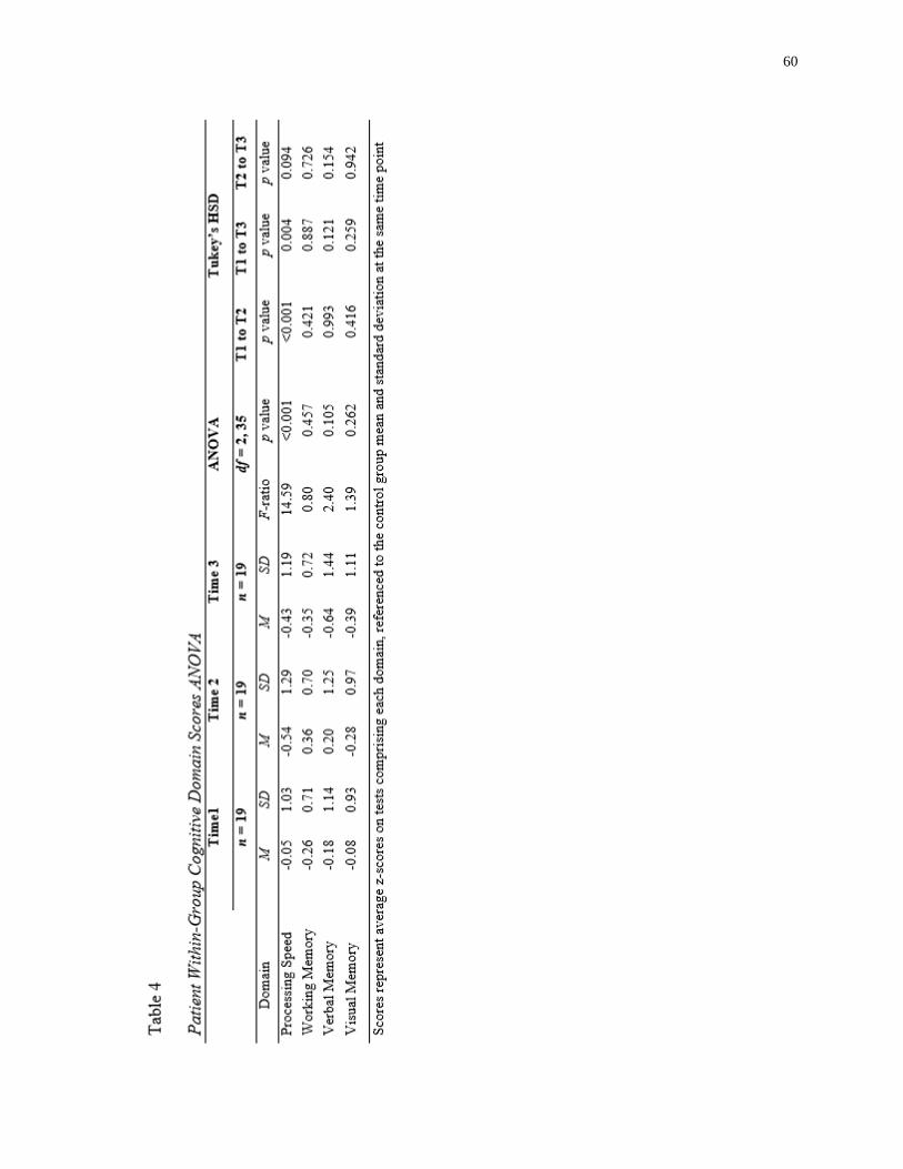
60
61
Cognitive functioning and relationship to grey matter volumes
Listed in Table 5 are the correlations between ROI grey matter volumes and the four
cognitive domains. Processing speed displayed a positive relationship with the whole-brain
composite (r = 0.61, p <0.001) and frontal, temporal, and occipital areas. Working memory
showed a positive relationship with the left medial orbitofrontal gyrus (r = 0.51, p = 0.007) and
the right middle frontal gyrus (r = 0.42, p < 0.05). Visual memory was positively related to grey
matter volume in the left inferior frontal operculum (r = 0.71, p = 0.009) and the right middle
frontal gyrus (r = 0.71, p < 0.009). There was no relationship between grey matter in the ROIs
and composite whole-brain with verbal memory.

62

63
Discussion
VBM analyses showed diffuse reductions in brain regions of breast cancer patients one
month after chemotherapy. This attenuation recovered in nearly half of the regions one year post-
chemotherapy. These results provide both evidence of a neural basis for CRCI and optimism for
the recovery from the injurious effects that chemotherapy appears to have on the brain.
Our primary hypothesis that grey matter alterations would be more pronounced and
distributed shortly after chemotherapy, and then partially resolve one year post-chemotherapy,
was supported. Diffuse grey matter alterations in the patient group approximately one month
after chemotherapy exposure are congruent with existing VBM studies that have shown
distributed grey matter disruption in the breast cancer population shortly after chemotherapy
treatment (Inagaki et al., 2007; McDonald et al., 2010; McDonald, Conroy, Smith, et al., 2012).
Additionally, our results support the converging evidence from both structural (Inagaki et al.,
2007; McDonald et al., 2010; McDonald, Conroy, Smith, et al., 2012) and functional studies (de
Ruiter et al., 2011; Kesler et al., 2009, 2011; Lopez Zunini et al., 2013; McDonald, Conroy,
Ahles, et al., 2012; Silverman et al., 2007) that the frontal lobes appear particularly sensitive to
chemotherapy. These findings are important in light of common reports of acute executive
function and working memory difficulties in patients subsequent to chemotherapy (Wefel &
Schagen, 2012) because these cognitive functions are subserved by the frontal lobes (Fletcher &
Henson, 2001). Our findings of both attenuated grey matter volume in the frontal lobes and the
positive relationship between GM reduction in these regions with poorer performance on
executive function, working memory, and visual memory strengthen the neuroanatomical
evidence of CRCI.
64
In contrast to one month post-chemotherapy, fewer regions displayed reduced GM at one
year post-treatment relative to baseline. Regions displaying persistent grey matter loss remained
bilaterally distributed in frontotemporal regions. Enduring frontal grey matter suppression was
found in the left anterior cingulate gyrus, left inferior frontal operculum, and right middle frontal
gyrus, congruent with a previous report of chronic frontal insult in chemotherapy-treated breast
cancer patients (McDonald et al., 2010). The region of the left superior temporal gyrus and left
insula that had pronounced reduction at T2 relative to T1 (see Figure 1) did not fully resolve. The
superior temporal gyrus and insula share efferent and afferent connections (Flynn, 1999) and this
may help explain the concomitant insults to these regions observed in the present study.
Interestingly, the right hippocampus displayed reduced grey matter, concordant with previous
studies that have demonstrated prolonged hippocampal compromise (Bergouignan et al., 2011;
Kesler et al., 2013; McDonald et al., 2010), but in contrast to a study by Yoshikawa et al. (2005)
that failed to find hippocampal insult.
The diffuse nature of grey matter modulation observed in this and similar studies, along
with some of the regional volume loss discordance across extant grey matter studies of
chemotherapy in breast cancer, may be attributable to the inclusion of heterogeneous
chemotherapy regimens. Although nearly all cytostatic agents have been associated with
neurobiological effects, the mechanisms and outcomes vary across treatments (for a review, see
(Seigers, Schagen, Van Tellingen, & Dietrich, 2013)). Some chemotherapeutic agents, such as
methotrexate, 5-fluorouracil, and cyclophosphamide, appear to have direct cytotoxic effects via
their ability to penetrate the blood-brain-barrier (BBB; Dietrich, 2010); however, other agents
appear to have indirect effects due to their inability to cross the BBB. A recent study by Kesler et
al. (2013) suggests that elevated pro-inflammatory cytokine expression, as seen in BBB
65
impermeable agents like doxorubicin, may have direct and indirect injurious effects on brain
structures. Our present study included both BBB permeable and impermeable chemotherapeutic
agents (see Table 1). It will be important for future studies to tease apart the differential effects
of various chemotherapy treatments on the brain.
Our secondary hypothesis that grey matter attenuation would be related to cognitive
functioning was supported. Although processing speed was positively related to distributed grey
matter volumes, the association was observed predominantly in frontotemporal regions. This
included the left insula and a portion of prefrontal areas with which the insula shares
bidirectional connections (Flynn, 1999), specifically, the left medial and inferior regions of the
orbitofrontal cortex (OFC) and the left inferior frontal operculum. The insula is critical for neural
communication between the prefrontal cortex and more posterior regions (Augustine, 1996), and
disruption to the insula and associated regions may underlie some of the cognitive difficulties
expressed by chemotherapy-exposed breast cancer patients.
Interestingly, processing speed and working memory were positively correlated with grey
matter volume in the medial orbitofrontal gyrus. This is a notable finding given the clinical
implications. Previous studies have associated the OFC with a range of cognitive processes
including decision-making (Plassmann, O’Doherty, & Rangel, 2010), emotion (Rolls &
Grabenhorst, 2008), and response inhibition (Horn, Dolan, Elliott, Deakin, & Woodruff, 2003).
Grey matter loss in this region of the OFC was significant for patients between pre-
chemotherapy exposure and one month post-treatment, consistent with the pattern of decreased
cognitive performance during the same interval. Our findings underscore the extensive impact
that exposure to chemotherapy may have on breast cancer patients. In light of our results, work
showing that the OFC is involved in processes beyond the cognitive domains selected in our
66
study points to a potential for chemotherapy exposure to adversely impact the lives of breast
cancer patients more widely than suggested by our results alone.
The strengths of this study include its longitudinal design and the administration of a
comprehensive battery of objective neuropsychological measures that covered a broad range of
cognitive processes. We acknowledge that there are limitations to our study that necessitate a
degree of caution when interpreting our findings. Primarily, we did not include a chemotherapy-
naïve comparison group and, consequently, we could not control for the potential influence of
cancer-related factors. Existing VBM studies that have included both a healthy control group and
a chemotherapy-naïve breast cancer control group have found no within-group grey matter
differences in these control groups in contrast to the decline observed in chemotherapy-exposed
breast cancer patients (McDonald et al., 2010; McDonald, Conroy, Smith, et al., 2012),
suggesting that grey matter alterations may stem from chemotherapy exposure.
The number of treatment cycles and types of surgery varied across breast cancer patients
(see Table 1). Given our limited sample size, we could not control for the influence of these
potentially confounding factors. Currently, one cross-sectional study has examined the effects of
a heterogeneous chemotherapy regimen on grey matter volumes in a large sample (Koppelmans
et al., 2012); it will be important for future longitudinal investigations to employ a similar
approach. Treatment-induced menopausal symptoms co-occur with cognitive impairment
following chemotherapy in breast cancer patients (Fan et al., 2005). Although our patients and
controls were closely matched at baseline, at T3 relative to T2 all patients were menopausal
while controls remained unchanged. As a result, we were unable to control for the effects of
menopausal status. From time T2 to T3, some patients received radiotherapy or commenced
hormonal therapy. Reports in the literature suggest that these treatments may perturb cognition
67
(Bender, Paraska, Sereika, Ryan, & Berga, 2001; Quesnel, Savard, & Ivers, 2009). However,
despite their administration, an overall improvement in GM volumes was observed from T2 to
T3, suggesting that these therapies may have had a negligible effect.
In summary, the present study demonstrated grey matter volume loss in diffuse brain
regions in breast cancer patients one month following chemotherapy treatment. One year
following treatment, grey matter was partially recovered. Grey matter volumes were related to
cognitive performance in the domains of processing speed, working memory, and visual
memory. Cognitive dysfunction was found to follow a similar course to grey matter changes,
particularly in the domain of processing speed. This study strengthens the evidence for the
relationship between brain alterations and objectively measured cognitive difficulties in breast
cancer patients exposed to chemotherapy. In addition to the burden of being diagnosed with a
life-threatening disease, breast cancer patients must contend with potential adverse side effects of
treatment. Cognitive and neurophysiological alterations touch many areas of survivors’ lives,
warranting future research to further elucidate the mechanisms of CRCI and to improve breast
cancer patients’ quality of life.
68
References
Ahles, T. A., Saykin, A. J., Furstenberg, C. T., Cole, B., Mott, L. A., Skalla, K., … Silberfarb, P.
M. (2002). Neuropsychologic impact of standard-dose systemic chemotherapy in long-term
survivors of breast cancer and lymphoma. Journal of Clinical Oncology, 20(2), 485–493.
Ahles, T. A., Saykin, A. J., McDonald, B. C., Furstenberg, C. T., Cole, B. F., Hanscom, B. S., …
Kaufman, P. A. (2007). Cognitive function in breast cancer patients prior to adjuvant
treatment. Breast Cancer Research and Treatment, 110(1), 143–152.
Andersson, J. L. R., Jenkinson, M., & Smith, S. (2007). Non-linear registration aka Spatial
normalisation FMRIB Technial Report TR07JA2. Retrieved from
http://fmrib.medsci.ox.ac.uk/analysis/techrep/tr07ja2/tr07ja2.pdf
Ashburner, J., & Friston, K. J. (2000). Voxel-based morphometry--the methods. NeuroImage,
11(6), 805–821.
Augustine, J. R. (1996). Circuitry and functional aspects of the insular lobe in primates including
humans. Brain Research Reviews, 22(3), 229–244.
Bender, C. M., Paraska, K. K., Sereika, S. M., Ryan, C. M., & Berga, S. L. (2001). Cognitive
function and reproductive hormones in adjuvant therapy for breast cancer: a critical review.
Journal of Pain and Symptom Management, 21(5), 407–424.
Benedict, R. (1997). Brief Visuospatial Memory Test—Revised Professional Manual. Lutz, FL:
Psychological Assessment Resources.
Benjamini, Y., & Hochberg, Y. (1995). Controlling the False Discovery Rate: A Practical and
Powerful Approach to Multiple Testing. Journal of the Royal Statistical Society. Series B,
Statistical Methodology, 57(1), 289–300.
69
Bergouignan, L., Lefranc, J. P., Chupin, M., Morel, N., Spano, J. P., & Fossati, P. (2011). Breast
cancer affects both the hippocampus volume and the episodic autobiographical memory
retrieval. PloS One, 6(10), e25349.
Boykoff, N., Moieni, M., & Subramanian, S. K. (2009). Confronting chemobrain: an in-depth
look at survivors’ reports of impact on work, social networks, and health care response.
Journal of Cancer Survivorship, 3(4), 223–232.
Brandt, J., & Benedict, R. (2001). Hopkins Verbal Learning Test—Revised Professional Manual.
Lutz, FL: Psychological Assessment Resources.
Brown, J. (1958). Some tests of the decay theory of immediate memory. The Quarterly Journal
of Experimental Psychology, 10(1), 12–21.
Collins, B., MacKenzie, J., Tasca, G. A., Scherling, C., & Smith, A. (2013). Cognitive effects of
chemotherapy in breast cancer patients: a dose–response study. Psycho-Oncology, 22(7),
1517–1527.
Conroy, S. K., McDonald, B. C., Smith, D. J., Moser, L. R., West, J. D., Kamendulis, L. M., …
Saykin, A. J. (2012). Alterations in brain structure and function in breast cancer survivors:
effect of post-chemotherapy interval and relation to oxidative DNA damage. Breast Cancer
Research and Treatment, 137(2), 493–502.
De Ruiter, M. B., Reneman, L., Boogerd, W., Veltman, D. J., Caan, M., Douaud, G., …
Schagen, S. B. (2012). Late effects of high-dose adjuvant chemotherapy on white and gray
matter in breast cancer survivors: converging results from multimodal magnetic resonance
imaging. Human Brain Mapping, 33(12), 2971–2983.
70
De Ruiter, M. B., Reneman, L., Boogerd, W., Veltman, D. J., van Dam, F. S. A. M., Nederveen,
A. J., … Schagen, S. B. (2011). Cerebral hyporesponsiveness and cognitive impairment 10
years after chemotherapy for breast cancer. Human Brain Mapping, 32(8), 1206–1219.
Delis, D., Kaplan, E., & Kramer, J. (2001). Delis Kaplan Executive Function System Test
Manual. San Antonio, TX: Psychological Corporation.
Dietrich, J. (2010). Chemotherapy Associated Central Nervous System Damage. In Chemo Fog
(pp. 77–85). Springer New York.
Douaud, G., Smith, S., Jenkinson, M., Behrens, T., Johansen-Berg, H., Vickers, J., … James, A.
(2007). Anatomically related grey and white matter abnormalities in adolescent-onset
schizophrenia. Brain: A Journal of Neurology, 130(9), 2375–2386.
Falleti, M. G., Sanfilippo, A., Maruff, P., Weih, L., & Phillips, K.-A. (2005). The nature and
severity of cognitive impairment associated with adjuvant chemotherapy in women with
breast cancer: a meta-analysis of the current literature. Brain and Cognition, 59(1), 60–70.
Fan, H. G. M., Houédé-Tchen, N., Yi, Q.-L., Chemerynsky, I., Downie, F. P., Sabate, K., &
Tannock, I. F. (2005). Fatigue, menopausal symptoms, and cognitive function in women
after adjuvant chemotherapy for breast cancer: 1- and 2-year follow-up of a prospective
controlled study. Journal of Clinical Oncology, 23(31), 8025–8032.
Ferguson, R. J., McDonald, B. C., Saykin, A. J., & Ahles, T. A. (2007). Brain structure and
function differences in monozygotic twins: possible effects of breast cancer chemotherapy.
Journal of Clinical, 25(25), 3866–3870.
Fischer, J., Jak, A., Kniker, J., Rudick, R., & Cutter, G. (2001). Administration and Scoring
Manual for the Multiple Sclerosis Functional Composite Measure (MSFC). National
Multiple Sclerosis. New York: National Multiple Sclerosis Society.
71
Fletcher, P. C., & Henson, R. N. A. (2001). Frontal lobes and human memory insights from
functional neuroimaging. Brain: A Journal of Neurology, 124, 849–881.
Flynn, F. G. (1999). Anatomy of the insula functional and clinical correlates. Aphasiology, 13(1),
55–78.
Good, C., Johnsrude, I., Ashburner, J., Henson, R., Fristen, K., & Frackowiak, R. S. J. (2002). A
voxel-based morphometric study of ageing in 465 normal adult human brains. In Biomedical
Imaging, 2002. 5th IEEE EMBS International Summer School on (p. 16 pp.–).
Gualtieri, C. T., & Johnson, L. G. (2006). Reliability and validity of a computerized
neurocognitive test battery, CNS Vital Signs. Archives of Clinical Neuropsychology, 21(7),
623–643.
Gualtieri, C. T., & Johnson, L. G. (2008). A computerized test battery sensitive to mild and
severe brain injury. Medscape Journal of Medicine, 10(4), 90.
Hakamata, Y., Matsuoka, Y., Inagaki, M., Nagamine, M., Hara, E., Imoto, S., … Uchitomi, Y.
(2007). Structure of orbitofrontal cortex and its longitudinal course in cancer-related post-
traumatic stress disorder. Neuroscience Research, 59(4), 383–389.
Horn, N. R., Dolan, M., Elliott, R., Deakin, J. F. W., & Woodruff, P. W. R. (2003). Response
inhibition and impulsivity: an fMRI study. Neuropsychologia, 41(14), 1959–1966.
Hosseini, S. M. H., Koovakkattu, D., & Kesler, S. R. (2012). Altered small-world properties of
gray matter networks in breast cancer. BMC Neurology, 12, 28.
Inagaki, M., Yoshikawa, E., Matsuoka, Y., Sugawara, Y., Nakano, T., Akechi, T., … Uchitomi,
Y. (2007). Smaller regional volumes of brain gray and white matter demonstrated in breast
cancer survivors exposed to adjuvant chemotherapy. Cancer, 109(1), 146–156.

72
Jim, H. S. L., Phillips, K. M., Chait, S., Faul, L. A., Popa, M. A., Lee, Y. H., … Small, B. J.
(2012). Meta-analysis of cognitive functioning in breast cancer survivors previously treated
with standard-dose chemotherapy. Journal of Clinical Oncology, 30(29), 3578–3587.
Kesler, S., Janelsins, M., Koovakkattu, D., Palesh, O., Mustian, K., Morrow, G., & Dhabhar, F.
S. (2013). Reduced hippocampal volume and verbal memory performance associated with
interleukin-6 and tumor necrosis factor-alpha levels in chemotherapy-treated breast cancer
survivors. Brain, Behavior, and Immunity, 30 Suppl, S109–16.
Kesler, S. R., Bennett, F. C., Mahaffey, M. L., & Spiegel, D. (2009). Regional brain activation
during verbal declarative memory in metastatic breast cancer. Clinical Cancer Research,
15(21), 6665–6673.
Kesler, S. R., Kent, J. S., & O’Hara, R. (2011). Prefrontal cortex and executive function
impairments in primary breast cancer. Archives of Neurology, 68(11), 1447–1453.
Koppelmans, V., Breteler, M. M. B., Boogerd, W., Seynaeve, C., Gundy, C., & Schagen, S. B.
(2012). Neuropsychological performance in survivors of breast cancer more than 20 years
after adjuvant chemotherapy. Journal of Clinical Oncology, 30(10), 1080–1086.
Koppelmans, V., de Ruiter, M. B., van der Lijn, F., Boogerd, W., Seynaeve, C., van der Lugt, A.,
… Schagen, S. B. (2012). Global and focal brain volume in long-term breast cancer
survivors exposed to adjuvant chemotherapy. Breast Cancer Research and Treatment,
132(3), 1099–1106.
Lezak, M. D., Howieson, D. B., Loring, D. W., Hannay, H. J., & Fischer, J. S. (2004).
Neuropsychological Assessment (4th ed.). New York: Oxford University Press.
López Zunini, R. A., Scherling, C., Wallis, N., Collins, B., MacKenzie, J., Bielajew, C., &
Smith, A. M. (2013). Differences in verbal memory retrieval in breast cancer chemotherapy
73
patients compared to healthy controls: a prospective fMRI study. Brain Imaging and
Behavior, 7(4), 460–477.
Maldjian, J. A., Laurienti, P. J., & Burdette, J. H. (2004). Precentral gyrus discrepancy in
electronic versions of the Talairach atlas. NeuroImage, 21(1), 450–455.
Maldjian, J. A., Laurienti, P. J., Kraft, R. A., & Burdette, J. H. (2003). An automated method for
neuroanatomic and cytoarchitectonic atlas-based interrogation of fMRI data sets.
NeuroImage, 19(3), 1233–1239.
McDonald, B. C., Conroy, S. K., Ahles, T. A., West, J. D., & Saykin, A. J. (2010). Gray matter
reduction associated with systemic chemotherapy for breast cancer: a prospective MRI
study. Breast Cancer Research and Treatment, 123(3), 819–828.
McDonald, B. C., Conroy, S. K., Ahles, T. A., West, J. D., & Saykin, A. J. (2012). Alterations in
brain activation during working memory processing associated with breast cancer and
treatment: a prospective functional magnetic resonance imaging study. Journal of Clinical
Oncology, 30(20), 2500–2508.
McDonald, B. C., Conroy, S. K., Smith, D. J., West, J. D., & Saykin, A. J. (2012). Frontal gray
matter reduction after breast cancer chemotherapy and association with executive symptoms:
a replication and extension study. Brain, Behavior, and Immunity, 30 Suppl, S117–25.
McDonald, B. C., & Saykin, A. J. (2013). Alterations in brain structure related to breast cancer
and its treatment: chemotherapy and other considerations. Brain Imaging and Behavior, 7(4),
374–387.
O’Farrell, E., MacKenzie, J., & Collins, B. (2013). Clearing the air: a review of our current
understanding of “chemo fog.” Current Oncology Reports, 15(3), 260–269.
74
Plassmann, H., O’Doherty, J. P., & Rangel, A. (2010). Appetitive and aversive goal values are
encoded in the medial orbitofrontal cortex at the time of decision making. The Journal of
Neuroscience, 30(32), 10799–10808.
Quesnel, C., Savard, J., & Ivers, H. (2009). Cognitive impairments associated with breast cancer
treatments: results from a longitudinal study. Breast Cancer Research and Treatment,
116(1), 113–123.
R Development Core Team. (2014). R: the r project for statistical computing (Version 3.0.3).
Rao, S. M., Leo, G. J., Bernardin, L., & Unverzagt, F. (1991). Cognitive dysfunction in multiple
sclerosis. I. Frequency, patterns, and prediction. Neurology, 41(5), 685–691.
Raudenbush, S., & Bryk, A. (2002). Hierarchical Linear Models: Applications and Data
Analysis Methods. SAGE Publications.
Raudenbush, S., Bryk, A., Cheong, Y., & Condon, R. (2011). HLM 7: Linear and Nonlinear
Modeling. Lincolnwood, IL: Scientific Software International.
Rolls, E. T., & Grabenhorst, F. (2008). The orbitofrontal cortex and beyond: from affect to
decision-making. Progress in Neurobiology, 86(3), 216–244.
Scherling, C., Collins, B., Mackenzie, J., Bielajew, C., & Smith, A. (2011). Pre-chemotherapy
differences in visuospatial working memory in breast cancer patients compared to controls:
an FMRI study. Frontiers in Human Neuroscience, 5, 122.
Scherling, C., Collins, B., Mackenzie, J., Bielajew, C., & Smith, A. (2012). Prechemotherapy
differences in response inhibition in breast cancer patients compared to controls: a functional
magnetic resonance imaging study. Journal of Clinical and Experimental Neuropsychology,
34(5), 543–560.
75
Scherling, C., Collins, B., MacKenzie, J., Lepage, C., Bielajew, C., & Smith, A. (2012).
Structural Brain Differences in Breast Cancer Patients Compared to Matched Controls Prior
to Chemotherapy. International Journal of Biology, 4(2), 3.
Scherling, C., & Smith, A. (2013). Opening up the Window into “Chemobrain”: A
Neuroimaging Review. Sensors , 13(3), 3169–3203.
Seigers, R., Schagen, S. B., Van Tellingen, O., & Dietrich, J. (2013). Chemotherapy-related
cognitive dysfunction: current animal studies and future directions. Brain Imaging and
Behavior, 7(4), 453–459.
Silverman, D. H. S., Dy, C. J., Castellon, S. A., Lai, J., Pio, B. S., Abraham, L., … Ganz, P. A.
(2007). Altered frontocortical, cerebellar, and basal ganglia activity in adjuvant-treated
breast cancer survivors 5-10 years after chemotherapy. Breast Cancer Research and
Treatment, 103(3), 303–311.
Smith, S. M. (2002). Fast robust automated brain extraction. Human Brain Mapping, 17(3), 143–
155.
Smith, S. M., Jenkinson, M., Woolrich, M. W., Beckmann, C. F., Behrens, T. E. J., Johansen-
Berg, H., … Matthews, P. M. (2004). Advances in functional and structural MR image
analysis and implementation as FSL. NeuroImage, 23 Suppl 1, S208–19.
Smith, S. M., & Nichols, T. E. (2008). Threshold-free cluster enhancement: addressing problems
of smoothing, threshold dependence and localisation in cluster inference. NeuroImage,
44(1), 83–98.
Stewart, A., Bielajew, C., Collins, B., Parkinson, M., & Tomiak, E. (2006). A meta-analysis of
the neuropsychological effects of adjuvant chemotherapy treatment in women treated for
breast cancer. The Clinical Neuropsychologist, 20(1), 76–89.

76
Stewart, A., Collins, B., Mackenzie, J., Tomiak, E., Verma, S., & Bielajew, C. (2008). The
cognitive effects of adjuvant chemotherapy in early stage breast cancer: a prospective study.
Psycho-Oncology, 17(2), 122–130.
Strauss, E., Sherman, E. M. S., & Spreen, O. (2006). A Compendium of Neuropsychological
Tests (3rd ed.). New York: Oxford University Press.
Tzourio-Mazoyer, N., Landeau, B., Papathanassiou, D., Crivello, F., Etard, O., Delcroix, N., …
Joliot, M. (2002). Automated anatomical labeling of activations in SPM using a macroscopic
anatomical parcellation of the MNI MRI single-subject brain. NeuroImage, 15(1), 273–289.
US Army. (1944). Army Individual Test Battery: Manual for Directions and Scoring. Manual of
Directions and Scoring. Washington, DC: War Department, Adjutant General’s Office.
Wechsler, D. (1997). Wechsler Adult Intelligence Scale (3rd edn). San Antonio, TX: Harcourt,
Brace, & Co.
Wefel, J. S., Saleeba, A. K., Buzdar, A. U., & Meyers, C. A. (2010). Acute and late onset
cognitive dysfunction associated with chemotherapy in women with breast cancer. Cancer,
116(14), 3348–3356.
Wefel, J. S., & Schagen, S. B. (2012). Chemotherapy-related cognitive dysfunction. Current
Neurology and Neuroscience Reports, 12(3), 267–275.
Wefel, J. S., Vardy, J., Ahles, T., & Schagen, S. B. (2011). International Cognition and Cancer
Task Force recommendations to harmonise studies of cognitive function in patients with
cancer. The Lancet Oncology, 12(7), 703–708.
Yoshikawa, E., Matsuoka, Y., Inagaki, M., Nakano, T., Akechi, T., Kobayakawa, M., …
Uchitomi, Y. (2005). No adverse effects of adjuvant chemotherapy on hippocampal volume
in Japanese breast cancer survivors. Breast Cancer Research and Treatment, 92(1), 81–84.

77
Yoshikawa, E., Matsuoka, Y., Yamasue, H., Inagaki, M., Nakano, T., Akechi, T., … Uchitomi,
Y. (2005). Prefrontal cortex and amygdala volume in first minor or major depressive episode
after cancer diagnosis. Biological Psychiatry, 59(8), 707–712.
78
Post-chemotherapy recovery of working memory brain activity and functional connectivity
in breast cancer: a prospective fMRI study
Chris Lepage, Carole Scherling, Nancy Wallis, Jeremy T. Moreau, Miranda Kiyomi Setoguchi,
Zahra Mawani, Joyce MacKenzie, Barbara Collins, and Andra Smith
Submitted for publication September 2015.
79
Abstract
Chemotherapy-related cognitive impairment (CRCI) has been demonstrated across
various cognitive domains subserved by frontoparietal brain regions. Working memory appears
to be particularly impacted in the short term; yet, the longitudinal course of dysfunction in this
cognitive domain remains to be fully explored. Neuroimaging investigations of the underlying
networks involved in CRCI are emerging, but much work remains to elucidate the affected
circuitry. The aim of this study was to prospectively examine working memory-related
functional connectivity in the frontoparietal network (FPN) of newly diagnosed breast cancer
patients (n = 23) and cancer-free controls (n = 23). Women were recruited to complete a
working memory task during functional magnetic resonance imaging before patient
chemotherapy, one month after therapy, and one year after treatment cessation. A seed in the
dorsolateral prefrontal cortex was used to assess functional connectivity within the FPN.
Between-group differences were present at one month after chemotherapy only, with patients
displaying broad hyperactivity in the frontal, parietal, and occipital regions while performing a
working memory task. Similarly, patients had reduced FPN connectivity between the left
dorsolateral prefrontal cortex and a left occipito-parietal region. This is the first CRCI
neuroimaging study to show a return to baseline levels of brain activity in chemotherapy-
treated breast cancer patients. This study adds to the accumulating evidence of acute post-
chemotherapy cognitive sequelae, and offers a neurofunctional account of the rebound effect of
working memory impairment reported in the neuropsychological literature of CRCI.
80
The advancement of treatment for breast cancer has resulted in an increase in
survivorship accompanied by a growing concern for the impact on cognition from adjuvant
therapies. Chemotherapy exposure is commonly associated with subjective reports of
diminished cognition in breast cancer patients (e.g., see (Pullens, De Vries, & Roukema 2010)).
Although the concordance between subjective cognitive complaints and the results of objective
testing is limited (Ganz et al. 2013; Pullens et al. 2010), an accumulating body of literature
supports the existence of objective cognitive decline over the course of treatment of breast
cancer (for a review, see (Wefel & Schagen 2012)).
Chemotherapy-related cognitive impairment (CRCI) is most pronounced during and
shortly after treatment, relative to baseline and long-term follow-up (Collins, MacKenzie, Tasca,
Scherling, & Smith 2013; Collins, Mackenzie, Tasca, Scherling, & Smith 2014; Jansen, Cooper,
Dodd, & Miaskowski 2011; Wefel, Lenzi, Theriault, Davis, & Meyers 2004), It has been
reported across a breadth of cognitive domains, including memory, attention, executive function,
and processing speed; however, the extent and frequency of the impairment across cognitive
domains is variable (Ahles et al. 2002; Hermelink et al. 2007; Hurria et al. 2006; Jansen et al.
2011; Quesnel, Savard, & Ivers 2009; Schagen, Muller, Boogerd, Mellenbergh, & van Dam
2006; Wefel, Saleeba, Buzdar, & Meyers 2010). A number of studies have found that working
memory may be particularly sensitive to the injurious effect of chemotherapy (Bender et al.
2006; Collins, Mackenzie, Stewart, Bielajew, & Verma 2009; Shilling, Jenkins, Morris, Deutsch,
& Bloomfield 2005; Stewart et al. 2008; Stewart, Bielajew, Collins, Parkinson, & Tomiak 2006)
suggesting that this cognitive domain should be a focus in studies that seek to better characterize
CRCI.
81
Neuroimaging has been used increasingly to investigate the neural mechanisms
underlying CRCI. Retrospective and prospective studies have reported brain structure (Abraham
et al., 2008; Conroy et al., 2012; de Ruiter et al., 2012; Deprez et al., 2011, 2012; Inagaki et al.
2007; Koppelmans et al., 2011; Lepage et al., 2014; McDonald, Conroy, Ahles, West, & Saykin,
2010; McDonald, Conroy, Smith, West, & Saykin, 2012; Scherling, Collins, MacKenzie, et al.,
2012) and function (Askren et al., 2014; Berman et al., 2014; Cimprich et al., 2010; de Ruiter et
al., 2011; Ferguson, McDonald, Saykin, & Ahles, 2007; Kesler, Bennett, Mahaffey, & Spiegel,
2009; Kesler, Kent, & O’Hara, 2011; López Zunini et al., 2013; McDonald, Conroy, Ahles,
West, & Saykin, 2012; Scherling, Collins, Mackenzie, Bielajew, & Smith, 2011, 2012;
Silverman et al., 2007) irregularities in breast cancer patients that generally follow the course of
impairment reported by studies of cognitive function. Although abnormal neural activation has
been reported across studies of various cognitive abilities - including executive function,
memory, and attention - working memory has been most commonly studied (for reviews, see
(McDonald & Saykin, 2013; Scherling & Smith, 2013)). Atypical activation patterns are most
typically seen in frontal and parietal regions in breast cancer patients (Conroy et al. 2012; de
Ruiter et al., 2011; Ferguson et al., 2007; Kesler et al., 2011; McDonald, Conroy, Ahles et al.,
2012). In addition to reflecting the neural demands of the tasks, these activation profiles may
reveal common brain networks that are particularly vulnerable to the effects of chemotherapy.
Studies of neural networks in breast cancer patients are few; however, emerging evidence
points to disrupted global brain network organization in chemotherapy-treated breast cancer
patients (Bruno, Hosseini, & Kesler, 2012; Hosseini, Koovakkattu, & Kesler, 2012). In a recent
longitudinal pilot study that used a working memory task, reduced functional connectivity was
found in the dorsal attention network one month after chemotherapy relative to baseline, which
82
partially recovered one year later (Dumas et al., 2013). In the same study, the authors also
reported persistent decreased functional connectivity in the default mode network that began one
month post-chemotherapy. These findings highlight the importance of combining emerging
analysis methods and common assessments to better characterize suspected cognitive deficits.
The frontoparietal network (FPN) is a cognitive and action control system that flexibly
recruits and updates neural hubs in order to guide adaptive behaviour across a range of cognitive
demands (Cole et al., 2013; Koziol, Barker, Joyce, & Hrin, 2014). Hubs within the FPN have
been uniquely associated with cognitive control and working memory (Harding, Yücel, Harrison,
Pantelis, & Breakspear, 2014). Thus, considering the irregular neural activation and network
profiles within frontoparietal regions of chemotherapy-treated breast cancer patients, the
investigation of the FPN may offer greater insight into the neural substrates of working memory
deficits in this population.
The purpose of this study was to prospectively compare functional connectivity within
the FPN during a task of working memory between breast cancer patients and healthy controls. It
was hypothesized that breast cancer patients would show reduced functional connectivity shortly
after treatment, and that there would be recovery one year later. A secondary aim of this study
was to replicate findings of abnormal neural activation during a commonly studied working
memory paradigm in the CRCI neuroimaging literature.
Methods
Participants
Twenty-three early-stage breast cancer patients and 23 healthy controls matched on age,
sex, and education were recruited via the Ottawa Hospital Regional Cancer Centre from a pool of
candidates participating in a prospective study of the effects of chemotherapy on cognition
83
(Collins et al. 2013). Patients were recruited after surgery but before commencing chemotherapy,
radiation, or hormone therapy. Each patient nominated her own control; when she was unable to
do so, a control was recruited through posters and internet advertisements. All participants were
required to be between the ages of 18 and 65 years, to be fluent in English, to possess at least a
grade-8 education, and to reside within 30-miles of Ottawa. Additional exclusion criteria were:
previous history of cancer or chemotherapy, psychiatric or neurological illness, substance abuse,
and MRI contraindications (e.g. metal implants). Specific to the breast cancer group, patients
with metastasis of disease beyond axillary lymph nodes were excluded. Various chemotherapy
regimens were accepted (Table 1). Two patients withdrew from the study after treatment onset.
At one year post-treatment, an additional patient withdrew and another was excluded from the
study due to a recurrence.
Assessment Protocol and Schedule
Neuropsychological measures were collected from the patients after surgery, but before
chemotherapy (Time 1), following each chemotherapy cycle, and one year following completion
of chemotherapy (Time 3). A detailed description of each testing session is provided elsewhere
(Collins et al. 2013). MRI measures were collected at Time 1, one month after the last
chemotherapy cycle (Time 2), and Time 3. Psychometric testing and MRI scanning sessions for
controls were yoked to a respectively matched patient.
MRI acquisition protocol
Imaging data were acquired using a 1.5 Tesla Siemens Magnetom Symphony MRI
scanner. A gradient echo localizer was acquired and used to prescribe a subsequent 3D Fast Low
Angle Shot spoiled gradient sequence, with TR/TE 22/9.2ms, flip angle 30º, field of view
256x256 mm. Whole brain echo planar fMRI based on the blood oxygen level-dependent effect
84
was performed using a gradient echo pulse sequence (TR/TE 3000/40ms, flip angle 90º, field of
view: 250x187.5,mm 64x64 matrix, slice thickness 5mm, 27 axial slices, bandwidth 2430 Hz per
pixel). As part of the imaging protocol, participants performed a total of four tasks aimed to
investigate different cognitive processes. The results of these investigations and studies of
structural data acquired during the imaging sessions have been published separately (Lepage et
al., 2014; López Zunini et al., 2013; Scherling et al., 2011; Scherling, Collins, Mackenzie, et al.,
2012; Scherling, Collins, MacKenzie, et al., 2012).
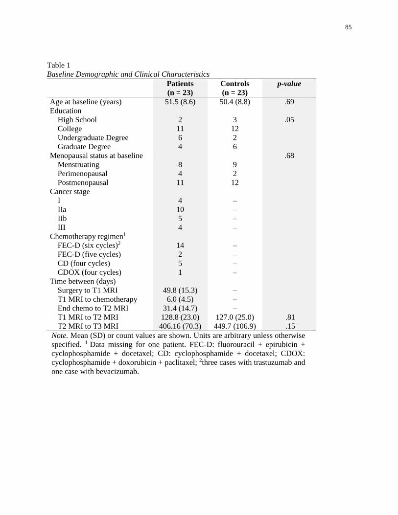
85
Table 1
Baseline Demographic and Clinical Characteristics
Patients
(n = 23)
Controls
(n = 23)
p-value
Age at baseline (years) 51.5 (8.6) 50.4 (8.8) .69
Education
High School 2 3 .05
College 11 12
Undergraduate Degree 6 2
Graduate Degree 4 6
Menopausal status at baseline .68
Menstruating 8 9
Perimenopausal 4 2
Postmenopausal 11 12
Cancer stage
I 4 –
IIa 10 –
IIb 5 –
III 4 –
Chemotherapy regimen1
FEC-D (six cycles)2 14 –
FEC-D (five cycles) 2 –
CD (four cycles) 5 –
CDOX (four cycles) 1 –
Time between (days)
Surgery to T1 MRI 49.8 (15.3) –
T1 MRI to chemotherapy 6.0 (4.5) –
End chemo to T2 MRI 31.4 (14.7) –
T1 MRI to T2 MRI 128.8 (23.0) 127.0 (25.0) .81
T2 MRI to T3 MRI 406.16 (70.3) 449.7 (106.9) .15
Note. Mean (SD) or count values are shown. Units are arbitrary unless otherwise
specified. 1 Data missing for one patient. FEC-D: fluorouracil + epirubicin +
cyclophosphamide + docetaxel; CD: cyclophosphamide + docetaxel; CDOX:
cyclophosphamide + doxorubicin + paclitaxel; 2three cases with trastuzumab and
one case with bevacizumab.
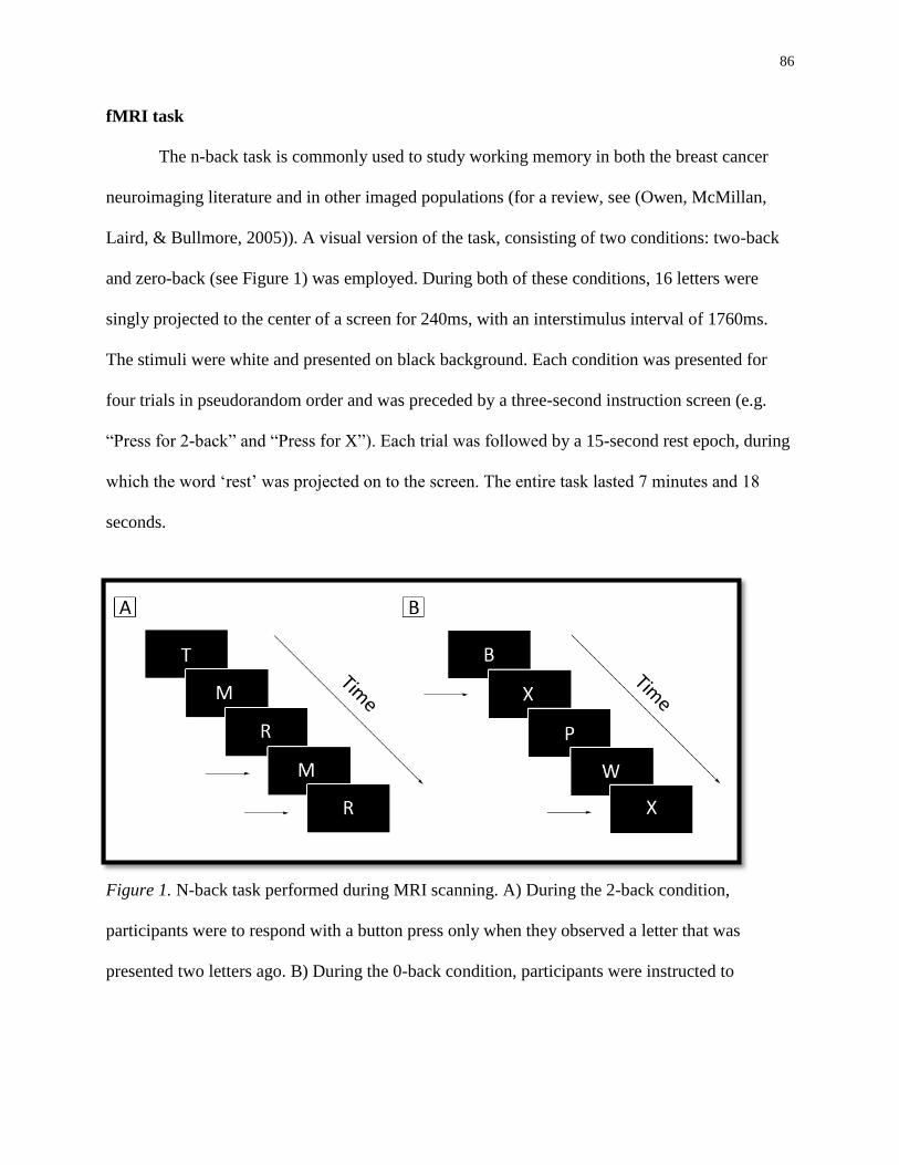
86
fMRI task
The n-back task is commonly used to study working memory in both the breast cancer
neuroimaging literature and in other imaged populations (for a review, see (Owen, McMillan,
Laird, & Bullmore, 2005)). A visual version of the task, consisting of two conditions: two-back
and zero-back (see Figure 1) was employed. During both of these conditions, 16 letters were
singly projected to the center of a screen for 240ms, with an interstimulus interval of 1760ms.
The stimuli were white and presented on black background. Each condition was presented for
four trials in pseudorandom order and was preceded by a three-second instruction screen (e.g.
“Press for 2-back” and “Press for X”). Each trial was followed by a 15-second rest epoch, during
which the word ‘rest’ was projected on to the screen. The entire task lasted 7 minutes and 18
seconds.
Figure 1. N-back task performed during MRI scanning. A) During the 2-back condition,
participants were to respond with a button press only when they observed a letter that was
presented two letters ago. B) During the 0-back condition, participants were instructed to
87
respond with a button press only when the letter ‘x’ appeared on screen. Horizontal arrows
indicate correct responses.
Neuropsychological assessment of working memory
Working memory was measured with the digit span and letter-number-sequencing
subtests of the Wechsler Adult Intelligence Scale-III (Wechsler 1997), Paced Auditory Serial
Addition Test (Fischer, Jak, Kniker, Rudick, & Cutter, 2001; Rao, Leo, Bernardin, & Unverzagt,
1991), Auditory Consonant Trigrams Test (Brown, 1958), Controlled Oral Word Association
Test (Delis, Kaplan, & Kramer, 2001), and CNS-Vital Signs Flexibility and Working Memory
indices (Gualtieri & Johnson, 2006, 2008). Raw scores from the traditional neuropsychological
tests and the index scores from the computerized cognitive tests were combined into a composite
summary score. This was done in order to limit the number of comparisons and, consequently,
reduce the risk of Type I errors. Raw test scores for each participant, on each cognitive measure,
and at each time point were standardized to the means and standard deviations of the
corresponding variables in the control group. Further elaboration on the construction of the
composite score is provided elsewhere (Collins et al., 2013).
Statistical Analyses
fMRI Task Activation Analyses. Statistical Parametric Mapping 8 (SPM8;
www.fil.ion.ucl.ac.uk/spm/software/spm8/) was used to preprocess the imaging data. The
functional images were realigned using least-squares minimization to correct for head motion,
coregistered and normalized using the segmented anatomical volume, and, finally, smoothed
using an isotropic Gaussian spatial filter (FWHM = 6 mm) to reduce noise effects. Where
needed, artifacts were detected and repaired using Art Detection Tools
(http://web.mit.edu/swg/software.htm). Statistical analyses of the fMRI data were conducted
88
using the SPM8 implementation of the general linear model (GLM). Contrast images comparing
working memory over the control condition (e.g. two-back > zero-back) were created for each
participant and used in random-effect analyses at the second-level. Between-group differences in
brain activation were calculated using an independent-samples t-test at Times 1 to 3 using a
height threshold of p < .05 and extent threshold of 50 voxels, corrected for multiple comparisons
(e.g. false discovery rate). In order to maximize our statistical power, between-group t-tests were
conducted at each time point with the maximum available number of patients, which reduced
over time due to attrition. This approach was retained for the functional connectivity analyses.
Functional Connectivity Analyses. The CONN toolbox (Whitfield-Gabrieli & Nieto-
Castanon, 2012) for SPM8 was used to conduct seed-to-voxel functional connectivity analyses.
Preprocessing of the imaging data progressed the same way as for the activation analyses;
however, additional steps were taken to reduce blood oxygen level-dependent contrast signal
noise sources that can lead to an increased risk of Type I errors in functional connectivity
analyses. Specifically, images were band-pass filtered to 0.01Hz~0.9Hz. Next, following the
implemented anatomical component-based noise correction (Behzadi, Restom, Liau, & Liu,
2007), participants’ images were regressed on six motion parameters, white matter, cerebrospinal
fluid, physiological noise sources (e.g. cardiac and respiratory effects), and task-effects and their
temporal derivatives. A 12mm spherical seed located in the dorsolateral prefrontal cortex
(dlPFC; MNI: -38, 30, 18) was used to examine FPN-related functional connectivity during
working memory processes. This region of the FPN has been previously associated with working
memory but not cognitive control (Harding et al., 2014). Within-subject voxel-wise bivariate
correlations were calculated between the time courses of the seed region and the whole-brain.
Resultant correlation maps were then subjected to Fisher Z-transformations. Between-group
89
comparisons (e.g. 2nd level GLM) were made at each time point using independent samples t-
tests, with a height threshold set at p < .01 and extent threshold of 50 voxels, corrected for
multiple comparisons.
Demographic Variables, Outcome Measures, and fMRI Task Performance.
Demographic and clinical characteristics were analyzed in SPSS 21.0 with t-tests and Chi-
square, where appropriate. Neuropsychological data and fMRI task performance data were
assessed using mixed design ANOVA.
Results
Baseline demographic and clinical characteristics are presented in Table 1. The groups
did not differ in age, education, menopausal status, or inter-scan intervals (p > .05).
fMRI Analysis
Patients and controls did not display significant working memory activation differences
at Times 1 and 3; however, patients had significantly greater activations at Time 2 in distributed
regions (see Table 2). Significant between-group differences of activation were found in the right
postcentral gyrus (Brodmann Area [BA] 43) extending to the right temporal lobe, the left
occipital pole (BA 17), the left parietal operculum (BA 40) extending to the left temporal lobe
and postcentral gyrus, and the left anterior cingulate gyrus (BA 32) extending to left prefrontal
regions (Figure 2).
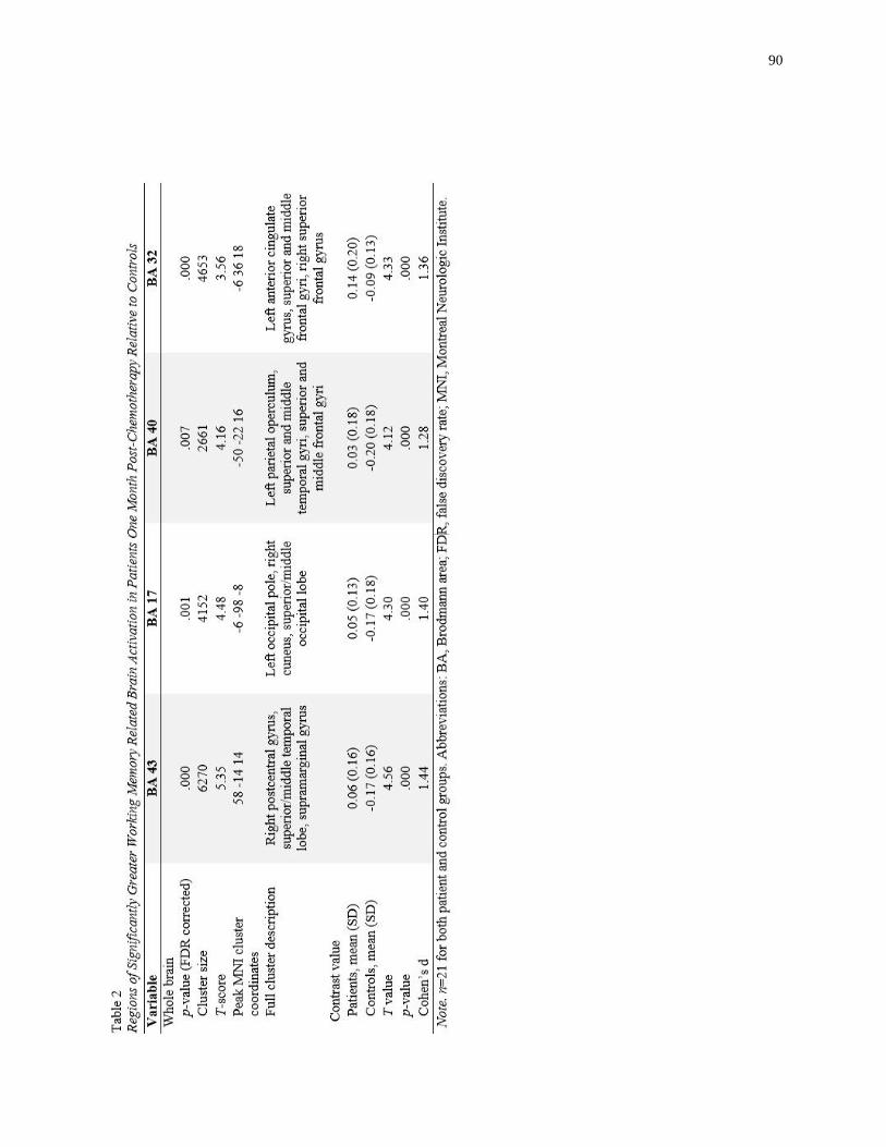
90
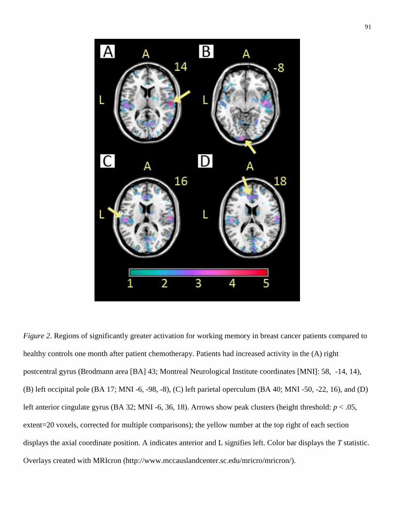
91
Figure 2. Regions of significantly greater activation for working memory in breast cancer patients compared to
healthy controls one month after patient chemotherapy. Patients had increased activity in the (A) right
postcentral gyrus (Brodmann area [BA] 43; Montreal Neurological Institute coordinates [MNI]: 58, -14, 14),
(B) left occipital pole (BA 17; MNI -6, -98, -8), (C) left parietal operculum (BA 40; MNI -50, -22, 16), and (D)
left anterior cingulate gyrus (BA 32; MNI -6, 36, 18). Arrows show peak clusters (height threshold: p < .05,
extent=20 voxels, corrected for multiple comparisons); the yellow number at the top right of each section
displays the axial coordinate position. A indicates anterior and L signifies left. Color bar displays the T statistic.
Overlays created with MRIcron (http://www.mccauslandcenter.sc.edu/mricro/mricron/).
92
Functional Connectivity
Comparisons were made between patients and controls to test whether they differed in
the degree of functional connectivity displayed in the FPN while performing the two-back
condition across Times 1 to 3. Similar to the activation analyses, there were no between group
differences at Times 1 and 3. At Time 2, patients demonstrated significantly attenuated
functional connectivity between the dlPFC and a cluster in the left occipito-parietal region (see
Figure 3).
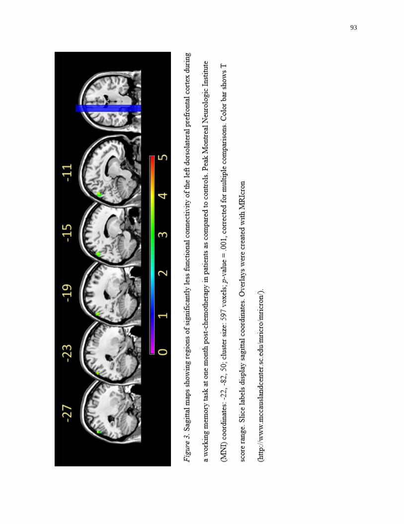
93
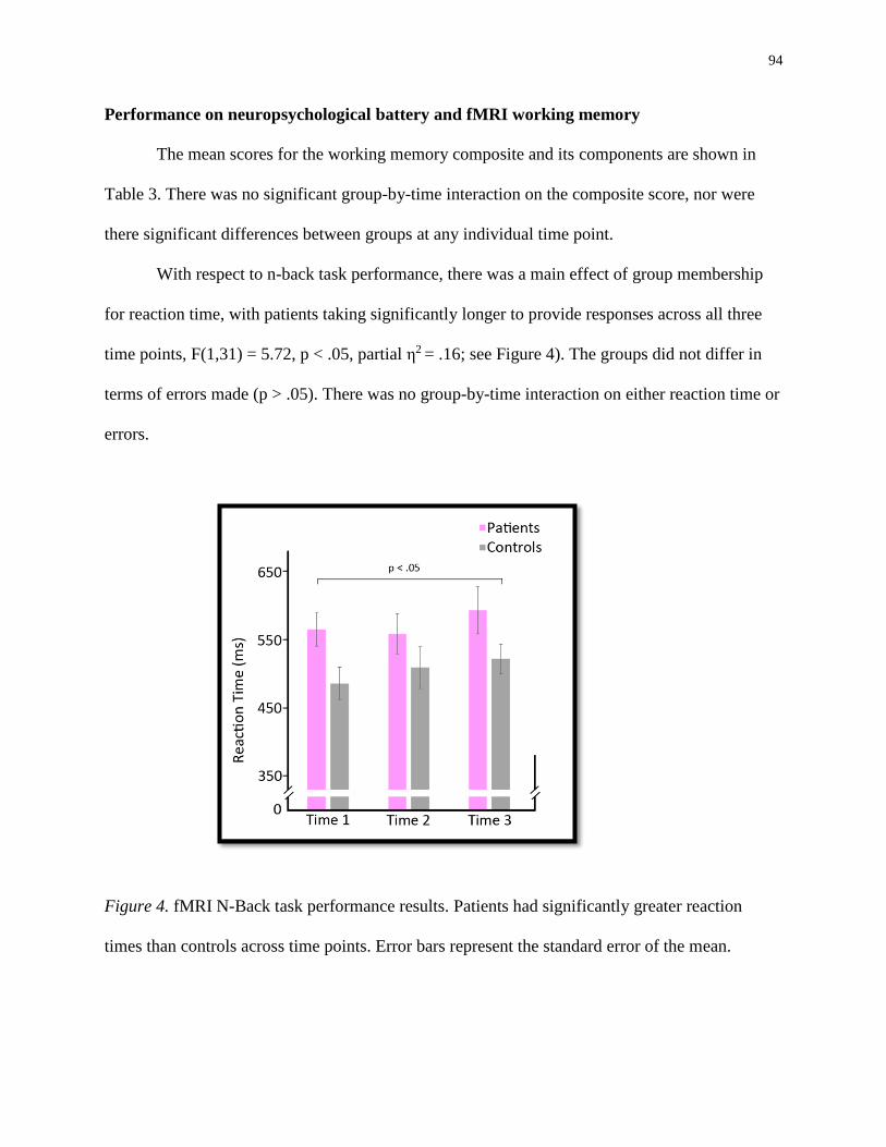
94
Performance on neuropsychological battery and fMRI working memory
The mean scores for the working memory composite and its components are shown in
Table 3. There was no significant group-by-time interaction on the composite score, nor were
there significant differences between groups at any individual time point.
With respect to n-back task performance, there was a main effect of group membership
for reaction time, with patients taking significantly longer to provide responses across all three
time points, F(1,31) = 5.72, p < .05, partial η2 = .16; see Figure 4). The groups did not differ in
terms of errors made (p > .05). There was no group-by-time interaction on either reaction time or
errors.
Figure 4. fMRI N-Back task performance results. Patients had significantly greater reaction
times than controls across time points. Error bars represent the standard error of the mean.
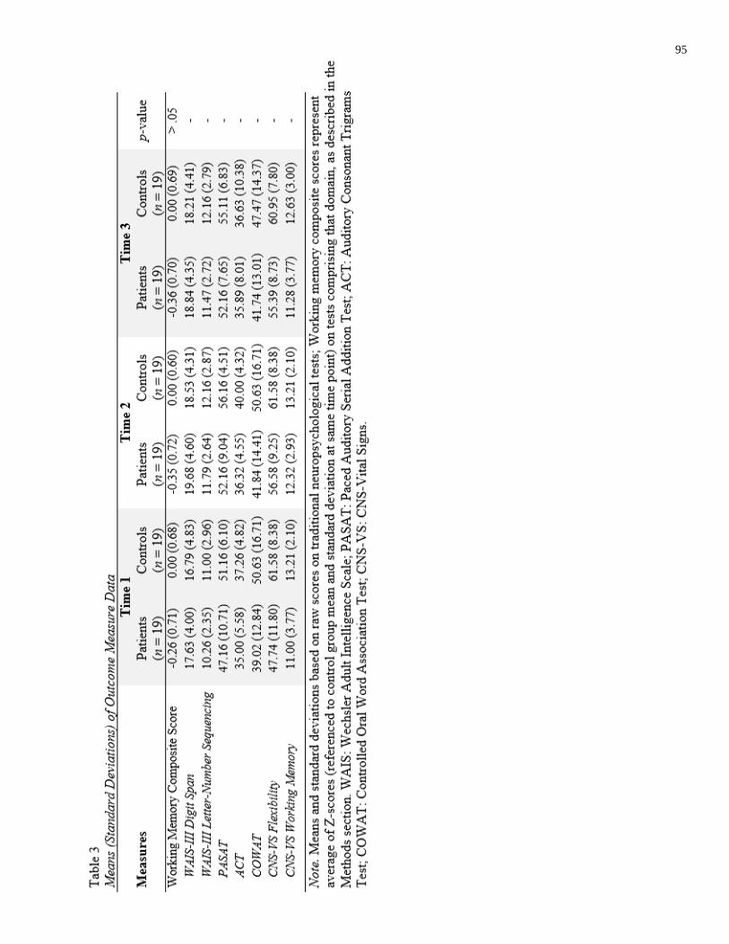
95
96
Discussion
This is the first study, to our knowledge, that has prospectively examined functional
connectivity in breast cancer patients compared to healthy controls. The findings from this study
play an important role in understanding the neural underpinnings of CRCI in this population.
Notably, we found that at an average of one month after chemotherapy, breast cancer patients
had reduced functional connectivity in the frontoparietal network, and that patients recruited
significantly broader brain regions when completing a working memory task. These differences
were not observed at baseline or one year post-chemotherapy, strengthening the evidence of the
acutely injurious effects of chemotherapy.
In examining the susceptibility of the FPN to chemotherapy, this study makes a unique
contribution to the CRCI neuroimaging literature. Although the FPN is broadly implicated in
flexibly allocating cognitive resources across various mental demands, the FPN is also critical
for working memory (Harding et al., 2014). In the present study at Time 2, patients had reduced
functional connectivity between a region of the FPN in the left dlPFC and the left occipito-
parietal region. Each of these regions has been shown to support differing functions of working
memory. The left dlPFC has been implicated in the manipulation of verbal information (Barbey,
Koenigs, & Grafman, 2013), whereas the occipito-parietal cortex has been associated with the
storage of verbal information during working memory tasks (Jonides et al., 1998). In addition,
previous work has found evidence of reduced white matter integrity in the left parietal region of
the superior longitudinal fasciculus (SLF) in breast cancer patients three to five months post-
chemotherapy (Deprez et al., 2012). The SLF is a white matter tract that supports communication
between the occipito-parietal and frontal regions (Schmahmann & Pandya, 2006), and it has been
related to working memory in healthy and pathological populations (Walsh et al., 2011). Taken

97
together, our finding, along with previous work, offers accumulating evidence of working
memory-related brain network disruption shortly after chemotherapy.
The pathological mechanism of injury to this network remains to be fully understood.
Although there is controversy surrounding the sensitivity of diffusion tensor imaging to detect
white matter demyelination (Wheeler-Kingshott & Cercignani, 2009), others have proposed that
this injurious process and axonal injury may be related to changes in the microstructure of white
matter in CRCI (de Ruiter et al., 2012; Deprez et al., 2012). Previous work has shown reductions
of myelin and oligodendrocyte precursors subsequent to the administration of 5-fluorouracil in
mice (Weng et al., 2014). Transiently reduced functional connectivity between areas connected
by the SLF may be a consequence of white matter demyelination of the tracts that support the
working memory component of the FPN. Subsequent recovery of functional connectivity in this
network may be attributable to remyelination, which has been reported in clinical populations
(for a review, see Franklin & ffrench-Constant, 2008; Barkhof et al., 2003).
Interestingly, although we found reduced functional connectivity within working
memory-related circuitry of the FPN, patients and controls did not differ in terms of correct and
incorrect responses made during the fMRI task. The groups differed in terms of reaction time;
however, this finding appears unrelated to chemotherapy, as it was significant across time,
including at pre-treatment. This is consistent with prior work that reported similar baseline
reaction time differences (Ahles et al., 2008).
Imaging results at Time 2 revealed expansive brain hyperactivations for the breast cancer
patients, with the most significant increases in neural activity occurring in the right postcentral
gyrus and left anterior cingulate gyrus regions. The anterior cingulate has long been recognized
for its role in action monitoring (Carter et al., 1998; MacDonald, Cohen, Stenger, & Carter,

98
2000), thus its increased activation during a working memory task at Time 2 in the patient group
may reflect increased attention to performance. In light of the similar performance between
groups during the working memory task (e.g. errors during the two-back condition), these
imaging findings support a hypothesis advanced by our group and others that breast cancer
patients may be engaging in neural compensation in order to perform at, or near, premorbid
levels (McDonald, Conroy, Ahles, et al., 2012; Scherling & Smith, 2013).
Although working memory composite scores from the neuropsychological battery were
not statistically different between breast cancer patients and controls, it is important to highlight
that patients did, on average, perform more poorly than controls cross-sectionally at each time
point. An absence of statistical significance raises the possibility that CRCI does not directly
impact working memory; however, others have reported significant differences between breast
cancer patients and controls in this domain one month after chemotherapy (Stewart et al. 2008),
and we observed a significant decline in working memory when analyzing data from the full
sample of breast cancer patients from which this subsample was drawn (Collins et al., 2014).
This suggests that failure to achieve statistical significance in the current study is due to a lack of
power resulting from our limited sample size. Considering our findings of broad neural
hyperactivations and disrupted functional connectivity shortly after treatment in the present
sample, these findings suggest that neuroimaging may be more sensitive than neuropsychological
measures to the subtle brain effects of peripheral chemotherapy administration. Interestingly,
Collins et al. (2014) reported that working memory significantly improved one year post-
chemotherapy, in line with our study’s finding of functional recovery at Time 3. Additionally,
we have previously reported that one-year post-chemotherapy, breast cancer patients from this
cohort displayed grey matter recovery in regions associated with working memory circuitry
99
(Lepage et al., 2014). Although cross-sectional CRCI studies have reported brain irregularities
and cognitive dysfunction in executive functions, processing speed, and memory many years
after chemotherapy (Ahles et al., 2002; de Ruiter et al., 2011, 2012; Koppelmans et al., 2011,
2012; Silverman et al., 2007; Yamada, Denburg, Beglinger, & Schultz, 2010), there is a paucity
of evidence of chronic working memory dysfunction. Taken together, these findings suggest that
working memory is acutely vulnerable to the cognitively injurious effects of chemotherapy, but
may not be a component of long-term CRCI.
A theme that is emerging from longitudinal neuroimaging studies of CRCI is one of
structural and functional brain abnormalities during and shortly after treatment, with some
degree of recovery in the year following chemotherapy cessation (Dumas et al., 2013; Lepage et
al., 2014; McDonald et al., 2010; McDonald, Conroy, Ahles, et al., 2012). To our knowledge,
this study is the first to report a complete return to baseline by one year post-treatment. A prior
longitudinal study of working memory in the breast cancer population found that despite some
recovery one year after chemotherapy, frontal hyperactivations and parietal hypoactivations were
still present during an auditory n-back task (McDonald, Conroy, Ahles, et al., 2012). The
variations in our findings may be attributable to the differing study characteristics, including the
presentation of stimuli (e.g. visual compared to auditory presentation), which has been shown to
elicit different frontoparietal activations in healthy controls (Crottaz-Herbette, Anagnoson, &
Menon, 2004). Similarly, we did not find any pre-treatment working memory-related functional
differences, in contrast to previous work by others and by our group (Cimprich et al., 2010;
López Zunini et al., 2013; McDonald, Conroy, Ahles, et al., 2012; Scherling et al., 2011). These
studies differed in terms of included chemotherapy regimens, working memory tasks employed,
and different comparative groups, such as healthy and chemotherapy-free breast cancer controls,

100
which may contribute to the variability in the findings across studies. Furthermore, differential
findings among studies may also be influenced by patients who are particularly vulnerable to
chemotherapy, even before the commencement of treatment. A recent prospective study has
found that pre-treatment brain functional inefficiency, signalled by broad spatial variance within
frontoparietal regions during working memory, significantly predicted cognitive complaints in
breast cancer patients (Askren et al., 2014). Previous work has also demonstrated a relationship
between reduced processing speed performance and lower cognitive reserve in chemotherapy-
treated breast cancer patients (Ahles et al., 2010); however, cognitive reserve and its influence on
CRCI remains an understudied topic. In light of work showing that some women experience
cognitive dysfunction prior to chemotherapy, future work should look to pre-treatment cognitive
reserve as a possible modulating factor of later CRCI.
Limitations of this study include the lack of a non-chemotherapy breast cancer control
group. While others have included such a control group in a prospective study and obtained
similar functional results (McDonald, Conroy, Ahles, et al., 2012), we could not control for
cancer-related factors. Although our participants were closely matched at baseline, all patients
became menopausal by Time 3, whereas controls remained unchanged with respect to
menopausal status. Subsequent to chemotherapy, treatment-induced menopausal symptoms have
been reported to co-occur alongside cognitive disruption (Fan et al., 2005); thus, future
longitudinal studies should account for menopausal status. The types of surgery, number of
cycles and type of chemotherapy varied among the patients (see Table 1). From Time 2 to Time
3, some patients received radiotherapy or commenced hormonal therapy. Although these
adjuvant treatments have been associated with cognitive decline (Bender, Paraska, Sereika,
Ryan, & Berga, 2001; Quesnel et al., 2009), the sample size of the present study precluded their

101
investigation. In order to overcome the shortcomings that are common between this and other
neuroimaging studies of CRCI, it will be important for future research to strive to be multicenter
and collaborative.
In summary, the current study offers the first prospective report of frontoparietal network
functional connectivity disruption in chemotherapy-treated breast cancer patients. Also unique to
this study is evidence of a neurofunctional return to baseline in working memory-related
circuitry around one year post-chemotherapy. Although there are still many questions to answer
about CRCI, this study provides hope for patients who experience trouble with working memory
both during and following chemotherapy. As women are being more informed about the
potential for CRCI as they begin treatment, this article provides evidence for recovery of the
neural underpinnings of working memory and, thus, may reduce the fear of permanent
consequences of treatment.
102
References
Abraham, J., Haut, M. W., Moran, M. T., Filburn, S., Lemiuex, S., & Kuwabara, H. (2008).
Adjuvant chemotherapy for breast cancer: effects on cerebral white matter seen in
diffusion tensor imaging. Clinical Breast Cancer, 8(1), 88–91.
doi:10.3816/CBC.2008.n.007
Ahles, T. A., Saykin, A. J., Furstenberg, C. T., Cole, B., Mott, L. A., Skalla, K., Whedon, M. B.,
Bivens, S., Mitchell, T., Greenberg, E. R., & Silberfarb, P. M. (2002). Neuropsychologic
impact of standard-dose systemic chemotherapy in long-term survivors of breast cancer
and lymphoma. Journal of Clinical Oncology, 20(2), 485–493. doi:
10.1200/JCO.20.2.485
Ahles, T. A., Saykin, A. J., McDonald, B. C., Furstenberg, C. T., Cole, B. F., Hanscom, B. S.,
Mulrooney, T. J., Schwartz, G. N., & Kaufman, P. A. (2008). Cognitive function in breast
cancer patients prior to adjuvant treatment. Breast Cancer Research and Treatment,
110(1), 143–152. doi: 10.1200/JCO.20.2.485
Ahles, T. A., Saykin, A. J., McDonald, B. C., Li, Y., Furstenberg, C. T., Hanscom, B. S.,
Mulrooney, T. J., Schwartz, G. N., & Kaufman, P. A. (2010). Longitudinal assessment of
cognitive changes associated with adjuvant treatment for breast cancer: impact of age and
cognitive reserve. Journal of Clinical Oncology, 28(29), 4434–4440.
doi:10.1200/JCO.2009.27.0827
Askren, M. K., Jung, M., Berman, M. G., Zhang, M., Therrien, B., Peltier, S., Ossher, L., Hayes,
D. F., Reuter-Lorenz, P. A., & Cimprich, B. (2014). Neuromarkers of fatigue and
cognitive complaints following chemotherapy for breast cancer: a prospective fMRI
103
investigation. Breast Cancer Research and Treatment, 147(2), 445–455.
doi:10.1007/s10549-014-3092-6
Barbey, A. K., Koenigs, M., & Grafman, J. (2013). Dorsolateral prefrontal contributions to
human working memory. Cortex, 49(5), 1195–1205. doi:10.1016/j.cortex.2012.05.022
Barkhof, F., Bruck, W., De Groot, C. J. A., Bergers, E., Hulshof, S., Geurts, J., Polman, C. H., &
van der Valk, P. (2003). Remyelinated lesions in multiple sclerosis: magnetic resonance
image appearance. Archives of Neurology, 60(8), 1073–1081.
doi:10.1001/archneur.60.8.1073
Behzadi, Y., Restom, K., Liau, J., & Liu, T. T. (2007). A component based noise correction
method (CompCor) for BOLD and perfusion based fMRI. NeuroImage, 37(1), 90–101.
doi:10.1016/j.neuroimage.2007.04.042
Bender, C M, Paraska, K. K., Sereika, S. M., Ryan, C. M., & Berga, S. L. (2001). Cognitive
function and reproductive hormones in adjuvant therapy for breast cancer: a critical
review. Journal of Pain and Symptom Management, 21(5), 407–424. doi:10.1016/S0885-
3924(01)00268-8
Bender, C. M., Sereika, S. M., Berga, S. L., Vogel, V. G., Brufsky, A. M., Paraska, K. K., &
Ryan, C. M. (2006). Cognitive impairment associated with adjuvant therapy in breast
cancer. Psycho-oncology, 15(5), 422–430. doi:10.1002/pon.964
Berman, M. G., Askren, M. K., Jung, M., Therrien, B., Peltier, S., Noll, D. C., Zhang, M.,
Ossher, L., Hayes, D. F., Reuter-Lorenz, P. A., & Cimprich, B. (2014). Pretreatment
worry and neurocognitive responses in women with breast cancer. Health Psychology,
33(3), 222–231. doi:10.1037/a0033425

104
Brown, J. (1958). Some tests of the decay theory of immediate memory. The Quarterly Journal
of Experimental Psychology, 10(1), 12–21. doi:10.1080/17470215808416249
Bruno, J., Hosseini, S. M. H., & Kesler, S. (2012). Altered resting state functional brain network
topology in chemotherapy-treated breast cancer survivors. Neurobiology of Disease,
48(3), 329–338. doi:10.1016/j.nbd.2012.07.009
Carter, C. S., Braver, T. S., Barch, D. M., Botvinick, M. M., Noll, D., & Cohen, J. D. (1998).
Anterior cingulate cortex, error detection, and the online monitoring of performance.
Science, 280(5364), 747–749. doi:10.1126/science.280.5364.747
Cimprich, B., Reuter-Lorenz, P., Nelson, J., Clark, P. M., Therrien, B., Normolle, D., Berman,
M. G., Hayes, D. F., Noll, D. C., Peltier, S., & Welsh, R. C. (2010). Prechemotherapy
alterations in brain function in women with breast cancer. Journal of Clinical and
Experimental Neuropsychology, 32(3), 324–331. doi:10.1080/13803390903032537
Cole, M. W., Reynolds, J. R., Power, J. D., Repovs, G., Anticevic, A., & Braver, T. S. (2013).
Multi-task connectivity reveals flexible hubs for adaptive task control. Nature
Neuroscience, 16(9), 1348–1355. doi:10.1038/nn.3470
Collins, B., Mackenzie, J., Stewart, A., Bielajew, C., & Verma, S. (2009). Cognitive effects of
chemotherapy in post-menopausal breast cancer patients 1 year after treatment. Psycho-
oncology, 18(2), 134–143. doi:10.1002/pon.1379
Collins, B., MacKenzie, J., Tasca, G. A., Scherling, C., & Smith, A. (2013). Cognitive effects of
chemotherapy in breast cancer patients: a dose–response study. Psycho-oncology, 22(7),
1517–1527. doi:10.1002/pon.3163
Collins, B., Mackenzie, J., Tasca, G. A., Scherling, C., & Smith, A. (2014). Persistent cognitive
changes in breast cancer patients 1 year following completion of chemotherapy. Journal
105
of the International Neuropsychological Society, 20(4), 370–379.
doi:10.1017/S1355617713001215
Conroy, S. K., McDonald, B. C., Smith, D. J., Moser, L. R., West, J. D., Kamendulis, L. M., …
Saykin, A. J. (2012). Alterations in brain structure and function in breast cancer
survivors: effect of post-chemotherapy interval and relation to oxidative DNA damage.
Breast Cancer Research and Treatment, 137(2), 493–502. doi:10.1007/s10549-012-2385-
x
Crottaz-Herbette, S., Anagnoson, R. T., & Menon, V. (2004). Modality effects in verbal working
memory: differential prefrontal and parietal responses to auditory and visual stimuli.
NeuroImage, 21(1), 340–351. doi:10.1016/j.neuroimage.2003.09.019
De Ruiter, M. B., Reneman, L., Boogerd, W., Veltman, D. J., Caan, M., Douaud, G., Lavini, C.,
Linn, S. C., Boven, E., van Dam, F. S. A. M., & Schagen, S. B. (2012). Late effects of
high-dose adjuvant chemotherapy on white and gray matter in breast cancer survivors:
converging results from multimodal magnetic resonance imaging. Human Brain
Mapping, 33(12), 2971–2983. doi:10.1002/hbm.21422
De Ruiter, M. B., Reneman, L., Boogerd, W., Veltman, D. J., van Dam, F. S. A. M., Nederveen,
A. J., Boven, E., & Schagen, S. B. (2011). Cerebral hyporesponsiveness and cognitive
impairment 10 years after chemotherapy for breast cancer. Human Brain Mapping, 32(8),
1206–1219. doi:10.1002/hbm.21102
Delis, D., Kaplan, E., & Kramer, J. (2001). Delis Kaplan Executive Function System Test
Manual. San Antonio, TX: Psychological Corporation.
Deprez, S., Amant, F., Smeets, A., Peeters, R., Leemans, A., Van Hecke, W., Verhoeven, J. S.,
Christiaens, M. R., Vandenberghe, M., & Sunaert, S. (2012). Longitudinal assessment of
106
chemotherapy-induced structural changes in cerebral white matter and its correlation with
impaired cognitive functioning. Journal of Clinical Oncology, 30(3), 274–281.
doi:10.1200/JCO.2011.36.8571
Deprez, S., Amant, F., Yigit, R., Porke, K., Verhoeven, J., Van den Stock, J., Smeets, A.,
Christiaens, M. R., Leemans, A., Van Hecke, W., Vandenberghe, J., Vandenbulcke, M.,
& Sunaert, S. (2011). Chemotherapy-induced structural changes in cerebral white matter
and its correlation with impaired cognitive functioning in breast cancer patients. Human
Brain Mapping, 32(3), 480–493. doi:10.1002/hbm.21033
Dumas, J. A., Makarewicz, J., Schaubhut, G. J., Devins, R., Albert, K., Dittus, K., & Newhouse,
P. A. (2013). Chemotherapy altered brain functional connectivity in women with breast
cancer: a pilot study. Brain Imaging and Behavior, 7(4), 524–532. doi:10.1007/s11682-
013-9244-1
Fan, H. G. M., Houédé-Tchen, N., Yi, Q.-L., Chemerynsky, I., Downie, F. P., Sabate, K., &
Tannock, I. F. (2005). Fatigue, menopausal symptoms, and cognitive function in women
after adjuvant chemotherapy for breast cancer: 1- and 2-year follow-up of a prospective
controlled study. Journal of Clinical Oncology, 23(31), 8025–8032.
doi:10.1200/JCO.2005.01.6550
Ferguson, R. J., McDonald, B. C., Saykin, A. J., & Ahles, T. A. (2007). Brain structure and
function differences in monozygotic twins: possible effects of breast cancer
chemotherapy. Journal of Clinical Oncology, 25(25), 3866–3870.
doi:10.1200/JCO.2007.10.8639
107
Fischer, J., Jak, A., Kniker, J., Rudick, R., & Cutter, G. (2001). Administration and Scoring
Manual for the Multiple Sclerosis Functional Composite Measure. New York: National
Multiple Sclerosis Society.
Franklin, R. J. M., & ffrench-Constant, C. (2008). Remyelination in the CNS: from biology to
therapy. Nature Reviews Neuroscience, 9(11), 839–855. doi:10.1038/nrn2480
Ganz, P. A., Kwan, L., Castellon, S. A., Oppenheim, A., Bower, J. E., Silverman, D. H. S., Cole,
S. W., Irwin, M. R., Ancoli-Israel, S., & Belin, T. R. (2013). Cognitive complaints after
breast cancer treatments: examining the relationship with neuropsychological test
performance. Journal of the National Cancer Institute, 105(11), 791–801.
doi:10.1093/jnci/djt073
Gualtieri, C. T., & Johnson, L. G. (2006). Reliability and validity of a computerized
neurocognitive test battery, CNS Vital Signs. Archives of Clinical Neuropsychology,
21(7), 623–643. doi:10.1016/j.acn.2006.05.007
Gualtieri, C. T., & Johnson, L. G. (2008). A computerized test battery sensitive to mild and
severe brain injury. Medscape Journal of Medicine, 10(4), 90.
Harding, I. H., Yücel, M., Harrison, B. J., Pantelis, C., & Breakspear, M. (2014). Effective
connectivity within the frontoparietal control network differentiates cognitive control and
working memory. NeuroImage, 106C, 144–153. doi:10.1016/j.neuroimage.2014.11.039
Hermelink, K., Untch, M., Lux, M. P., Kreienberg, R., Beck, T., Bauerfeind, I., & Münzel, K.
(2007). Cognitive function during neoadjuvant chemotherapy for breast cancer: results of
a prospective, multicenter, longitudinal study. Cancer, 109(9), 1905–1913.
doi:10.1002/cncr.22610

108
Hosseini, S. M. H., Koovakkattu, D., & Kesler, S. R. (2012). Altered small-world properties of
gray matter networks in breast cancer. BMC Neurology, 12, 28. doi:10.1186/1471-2377-
12-28
Hurria, A., Rosen, C., Hudis, C., Zuckerman, E., Panageas, K. S., Lachs, M. S., Witner, M., van
Gorp, W. G., Fornier, M., D’Andrea, G., Moasser, M., Dang, C., Van Poznak, C., Hurria,
A., & Holland, J. (2006). Cognitive function of older patients receiving adjuvant
chemotherapy for breast cancer: a pilot prospective longitudinal study. Journal of the
American Geriatrics Society, 54(6), 925–931. doi:10.1111/j.1532-5415.2006.00732.x
Inagaki, M., Yoshikawa, E., Matsuoka, Y., Sugawara, Y., Nakano, T., Akechi, T., Wada, N.,
Imoto, S., Murakami, K., & Uchitomi, Y. (2007). Smaller regional volumes of brain gray
and white matter demonstrated in breast cancer survivors exposed to adjuvant
chemotherapy. Cancer, 109(1), 146–156. doi:10.1002/cncr.22368
Jansen, C. E., Cooper, B. A., Dodd, M. J., & Miaskowski, C. A. (2011). A prospective
longitudinal study of chemotherapy-induced cognitive changes in breast cancer patients.
Supportive Care in Cancer, 19(10), 1647–1656. doi:10.1007/s00520-010-0997-4
Jonides, J., Schumacher, E. H., Smith, E. E., Koeppe, R. A., Awh, E., Reuter-Lorenz, P. A.,
Awh, E., Reuter-Lorenz, P. A., Marshetz, C., & Willis, C. R. (1998). The role of parietal
cortex in verbal working memory. The Journal of Neuroscience, 18(13), 5026–5034.
Kesler, S. R., Bennett, F. C., Mahaffey, M. L., & Spiegel, D. (2009). Regional brain activation
during verbal declarative memory in metastatic breast cancer. Clinical Cancer Research,
15(21), 6665–6673. doi:10.1158/1078-0432.CCR-09-1227
109
Kesler, S. R., Kent, J. S., & O’Hara, R. (2011). Prefrontal cortex and executive function
impairments in primary breast cancer. Archives of Neurology, 68(11), 1447–1453.
doi:10.1001/archneurol.2011.245
Koppelmans, V., Breteler, M. M. B., Boogerd, W., Seynaeve, C., Gundy, C., & Schagen, S. B.
(2012). Neuropsychological performance in survivors of breast cancer more than 20 years
after adjuvant chemotherapy. Journal of Clinical Oncology, 30(10), 1080–1086.
doi:10.1200/JCO.2011.37.0189
Koppelmans, V., de Ruiter, M. B., van der Lijn, F., Boogerd, W., Seynaeve, C., van der Lugt, A.,
Vrooman, H., Niessen, W. J., Breteler, M. M. B., & Schagen, S. B. (2011). Global and
focal brain volume in long-term breast cancer survivors exposed to adjuvant
chemotherapy. Breast Cancer Research and Treatment, 132(3), 1099–1106.
doi:10.1007/s10549-011-1888-1
Koziol, L. F., Barker, L. A., Joyce, A. W., & Hrin, S. (2014). Structure and function of large-
scale brain systems. Applied Neuropsychology Child, 3(4), 236–244.
doi:10.1080/21622965.2014.946797
Lepage, C., Smith, A. M., Moreau, J., Barlow-Krelina, E., Wallis, N., Collins, B., MacKenzie, J.,
& Scherling, C. (2014). A prospective study of grey matter and cognitive function
alterations in chemotherapy-treated breast cancer patients. SpringerPlus, 3, 444.
doi:10.1186/2193-1801-3-444
López Zunini, R. A., Scherling, C., Wallis, N., Collins, B., MacKenzie, J., Bielajew, C., &
Smith, A. M. (2013). Differences in verbal memory retrieval in breast cancer
chemotherapy patients compared to healthy controls: a prospective fMRI study. Brain
Imaging and Behavior, 7(4), 460–477. doi:10.1007/s11682-012-9213-0
110
MacDonald, A. W., 3rd, Cohen, J. D., Stenger, V. A., & Carter, C. S. (2000). Dissociating the
role of the dorsolateral prefrontal and anterior cingulate cortex in cognitive control.
Science, 288(5472), 1835–1838. doi:10.1126/science.288.5472.1835
McDonald, B. C., Conroy, S. K., Ahles, T. A., West, J. D., & Saykin, A. J. (2010). Gray matter
reduction associated with systemic chemotherapy for breast cancer: a prospective MRI
study. Breast Cancer Research and Treatment, 123(3), 819–828. doi:10.1007/s10549-
010-1088-4
McDonald, B. C., Conroy, S. K., Ahles, T. A., West, J. D., & Saykin, A. J. (2012). Alterations in
brain activation during working memory processing associated with breast cancer and
treatment: a prospective functional magnetic resonance imaging study. Journal of
Clinical Oncology, 30(20), 2500–2508. doi:10.1200/JCO.2011.38.5674
McDonald, B. C., Conroy, S. K., Smith, D. J., West, J. D., & Saykin, A. J. (2012). Frontal gray
matter reduction after breast cancer chemotherapy and association with executive
symptoms: a replication and extension study. Brain, Behavior, and Immunity, 30 Suppl,
S117–25. doi:10.1016/j.bbi.2012.05.007
McDonald, B. C., & Saykin, A. J. (2013). Alterations in brain structure related to breast cancer
and its treatment: chemotherapy and other considerations. Brain Imaging and Behavior,
7(4), 374–387. doi:10.1007/s11682-013-9256-x
Owen, A. M., McMillan, K. M., Laird, A. R., & Bullmore, E. (2005). N-back working memory
paradigm: a meta-analysis of normative functional neuroimaging studies. Human Brain
Mapping, 25(1), 46–59. doi:10.1002/hbm.20131
111
Pullens, M. J. J., De Vries, J., & Roukema, J. A. (2010). Subjective cognitive dysfunction in
breast cancer patients: a systematic review. Psycho-oncology, 19(11), 1127–1138.
doi:10.1002/pon.1673
Quesnel, C., Savard, J., & Ivers, H. (2009). Cognitive impairments associated with breast cancer
treatments: results from a longitudinal study. Breast Cancer Research and Treatment,
116(1), 113–123. doi:10.1007/s10549-008-0114-2
Rao, S. M., Leo, G. J., Bernardin, L., & Unverzagt, F. (1991). Cognitive dysfunction in multiple
sclerosis. I. Frequency, patterns, and prediction. Neurology, 41(5), 685–691.
doi:10.1212/WNL.41.5.685
Schagen, S. B., Muller, M. J., Boogerd, W., Mellenbergh, G. J., & van Dam, F. S. A. M. (2006).
Change in cognitive function after chemotherapy: a prospective longitudinal study in
breast cancer patients. Journal of the National Cancer Institute, 98(23), 1742–1745.
doi:10.1093/jnci/djj470
Scherling, C., Collins, B., Mackenzie, J., Bielajew, C., & Smith, A. (2011). Pre-chemotherapy
differences in visuospatial working memory in breast cancer patients compared to
controls: an FMRI study. Frontiers in Human Neuroscience, 5, 122.
doi:10.3389/fnhum.2011.00122
Scherling, C., Collins, B., Mackenzie, J., Bielajew, C., & Smith, A. (2012). Prechemotherapy
differences in response inhibition in breast cancer patients compared to controls: a
functional magnetic resonance imaging study. Journal of Clinical and Experimental
Neuropsychology, 34(5), 543–560. doi:10.1080/13803395.2012.666227
112
Scherling, C., Collins, B., MacKenzie, J., Lepage, C., Bielajew, C., & Smith, A. (2012).
Structural brain differences in breast cancer patients compared to matched controls prior
to chemotherapy. International Journal of Biology, 4(2), 3. doi:10.5539/ijb.v4n2p3
Scherling, C., & Smith, A. (2013). Opening up the window into “chemobrain”: a neuroimaging
review. Sensors, 13(3), 3169–3203. doi:10.3390/s130303169
Schmahmann, J. D., & Pandya, D. N. (2006). Superior Longitudinal Fasciculus and Arcuate
Fasciculus. In Fiber Pathways of the Brain (pp.393-408). New York: Oxford University
Press. doi:10.1093/acprof:oso/9780195104233.003.0013
Shilling, V., Jenkins, V., Morris, R., Deutsch, G., & Bloomfield, D. (2005). The effects of
adjuvant chemotherapy on cognition in women with breast cancer—preliminary results of
an observational longitudinal study. Breast, 14(2), 142–150.
doi:10.1016/j.breast.2004.10.004
Silverman, D. H. S., Dy, C. J., Castellon, S. A., Lai, J., Pio, B. S., Abraham, L., Waddell, K.,
Petersen, L., Phelps, M. E., & Ganz, P. A. (2007). Altered frontocortical, cerebellar, and
basal ganglia activity in adjuvant-treated breast cancer survivors 5-10 years after
chemotherapy. Breast Cancer Research and Treatment, 103(3), 303–311.
doi:10.1007/s10549-006-9380-z
Stewart, A., Bielajew, C., Collins, B., Parkinson, M., & Tomiak, E. (2006). A meta-analysis of
the neuropsychological effects of adjuvant chemotherapy treatment in women treated for
breast cancer. The Clinical Neuropsychologist, 20(1), 76–89.
doi:10.1080/138540491005875
113
Stewart, A., Collins, B., Mackenzie, J., Tomiak, E., Verma, S., & Bielajew, C. (2008). The
cognitive effects of adjuvant chemotherapy in early stage breast cancer: a prospective
study. Psycho-oncology, 17(2), 122–130. doi:10.1002/pon.1210
Walsh, M., Montojo, C. A., Sheu, Y.-S., Marchette, S. A., Harrison, D. M., Newsome, S. D.,
Zhou, F., Shelton, A. L., & Courtney, S. M. (2011). Object working memory
performance depends on microstructure of the frontal-occipital fasciculus. Brain
Connectivity, 1(4), 317–329. doi:10.1089/brain.2011.0037
Wechsler, D. (1997). Wechsler Adult Intelligence Scale (3rd edn). San Antonio, TX: Harcourt,
Brace, & Co.
Wefel, J. S., Lenzi, R., Theriault, R. L., Davis, R. N., & Meyers, C. A. (2004). The cognitive
sequelae of standard-dose adjuvant chemotherapy in women with breast carcinoma:
results of a prospective, randomized, longitudinal trial. Cancer, 100(11), 2292–2299.
doi:10.1002/cncr.20272
Wefel, J. S., Saleeba, A. K., Buzdar, A. U., & Meyers, C. A. (2010). Acute and late onset
cognitive dysfunction associated with chemotherapy in women with breast cancer.
Cancer, 116(14), 3348–3356. doi:10.1002/cncr.25098
Wefel, J. S., & Schagen, S. B. (2012). Chemotherapy-related cognitive dysfunction. Current
Neurology and Neuroscience Reports, 12(3), 267–275. doi:10.1007/s11910-012-0264-9
Weng, Q., Tan, B., Wang, J., Wang, J., Zhou, H., Shi, J., He, Q., & Yang, B. (2014). 5-
Fluorouracil causes severe CNS demyelination by disruption of
TCF7L2/HDAC1/HDAC2 complex in adolescent mice. Toxicology, 325(0), 144–150.
doi:10.1016/j.tox.2014.08.011

114
Wheeler-Kingshott, C. A. M., & Cercignani, M. (2009). About “axial” and “radial” diffusivities.
Magnetic Resonance in Medicine, 61(5), 1255–1260. doi:10.1002/mrm.21965
Whitfield-Gabrieli, S., & Nieto-Castanon, A. (2012). Conn: a functional connectivity toolbox for
correlated and anticorrelated brain networks. Brain Connectivity, 2(3), 125–141.
doi:10.1089/brain.2012.0073
Yamada, T. H., Denburg, N. L., Beglinger, L. J., & Schultz, S. K. (2010). Neuropsychological
outcomes of older breast cancer survivors: cognitive features ten or more years after
chemotherapy. The Journal of Neuropsychiatry and Clinical Neurosciences, 22(1), 48–
54. doi:10.1176/appi.neuropsych.22.1.48
115
General Discussion
The overarching goal of this thesis was to use contemporary neuroimaging techniques to
better characterize the chemotherapy-related neurophysiology underlying the cognitive
disruption experienced by chemotherapy-treated breast cancer patients. By prospectively
investigating the relationship of both brain structure and function to cognitive functioning in
chemotherapy-exposed breast cancer patients, this thesis provides novel insights about the extent
of CRCI. To achieve its goal, the thesis comprised two correlational studies in which brain
structure and function were contrasted between breast cancer patients and cancer-free controls
before patients began chemotherapy, as they completed it, and one-year after treatment.
In the following sections, both studies of the thesis are recapitulated, with special
attention drawn to their principal findings. The limitations of the works are then discussed and
blended with suggestions for future research. The final section offers an overall summary and
concludes the thesis.
Grey matter volumes and overall cognitive function
The first study (Study 1) had a primary aim of investigating the relationship between grey
matter alterations and cognitive function. Although previous work established a link between
grey matter fluctuations and cognitive compromise in chemotherapy-treated breast cancer
patients (Conroy et al., 2012; McDonald, Conroy, Ahles, West, & Saykin, 2010; McDonald,
Conroy, Smith, West, & Saykin, 2012), a prospective study incorporating a robust
neuropsychological battery was lacking in the literature. Thus, Study 1 served the important
purpose of characterizing, not only the areas of the brain that may be negatively impacted over
time by peripheral chemotherapy, but also how these areas relate to cognitive function.
116
With respect to the course of grey matter attenuation, the hypothesis that greater
disruption would be greatest shortly after chemotherapy, and partially resolve one year later, was
supported. Given the paucity of longitudinal grey matter studies in the CRCI literature, analyses
comprised whole-brain volumes rather than a priori regions-of-interest. Approximately one
month after chemotherapy completion, breast cancer patients had grey matter volume loss in
widespread regions. Medial and inferior frontal regions, along with temporal areas were the
greatest to be acutely impacted, in line with previous studies (Inagaki et al., 2007; McDonald et
al., 2010; McDonald et al., 2012). Similar to prior work (de Ruiter et al., 2012) showing parietal
volume reductions, the paracentral lobule and the precuneus within the right hemisphere were
also disrupted shortly after chemotherapy. The functional neuroimaging literature of CRCI
highlights abnormal task-related activations primarily in frontal and parietal regions (for a
review, see (Pomykala, de Ruiter, Deprez, McDonald, & Silverman, 2013)). Since those regions
are commonly recruited during cognitive processes frequently explored in CRCI (e.g. working
memory), it remained unclear whether the circumscribed nature of the irregularities reflected
task demands, or whether frontoparietal regions might instead be more sensitive to chemotherapy
than other areas. In absence of frontoparietal-driven demands, Study 1 lends support for the
latter, adding to the evidence from other structural studies showing increased chemotherapy
susceptibility in these regions (de Ruiter et al., 2012; Inagaki et al., 2007; McDonald et al.,
2012).
Cognitive function was assessed using a comprehensive neuropsychological battery and
was categorized as follows: information processing speed, working memory, verbal memory, and
visual memory. Although the larger sample from which participants of Study 1 were drawn
displayed significantly decreased working memory over the course of treatment (Collins,
117
MacKenzie, Tasca, Scherling, & Smith, 2013), information processing speed was the only
domain to reach significance in the Study 1 participants. Mean scores on the three other domains
tended to decrease, but the differences were not significant. Information processing speed
displayed the greatest relationship with a range of disrupted areas, particularly in prefrontal,
temporal, and superior parietal areas. Although less robust, visual memory was related to areas in
the prefrontal cortex, and working memory was positively associated with bilateral frontal grey
matter. Verbal memory did not show an association with regions of reduced grey matter.
Acute grey matter volume losses tended to resolve by one-year post-chemotherapy.
However, continued disruption was noted largely within inferior and medial frontal regions, the
anterior cingulate, the middle frontal gyrus, and in medial temporal areas. Medial frontal regions
have been previously shown to have persistent reductions of grey matter volume following
chemotherapy (McDonald et al., 2010). Study 1 found working memory was positively
associated with the right middle frontal gyrus, a region demonstrating sustained hypoactivity
during a working memory task in breast cancer survivors one year after treatment (McDonald et
al., 2012). Taken together, the findings from Study 1 suggest that a potential predilection for the
prefrontal cortex by chemotherapy may underlie the working memory deficits experienced by
breast cancer patients.
Working memory and the frontoparietal network
The aim of the second study (Study 2) was to elucidate the neural circuitry of the working
memory impingement associated with chemotherapy. As the most acutely vulnerable cognitive
domain in the context of CRCI (Stewart, Bielajew, Collins, Parkinson, & Tomiak, 2006),
working memory has been the subject of increasing investigation using functional neuroimaging;
however, prospective studies remain scarce. Study 2 sought to replicate previous studies of
118
working memory task-related activity over the course of treatment, and to incorporate cutting
edge analytical methods to explore a functional network within commonly disrupted brain
regions.
Prior to treatment, breast cancer patients and controls did not display any functional
differences, contrary to prior reports of baseline working memory related frontal, insular, and
diencephalic hyperactivations, and parietal hypoactivations (McDonald et al., 2012; Scherling,
Collins, Mackenzie, Bielajew, & Smith, 2011). By one month after treatment, breast cancer
patients had expansive working memory associated hyperactivations in frontal, temporal, and
parietal regions when compared to controls. Remarkably, these hyperactivations resolved by
one-year post-treatment. Although a previous longitudinal study reported a partial return to
baseline working memory activations at the one-year post-chemotherapy interval (McDonald et
al., 2012), two important distinctions should be noted. First, McDonald et al. (2012) reported that
breast cancer patients had baseline working memory hyperactivations in frontal and parietal
regions, and decreased inferior frontal activity by one-month post-treatment. Thus, the
directionality of activity between Study 2 and that study is opposite. Next, Study 2 is the first to
report a complete resolution of abnormal functional activity by the one-year interval. This
finding is supported by recovery of working memory in the larger sample of chemotherapy-
treated breast cancer patients from which the Study 2 cohort was drawn (Collins, Mackenzie,
Tasca, Scherling, & Smith, 2014).
The frontoparietal network (FPN) was explored using functional connectivity - an
analysis method to correlate neural activity between regions across a timeseries (Friston &
Buchel, 2003). Thus, this technique allows for inference based on the co-activation of regions,
which can suggest brain network function. Beginning with a region in the left dorsolateral
119
prefrontal cortex (dlPFC), the timeseries across the entire brain was explored to determine areas
significantly activated alongside this region. Given the selected region of the dlPFC was chosen
for its role in working memory (Harding, Yücel, Harrison, Pantelis, & Breakspear, 2014), it was
not surprising to find that it was functionally correlated in time to an area in the left superior
parieto-occipital region. However, it was noteworthy that this connection was significantly
disrupted in breast cancer patients one-month post-treatment, but not at baseline and one-year
follow-up. Previous work has illustrated a similar pattern within the dorsal attention network,
with disruption between frontoparietal regions (Dumas et al., 2013). However, in that study, an
identical analysis of the default mode network, disruption between the posterior cingulate cortex
and precuneus persisted to the final assessment interval. Thus, Study 2 adds to the accumulating
evidence that, despite a pronounced, acute disruption in working memory substrates, regions
supporting this cognitive function recover over time, in line with neuropsychological
performance in this domain (Collins et al., 2014).
Overall, this thesis adds to the growing body of CRCI neuroimaging literature that
identifies the short-term post-treatment period to be the most susceptible to neural and cognitive
disruption. Study 1 makes a novel contribution to the field of CRCI research by exploring the
relationship of grey matter attenuation and a number of objectively assessed cognitive domains.
Study 2 is the first prospective functional connectivity study conducted in the CRCI literature. It
is also the first to demonstrate a recovery to pre-treatment neural activity, despite short-term
working memory circuitry abnormalities. An overarching strength of both studies was the
inclusion of a comparison group that was matched to the patient group on age and education,
thus controlling for potential confounding by these variables.
Limitations and future directions
120
Although the studies of this thesis contribute novel findings to the CRCI literature, some
limitations herein warrant acknowledgement. Since both studies were conducted on the same
sample, and neuroimaging data were collected during the same sessions for each study, most of
the limitations apply equally to both investigations. Although patients and controls were closely
aligned on important characteristics, such as age, and education, they were not matched on
disease and treatment. The control group comprised cancer- and chemotherapy-free women.
With the absence of a disease-matched group, the influence of potentially cancer-related
processes on cognition could not be controlled. Despite this limitation, the findings of both
studies are congruent with previous studies of grey matter volume (McDonald et al., 2010), and
working memory fMRI investigations that included chemotherapy-free breast cancer controls
(McDonald, Conroy, Ahles, et al., 2012).
Within the patient group, there was variability in terms of the number of cycles, and types
of chemotherapy. Teasing apart differential contributions to CRCI by various chemotherapy
agents remains a challenge in human studies. Animal studies indicate that the magnitude of
neurotoxicity is contingent on the chemotherapy agent administered, in addition to other
variables, including animal model, and type of test used (for a review, see (Seigers & Fardell,
2011)). In the context of human studies of CRCI, some neuroimaging and neuropsychological
studies have explored the effects of single regimes (e.g. (Collins et al., 2013; Koppelmans,
Breteler, et al., 2012; Koppelmans, de Ruiter, et al., 2012)); however, comparing the effects of
different agents remains a challenge.
Similarly, some patients received radiotherapy or hormonal therapy as treatment
progressed. Although these adjuvant treatments have been associated with cognitive decline
(Bender, Paraska, Sereika, Ryan, & Berga, 2001; Collins, Mackenzie, Stewart, Bielajew, &
121
Verma, 2009; Quesnel, Savard, & Ivers, 2009), sample size limitations prevented their
exploration in this thesis. Interestingly, despite the potential confounding effects of these
treatments, patients experienced a recovery of grey matter, and working memory related neural
activity patterns. In addition, although estrogen and antiestrogen treatments appear to negatively
influence verbal memory in particular (Collins et al., 2009; Ryan, Scali, Carriere, Ritchie, &
Ancelin, 2008), this ability was not related to regions of grey matter loss reported in Study 1. In
all, these findings suggest the potential effects of hormonal and radiation therapy may have been
minimal, if they existed.
Participants were closely matched prior to patient treatment; however, patients became
menopausal over the course of chemotherapy, whereas controls remained unchanged in this
regard. Treatment-induced menopausal symptoms are associated with cognitive compromise
following chemotherapy in breast cancer patients (Conroy, McDonald, Ahles, West, & Saykin,
2013; Fan et al., 2005), warranting further research to elucidate this confounding variable.
Both Study 1 and 2 are part of a larger research project from which several studies have
emerged. A striking difference between Study 2 and previously published fMRI studies
conducted by our research group is the absence of pre-treatment activation differences. A pre-
treatment visual working memory study by our group revealed frontal, insular, thalamic, and
midbrain neural hyperactivations in breast cancer patients (Scherling et al., 2011). Similarly, pre-
chemotherapy response inhibition in the same sample revealed abnormal frontal and cerebellar
activations (Scherling, Collins, Mackenzie, Bielajew, & Smith, 2012). Verbal recognition
associated pre-treatment insular, orbitofrontal, middle temporal, and anterior cingulate
hyperactivations have also been found in our sample (López Zunini et al., 2013). These studies
support work from other groups that have demonstrated pre-chemotherapy neuropsychological
122
performance (Wefel, Lenzi, Theriault, Davis, & Meyers, 2004) and brain function (Cimprich et
al., 2010) disruptions in breast cancer patients. The work from our group has demonstrated that
variables such as estrogen, cortisol, metacognition, days since surgery, depression, anxiety, task
errors (commission and omission), reaction time, and analytic approach may impact activation
profiles. A lack of baseline group differences in Study 2 may reflect the omission of covariates
during analysis, and subtle differences in cognitive demands between fMRI tasks. The use of
covariates in Studies 1 and 2 was restricted in an attempt to protect from Type I errors. To tease
apart the influence of the many factors that have been suggested to moderate CRCI-related brain
activity, multisite collaborations are needed to pool together larger samples.
A number of risk factors and postulated mechanisms of impairment were outlined in the
general discussion, but were not examined in this thesis. These offer ripe opportunities for future
research, especially when combined with contemporary neuroimaging techniques. One of the
most salient lines of inquiry in CRCI pertains to the mystery of how chemotherapy exerts
neurotoxic effects. The blood-brain barrier (BBB) is remarkable in its ability to prevent
substances from coming into contact with brain parenchyma and continues to stifle the use of
cytotoxic agents in the treatment of brain tumours (Deeken & Löscher, 2007). In spite of
evidence from animal studies that certain commonly used agents, such as 5-fluorouracil, can
penetrate the rodent BBB (Sakane, Yamashita, Yata, & Sezaki, 1999), evidence in humans is
lacking.
Recent work using dynamic contrast-enhanced MRI in the field of Alzheimer’s research
suggests that age-related breakdown of the BBB can occur in the hippocampus (Montagne et al.,
2015). Although Montagne et al. observed more pronounced BBB breakdown in MCI patients in
contrast with neurologically healthy controls, their findings have important implications for
123
CRCI research. The use of dynamic contrast-enhanced MRI is relatively nascent in MRI research
(Armitage, Farrall, Carpenter, Doubal, & Wardlaw, 2011; Heye, Culling, Valdés Hernández,
Thrippleton, & Wardlaw, 2014); however, the use of this technology in CRCI may elucidate how
neurotoxicity via peripheral chemotherapy administration occurs. Next, age-related reductions in
the protection provided by the BBB are portentous for a disease that increases in incidence as
women age. Future work should investigate whether age-associated weakening of the BBB
increases the risk of CRCI in breast cancer patients as they age. Finally, the hippocampus is
disrupted in function and size in human and animal studies of CRCI (Bergouignan et al., 2011;
Christie et al., 2012; de Ruiter et al., 2011; Kesler, Janelsins, Koovakkattu, & Palesh, 2013;
Seigers et al., 2009). Study 1 of this thesis found the right hippocampus had persistent volume
loss one-year post-treatment and was marginally significantly associated with processing speed.
Others have linked chemotherapy-related hippocampal volume loss to memory retrieval deficits
(de Ruiter et al., 2011; Kesler et al., 2013). Future studies should investigate the role of age-
related hippocampal BBB disruption and CRCI, particularly in light of studies showing intact
hippocampi post-treatment (Koppelmans et al., 2012; Yoshikawa et al., 2005). These mixed
findings in the CRCI literature may be associated to age-related BBB disruption.
Cancer patients have increased levels of cytokines compared to healthy controls, with
evidence that chemotherapy induces an even greater presence of these proteins (Vardy et al.,
2007). Among other functions, cytokines instigate inflammation, and oxidative stress in the
brains of animal models (Joshi et al., 2010; Tangpong et al., 2007). In breast cancer patients,
alterations of cytokine levels (e.g. interleukin-6, tumour necrosis factor-alpha) have been
associated with hippocampal size and verbal memory performance (Kesler et al., 2013). As well,
increased levels of pro-inflammatory cytokines have been associated with cognitive complaints
124
and abnormal brain metabolism (Ganz et al., 2013; Pomykala, Ganz, et al., 2013). There is
evidence that chemotherapeutic agents frequently used to treat breast cancer modulate cytokine
responses differently (Janelsins et al., 2012) and that increases in some, coupled with decreases
in others, are associated with reduced volume in grey matter structures (Kesler et al., 2013).
Thus, although this thesis did not examine the effects of cytokines, they are increasingly being
recognized as a potential candidate mechanism for CRCI, and they should be further examined in
future studies.
Genetic vulnerability and cognitive reserve have been identified as potential risk factors
for CRCI; however, their contributions to CRCI are only beginning to be investigated (Ahles et
al., 2003, 2014; Ferguson, McDonald, Saykin, & Ahles, 2007). Although these factors were not
explored in this thesis, their potential influence should be studied in the future.
As the list of potential confounds that were presented in this thesis suggests, CRCI is a
complex, multifaceted phenomenon. With respect to neuroimaging investigations, sample sizes
often limit researchers’ ability to explore the role of numerous variables due to the inherently
expensive nature of such studies. In the CRCI literature, sample sizes can be further limited by
the challenge of recruiting patients experiencing a life-threatening illness and its disruptive
treatments. With an aim to better characterize CRCI, related neuroimaging studies would benefit
from multi-centre collaborations. This would allow researchers to pool together imaging data in
order to have adequate statistical power to tease apart the roles of confounds that have yet to be
fully understood.
As this thesis illustrates, neuroimaging is an effective tool that can describe brain
morphology and function related to cognitive decline. MRI is also a useful tool to explore
relationships between variables of interest, such as cytokine levels, age, etc., and brain related

125
activity and structure, both statically and over time. In the context of increasing our
understanding of CRCI, a much-anticipated next step for MRI technology would be to predict
who might be expected to experience cognitive decline and recovery. In psychiatric populations,
functional neuroimaging has been used to improve the prediction of treatment response using
pre-treatment task-related neural signatures (Doehrmann et al., 2013) and metabolic activity
(Fan, Resnick, Wu, & Davatzikos, 2008). These approaches may be well suited to extend the
application of research paradigms into clinical practice, increasing evidence-based treatment
considerations for cancer patients. Thus far, emerging analytical approaches to MRI data have
shown promising results in terms of differentiating chemotherapy-treated from chemotherapy-
free patients and healthy controls (Kesler et al., 2013). The predictive potential of MRI is an
exciting and nascent area in CRCI research that deserves increased attention.
With the accrual of CRCI evidence, research should also look beyond description and
prediction of CRCI and into remediation of treatment-related cognitive dysfunction. Increased
prediction of those who will be at risk of CRCI will be helpful to make treatment-related
decisions; however, chemotherapy is likely to continue to be used as an adjuvant treatment for
the foreseeable future. Thus, current issues facing cancer patients must be addressed. A limited
number of studies have endeavoured to explore the impact of cognition-oriented psychological
interventions in breast cancer survivors (for a review, see (Sleight, 2015)). Although the results
are mixed, promise has been found in terms of improved executive function (e.g. cognitive
flexibility, verbal fluency, and processing speed) (Kesler et al., 2013), and verbal memory
(McDougall, Becker, Acee, Vaughan, & Delville, 2011).
Incorporating MRI-based interventions into existing treatment approaches may offer an
opportunity to improve outcomes in chemotherapy-treated cancer patients with cognitive
126
dysfunction. Neurofeedback using real-time fMRI allows participants to exert cognitive control
over regional blood oxygen level-dependent signals, with the aim of altering outcomes and
behaviour (for a comprehensive review, see (Sulzer et al., 2013)). Of interest, there is
preliminary evidence that the dlPFC can be self-regulated with the assistance of real-time fMRI
feedback (Zhang, Yao, Zhang, Long, & Zhao, 2013). Notably, Zhang et al. reported that, in
addition to showing increased activity in the left dlPFC, the experimental group made significant
improvements on working memory measures. Conversely, the control group was unchanged in
focal brain activity or working memory performance. In light of findings of Study 2, real-time
fMRI provides an interesting opportunity to mediate short-term working memory neural
dysfunction in breast cancer patients.
Conclusion
Chemotherapy remains the primary post-surgical intervention for breast cancer patients.
Both the prevalence of breast cancer, and the associated negative side-effects related to its
treatment compel research institutions and their scientists to uncover ways to improve the quality
of life for the millions of impacted women worldwide. An area that has received increasing
attention is the influence of chemotherapy on cognition. With an impairing effect on cognitive
abilities such as working memory, executive function, processing speed, and learning and
memory, chemotherapy is a Trojan horse treatment.
The strength of evidence for its damaging reach on cognition has accumulated, with
longitudinal, prospective studies clarifying the course and related factors of chemotherapy-
associated cognitive decline. Neuroimaging studies have contributed to the characterization of
CRCI, but longitudinal studies of this nature are few. This thesis aimed to provide further
127
clarification on the structural and functional consequences of chemotherapy, and did so with two
prospective investigations.
As has been emerging from the CRCI literature, this thesis found that the injurious effects
of chemotherapy are most evident shortly after treatment completion. Novel findings were made
in each included study. The first study discovered an association between broad regions of grey
matter volume loss with neuropsychological functioning across a number of domains. The
second study provided evidence of working memory related brain disruption that existed only at
the short-term post-treatment interval. A noteworthy return to baseline activity was noted by one-
year after treatment, an as-yet unseen finding in the CRCI literature.
In summary, the studies of this thesis contribute to the growing body of literature that
describes the course of disruption in the neural substrates of the cognitive abilities that are
compromised following adjuvant chemotherapy. Despite an observed longer-term recovery, the
short-term brain disruption experienced by breast cancer patients warrants increased attention.
Neuroimaging research is well poised to make improvements in the care of breast cancer
patients. It is hoped that future research will use this technology to inform treatment decisions
and be used to alleviate chemotherapy-related cognitive impairment.
This research can aid patients and their doctors become better informed about the
potential cognitive sequelae of chemotherapy. Women slated to undergo chemotherapy should be
made aware that they might experience cognitive difficulties associated with treatment.
Moreover, for women who experience working memory related difficulties during and after
chemotherapy, it is hoped that this research will offer a degree of optimism for a potential
resolution of the treatment-related cognitive dysfunction.
128
References
Ahles, T. A., Li, Y., McDonald, B. C., Schwartz, G. N., Kaufman, P. A., Tsongalis, G. J., …
Saykin, A. J. (2014). Longitudinal assessment of cognitive changes associated with
adjuvant treatment for breast cancer: the impact of APOE and smoking. Psycho-Oncology,
23(12), 1382–1390.
Ahles, T. A., Saykin, A. J., Noll, W. W., Furstenberg, C. T., Guerin, S., Cole, B., & Mott, L. A.
(2003). The relationship of APOE genotype to neuropsychological performance in long-
term cancer survivors treated with standard dose chemotherapy. Psycho-Oncology, 12(6),
612–619.
Armitage, P. A., Farrall, A. J., Carpenter, T. K., Doubal, F. N., & Wardlaw, J. M. (2011). Use of
dynamic contrast-enhanced MRI to measure subtle blood–brain barrier abnormalities.
Magnetic Resonance Imaging, 29(3), 305–314.
Bender, C. M., Paraska, K. K., Sereika, S. M., Ryan, C. M., & Berga, S. L. (2001). Cognitive
function and reproductive hormones in adjuvant therapy for breast cancer: a critical review.
Journal of Pain and Symptom Management, 21(5), 407–424.
Bergouignan, L., Lefranc, J. P., Chupin, M., Morel, N., Spano, J. P., & Fossati, P. (2011). Breast
cancer affects both the hippocampus volume and the episodic autobiographical memory
retrieval. PloS One, 6(10), e25349.
Christie, L.-A., Acharya, M. M., Parihar, V. K., Nguyen, A., Martirosian, V., & Limoli, C. L.
(2012). Impaired cognitive function and hippocampal neurogenesis following cancer
chemotherapy. Clinical Cancer Research: An Official Journal of the American Association
for Cancer Research, 18(7), 1954–1965.
Cimprich, B., Reuter-Lorenz, P., Nelson, J., Clark, P. M., Therrien, B., Normolle, D., … Welsh,
129
R. C. (2010). Prechemotherapy alterations in brain function in women with breast cancer.
Journal of Clinical and Experimental Neuropsychology, 32(3), 324–331.
Collins, B., Mackenzie, J., Stewart, A., Bielajew, C., & Verma, S. (2009). Cognitive effects of
chemotherapy in post-menopausal breast cancer patients 1 year after treatment. Psycho-
Oncology, 18(2), 134–143.
Collins, B., MacKenzie, J., Tasca, G. A., Scherling, C., & Smith, A. (2013). Cognitive effects of
chemotherapy in breast cancer patients: a dose–response study. Psycho-Oncology, 22(7),
1517–1527.
Collins, B., Mackenzie, J., Tasca, G. A., Scherling, C., & Smith, A. (2014). Persistent cognitive
changes in breast cancer patients 1 year following completion of chemotherapy. Journal of
the International Neuropsychological Society: JINS, 20(4), 370–379.
Conroy, S. K., McDonald, B. C., Ahles, T. A., West, J. D., & Saykin, A. J. (2013).
Chemotherapy-induced amenorrhea: a prospective study of brain activation changes and
neurocognitive correlates. Brain Imaging and Behavior, 7(4), 491–500.
Conroy, S. K., McDonald, B. C., Smith, D. J., Moser, L. R., West, J. D., Kamendulis, L. M., …
Saykin, A. J. (2012). Alterations in brain structure and function in breast cancer survivors:
effect of post-chemotherapy interval and relation to oxidative DNA damage. Breast Cancer
Research and Treatment, 137(2), 493–502.
De Ruiter, M. B., Reneman, L., Boogerd, W., Veltman, D. J., Caan, M., Douaud, G., …
Schagen, S. B. (2012). Late effects of high-dose adjuvant chemotherapy on white and gray
matter in breast cancer survivors: converging results from multimodal magnetic resonance
imaging. Human Brain Mapping, 33(12), 2971–2983.
De Ruiter, M. B., Reneman, L., Boogerd, W., Veltman, D. J., van Dam, F. S. A. M., Nederveen,
130
A. J., … Schagen, S. B. (2011). Cerebral hyporesponsiveness and cognitive impairment 10
years after chemotherapy for breast cancer. Human Brain Mapping, 32(8), 1206–1219.
Deeken, J. F., & Löscher, W. (2007). The blood-brain barrier and cancer: transporters, treatment,
and Trojan horses. Clinical Cancer Research, 13(6), 1663–1674.
Doehrmann, O., Ghosh, S. S., Polli, F. E., Reynolds, G. O., Horn, F., Keshavan, A., … Gabrieli,
J. D. (2013). Predicting treatment response in social anxiety disorder from functional
magnetic resonance imaging. JAMA Psychiatry , 70(1), 87–97.
Dumas, J. A., Makarewicz, J., Schaubhut, G. J., Devins, R., Albert, K., Dittus, K., & Newhouse,
P. A. (2013). Chemotherapy altered brain functional connectivity in women with breast
cancer: a pilot study. Brain Imaging and Behavior, 7(4), 524–532.
Fan, H. G. M., Houédé-Tchen, N., Yi, Q. L., Chemerynsky, I., Downie, F. P., Sabate, K., &
Tannock, I. F. (2005). Fatigue, menopausal symptoms, and cognitive function in women
after adjuvant chemotherapy for breast cancer: 1- and 2-year follow-up of a prospective
controlled study. Journal of Clinical Oncology, 23(31), 8025–8032.
Fan, Y., Resnick, S. M., Wu, X., & Davatzikos, C. (2008). Structural and functional biomarkers
of prodromal Alzheimer’s disease: a high-dimensional pattern classification study.
NeuroImage, 41(2), 277–285.
Ferguson, R. J., McDonald, B. C., Saykin, A. J., & Ahles, T. A. (2007). Brain structure and
function differences in monozygotic twins: possible effects of breast cancer chemotherapy.
Journal of Clinical Oncology, 25(25), 3866–3870.
Friston, K., & Buchel, C. (2003). Functional Connectivity. In R. S. J. Frackowiak, K. J. Friston,
C. D. Frith, R. J. Dolan, C. J. Price, S. Zeki, … W. D. Penny (Eds.), Human Brain Function
(pp. 999–1018). London: Elsevier.
131
Ganz, P. A., Bower, J. E., Kwan, L., Castellon, S. A., Silverman, D. H. S., Geist, C., … Cole, S.
W. (2013). Does tumor necrosis factor-alpha (TNF-α) play a role in post-chemotherapy
cerebral dysfunction? Brain, Behavior, and Immunity, 30 Suppl, S99–108.
Harding, I. H., Yücel, M., Harrison, B. J., Pantelis, C., & Breakspear, M. (2014). Effective
connectivity within the frontoparietal control network differentiates cognitive control and
working memory. NeuroImage, 106C, 144–153.
Heye, A. K., Culling, R. D., Valdés Hernández, M. del C., Thrippleton, M. J., & Wardlaw, J. M.
(2014). Assessment of blood–brain barrier disruption using dynamic contrast-enhanced
MRI. A systematic review. NeuroImage: Clinical, 6, 262–274.
Inagaki, M., Yoshikawa, E., Matsuoka, Y., Sugawara, Y., Nakano, T., Akechi, T., … Uchitomi,
Y. (2007). Smaller regional volumes of brain gray and white matter demonstrated in breast
cancer survivors exposed to adjuvant chemotherapy. Cancer, 109(1), 146–156.
Janelsins, M. C., Mustian, K. M., Palesh, O. G., Mohile, S. G., Peppone, L. J., Sprod, L. K., …
Morrow, G. R. (2012). Differential expression of cytokines in breast cancer patients
receiving different chemotherapies: implications for cognitive impairment research.
Supportive Care in Cancer, 20(4), 831–839.
Joshi, G., Aluise, C. D., Cole, M. P., Sultana, R., Pierce, W. M., Vore, M., … Butterfield, D. A.
(2010). Alterations in brain antioxidant enzymes and redox proteomic identification of
oxidized brain proteins induced by the anti-cancer drug adriamycin: implications for
oxidative stress-mediated chemobrain. Neuroscience, 166(3), 796–807.
Kesler, S. R., Janelsins, M., Koovakkattu, D., Palesh, O., Mustian, K., Morrow, G., & Dhabhar,
F. S. (2013). Reduced hippocampal volume and verbal memory performance associated
with interleukin-6 and tumor necrosis factor-alpha levels in chemotherapy-treated breast
132
cancer survivors. Brain, Behavior, and Immunity, 30 Suppl, S109–16.
Kesler, S. R., Wefel, J. S., Hosseini, S. M. H., Cheung, M., Watson, C. L., & Hoeft, F. (2013).
Default mode network connectivity distinguishes chemotherapy-treated breast cancer
survivors from controls. Proceedings of the National Academy of Sciences of the United
States of America, 110(28), 11600–11605.
Kesler, S. R., Hadi Hosseini, S. M., Heckler, C., Janelsins, M., Palesh, O., Mustian, K., &
Morrow, G. (2013). Cognitive training for improving executive function in chemotherapy-
treated breast cancer survivors. Clinical Breast Cancer, 13(4), 299–306.
Koppelmans, V., Breteler, M. M. B., Boogerd, W., Seynaeve, C., Gundy, C., & Schagen, S. B.
(2012). Neuropsychological performance in survivors of breast cancer more than 20 years
after adjuvant chemotherapy. Journal of Clinical Oncology, 30(10), 1080–1086.
Koppelmans, V., de Ruiter, M. B., van der Lijn, F., Boogerd, W., Seynaeve, C., van der Lugt, A.,
… Schagen, S. B. (2012). Global and focal brain volume in long-term breast cancer
survivors exposed to adjuvant chemotherapy. Breast Cancer Research and Treatment,
132(3), 1099–1106.
López Zunini, R. A., Scherling, C., Wallis, N., Collins, B., MacKenzie, J., Bielajew, C., &
Smith, A. M. (2013). Differences in verbal memory retrieval in breast cancer chemotherapy
patients compared to healthy controls: a prospective fMRI study. Brain Imaging and
Behavior, 7(4), 460–477.
McDonald, B. C., Conroy, S. K., Ahles, T. A., West, J. D., & Saykin, A. J. (2010). Gray matter
reduction associated with systemic chemotherapy for breast cancer: a prospective MRI
study. Breast Cancer Research and Treatment, 123(3), 819–828.
McDonald, B. C., Conroy, S. K., Ahles, T. A., West, J. D., & Saykin, A. J. (2012). Alterations in
133
brain activation during working memory processing associated with breast cancer and
treatment: a prospective functional magnetic resonance imaging study. Journal of Clinical
Oncology, 30(20), 2500–2508.
McDonald, B. C., Conroy, S. K., Smith, D. J., West, J. D., & Saykin, A. J. (2012). Frontal gray
matter reduction after breast cancer chemotherapy and association with executive
symptoms: a replication and extension study. Brain, Behavior, and Immunity, 30 Suppl,
S117–25.
McDougall, G. J., Becker, H., Acee, T. W., Vaughan, P. W., & Delville, C. L. (2011). Symptom
management of affective and cognitive disturbance with a group of cancer survivors.
Archives of Psychiatric Nursing, 25(1), 24–35.
Montagne, A., Barnes, S. R., Sweeney, M. D., Halliday, M. R., Sagare, A. P., Zhao, Z., …
Zlokovic, B. V. (2015). Blood-brain barrier breakdown in the aging human hippocampus.
Neuron, 85(2), 296–302.
Pomykala, K. L., de Ruiter, M. B., Deprez, S., McDonald, B. C., & Silverman, D. H. S. (2013).
Integrating imaging findings in evaluating the post-chemotherapy brain. Brain Imaging and
Behavior, 7(4), 436–452.
Pomykala, K. L., Ganz, P. A., Bower, J. E., Kwan, L., Castellon, S. A., Mallam, S., …
Silverman, D. H. S. (2013). The association between pro-inflammatory cytokines, regional
cerebral metabolism, and cognitive complaints following adjuvant chemotherapy for breast
cancer. Brain Imaging and Behavior, 7(4), 511–523.
Quesnel, C., Savard, J., & Ivers, H. (2009). Cognitive impairments associated with breast cancer
treatments: results from a longitudinal study. Breast Cancer Research and Treatment,
116(1), 113–123.
134
Ryan, J., Scali, J., Carriere, I., Ritchie, K., & Ancelin, M. L. (2008). Hormonal treatment, mild
cognitive impairment and Alzheimer’s disease. International Psychogeriatrics, 20(1), 47–
56.
Sakane, T., Yamashita, S., Yata, N., & Sezaki, H. (1999). Transnasal delivery of 5-fluorouracil
to the brain in the rat. Journal of Drug Targeting, 7(3), 233–240.
Scherling, C., Collins, B., Mackenzie, J., Bielajew, C., & Smith, A. (2011). Pre-chemotherapy
differences in visuospatial working memory in breast cancer patients compared to controls:
an FMRI study. Frontiers in Human Neuroscience, 5(122), 1–21.
Scherling, C., Collins, B., Mackenzie, J., Bielajew, C., & Smith, A. (2012). Prechemotherapy
differences in response inhibition in breast cancer patients compared to controls: a
functional magnetic resonance imaging study. Journal of Clinical and Experimental
Neuropsychology, 34(5), 543–560.
Seigers, R., & Fardell, J. E. (2011). Neurobiological basis of chemotherapy-induced cognitive
impairment: a review of rodent research. Neuroscience and Biobehavioral Reviews, 35(3),
729–741.
Seigers, R., Schagen, S. B., Coppens, C. M., van der Most, P. J., van Dam, F. S. A. M.,
Koolhaas, J. M., & Buwalda, B. (2009). Methotrexate decreases hippocampal cell
proliferation and induces memory deficits in rats. Behavioural Brain Research, 201(2),
279–284.
Sleight, A. (2015). Coping with cancer-related cognitive dysfunction: a scoping review of the
literature. Disability and Rehabilitation, 1–9. Retrieved from:
http://www.ncbi.nlm.nih.gov/pubmed/25885669
Stewart, A., Bielajew, C., Collins, B., Parkinson, M., & Tomiak, E. (2006). A meta-analysis of
135
the neuropsychological effects of adjuvant chemotherapy treatment in women treated for
breast cancer. The Clinical Neuropsychologist, 20(1), 76–89.
Sulzer, J., Haller, S., Scharnowski, F., Weiskopf, N., Birbaumer, N., Blefari, M. L., … Sitaram,
R. (2013). Real-time fMRI neurofeedback: progress and challenges. NeuroImage, 76, 386–
399.
Tangpong, J., Cole, M. P., Sultana, R., Estus, S., Vore, M., St. Clair, W., … Butterfield, D. A.
(2007). Adriamycin-mediated nitration of manganese superoxide dismutase in the central
nervous system: insight into the mechanism of chemobrain. Journal of Neurochemistry,
100(1), 191–201.
Vardy, J. L., Booth, C., Pond, G. R., Zhang, H., Galica, J., Dhillon, H., … Tannock, I. F. (2007).
Cytokine levels in patients (pts) with colorectal cancer and breast cancer and their
relationship to fatigue and cognitive function. Journal of Clinical Oncology, 25(18_suppl),
9070.
Wefel, J. S., Lenzi, R., Theriault, R. L., Davis, R. N., & Meyers, C. A. (2004). The cognitive
sequelae of standard-dose adjuvant chemotherapy in women with breast carcinoma: results
of a prospective, randomized, longitudinal trial. Cancer, 100(11), 2292–2299.
Yoshikawa, E., Matsuoka, Y., Inagaki, M., Nakano, T., Akechi, T., Kobayakawa, M., …
Uchitomi, Y. (2005). No adverse effects of adjuvant chemotherapy on hippocampal volume
in Japanese breast cancer survivors. Breast Cancer Research and Treatment, 92(1), 81–84.
Zhang, G., Yao, L., Zhang, H., Long, Z., & Zhao, X. (2013). Improved working memory
performance through self-regulation of dorsal lateral prefrontal cortex activation using real-
time fMRI. PloS One, 8(8), e73735.