Embed Size (px)
Citation preview

are greater than in the first trimester, with maternal mor-tality being up to 5 times higher.2
Despite the advantages of CVS, the procedure hasstruggled to become universally accepted. This has beendue predominantly to a perception that the sampling andlaboratory procedures are more complex than amniocen-tesis. In addition, there have been concerns that the proce-dure may induce fetal limb defects. Recently, however,enthusiasm for CVS has been renewed. First, contempora-neous studies have demonstrated the accuracy of the labo-ratory results, the reliability of the sampling, and the safetyof the procedure if performed after 10 weeks of gestationby experienced operators. Second, studies have establishedthe superior safety to CVS over first trimester amniocente-sis.3-5 Additionally, over the last decade, the complicationrates of CVS and mid-trimester amniocentesis are compa-rable due to the reduction of CVS problems.6 Third, therecent success of first trimester screening for fetal chromo-somal abnormalities provides an impetus for a firsttrimester diagnostic procedure for fetal karyotype.7
CONCEPTS AND INDICATIONS FOR CHORIONIC VILLUS SAMPLING
For years, prenatal diagnosis has relied on the analysis ofamniotic fluid fibroblasts as an indirect reflection of thefetal genetic makeup. Similarly, chorionic villi are fetal inorigin, and as such are also an appropriate and usefulsource of tissue for the evaluation of fetal genetic disease.Their cytogenetic, molecular, and biochemical propertiesreflect those of the fetus. In addition, the villi are partlycomposed of cytotrophoblast cells, which are an activelydividing source of spontaneous mitoses that can be used toobtain a rapid chromosomal analysis. Finally, villi can beeasily obtained without requiring puncture of the chorionor amnion membrane.
With the exception of α-fetoprotein analysis, the indica-tions for CVS are essentially the same as those for amniocen-tesis. The major indications are listed in Table 24-1.Advanced maternal age (older than 35 years) is the most com-mon indication, accounting for 90% of procedures.8 In addi-tion, parents who have previously had a child with a chromo-somal abnormality that may recur are likely to request earlyinvasive testing, as are couples who are carriers of chromo-some translocations or autosomal recessive biochemical or
715Chapter 24 Chorionic Villus Sampling
Ronald J. Wapner ● Eugene C. Toy
Chapter 24
CHORIONIC VILLUS SAMPLING
Definitions
1. Chorionic villus sampling (CVS): an invasive proce-dure performed for first-trimester prenatal diagnosis.CVS is typically performed between 70 and 91 daysafter the LMP. In the procedure, tissue is withdrawnfrom the villi (vascular fingers) of the chorion, a partof the placenta, and examined.
2. Chorion frondosum: the cellular, outermost extraem-bryonic membrane, composed of trophoblastic cells,and develops villi, and forms the fetal component of theplacenta.
3. Fluorescent in situ hybridization (FISH): rapidmethod of assessing targeted chromosomal abnor-malities such as trisomy 21, 18, or 13.
4. First-trimester screening: a method of screening forchromosomal abnormalities in the first trimesterusing two biochemical tests, PAPP-A and hCG as wellas sonographic nuchal translucency measurement.
INTRODUCTION
Sonographically guided chorionic villus sampling (CVS)has been available in the United States since the early1980s and has offered couples at genetic risk an early andrapid prenatal diagnosis.1 The procedure, which can beperformed as early as 10 weeks of menstrual age, providespreliminary cytogenetic results within 48 hours and finalculture results within 7 days. In contrast, genetic amnio-centesis is not routinely performed until approximately 16weeks of menstrual age with an additional 7 to 10 daysrequired to culture the amniotic fluid cells. Fluorescent insitu hybridization (FISH) can be used for both of thesetechniques. Thus, the pregnancy is nearly half completedbefore a definitive diagnosis can be established withamniocentesis. If a significant fetal abnormality is identi-fied, the prospective parents must make a difficult choiceof whether to continue or terminate the pregnancy.Postponing this decision until the mid-trimester isextremely difficult because fetal movement has been per-ceived and significant bonding between the parent andfetus has occurred. In addition, the pregnancy is publicknowledge, thereby precluding an element of privacy indecision making. If termination is chosen, maternal risks
Fleischer_Ch24.qxd 6/23/10 5:08 AM Page 715

molecular diseases. First-trimester prenatal diagnosis is oftenrequested by women who carry sex-linked diseases becauseof the 50% recurrence risk in male offspring. Recently, screen-ing for trisomies 21 and 18 in the first trimester has becomepossible by using a combination of biochemical analysis(pregnancy-associated plasma protein A [PAPP-A] andhuman chorionic gonadotropin [hCG]) and measurement ofthe fetal nuchal translucency.7 If the preliminary workdemonstrating almost 90% sensitivity is substantiated, a posi-tive screen could become a major indication for CVS.
HISTORY OF CHORIONIC VILLUS SAMPLING
First-trimester prenatal diagnosis is not a new concept.The ability to sample and analyze villus tissue was demon-strated more than 25 years ago by the Chinese who, in anattempt to develop a technique for fetal sex determination,inserted a thin catheter into the uterus guided only by tac-tile sensation.9 When resistance from the gestational sacwas felt, suction was applied and small pieces of villi aspi-rated. Although this approach seems relatively crude bytoday’s standards of ultrasonically guided invasive proce-dures, the diagnostic accuracy and low miscarriage ratedemonstrated the feasibility of first-trimester sampling.
In 1968, Hahnemann and Mohr attempted blind tran-scervical (TC) trophoblast biopsy in 12 patients using a 6-mm-diameter instrument.10 Although successful tissueculture was obtained, half of these subjects subsequentlyaborted. In 1973, Kullander and Sandahl used a 5-mm-diameter fiberoptic endocervoscope with biopsy forceps toperform TC CVS in patients requesting pregnancy termi-nation.11 Although tissue culture was successful in approx-imately half of the cases, 2 of the subjects subsequentlybecame septic.
In 1974, Hahnemann described further experience withfirst-trimester prenatal diagnosis using a 2.5-mm hystero-scope and cylindrical biopsy knife.12 Once again, significantcomplications, including inadvertent rupture of the amnioticsac, were encountered. By this time, the safety of mid-trimester genetic amniocentesis had become well established,and further attempts at first-trimester prenatal diagnosis weretemporarily abandoned in the Western hemisphere.
Two technological advances occurred in the early1980s to allow reintroduction of CVS. The first of thesewas the development of real-time sonography, makingcontinuous guidance possible. At the same time, samplinginstruments were miniaturized and refined. In 1982, Kazyet al reported the first TC CVS performed with real-time
sonographic guidance.13 That same year, Old reported thefirst-trimester diagnosis of β-thalassemia major using DNAfrom chorionic villi obtained by sonographically guided TCaspiration with a 1.5-mm-diameter polyethylene catheter.14
Using a similar sampling technique, Brambati and Simonidiagnosed trisomy 21 at 11 weeks of gestation.15
After these preliminary reports, several CVS programswere established in both Europe and the United States,with the outcomes informally reported to a World HealthOrganization (WHO)-sponsored registry maintained atJefferson Medical College. This registry and single-centerreports were used to estimate the safety of CVS until 1989,when 2 prospective multicentered studies, 1 fromCanada16 and 1 from the United States,17 were publishedand confirmed the safety of the procedure.
CHORIONIC VILLUS SAMPLING: THEPROCEDURE
Procedure-Related Anatomy (Figure 24-1)
Between 9 and 12 weeks after the last menstrual period, thedeveloping gestation does not yet fill the uterine cavity.The sac is surrounded by the thick leathery chorionicmembranes within which are both the amniotic cavity andthe extraembryonic coelom. The amniotic cavity containsthe embryo and is enclosed by the thin, whispy, freelymobile amniotic membrane. The extraembryonic coelomis located between the amniotic and chorionic membranes,contains a tenacious mucoid-like substance, and disap-pears as the amniotic sac grows toward the chorion and the2 membranes become juxtaposed.
Before 9 weeks, chorionic villi cover the entire outer sur-face of the gestational sac. As growth continues, the develop-ing sac begins to fill the uterine cavity, and most villi regressexcept at the implantation site, where they are associated withthe decidua basalis (see Figure 24-1). Villi in this area rapidlyproliferate to form the chorion frondosum, or fetal compo-nent of the placenta. Between 9 and 12 weeks of gestation, thevilli float freely within the blood of the intervillus space andare only loosely anchored to the underlying decidua basalis.
Sampling Techniques
Sampling by CVS is generally performed between 70 and 91 daysafter the last menstrual period. This window is chosen to mini-mize the background spontaneous miscarriage rate that is higherin early pregnancy, yet still allows sufficient time for results to beavailable within the first trimester. The chorion frondosum iseasily localized by ultrasound as a hyperechoic homogeneousarea by this gestational age (Figure 24-2). In addition, fusion ofthe amnion and chorion has not yet occurred, thereby decreas-ing the risk of amnion rupture during the procedure. Samplingsignificantly earlier in gestation may be associated with anincreased risk of fetal abnormalities and should not routinely bedone.18 Transcervical sampling may be more difficult after 12weeks of menstrual age due to the increasing distance betweenthe cervix and placental site as uterine growth continues.
Chorionic villus sampling can be performed by eitherthe TC or the transabdominal (TA) approach (Figure 24-3).The techniques are equally safe and efficacious, and themajority of patients can be sampled by either technique.19 In
716 Part 3 RISK ASSESSMENT AND THERAPY
Maternal age: 35 years or older at estimated date of delivery
Previous child with nondisjunctional chromosome abnormality
Parent is carrier of balanced translocation or other chromosome
disorder
Both parents are carriers of autosomal recessive disease
Women who are carriers of a sex-linked disease
Positive first-trimester screen for trisomy 21 or 18
MAJOR INDICATIONS FORCHORIONIC VILLUS SAMPLINGTable 24-1
Fleischer_Ch24.qxd 6/23/10 5:08 AM Page 716

most cases, physician or patient preference will dictatewhich approach is used; however, in approximately 3% to5% of patients, clinical circumstances will support oneapproach over the other (Table 24-2) requiring operators tobe proficient in both.19,20 Transcervical CVS is preferredwhen the placenta is located on the posterior uterine wall,whereas TA sampling is particularly useful when the pla-centa is implanted in a fundal or high anterior location.Transcervical sampling has the advantage of minimalpatient discomfort but is somewhat more difficult to learn.21
Both approaches are best performed by using a 2-persontechnique, with one individual performing the sampling andthe other guiding the ultrasound. Communication betweenthe sonographer and sampler is imperative, and the bestresults have come from centers in which a limited numberof samplers and sonographers perform CVS.
717Chapter 24 Chorionic Villus Sampling
Figure 24-2. Sonogram at 10.8 weeks of gestation. The chorion frondo-sum (placenta) is located posteriorly and appears as a homogeneoushyperechoic area.
A
B
3.5 mHz sector transducerwith biopsy guide
20 cc syringewith 5 cc RPMI
20 cc syringewith 5 cc RPMI
Tenaculum(optional)
Portexcatheterint. diam.89 mm
20 G spinalneedle
int diam- .58 mm
Transabdominal chorionic villus sampling
Transcervical chorionic villus sampling
Figure 24-3. Diagram illustrating the technique of sonographicallyguided chorionic villus sampling: (A) transabdominal sampling and(B) transcervical sampling.
Yolk sac
Chorion frondosum
Deciduous border
Chorion laeveAmniotic
membrane
Extraembryonicborder
Figure 24-1. Diagram of first-trimester pregnancy illustrating relevant anatomic landmarks.
Fleischer_Ch24.qxd 6/23/10 5:09 AM Page 717

Transcervical Sampling
Transcervical CVS is performed by using a polyethylenecatheter through which a stainless-steel malleable stylethas been inserted. The stylet fits snugly through thecatheter and provides sufficient rigidity for adequate pas-sage through the cervix and into the frondosum. Thestylet has a rounded, blunt end that protrudes slightlybeyond the end of the catheter to prevent sharp edges thatmay potentially perforate the membranes. The catheterhas a luerlock end to accommodate a syringe. TheTrophcan catheter (Portex Company, Concord, MA,USA) had been the one most frequently used in the UnitedStates. However, this catheter has recently been removedfrom the market by the manufacturer, leaving the cathetermanufactured by the Cook Company (Spencer, IN, USA)as the only commercially available TC sampling device(Figure 24-4).
Before performing the CVS procedure, ultrasoundscanning confirms fetal viability and establishes the area ofthe chorion frondosum. An approach is mentally mappedthat allows catheter placement parallel to the chorionicmembrane. Uterine contractions may be present andobstruct or alter the sampling path (Figure 24-5). Theymay also alter the appearance and location of the placentaby pulling it into unusual locations. When contractionssignificantly interfere with a proposed sampling path,delaying the procedure for 15 to 30 minutes until theyabate is suggested. The presence of large placental lakesshould also be noted so they can be avoided, because sam-pling through these lakes has been associated withincreased postprocedure bleeding.22
The maternal bladder should be sufficiently full toprovide an acoustic window through which the vagina,cervix, and uterus can be visualized. Overfilling makesretrieval more difficult by increasing patient discomfort
718 Part 3 RISK ASSESSMENT AND THERAPY
Transcervical Transabdominal
Relative contraindications Cervical polyps, active cervical, or Interceding bowel
vaginal herpes
Ease of learning Somewhat more complex than Adaptation of amniocentesis technique but
transabdominal approach learning curve still required
Sample size Large sample; includes whole villi Smaller sample; includes many small pieces
Patient discomfort Minimal to absent Moderate
Placental location Better for posterior placenta Better for fundal placenta
COMPARISON OF TRANSCERVICAL AND TRANSABDOMINAL CHORIONIC VILLUS SAMPLING PROCEDURESTable 24-2
Figure 24-4. Cook catheter used for transcervical chorionic villus sam-pling. Note the general curvature of the distal end, which is aligned withthe notch on the handle. This allows the operator to be aware of the direc-tion of the curve.
Uterine contraction CVS catheter
Figure 24-5. Transcervical chorionic villus sampling catheter forcedanteriorly by posterior uterine contraction.
Fleischer_Ch24.qxd 6/23/10 5:09 AM Page 718

and displacing the uterus out of the pelvis, which extendsand fixes the sampling path.
The procedure is performed in the lithotomy positionon a standard examination table with foot stirrups. Aspeculum is inserted, and the vagina and cervix arecleansed with antiseptic solution. The catheter is preparedby slightly curving its distal 3- to 5-cm part with theguidewire in place to allow easy insertion through thecervix. In most cases, only a minimal amount of curvatureis required. The cervical canal is then reimaged by ultra-sound, and the catheter is introduced through the cervixuntil loss of resistance at the internal os is felt. Once thesonographer clearly identifies the catheter tip, it is guidedby real-time sector scanning to the placental site (Figure24-6A). The catheter is directed by gently maneuvering thecurved periphery of the gestational sac. A greater amountof upward or downward movement of the tip can beaccomplished by manipulating the speculum to redirectthe angle of approach. Severe bending of the stylet is rarely,if ever, required, but occasionally use of a single-toothtenaculum on the cervix is needed to alter uterine position.
Insertion of the catheter in the correct tissue planebetween the inner uterine wall and gestational sac is criti-cal to safe sampling. Although sonographic guidance iscrucial, tactile sensation is equally important. The cathetercan be easily advanced if it is in the proper tissue plane,whereas resistance is encountered if it is against the chori-onic membrane or uterine wall. A gritty sensation is felt ifthe catheter is inserted too deeply into the decidua. Slightreadjustment of the angle of direction corrects the prob-lem. To ensure an adequate sample, the catheter should beadvanced through the full length of the placenta. Theguidewire is then removed, and a 20-cc syringe containingapproximately 5 cc of a collection medium is attached. Thesample is collected by aspiration using negative pressure asthe catheter is slowly withdrawn. Slight distortion of theplacental surface may be noted sonographically during thisprocess, and larger villus fragments may be visualized asthey pass through the catheter lumen.
Transabdominal Chorionic Villus Sampling
Two techniques for TA sampling are presently used. In thesingle-needle approach a 20-gauge spinal needle is used.23
Alternatively, some operators perform a double-needletechnique that uses an outer guide needle (18-gauge thinwall or a 16- to 17-gauge standard spinal needle) and asmaller sampling needle (20 gauge).24 In general, a 3.5-inch-long needle is sufficient for most patients, but a 5- or 6-inch-long needle should be available for very obese women.
With the single-needle technique, a sampling path ischosen so that the tip of the needle passes within thechorion frondosum parallel to the chorionic membrane.Intervening bowel and bladder must be avoided. The needletip is first inserted into the myometrium and then redirectedparallel to the membrane (Figure 24-7). As with cervicalsampling, the needle should be passed through as much vil-lus tissue as possible and remain parallel to the chorionicmembrane to avoid inadvertent puncture (Figure 24-6B).Once appropriately placed within the placenta, the styletis removed and a syringe containing 5 cc of media isattached. Under continuous suction, 4 or 5 to-and-fro passes
719Chapter 24 Chorionic Villus Sampling
A
B
Figure 24-6. Sonogram illustrating sonographically guided transcervi-cal chorionic villus sampling at 11.5 weeks of menstrual age. A: The tip ofthe catheter is visible at the internal os before farther advancement.B: The catheter is correctly placed within the corion frondosum parallelto the chorionic membrane.
Figure 24-7. Sonogram illustrating transabdominal chorionic villussampling. The needle is parallel to the chorionic plate.
Fleischer_Ch24.qxd 6/23/10 5:09 AM Page 719

within the frondosum are made. The needle is then removedfrom the abdomen while suction is continued. This “vacu-uming” technique is required to ensure retrieval of sufficientvillus tissue because the diameter of the 20-gauge needle isslightly smaller than that of a TC catheter.
The 2-needle technique uses a slightly larger-gaugespinal needle as a trocar, which is inserted into themyometrium. A thinner (19- to 20-gauge) and longer sam-pling needle is passed through the trocar into the chorionfrondosum. The stylet of the sampling needle is thenreplaced with a syringe, and sampling is performed as witha single needle.
Both TA sampling approaches appear to be equallysafe. The 2-needle technique is theoretically less traumaticbecause the outer trocar remains still during sampling. Italso has the advantage of allowing the operator to obtainadditional villi by reinserting the sampling needle withoutrequiring a second skin puncture. The single-needleapproach is quicker, less uncomfortable, able to retrieveadequate tissue with minimal insertions, and appears to bethe technique that has gained widest acceptance. Bothtechniques have a learning curve, and operator experiencedoes seem to have a bearing on fetal loss rate.25
Confirmation of Adequate Tissue Retrieval
The presence of adequate villus tissue can usually be con-firmed by visual inspection of the syringe contents, butoccasionally the sample may need to be evaluated under adissecting microscope. Samples typically contain a mixtureof predominantly villi with a small amount of maternallyderived decidua. The chorionic villi appear as free-floating,white structures with fluffy, filiforme branches (Figure 24-8A). Contaminating decidua tissue has a more amorphousappearance and lacks distinct branches. Although these 2tissues can usually be grossly distinguished by virtue of theirrespective morphology, confirmation under a dissectingmicroscope is required if there is uncertainty that adequate
villi have been retrieved. Microsopically, the villi have a dis-tinctive branched appearance. Their surface is punctuatedby small buds consisting of an outer syncytiotrophoblastcovering and a core of mitotically active cytotrophoblastcells (Figure 24-8B). Within the center of each villus is themesenchymal core, through which capillaries carrying fetalblood cells course.
A minimum of 5 mg of villus tissue is required for mostgenetic analyses. If insufficient villi are present with the ini-tial attempt, a second aspiration may be performed withoutadditional risk.8 Pregnancy loss rates increase significantlywhen more than 2 insertions are required, and may be ashigh as 10% if 3 attempts are made.17,26 Therefore, a thirdpass should only be attempted if successful retrieval seemscertain. Before a third attempt, the anatomic relationshipsshould be reevaluated, interfering contractions should haveabated, and consideration should be given to sampling bythe alternative route. In most experienced centers, morethan 99% of patients can be successfully sampled with 2 orfewer insertions. In our center, we have not had a failedprocedure in our last 15,000 patients.
Patients may resume normal physical activity afterCVS, although strenuous exercise should be avoided for 24hours. Sexual abstinence is recommended for a shortperiod of time to minimize any risk of ascending infection.Patients may have some mild vaginal bleeding after CVS;therefore, they should be counseled about this possibilitybefore sampling.
RISKS ASSOCIATED WITH CHORIONIC VILLUS SAMPLING
Bleeding
Vaginal bleeding is uncommon after TA CVS but is seen in7% to 10% of patients sampled transcervically. Minimalspotting is a common occurrence and may occur in almostone-third of women sampled by the TC route.17 In most
720 Part 3 RISK ASSESSMENT AND THERAPY
A B
Figure 24-8. A: Photograph of chorionic villus fragments in a Petri dish after collection by chorionic villus sampling. B: Magnified image of chorionicvillus. Note the cytotrophoblastic bud. Within the center of the villus is the mesenchymal core and fetal blood vessels.
Fleischer_Ch24.qxd 6/23/10 5:09 AM Page 720

cases, the bleeding is self-limited and the pregnancy out-come is excellent. However, a subchorionic hematomamay be visualized immediately after sampling in up to 4%of TC samples.27 The hematoma usually disappears beforethe 16th week of pregnancy and is only rarely associatedwith adverse outcome. Of the more than 15,000 CVS pro-cedures performed in our center, we have never needed toterminate a pregnancy or admit a patient for excessivepostprocedural bleeding.
Cases of heavy bleeding and resulting hematoma for-mation occur from accidental placement of the TCcatheter into the vascular decidua basalis underlying thechorion frondosum. In extreme cases, the development ofthe hematoma can actually be seen on ultrasound. In mostof these cases, a gritty feeling indicates penetration into thedecidual layer. Careful attention to the feel of the catheterand avoidance of unnecessary manipulation can preventmost of these hemorrhagic episodes and minimize thiscomplication.
Infection
Since the initial development of TC CVS, there has beenconcern that transvaginal passage of an instrument wouldintroduce vaginal flora into the uterus. This possibility wasconfirmed by cultures that isolated bacteria from up to 30%of catheters used for CVS.28-30 However, in clinical practice,the incidence of post-CVS chorioamnionitis is low.16,17,31,32
In a recently published US study of more than 2000 cases ofTC CVS, infection was suspected as a possible etiology ofpregnancy loss in only 0.3% of cases.17 Infection after TACVS also occurs and has been demonstrated, at least insome cases, to be secondary to bowel flora introduced byinadvertent puncture by the sampling needle.
In our own series of more than 15,000 procedures inwhich prophylactic antibodies are not used, we have notobserved any cases of chorioamnionitis requiring uterineevacuation. Our incidence of periabortion chorioamnioni-tis was 0.08% for both TC and TA sampling; this rate isabout the same as that seen in series of spontaneous abor-tions that have not been sampled.33,34 At present, becauseof the clinically low incidence of post-CVS chorioamnioni-tis, routine pre-CVS vaginal or cervical cultures for anyorganism other than gonococcus is not indicated.
Early in the development of TC CVS, 2 life-threatening pelvic infections were reported.35,36 Eachinitially presented with a mild prodrome of maternal myal-gias and low-grade fever without localized adnexal or uter-ine tenderness and subsequently led to maternal sepsis.Both occurred early in the respective center’s experience,and in both the same catheter was used for repeat inser-tions. Since these reports, a practice of using a new sterilecatheter for each insertion has been universally adopted,with only exceedingly rare reports of serious infectiouscomplications.
Ruptured Membranes
Acute rupture of the membranes, documented by eitherobvious gross fluid leakage or a decrease in measurableamnionic fluid on ultrasound evaluation, is a very rare
complication of CVS.17,37 In our own experience, acuterupture of the membranes has not occurred. Experimentalattempts to rupture membranes intentionally with a TCcatheter have confirmed that the chorion can withstandsignificant punishment without perforation.
Gross rupture of the membranes days to weeks afterthe procedure is acknowledged as a possible post-CVScomplication. Delayed rupture can result from eithermechanical injury to the chorion at the time of samplingwith rupture from exposure of the amnion, or chronic irri-tation or inflammation from a hematoma on low-gradeinfection, allowing exposure of the amnion to subsequentdamage or infection. One group reported a 0.3% incidenceof delayed rupture of the membranes after CVS,32 a rateconfirmed by Brambati et al.27
Unexplained mid-trimester oligohydramnios has beensuggested as a rare complication of TC CVS and may occurfrom delayed chorioamnion rupture with slow leakage ofamniotic fluid.37 These cases are frequently associated withpostprocedure bleeding and an elevated maternal serumα-fetoprotein (MSAFP). Operator experience willmarkedly reduce the risk of this complication, probably bydecreasing hematoma formation with its potential to serveas either a nidus for a smoldering infection or a chemicalirritant of the membranes.
Elevated MSAFP
An acute rise in MSAFP after CVS has been consistentlyreported, implying a detectable degree of fetal maternalbleeding.38-40 The elevation is transient, occurs more fre-quently after TA CVS, and appears to be dependent on thequantity of tissue aspirated.40 Some studies have alsodemonstrated a correlation between the degree of eleva-tion and the incidence of pregnancy loss.41 Levels will dropto normal ranges by 16 to 18 weeks, which allows neuraltube defect (NTD) serum screening to proceed accordingto usual prenatal protocols.
Rh Isoimmunization
In Rh-negative women, the otherwise negligible fetalmaternal bleeding that follows CVS accrues special impor-tance because Rh-positive cells in volumes as low as 0.1 mLhave been shown to cause Rh sensitization.42 Because allwomen with even a single pass of a catheter or needleshow detectable rises in MSAFP, it seems prudent thatall Rh-negative nonsensitized women undergoing CVSreceive Rho (D) immunoglobulin subsequent to theprocedure.
The potential for a CVS-induced maternal-to-fetaltransfusion to worsen already existing Rh immunizationhas been described, suggesting that sampling sensitizedpatients represents a contraindication to the procedure.43
Pregnancy Loss
Multiple reports from individual centers have demonstratedthe safety and low pregnancy loss rates after CVS.8,44-51 Inexperienced centers, the rate of miscarriage from the timeof CVS until 28 weeks of gestation is approximately 2%
721Chapter 24 Chorionic Villus Sampling
Fleischer_Ch24.qxd 6/23/10 5:09 AM Page 721

to 3%.19 However, to determine the incidence of procedure-induced pregnancy loss, adjustments for the relativelyhigh background loss at this gestational age must bemade.
First-trimester spontaneous abortion in women notundergoing CVS is a common event, occurring in 1 inevery 6 clinically recognized pregnancies.52 However, mis-carriage rates after ultrasound confirmation of a viable ges-tation are expected to be less. Simpson et al reported that,when ultrasound confirmation of fetal viability was notedat 8 weeks, 3.2% of 220 women with a mean age of 30 yearsaborted.53 Christiaens and Stoutenbeek noted a 3.3% fetalloss rate in 274 women with proven fetal viability at 10weeks.54 Because the majority of women undergoing CVSare older than 35 years and the spontaneous miscarriagerate increases with advancing maternal age, this variablemust also be considered. Wilson et al found a total fetalloss rate after proven viability by first-trimester ultra-sonography of 1.4% in women younger than 30 years, 2.6%in those between 30 and 34 years old, and 4.3% in womenolder than 35 years.55 It appears that the best estimate ofthe background spontaneous miscarriage rate in a populationof women similar to those undergoing CVS is approximately2% to 3%. Although this rate is similar to the postproce-dure loss rate in other centers, a randomized clinical trialis necessary to quantify the procedure-induced risk precisely.Unfortunately, no randomized comparison of sampledwith unsampled patients is likely; however, comparisons toamniocentesis have been performed.
Because the background loss rate is higher in the first-trimester than in the second, any comparison of CVS tosecond-trimester amniocentesis must enroll all patientsbefore the gestational age at which CVS is performed. Thetotal loss rates can then be compared. All losses must beincluded, whether from a spontaneous miscarriage or aninduced termination for abnormal results. This approacheliminates any bias that may occur when comparing proce-dures performed at significantly different gestational ages,and also takes into account cytogenetically abnormalembryos that miscarry before an amniocentesis, whichwould be electively terminated after CVS.
The largest demonstrations of data evaluating the rela-tive safety of CVS and amniocentesis come from 3 recentcollaborative reports. In 1989, the Canadian CollaborativeCVS-Amniocentesis Clinical Trial Group reported its expe-rience with a prospective, randomized trial comparing TCCVS with second-trimester amniocentesis.16 During thestudy period, patients across Canada were only able toundergo CVS in conjunction with the randomized protocol.There were 7.6% fetal losses (spontaneous abortions,induced abortions, and late losses) in the CVS group and7.0% in the amniocentesis group. Thus, in desired pregnan-cies, an excess loss rate of 0.6% for CVS over amniocentesiswas obtained; this difference was not statistically significant.
Two months after the publication of the Canadianexperience, the first American collaborative reportappeared.17 This study was a prospective, although non-randomized, trial of more than 2200 women who choseeither TC CVS or second-trimester amniocentesis.Patients in both groups were recruited in the first-trimester of pregnancy. As in the Canadian study, advanced
maternal age was the primary indication for prenatal test-ing. When the loss rates were adjusted for slight groupdifferences in maternal and gestational ages at enrollment,an excess pregnancy loss rate of 0.8% referable to CVS overamniocentesis was calculated, which was not statisticallysignificant.
Whereas both North American trials showed no statis-tical difference in pregnancy loss when CVS was comparedwith amniocentesis, a prospective, randomized collabora-tive comparison of more than 3200 pregnancies sponsoredby the European MRC Working Party on the Evaluation ofCVS demonstrated a 4.6% greater pregnancy loss rate afterCVS (95% confidence interval [CI], 1.6% to 7.5%).36 Thisdifference reflected more spontaneous deaths before 28weeks of gestation (2.9%), more terminations of pregnancyfor chromosomal anomalies (1.0%), and more neonataldeaths (0.3%) in the CVS group.
The factors responsible for the discrepant resultsbetween the European and North American studiesremain uncertain, but it is probable that inadequate oper-ator experience with CVS accounted for a large part of thisdifference. Whereas the US trial consisted of 7 centers andthe Canadian trial 11 centers, the European trial included31 sampling sites. There were, on average, 325 cases percenter in the US study, 106 in the Canadian study, and only52 in the European trial. Although no significant change inpregnancy loss rate was demonstrated during the course ofthe European trial, it appears that the learning curve forboth TC and TA CVS may exceed 400 or more cases.56,57
Operators having performed fewer than 100 cases mayhave 2 or 3 times the postprocedure loss rate of operatorswho have performed more than 1000 procedures.
The consensus of the recent literature indicates thatwith experienced operators, the procedural complicationrates with CVS and amniocentesis is comparable; however,CVS is more difficult to learn.5
There have been similar comparisons between CVS andearly amniocentesis, defined as amniocentesis performedbefore 14 weeks of gestation. In these comparisons of 2 first-trimester procedures, consideration of gestational age differ-ences is not necessary. Nicolaides et al compared TA CVSwith amniocentesis performed between 10 and 13 weeks andgestation.58 In this prospective comparison, the spontaneousloss rate was significantly higher after early amniocentesis(5.3%) than after CVS (2.3%). Also, a significant increase inthe incidence of talipes equinovarus was seen after earlyamniocentesis. In another recent comparison, Sundberg et alrandomized patients to either amniocentesis between 11 and13 weeks or TA CVS between 10 and 12 weeks.3 Althoughthe initial end point of this trial was intended to be pregnancyloss, the trial was stopped early because of an increased riskof talipes equinovarus in the early amniocentesis group.Although the power of the trial to compare fetal loss rateswas limited by the incomplete sample, no significant differ-ence was demonstrated. The amniocentesis loss rate, how-ever, was 0.6% higher. Leakage of amniotic fluid after sam-pling occurred significantly more frequently after amniocen-tesis. Overall, the higher loss rates, increased risk of fluidleakage, and subsequent club foot deformity with earlyamniocentesis suggest that CVS is the preferred techniquefor first-trimester sampling.
722 Part 3 RISK ASSESSMENT AND THERAPY
Fleischer_Ch24.qxd 6/23/10 5:09 AM Page 722

PREGNANCY LOSS: TRANSCERVICAL VERSUSTRANSABDOMINAL CHORIONIC VILLUSSAMPLING
Randomized trials have compared the TC and TAapproaches.19,57,59-61 The US collaborative CVS project per-formed a randomized, prospective study and found no differ-ence in the postprocedure pregnancy loss rates between the2 approaches (TC, 2.5%; TA, 2.3%).19 Equally important wasthat the overall post-CVS loss rate in the study (2.5%) was0.8% lower than that in the initial US study, which comparedCVS with second-trimester amniocentesis. Because 0.8% wasthe quantitative difference in loss rates between amniocente-sis and CVS in the original study, this finding suggests that,when centers become equivalently experienced, amniocente-sis and CVS may have the same risk of pregnancy loss.
Smidt-Jensen et al, pioneers of TA CVS, added addi-tional information to the comparative safety of the proce-dures.61 In a prospective, randomized study, they found nodifference in pregnancy loss between TA CVS and second-trimester amniocentesis, but did demonstrate an increasedrisk for TC CVS, the procedure for which their center wasleast experienced. Chueh et al, in a retrospective review ofmore than 9000 CVS procedures, showed that in their cen-ter TC CVS had a slightly greater risk of pregnancy lossthan TA sampling.62 It appears safe to speculate that fetalloss rates between TC and TA sampling will be similar inmost centers once equivalent expertise is gained witheither approach. Integration of both methods into the pro-gram of any single center will offer the most complete,practical, and safe approach to first-trimester diagnosis.
RISK OF FETAL ABNORMALITIES AFTERCHORIONIC VILLUS SAMPLING
It has recently been suggested that CVS may be associatedwith the occurrence of specific fetal malformations. The firstsuggestion of this was reported by Firth et al63 In a series of539 CVS-exposed pregnancies, they identified 5 infants withsevere limb abnormalities, all of which came from a cohort of289 pregnancies sampled at 66 days of gestation or less. Fourof these infants had the unusual and rare oromandibular-limb hypogenesis syndrome, and the fifth had a terminaltransverse limb reduction defect. Oromandibular-limbhypogenesis syndrome occurs with a birth prevalence of 1per 175,000 live births,64 and limb reduction defects occur in1 per 1690 births.65 Therefore, the occurrence of theseabnormalities in more than 1% of CVS-sampled cases raisedstrong suspicion of an association. In this initial report, all ofthe limb abnormalities followed TA sampling performedbetween 55 and 66 days of gestation.
Subsequent to this initial report, others added supportingcases to this list. Using the Italian multicenter birth defectsregistry, Mastroiacovo et al reported, in a case control study,an odds ratio of 11.3 (CI 5.6 to 2.13) for transverse limbabnormalities after first-trimester CVS.66 When stratified bygestational age at sampling, pregnancies sampled before 70days had a 19.7% increased risk of transverse limb reductiondefects, whereas patients sampled later did not demonstrate asignificantly increased risk. Other single-center and case con-trol studies, however, have been inconclusive about an associ-ation of CVS with limb reduction defects, with the majoritydemonstrating no increased risk (Table 24-3).
723Chapter 24 Chorionic Villus Sampling
No Association Association
n Post-CVS n Post-CVS Reference Liveborns n LRDs Reference Liveborns n LRDs
Jahoda et al.120 3973 3 Burton et al.131 394 4
Halliday et al.121 2071 3* Mastroiacovo et al.132 2759 3
Canadian group13 905 0 Bissonnette et al.129‡ 507 5
Schloo et al.122 3120 2
Monni et al.123 2752 2
Blakemore et al.124 3709 3
Silver et al.125 1048 1∗
Mahoney et al.126 4588 8∗∗
Jackson et al.127 12,863 5
Smidt-Jensen et al.128 2624 0
Bissonnette et al.129‡ 269 0
Case Control Studies OR CI OR CI
Dolk et al.130 1.8 0.7–5 Mastroiacovo and Botto133 19 9–37
Williams et al.73 Williams et al.73
Overall LRD 1.7 0.4–6 Terminal Digital LRD 6.4 1.1–38
Transverse LRD 4.7 0.8–28
∗Uncertain association: There was no statistical increase in LRDs, but absolute incidence was higher than general risk. †Includes known syndromal defects. ‡Single report comparing two sampling sites. §Less than 76 days. CI, confidence interval; OR, odds ratio.
STUDIES EVALUATING THE ASSOCIATION OF CHORIONIC VILLUS SAMPLING (CVS) AND LIMB REDUCTION DEFECT (LRD): PROCEDURES PERFORMED AFTER 63 DAYS Table 24-3
Fleischer_Ch24.qxd 6/23/10 5:09 AM Page 723
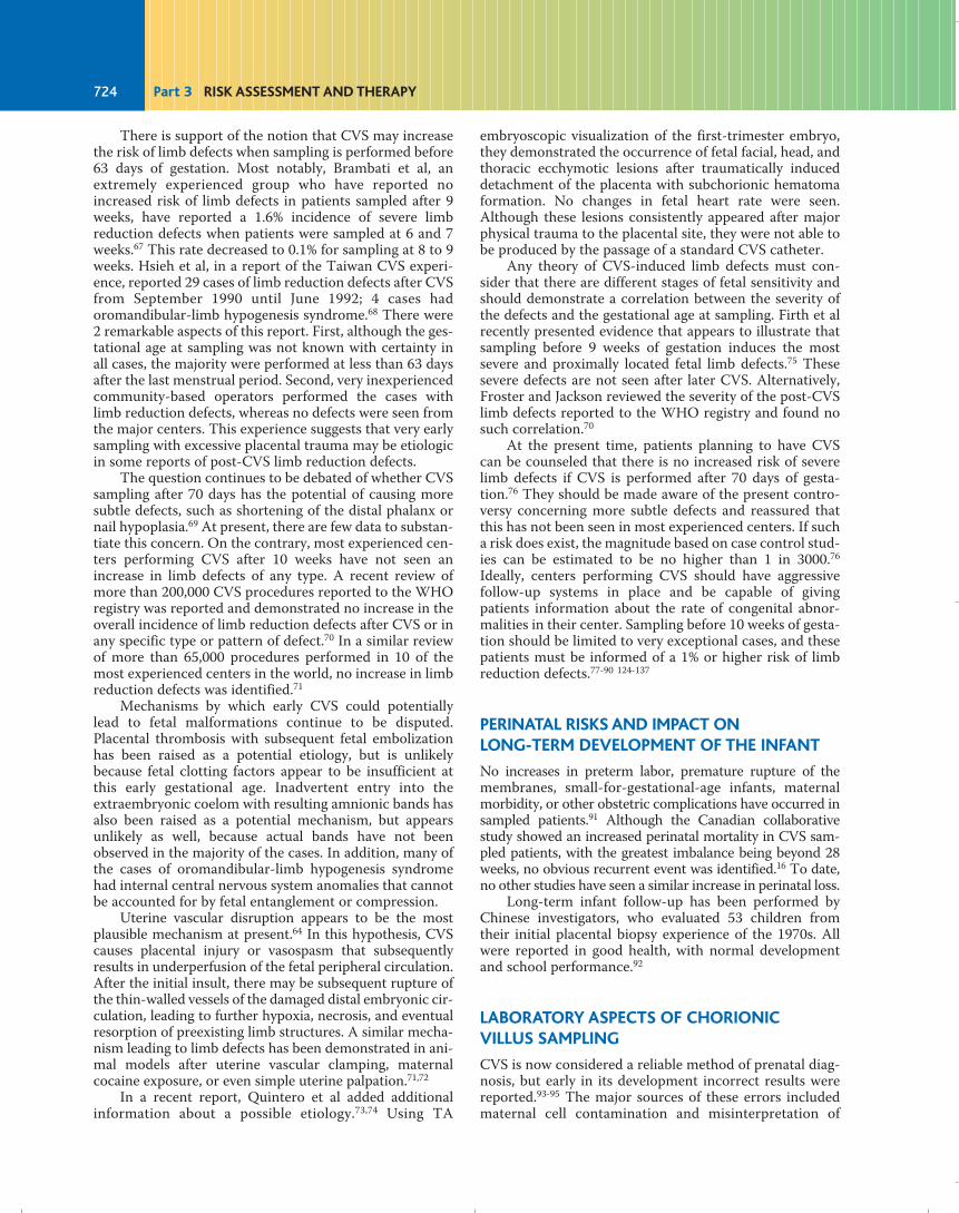
There is support of the notion that CVS may increasethe risk of limb defects when sampling is performed before63 days of gestation. Most notably, Brambati et al, anextremely experienced group who have reported noincreased risk of limb defects in patients sampled after 9weeks, have reported a 1.6% incidence of severe limbreduction defects when patients were sampled at 6 and 7weeks.67 This rate decreased to 0.1% for sampling at 8 to 9weeks. Hsieh et al, in a report of the Taiwan CVS experi-ence, reported 29 cases of limb reduction defects after CVSfrom September 1990 until June 1992; 4 cases hadoromandibular-limb hypogenesis syndrome.68 There were2 remarkable aspects of this report. First, although the ges-tational age at sampling was not known with certainty inall cases, the majority were performed at less than 63 daysafter the last menstrual period. Second, very inexperiencedcommunity-based operators performed the cases withlimb reduction defects, whereas no defects were seen fromthe major centers. This experience suggests that very earlysampling with excessive placental trauma may be etiologicin some reports of post-CVS limb reduction defects.
The question continues to be debated of whether CVSsampling after 70 days has the potential of causing moresubtle defects, such as shortening of the distal phalanx ornail hypoplasia.69 At present, there are few data to substan-tiate this concern. On the contrary, most experienced cen-ters performing CVS after 10 weeks have not seen anincrease in limb defects of any type. A recent review ofmore than 200,000 CVS procedures reported to the WHOregistry was reported and demonstrated no increase in theoverall incidence of limb reduction defects after CVS or inany specific type or pattern of defect.70 In a similar reviewof more than 65,000 procedures performed in 10 of themost experienced centers in the world, no increase in limbreduction defects was identified.71
Mechanisms by which early CVS could potentiallylead to fetal malformations continue to be disputed.Placental thrombosis with subsequent fetal embolizationhas been raised as a potential etiology, but is unlikelybecause fetal clotting factors appear to be insufficient atthis early gestational age. Inadvertent entry into theextraembryonic coelom with resulting amnionic bands hasalso been raised as a potential mechanism, but appearsunlikely as well, because actual bands have not beenobserved in the majority of the cases. In addition, many ofthe cases of oromandibular-limb hypogenesis syndromehad internal central nervous system anomalies that cannotbe accounted for by fetal entanglement or compression.
Uterine vascular disruption appears to be the mostplausible mechanism at present.64 In this hypothesis, CVScauses placental injury or vasospasm that subsequentlyresults in underperfusion of the fetal peripheral circulation.After the initial insult, there may be subsequent rupture ofthe thin-walled vessels of the damaged distal embryonic cir-culation, leading to further hypoxia, necrosis, and eventualresorption of preexisting limb structures. A similar mecha-nism leading to limb defects has been demonstrated in ani-mal models after uterine vascular clamping, maternalcocaine exposure, or even simple uterine palpation.71,72
In a recent report, Quintero et al added additionalinformation about a possible etiology.73,74 Using TA
embryoscopic visualization of the first-trimester embryo,they demonstrated the occurrence of fetal facial, head, andthoracic ecchymotic lesions after traumatically induceddetachment of the placenta with subchorionic hematomaformation. No changes in fetal heart rate were seen.Although these lesions consistently appeared after majorphysical trauma to the placental site, they were not able tobe produced by the passage of a standard CVS catheter.
Any theory of CVS-induced limb defects must con-sider that there are different stages of fetal sensitivity andshould demonstrate a correlation between the severity ofthe defects and the gestational age at sampling. Firth et alrecently presented evidence that appears to illustrate thatsampling before 9 weeks of gestation induces the mostsevere and proximally located fetal limb defects.75 Thesesevere defects are not seen after later CVS. Alternatively,Froster and Jackson reviewed the severity of the post-CVSlimb defects reported to the WHO registry and found nosuch correlation.70
At the present time, patients planning to have CVScan be counseled that there is no increased risk of severelimb defects if CVS is performed after 70 days of gesta-tion.76 They should be made aware of the present contro-versy concerning more subtle defects and reassured thatthis has not been seen in most experienced centers. If sucha risk does exist, the magnitude based on case control stud-ies can be estimated to be no higher than 1 in 3000.76
Ideally, centers performing CVS should have aggressivefollow-up systems in place and be capable of givingpatients information about the rate of congenital abnor-malities in their center. Sampling before 10 weeks of gesta-tion should be limited to very exceptional cases, and thesepatients must be informed of a 1% or higher risk of limbreduction defects.77-90 124-137
PERINATAL RISKS AND IMPACT ON LONG-TERM DEVELOPMENT OF THE INFANT
No increases in preterm labor, premature rupture of themembranes, small-for-gestational-age infants, maternalmorbidity, or other obstetric complications have occurred insampled patients.91 Although the Canadian collaborativestudy showed an increased perinatal mortality in CVS sam-pled patients, with the greatest imbalance being beyond 28weeks, no obvious recurrent event was identified.16 To date,no other studies have seen a similar increase in perinatal loss.
Long-term infant follow-up has been performed byChinese investigators, who evaluated 53 children fromtheir initial placental biopsy experience of the 1970s. Allwere reported in good health, with normal developmentand school performance.92
LABORATORY ASPECTS OF CHORIONIC VILLUS SAMPLING
CVS is now considered a reliable method of prenatal diag-nosis, but early in its development incorrect results werereported.93-95 The major sources of these errors includedmaternal cell contamination and misinterpretation of
724 Part 3 RISK ASSESSMENT AND THERAPY
Fleischer_Ch24.qxd 6/23/10 5:09 AM Page 724

mosaicism confined to the placenta. Today, genetic evalu-ation of chorionic villi provides a high degree of successand accuracy, in particular with regard to the diagnosis ofcommon trisomies.96,97 In 1990, the US collaborative studyreported a 99.7% rate of successful cytogenetic diagnosis,with 1.1% of the patients requiring a second diagnostictest, such as amniocentesis or fetal blood analysis to fur-ther interpret the results.96 In most cases, the additionaltesting was required to delineate the clinical significance ofmosaic or other ambiguous results (76%), and laboratoryfailure (21%) and maternal cell contamination (3%) alsorequired follow-up testing. Continued experience hasalmost eliminated maternal cell contamination as a sourceof clinical errors. In addition, we now have a better under-standing of the biology of the placenta so that confined pla-cental mosaicism no longer leads to incorrect diagnosis,but provides us with information predictive of pregnancyoutcome and can serve as a clue to the presence of uni-parental disomy. Therefore, an understanding of villusmorphology and CVS laboratory techniques is required toprovide correct clinical interpretation.
Chorionic villi have 3 major components: (1) an outerlayer of hormonally active and invasive syncytiotro-phoblast, (2) a middle layer of cytotrophoblast from whichsyncytiotrophoblast is derived, and (3) an inner mesoder-mal core containing blood, capillaries for oxygen, andnutrient exchange (Figure 24-8B). After collection, the villiare cleaned of any adherent decidua and then exposed totrypsin to digest and separate the cytotrophoblast from theunderlying mesodermal core. The cytotrophoblast has ahigh mitotic index, with many spontaneous mitoses avail-able for immediate chromosomal analysis. The liquid sus-pension containing the cytotrophblast is either droppedimmediately onto a slide for analysis or may undergo a
short incubation.98-100 This “direct” chromosomal prepara-tion can provide preliminary results within 2 to 3 hours.However, most laboratories now use overnight incubationto improve karyotype quality and thus report results within2 to 4 days (Figure 24-9). The remaining villus core is placedin tissue culture and is typically ready for harvest and chro-mosome analysis within 1 week.101 The direct method hasthe advantage of providing a rapid result and minimizingthe decidual contamination, whereas tissue culture is betterfor interpreting discrepancies between the cytotrophoblastand the actual fetal state. Ideally, both the direct and culturemethods should be used because they each evaluate slightlydifferent tissue sources. Abnormalities in either may haveclinical implications. However, the direct preparation islabor intensive, adds additional cost, and is not routinelyavailable in some laboratories.
MATERNAL CELL CONTAMINATION
Chorionic villus samples typically contain a mixture of pla-cental villi and maternally derived decidua. Although spec-imens are thoroughly washed and inspected under amicroscope after collection, some maternal cells mayremain and grow in the culture. As a result, 2 cell lines, onefetal and the other maternal, may be identified. In othercases, the maternal cell line may completely overgrow theculture, thereby leading to diagnostic errors includingincorrect sex determination8,102-104 and potentially to false-negative diagnoses, although there are no publishedreports of the latter. Direct preparations of chorionic villiare generally thought to prevent maternal cell contamina-tion,100, 103 whereas long-term culture has a contaminationrate ranging from 1.8% to 4%.104 Because, in contrast tocytotrophoblast, maternal decidua has a low mitotic index,
725Chapter 24 Chorionic Villus Sampling
Cytotrophoblasticcell column
Cytotrophoblast
Mesenchymal core
Syncytiotrophoblast
Villus tissuetrypsin
Mesenchymalcore
Cytotrophoblastsuspension
Villusculture
Directpreparation
A B
Figure 24-9. A: Diagram of normal villus architecture. B: Diagram outlining the laboratory technique for chorionicvillus sampling direct chromosomal preparation and villus culture.
Fleischer_Ch24.qxd 6/23/10 5:09 AM Page 725

it is highly desirable for laboratories to offer a direct chro-mosomal preparation and a long-term culture on all sam-ples of chorionic villus. Even in culture, the contaminatingcells are easily identified as maternal and should not lead toclinical errors. Interestingly, for reasons still uncertain,maternal cell contamination occurs more frequently inspecimens retrieved by the TC route.104
Contamination of samples with significant amounts ofmaternal decidual tissue is almost always due to small sam-ple size, making selection of appropriate tissue difficult. Inexperienced centers in which adequate quantities of villiare available, this problem has disappeared. Choosing onlywhole, clearly typical villus material and discarding anyatypical fragments, small pieces, or fragments with adher-ent decidua will avoid confusion.105 Therefore, if the initialaspiration is small, a second pass should be performedrather than risk inaccurate results. When proper care istaken and good cooperation and communication existsbetween the sampler and the laboratory, even smallamounts of contaminating maternal tissue can be absent.
Fluorescent in situ hybridization (FISH) for commonchromosomal abnormalities can be helpful in reaching arapid diagnosis (within hours) without the concern formaternal contamination.
CONFINED PLACENTAL MOSAICISM
The second major source of potential diagnostic errorassociated with CVS is mosaicism confined to the placenta.Although the fetus and placenta have a common ancestry,chorionic villus tissue will not always reflect fetal geno-type.96,106 Although there was concern that this mightinvalidate CVS as a prenatal diagnostic tool, subsequentinvestigations have led to a clearer understanding of villusbiology so that accurate clinical interpretation is now pos-sible. This understanding has also revealed new informa-tion about the etiology of pregnancy loss, discovered a newcause of intrauterine growth retardation, and clarified thebasic mechanism of uniparental disomy.
Discrepancies between the cytogenetics of the placentaand fetus occur because the cells contributing to the chori-onic villi become separate and distinct from those formingthe embryo in early development. Specifically, at approxi-mately the 32- to 64-cell stage, only 3 to 4 become compart-mentalized into the inner cell mass (ICM) to form theembryo, and the remainder become precursors of theextraembryonic tissues.107 Mosaicism can then occurthrough 2 possible mechanisms.108 An initial meiotic errorin one of the gametes can lead to a trisomic conceptus thatnormally would spontaneously abort. However, if one of theearly aneuploid precursor cells loses one of the chromo-somes contributing to the trisomic set during subsequentmitotic divisions, the embryo can be “rescued” by reductionof a portion of its cells to disomy. This will result in a mosaicmorula, with the percentage of normal cells dependent onthe cell division at which rescue occurred. More abnormalcells will be present when correction is delayed to the sec-ond or a subsequent cell division. Because the majority ofcells in the morula proceed to the trophoblast cell lineage(processed by the direct preparation), it is highly probablethat that lineage will continue to contain a significant number
of trisomic cells. Alternatively, because only a small numberof cells are incorporated into the ICM, involvement of thefetus will depend on the chance distribution of the aneu-ploid progenitor cells. Involvement of the mesenchymalcore of the villus, which also evolves from the ICM, is simi-larly dependent on this random cell distribution.Noninvolvement of the fetal cell lineage will produce con-fined placental mosaicism (CPM) in which the trophoblastand perhaps the extraembryonic mesoderm will have aneu-ploid cells, but the fetus will be euploid.
Alternatively, mitotic postzygotic errors can producemosaicism, with the distribution and percentage of aneu-ploid cells in the morula or blastocyst dependent on the tim-ing of nondisjunction. If mitotic errors occur early in thedevelopment of the morula, they may segregate to the ICMand have the same potential to produce an affected fetus asdo meoitic errors. Mitotic errors occurring after primary celldifferentiation and compartmentalization has been com-pleted lead to cytogenetic abnormalities in only one lineage.
Meiotic rescue can lead to uniparental disomy (UPD).This occurs because the original trisomic cell contained 2chromosomes from one parent and 1 from the other. Afterrescue, there is a theoretical 1 in 3 chance that the result-ing pair of chromosomes came from the same parent,which is called uniparental disomy. UPD may have clinicalconsequences if the chromosomes involved carryimprinted genes in which expression is based on the parentof origin. For example, Prader-Willi syndrome may resultfrom uniparental maternal disomy for chromosome 15.Therefore, a CVS diagnosis of confined placentalmosaicism for trisomy 15 may be the initial clue that UPDcould be present and lead to an affected child.109,110
Because of this, all cases in which CVS reveals trisomy 15(either complete or mosaic) should be evaluated for UPDby subsequent amniotic fluid analysis. In addition to chro-mosome 15, chromosomes 7, 11, 14, and 22 are felt to beimprinted and require follow-up.111
Recently, there has been evidence that confined pla-cental mosaicism (unassociated with UPD) can alter pla-cental function and lead to fetal growth failure or perinataldeath.108,112-117 The exact mechanism by which abnormalcells within the placenta alter fetal growth or lead to fetaldeath is unknown. However, the effect may be limited tospecific chromosomes. For example, CPM for chromo-some 16 leads to severe intrauterine growth restriction,prematurity, or perinatal death, with fewer than 30% ofpregnancies resulting in normal full-term infants appropri-ate for gestational age.118-125
CVS mosaic results require diligent follow-up byamniocentesis or fetal sampling to determine their clinicalsignificance because, in most cases, if the mosaic resultsare confined to the placenta, fetal development will be nor-mal. However, if the mosaic cell line also involves the fetus,there may be significant phenotypic consequences.Mosaicism occurs in about 1% of all CVS samples97, 104
,121,122 but is confirmed in the fetus in only 10% to 40% ofthese cases. The probability of fetal involvement appears tobe related to the tissue source in which the aneuploid cellswere detected and the specific chromosome involved.110
Mesenchymal core culture results are more likely thandirect preparation to reflect a true fetal mosaicism.
726 Part 3 RISK ASSESSMENT AND THERAPY
Fleischer_Ch24.qxd 6/23/10 5:09 AM Page 726

In a recent review, Phillips et al demonstrated thatautosomal mosaicism involving common trisomies (ie, 21,18, and 13) was confirmed in the fetus in 19% of cases,whereas uncommon trisomies involved the fetus in only3%.123 When sex chromosome mosaicism was found in theplacenta, the abnormal cell line was confirmed in the fetusin 16% of cases. When a nonfamilial marker chromosomewas involved, it was confirmed in the fetus in more thanone-fourth of cases, whereas mosaic polyploidy was con-firmed in only 1 of 28 cases. Chromosomal structuralabnormalities were confirmed in 8.6% of cases.
When placental mosaicism is discovered, amniocente-sis is frequently performed to elucidate the extent of fetalinvolvement. When mosaicism is limited to the directpreparation only, amniocentesis appears to correlate per-fectly with fetal genotype.123 However, when a mosaicism isobserved in tissue culture, both false-positive and false-negative amniocentesis results occur. In these cases, amnio-centesis will predict the true fetal karyotype in approxi-mately 94% of cases.123 Most importantly, these discrepan-cies may involve the common autosomal trisomies. Therehave been 3 cases reported of mosaic trisomy 21 on villusculture and a normal amniotic fluid analysis, followed by afetus or newborn with mosaic aneuploidy.96
At present, the following clinical recommendationsmay be used to assist in the evaluation of CVS mosaicism.Analysis of CVS samples should, if possible, include bothdirect preparation and tissue culture. Although the directpreparation is less likely to be representative of the fetus,its use will minimize the likelihood of maternal cell con-tamination, and if culture fails, a nonmosaic normal directpreparation result can be considered conclusive, althoughrare cases of false-negative results for trisomies 21 and 18have been reported.124-128 If mosaicism is found on eitherculture or direct preparation, follow-up amniocentesisshould be offered. Under no circumstances should a deci-sion to terminate a pregnancy be based entirely on a CVSmosaic result. For CVS mosaicism involving sex chromo-some abnormalities, polyploidy, marker chromosomes,structural rearrangements, and uncommon trisomies, thepatient can be reassured if amniocentesis results areeuploid and detailed ultrasonographic examination is nor-mal. However, no guarantees should be made and, asdescribed above, in certain cases testing for UPD will beindicated. If common trisomies 21, 18, and 13 are involved,amniocentesis should be offered, but the patient must beadvised of the possibilities of a false-negative result.Follow-up may include detailed ultrasonography, fetalblood sampling, or fetal skin biopsy. At present, the predic-tive accuracy of these additional tests is uncertain.
BIOCHEMICAL AND DNA PROCEDURES
Most biochemical and molecular diagnoses that can bemade from amnionic fluid or cultured amniocytes can alsobe made from chorionic villi. In many cases, the results willbe available more rapidly and more efficiently by usingvilli, because sufficient enzyme or DNA is present in villussamples to allow direct analysis rather than wait for tissueculture. For example, the analysis of Tay-Sachs disease canbe performed in less than 30 minutes using fresh villi.129
A discussion of individual biochemical or moleculardiagnoses is beyond the scope of this chapter and isimpractical because techniques are changing so rapidly. Aregistry of diagnoses performed by CVS is kept andupdated through the WHO by Dr Hans Galjaard inRotterdam, The Netherlands, and a published summary ofthe early worldwide experience is available.130
It cannot be assumed that biochemical or molecularresults from villus tissue will always be a true reflection ofthe fetal state. Recently, misdiagnosis of the peroxisomaldisorder, X-linked adrenoleukodystrophy, from culturedvillus cells has been reported.131 In addition, tests requiringdetermination of DNA methylation status, such as that forfragile X,132 are also not always reliable in villus tissue. Thisdoes not, however, preclude CVS from making these pre-natal diagnoses because other molecular approaches canbe used. It does emphasize that all tests on villus tissuemust be validated by testing sufficient numbers of affectedand unaffected pregnancies before being used clinically.Because of the rarity and unique aspects of most biochem-ical and molecular disorders, specific diagnoses are usuallyperformed by only a few laboratories. Before performing aCVS, the clinician should contact the center analyzing thetissue so that the details of testing can be discussed.
CHORIONIC VILLUS SAMPLING IN MULTIPLEGESTATIONS
Chorionic villus sampling is a safe and effective approach toexamining twins. Not only does it provide results early inpregnancy, but, if discordancy is discovered, the medicaland psychological difficulties encountered with selectivetermination can be minimized. However, it can be techni-cally more demanding because it requires an experiencedoperator and sonographer. The ideal time to perform a twinCVS is similar to that for singletons. Ultrasound initiallyidentifies placental locations, determines chorionicity, andconfirms fetal sizes and viability. Sampling of each sac isindependently performed by either a TC or TA approach,with separate passes of a new sampling instrument for eachattempt. Because no unique marker is available to ensurethat the samples have been retrieved from distinct placen-tas, it is imperative that insertion of the instrument intoeach frondosum is certain. Longitudinal and transversescanning planes should be used to ensure proper location.If any doubt exists, a repeat procedure is required, but withincreased experience, the need for repeat procedure israre.133
Contamination of one sample with villi from the othersac is possible and occurs most commonly when retrievalis performed near the dividing membrane, or if a needle orcatheter is dragged through one frondosum while sam-pling another. When the chorions appear fused, samplingnear the cord insertion sites, with avoidance of the area ofconfluence of the 2 placentas, should prevent contamina-tion and ensure sampling of each fetus. A combination ofTC and TA sampling can minimize co-twin contaminationby ensuring unique sampling paths. For example, if bothchorion frondosa are situated along the anterior uterinewall, the lower one can be sampled transcervically and the
727Chapter 24 Chorionic Villus Sampling
Fleischer_Ch24.qxd 6/23/10 5:09 AM Page 727

upper transabdominally without contaminating eithersample. Despite meticulous sampling techniques, co-twincontamination occurs in up to 4% of cases120 but is rarelyof clinical concern. If the cytogenetic laboratory is aware ofthe presence of a multiple gestation, the presence of bothnormal and aneuploid cells in the sample will be correctlyinterpreted. The possibility of contamination is of greaterconsequence if sampling is being performed for biochemi-cal analysis, where a small amount of contaminating tissuecould lead to an incorrect diagnosis. For this reason,instead of pooling a sample, we recommend analyzingindividual villi when biochemical studies are to be per-formed. An experienced sampler and an aware and knowl-edgeable laboratory are extremely important in such cases.
The need to map the placental location clearly andaccurately is mandatory, because the risk of discordantresults is higher early in the pregnancy and selective termi-nation may be required. The relative positions of the pla-centas will remain stable over the time frame that resultsare obtained, so that identification of the affected fetus canusually be determined even 2 or 3 weeks after the proce-dure. However, if there is uncertainty, a repeat CVS withdirect villus analysis can confirm the position immediatelybefore the termination.
Procedure-related loss rates after CVS sampling oftwins are well studied. In experienced centers, noincreased procedure-related loss risks are seen comparedwith second trimester amniocentesis.133,134 Table 24-4presents overall pregnancy loss rates to 28 weeks of 1.6% to2.8% in sampled twins. When compared with a contempo-raneously sampled group of patients choosing either CVSor second trimester amniocentesis, we found no differencein the overall risk of pregnancy loss (2.9% amniocentesis vs3.2% CVS). There was, however, a slightly increased risk oflosing one fetus in the group sampled by amniocentesis.133
The choice of the appropriate technique for samplingtwins depends on a number of factors including locallyavailable skill and expertise. In centers in which amniocen-tesis and CVS are both available, CVS may be the preferredapproach because it provides results 1 month sooner, thusproviding earlier reassurance. When discordant results areencountered, complications and loss rates are decreased
when selective termination is performed before 16 weeksof gestation.135
CHORIONIC VILLUS SAMPLING ANDMULTIFETAL PREGNANCY REDUCTION
Multifetal pregnancy reduction (MFPR) to improve perina-tal outcome is an unfortunate but accepted part of repro-ductive medicine. As with selective termination, MFPR ismost safely performed in the first-trimester.136 Therefore, inhigh-order gestations at increased risk for a genetic abnor-mality, CVS before a reduction can avoid the potential needfor a later selective termination. The CVS can be performedbetween 10 and 11.5 weeks of gestation, and a rapid kary-otype can be available within 24 to 48 hours, after which theMFPR is performed. Villus mesenchymal core tissue cul-ture is also performed, but results are usually not availablefor 7 to 10 days. Because of the small increased risk of con-fined placental mosaicism with the direct preparation,awaiting culture results when time allows is suggested. Thepositions of the sampled fetuses will be the same when thepatient returns for the MFPR. In most cases, only the 2fetuses most likely to remain after the MFPR are sampled.If an abnormality is identified, an additional fetus can besampled at the time of the MFPR and the patient can returnif this is also abnormal. Of 745 MFPR reductions performedat our center, 254 had an initial CVS. Abnormal chromoso-mal results were present in approximately 2.5% of pregnan-cies, and the abnormal fetus was terminated as part of thereduction procedure. The pregnancy loss rate to 24 weeksof gestation of those having a preceding CVS was 5.5% ver-sus 5.6% in those having only MFPR. This encouraging out-come is similar to that described by Brambati et al137 inwhich the cohort of patients undergoing CVS before MFPRdemonstrated no increased risk of pregnancy loss, prema-turity, or small-for-gestational-age infants.
SUMMARY
Chorionic villus sampling is a safe technique for first-trimester prenatal diagnosis of genetic disorders. Real-timesonography and technologic advances of sampling instru-ments have been critical in establishing a safe technique forretrieving villus tissue for genetic analysis. Clinical trialssuggest that CVS carries a low risk of pregnancy loss, whichis comparable to that of second-trimester amniocentesis.An understanding of the laboratory techniques and humanembryology is essential in avoiding diagnostic errors relatedto confined placental mosaicism.
To maximize outcome, CVS should be performed byan experienced team of physicians, ultrasonographers, andgenetic laboratory technicians. Before initiating a CVSprogram, operators should have considerable experiencein the placement of the catheter, which can be achievedeither in a formal training program with observation of 50procedures followed by close hands-on supervision ofanother 100 cases,8 or by supervised practice on pregnan-cies undergoing subsequent abortion. In addition, it isadvisable to limit the number of ultrasonographers
728 Part 3 RISK ASSESSMENT AND THERAPY
Success Pregnancy Lossn Rate Rate to 28 Weeks
CVS
Wapner et al.115∗ 440† 100% 2.8%
Pergament et al.116 128 99.2% 2.4%
Brambati et al.119 66 100% 1.6%
Amniocentesis
Wapner et al.115∗ 73 100 2.9%
∗Contemporaneously collected comparison of CVS with amniocentesis in asingle center. †Additional cases added since original publication.
SAFETY OF CHORIONIC VILLUS SAMPLING (CVS) WITH TWINS COMPARED WITH AMNIOCENTESIS
Table 24-4
Fleischer_Ch24.qxd 6/23/10 5:09 AM Page 728

assigned to assist in this procedure, because sampling suc-cess is equally dependent on skillful ultrasound guidance.Similar to the physician retrieving the villi, the guidingsonographer should be knowledgeable in the didactics ofCVS sampling and should obtain adequate hands-on train-ing before beginning work in this area. This can beachieved in a formal training program or by visiting cen-ters performing this procedure.8 In this setting, CVS willcontinue to be an important, reliable, and safe contributorto prenatal genetic diagnosis.
12. Hahnemann N. Early prenatal diagnosis: a study of biopsy tech-niques and cell culturing from extraembryonic membranes. ClinGenet 1974;6:294-306.
13. Kazy Z, Rozovsky I, Bakhaey V. Chorion biopsy in early pregnancy:a method of early prenatal diagnosis for inherited disorders. PrenatDiagn 1982;2:39-45.
14. Old J. First trimester fetal diagnosis for haemoglobinopathies: threecases. Lancet 1982;2:1413-6.
15. Brambati B, Simoni G. Diagnosis of fetal trisomy 21 in firsttrimester. Lancet 1983;1:586.
16. Canadian Collaborative CVS-Amniocentesis Clinical Trial Group.Multicentre randomized clinical trial of chorion villus sampling andamniocentesis. Lancet 1989;i:1.
17. Rhoads GG, Jackson IG, Schlesselman SE, et al. The safety and effi-cacy of chorionic villus sampling for early prenatal diagnosis of cyto-genetic abnormalities. N Engl J Med 1989;320:609-9.
18. Committee on Genetics. ACOG Committee Opin 1995;160.19. Jackson LG, Zachary JM, Fowler SE, et al. Randomized comparison
of transcervical and transabdominal chorionic villus sampling. NEngl J Med 1992;327:594-8.
20. Silver RK, MacGregor SN, Sholl JS, et al. Initiating a chorionic vil-lus sampling program. Relying on placental location as the pri-mary determinant of the sampling route. J Reprod Med 1990;35:964-8.
21. Brambati B, Oldrini A, Lanzani A. Transabdominal and trans-cervical chorionic villus sampling: efficiency and risk evaluation of2,411 cases. Am J Med Genet 1990;35: 160-4.
22. Liu DT, Agbaje R, Preston C, Savage J. Intraplacental sonolucentspaces: Incidences and relevance to chorionic villus sampling.Prenat Diagn 1991;11:805.
23. Brambati B, Oldrini A, Lanzani A. Transabdominal chorionic villussampling: a freehand ultrasound-guided technique. Am J ObstetGynecol 1987;157:134.
24. Smidt-Jensen S, Hahnemann N. Transabdominal chorionic villussampling for fetal genetic diagnosis. Technical and obstetric evalua-tion of 100 cases. Prenat Diagn 1988;8:7.
25. Silver RK, MacGregor SN, Sholl JS, Hobart ED, Waldee JK. An eval-uation of the chorionic villus sampling learning curve. Am J ObstetGynecol 1990;163:917-22.
26. Jackson LG, Wapner RJ. Risks of chorionic villus sampling. ClinObstet Gynecol 1987;1:513.
27. Brambati B, Oldrini A, Ferrazzi E, et al. Chorionic villus sampling: ananalysis of the obstetric experience of 1000 cases. Prenat Diagn1987;7:157-69.
28. Brambati B, Matarrelli M, Varotto F. Septic complications afterchorionic villus sampling. Lancet 1987;i(8543):1212a.
29. Wass D, Bennett MJ. Infection and chorionic villus sampling. Lancet1985;ii:338.
30. Scialli AR, Neugebauer DL, Fabro SE. Microbiology of the endcervixin patients undergoing chorionic villus sampling. In: Fracearo M,Simoni G, Brambati B, eds. First Trimester Fetal Diagnosis. Berlin:Springer-Verlag, 1985:69-73.
31. Brambati B, Varotti F. Infection and chorionic villus sampling.Lancet 1985;ii:609.
32. Hogge WA, Schonberg SA, Golbus MS. Chorionic villus sampling:experience of the first 1000 cases. Am J Obstet Gynecol 1986;154:1249.
33. Gilmore DH, McNay MB. Spontaneous fetal loss rate in early preg-nancy. Lancet 1985;i:107.
34. Wilson RD, Kendrick V, Witmann BK. Risks of spontaneous abor-tion in ultrasonographically normal pregnancies. Lancet 1984;ii:920.
35. Barela A, Kleinman GE, Golditch IM, et al. Septic shock with renalfailure after chorionic villus sampling. Am J Obstet Gynecol1986;154:1120.
36. Blakemore KJ, Mahoney MJ, Hobbins JC. Infection and chorionicvillus sampling. Lancet 1985;2:339.
37. Cheng EY, Luth DA, Hickok D, et al. Transcervical chorionic villussampling and midtrimester oligohydramnios. Am J Obstet Gynecol1991;165:1063.
38. Blakemore KJ, Baumgarten A, Schoenfeld-Dimaio M, et al. Rise inmaternal serum alpha-fetoprotein concentration after chorionic vil-lus sampling. Lancet 1985;ii:339.
39. Bramabati B, Guercilena S, Bonacchi I, et al. Fetomaternal transfu-sion after chorionic villus sampling: clinical implications. HumReprod 1986;1:37.
729Chapter 24 Chorionic Villus Sampling
1. Chorionic villus sampling has a low complication ratecomparable to mid-trimester amniocentesis in experi-enced hands. The learning curve is more difficult withCVS.
2. CVS is the preferable method of fetal aneuploidy test-ing in the first trimester.
3. CVS is generally performed after 9 completed weeksof gestation. In this setting, there does not seem to bean increased risk of limb reduction defects.
4. CVS may be performed either transabdominally ortranscervically.
5. Caution should be exercised in the interpretation of amosaic of chromosomal findings from CVS.
6. The complications of CVS include miscarriage, bleed-ing, rupture of membranes, and infection.
KEY POINTS
REFERENCES1. American College of Obsetetricians and Gynecologists. Invasive
prenatal testing for aneuploidy. ACOG Practice Bulletin 88,Washington, DC: Dec 2007.
2. Grimes DA, Schultz KF. Morbity and mortality from secondtrimester abortion. J Repro Med 1985;30:505-14.
3. Sundberg K, Bang J, Smidt-Jensen S, et al. Randomized study of riskof fetal loss related to early amniocentesis versus chorionic villussampling. Lancet 1997;350:697-703.
4. CEMAT Group. Randomized trial to assess safety and fetal out-comes of early and midtrimester amniocentesis. Lancet 1998;351:1435.
5. Evans MI, Andriole S. Chorionic villus sampling and amniocentesisin 2008. Curr Opin Obstet Gynecol 2008;20(1):164-8.
6. Caughey AB, Hopkins LM, Norton ME. Chorionic villus samplingcompared with amniocentesis and the difference in the rate of preg-nancy loss. Obstet Gynecol 2006;18:612-6.
7. Spencer K, Souter V, Tul N, et al. A screening program for trisomy21 at 10-14 weeks using fetal nuchal translucency, maternal serumfree B-human chorionic gonadotrophin and pregnancy-assoicartedplasma protein A. Ultrasound Obstet Gynecol 1991;13:231-7.
8. Boehm FH, Salyer SL, Dev VG, et al. Chorionic villus sampling: qual-ity control-m-A continuous improvement model. Am J ObstetGynecol 1993;168:1766-77.
9. Department of Obstetrics and Gynecology. Tietung Hospital ofAnshan Iron and Steel Co. Anshan, China. Fetal sex prediction bysex chromatin of chorionic villi cells during early pregnancy.Chinese Med J 1975;1:117.
10. Hahnemann N, Mohr J. Genetic diagnosis in the embryo by meansof biopsy from extraembryonic membranes. Bull Eur Soc HumGenet 1968;2:23-9.
11. Kullander S, Sandahl B. Fetal chromosome analysis after transcervi-cal placental biopsies during early pregnancy. Acta Obstet GynecolScand 1973;52:355-9.
Fleischer_Ch24.qxd 6/23/10 5:09 AM Page 729

40. Shulman LP, Meyers CM, Simpson JL, et al. Fetomaternal transfu-sion depends on amount of chorionic villi aspirated but not onmethod of chorionic villus sampling. Am J Obstet Gynecol1990;162:1185.
41. Smidt-Jensen S, Philip J, Zachary J, et al. Implications of maternalserum alpha-fetoprotein elevation caused by transabdominal andtranscervical CVS. Prenat Diagn 1994;14:35-46.
42. Zipursky A, Israels LG. The pathogenesis and prevention of Rhimmunization. Can Med Assoc J 1967;97:1245-57.
43. Moise KJ, Carpenter RJ. Increased severity of fetal hemolytic diseasewith known Rhesus alloimmunization after first trimester transcer-vical chorionic villus biopsy. Fetal Diagn Ther 1990;5:76.
44. Clark BA, Bissonnette J, Olson SB, et al. Pregnancy loss in a smallchorionic villus sampling series. Am J Obstet Gynecol 1989;161:301.
45. Green JE, Dorfman A, Jones SL, et al. Chorionic villus sampling:experience with an initial 940 cases. Obstet Gynecol 1988;71:208.
46. Gustavii B, Claesson V, Kristoffersson U, et al. Risk of miscarriageafter chorionic biopsy is probably not higher than after amniocente-sis. Lakartidningen 1989;86:4221.
47. Jahoda MGJ, Pijpers L, Reuss A, et al. Evaluation of transcervicalchorionic villus sampling with a completed follow-up of 1550 con-secutive pregnancies. Prenat Diagn 1989;9:621.
48. Young SR, Shipley CF, Wade RV, et al. Single-center comparison ofresults of 1000 prenatal diagnoses with chorionic villus samplingand 1000 diagnoses with amniocentesis. Am J Obstet Gynecol1991;165:255.
49. Hogge WA, Schonberg SA, Golbus MS. Chorionic villus sampling:experience of the first 1000 cases. Am J Obstet Gynecol 1986;154:1249-52.
50. Jackson LG. CVS Newslett 1988;25-7.51. Wade RV, Young SR. Analysis of fetal loss after transcervical chori-
onic villus sampling: a review of 719 patients. Am J Obstet Gynecol1989;161:513-9.
52. Warburton D, Fraser FC. Spontaneous abortion risks in man: datafrom reproductive histories collected in a medical genetics unit.Hum Genet 1964;16:1-25.
53. Simpson JL, Mills ML, Holmes LB, et al. Low fetal loss rates afterultrasound-proved viability in early pregnancy. JAMA 1987;258:2555-7.
54. Christiaens GC, Stoutenbeeck P. Spontaneous abortion in provenintact pregnancies. Lancet 1984;2:571-2.
55. Wilson RD, Kendrick V, Wirtman BP, et al. Spontaneous abortionand pregnancy outcome after normal first-trimester ultrasoundexamination. Obstet Gynecol 1986;67:352-5.
56. Saura R, Gauthier B, Taine L, et al. Operator experiences and fetalloss rate in transabdominal CVS. Prenat Diagn 1994;14:70.
57. Wapner RJ, Barr MA, Heeger S, et al. Chorionic villus sampling: a10-year, over 13,000 consecutive case experience (abstract).Presented at the First Annual Meeting of the American College ofMedical Genetics, Orlando, Florida, March 1994.
58. Nicolaides K, Brizot M de L, Patel F, Snijders R. Comparison ofchorionic villus sampling and amniocentesis for fetal karyotyping at10-13 weeks’ gestation. Lancet 1994;344(8920):435.
59. Brambati B, Lanzani A, Tului L. Transabdominal and transcervicalchorionic villus sampling: efficacy and risk evaluation of 2411 cases.Am J Hum Genet 1990;35:160.
60. Brambati B, Terzian E, Tognoni G. Randomized clinical trial oftransabdominal versus transcervical chorionic villus sampling meth-ods. Prenat Diagn 1991;11:285.
61. Smidt-Jensen S, Permin M, Philip J. Sampling success and risk bytransabdominal chorionic villus sampling, transcervical chorionicvillus sampling and amniocentesis: a randomized study. UltrasoundObstet Gynecol 1991;1:86.
62. Chueh JT, Goldberg JD, Wohlfered MM, Golbus MS. Comparison oftranscervical and transabdominal chorionic villus sampling lossrates in nine thousand cases from a single center. Am J ObstetGynecol 1995;173:1277.
63. Firth HV, Boyd P, Chamberlain P, et al. Severe limb abnormalitiesafter chorion villus sampling at 56-66 days gestation. Lancet1991;337:726.
64. Hoyme F, Jones KL, Van Allen MI, et al. Vascular pathogenesis oftransverse limb reduction defects. J Pediatr 1982;101:839.
65. Foster-Iskenius U, Baird P. Limb reduction defects in over 1,000,000consecutive live births. Teratology 1989;39:127.
66. Mastroiacovo P, Botto LD, Cavalcanti DP. Limb anomalies followingchorionic villus sampling: a registry based case control study. Am JMed Genet 1992;44:856-63.
67. Brambati B, Simoni G, Traui M. Genetic diagnosis by chorionic vil-lus sampling before 8 gestational weeks: efficiency, reliability, andrisks on 317 completed pregnancies. Prenat Diagn 1992;12:784-99.
68. Hsieh FJ, Shyu MK, Sheu BC, et al. Limb defects after chorionic vil-lus sampling. Obstet Gynecol 1995;85:84.
69. Burton BK, Schultz CJ, Burd LI. Spectrum of limb disruption defectsassociated with chorionic villus sampling. Pediatrics 1993;91:989-93.
70. Froster UG, Jackson L. Limb defects and chorionic villus sampling:results from an international registry. 1992-1994. Lancet 1996;347(9000):489.
71. Brent RL. Relationship between uterine vascular clamping, vasculardisruption syndrome and cocaine teratology. Teratology 1990;41:757.
72. Webster W, Brown-Woodman T. Cocaine as a cause of congenitalmalformations of vascular origin: experimental evidence in the rat.Teratology 1990;41:689.
73. Quintero R, Romero R, Mahoney M, et al. Fetal haemorrhagiclesions after chorionic villus sampling. Lancet 1992;339:193.
74. Quintero RA, Romero R, Mahoney MJ, et al. Embryoscopic demon-stration of hemoagic lesions on the human embryo after placentaltauma. Am J Obstet Gynecol 1993;168:756-9.
75. Firth HV, Boyd PA, Chamberlain PF, et al. Analysis of limb reduc-tion defects in babies exposed to chorionic villus sampling. Lancet1994;343(8905):1069.
76. Olney RS, Khoury MJ, Alo CJ, et al. Increased risk for transverse dig-ital deficiency after chorionic villus sampling: results of the UnitedStates Multistate Case-Control Study. 1988-1992. Teratology1995;5(1):20-9.
77. Williams J, Medearis AL, Bear MD, et al. Chorionic villus samplingis associated with normal fetal growth. Am J Obstet Gynecol 1987;157:708.
78. Jahoda MGJ, Brandenberg H, Cohen-Overbeek TE, et al. Terminaltransverse limb defects and early chorionic villus sampling: evalua-tion of 4,300 cases with completed follow-up. Am J Med Genet1993;46:483.
79. Halliday J, Lumley J, Sheffield LJ, et al. Limb deficiencies, chorionvillus sampling, and advanced maternal age. Am J Med Genet1993;47:1096.
80. Schloo R, Miney P, Holzgreve W, et al. Distal limb deficiency follow-ing chorionic villus sampling? Am J Med Genet 1992;42:404.
81. Monni G, Ibba RM, Lai R, et al. Limb-reduction defects and chorionvillus sampling. Lancet 1991;337:1091.
82. Blakemore K, Filkins K, Luthy DA, et al. Cook Obstetrics andGynecology Catheter Multicenter Chorionic Villus Sampling Trial:Comparison of birth defects with expected rates. Am J ObstetGynecol 1993;169:1022.
83. Silver RK, Macgregor SN, Muhlbach LH, et al. Congenital malfor-mations subsequent to chorionic villus sampling: outcome analysisof 1048 consecutive procedures. Prenat Diagn 1994;14:421.
84. Mahoney MJ for the USNICHD Collaborative CVS Study Group.Limb abnormalities and chorionic villus sampling. Lancet1991;37:1422.
85. Jackson LG, Wapner RJ, Brambati B. Limb abnormalities and chori-onic villus sampling. Lancet 1991;337:1423.
86. Smidt-Jensen S, Permin M, Philip J, et al. Randomized comparisonof amniocentesis and transabdominal and transcervical chorionicvillus sampling. Lancet 1992;340:1237.
87. Bissonnette JM, Busch WL, Buckmaster JG, et al. Factors associatedwith limb anomalies after chorionic villus sampling (letter). PrenatDiagn 1993;13:1163-5.
88. Dolk H, Bertrend F, Lechat MF. Chorionic villus sampling and limbabnormalities. The EUROCAT Working Group. Lancet 1992;339:876.
89. Burton BK, Schulz CH, Burd LI. Limb anomalies associated withchorionic villus sampling. Obstet Gynecol 1992;79:726.
90. Mastroiacovo P, Tozzi AE, Agosti S, et al. Transverse limb reductiondefects after chorion villus sampling: a retrospective cohort study.Prenat Diagn 1993;13:1051.
91. Mastroiacovo P, Botto LD. Chorionic villus sampling and transverselimb deficiencies: maternal age is not a confounder. Am J MedGenet 1994;53:182.
730 Part 3 RISK ASSESSMENT AND THERAPY
Fleischer_Ch24.qxd 6/23/10 5:09 AM Page 730

92. Angue H, Bingru Z, Hong W. Long-term follow-up results afteraspiration of chorionic villi during early pregnancy. In: Fraccaro M,Simoni G, Brambati B, eds. First Trimester Fetal Diagnosis. NewYork: Springer, 1995:1.
93. Martin AO, Simpson JL, Rosinksy BJ, et al. Chorionic villus samplingin continuing pregnancies II: cytogenetic reliability. Am J ObstetGynecol 1986;154:1653-62.
94. Simoni G, Gimelli G, Cuoco C, et al. Discordance between prenatalcytogenetic diagnosis after chorionic villi sampling and chromoso-mal constitution of the fetus. In: Fraccaro M, Simoni G, Brambati B,eds. First Trimester Fetal Diagnosis. Heidelberg: Springer-Verlag,1985:137-43.
95. Cheung SW, Crane JP, Beaver HA, et al. Chromosome mosaicismand maternal cell contamination in chorionic villi. Prenat Diagn1987;7:535-42.
96. Ledbetter DH, Martin AO, Verlinsky Y, et al. Cytogenetic results ofchorionic villus sampling: high success rate and diagnostic accuracyin the United States collaborative study. Am J Obstet Gynecol1990;162:495.
97. Mikkelsen M, Ayme S. Chromosomal findings in chorionic villi. In:Vogel F, Sperling K, eds. Human Genetics. Heidelberg: Springer-Verlag, 1987:597.
98. Simoni G, Brambati B, Danesino C, et al. Efficient direct chromo-some analyses and enzyme determinations from chorionic villi sam-ples in the first trimester of pregnancy. Hum Genet 1983;63:349.
99. Ford JH, Jahnke AB. Handling of chorionic villus for direct chromo-some studies. Lancet 1983;2:1491.
100. Gregson NM, Seabright N. Handling of chorionic villi for directchromosome studies. Lancet 1983;2:1491.
101. Chang HC, Jones OW, Masui H. Human amniotic fluid cells grownin a hormone-supplemented medium: suitability for prenatal diag-nosis. Proc Natl Acad Sci U S A 1982;79:4795.
102. Hogge WA, Schonberg SA, Golbus MS. Prenatal diagnosis by chori-onic villus sampling: lessons of the first 600 cases. Prenat Diagn1985;5:393.
103. Williams J, Madearis AL, Chun WH, et al. Maternal cell contamina-tion in cultured chorionic villi: comparison of chromosome Q-polymorphisms derived from villi, fetal skin, and maternal lym-phocytes. Prenal Diagn 1987;7:315.
104. Ledbetter DH, Zachary JL, Simpson MS, et al. Cytogenetic resultsfrom the U.S. collaborative study on CVS. Prenat Diagn 1992;12:317.
105. Elles RG, Williamson R, Niazi D, et al. Absence of maternal contam-ination of chorionic villi used for fetal gene analysis. N Engl J Med1983;308:1433.
106. Karkut I, Zakrzewski S, Sperling K. Mixed karyotypes obtained bychorionic villi analysis: mosaicism and maternal contamination. In:Fraccaro M, Simoni G, Brambati B, eds. First Trimester FetalDiagnosis. Heidelberg: Springer-Verlag, 1985:144-6.
107. Markert C, Petters R. Manufactured hexaparenteral mice show thatadults are derived from three embryonic cells. Science 1978;202:56.
108. Wolstenholine J. Confined placental mosaicism for trisomies 2, 3, 7,8, 9, 16, and 22: their incidence, likely origins, and mechanisms forcell lineage compartmentalization. Prenat Diag 1996;16:511.
109. Cassidy SB, Lai LW, Erickson RP, et al. Trisomy 15 with loss of thepaternal 15 as a cause of Prader-Willi syndrome due to maternal dis-omy. Am J Hum Genet 1992;51:701.
110. Purvis-Smith SG, Sayille T, Manass S, et al. Uniparental disomy 15resulting from “correction” of an initial trisomy 15. Am J HumGenet 1992;50:1348.
111. Ledbetter DH, Engel E. Uniparental disomy in humans: develop-ment of an imprinting map and its implications for prenatal diagno-sis. Hum Mol Genet 1995;4:1757-64.
112. Kalousek DK, Dill FJ, Pantzar T, et al. Confined chorionic mosaicismin prenatal diagnosis. Hum Genet 1987;77:163.
113. Johnson A, Wapner RJ, Davis GH, et al. Mosaicism in chorionic vil-lus sampling: an association with poor perinatal outcome. ObstetGynecol 1990;75:573.
114. Wapner RJ, Simpson MS, Golbus MS, et al. Chorionic mosaicism:association with fetal loss but not with adverse perinatal outcome.Prenat Diagn 1992;12:347.
115. Goldberg JD, Proter AE, Golbus MS. Current assessment of fetallosses as a direct consequence of chorionic villus sampling. Am JMed Genet 1990;35:174.
116. Kalousek DK, Howard-Pebbles PN, Olson SB, et al. Confirmation ofCVS mosaicism in term placentae and high frequency of intrauter-ine growth retardation association with confined placentalmosaicism. Prenat Diagn 1991;11:743.
117. Worton RG, Stern RA. Canadian collaborative study of mosaicism inamniotic fluid cell cultures. Prenat Diagn 1984;4:131.
118. Kalousek DK, Langlois S, Barrett I, et al. Uniparental disomy forchromosome 16 in humans. Am J Hum Genet 1993;52:8.
119. Post JG, Nijhuis JG. Trisomy 16 confined to the placenta. PrenatDiagn 1992;12:1001.
120. BENN P. Trisomy 16 and trisomy 16 mosaicism: a review. Am J MedGenet 1998;79:121-33.
121. Vejerslev LO, Mikkelsen M. The European collaborative study onmosaicism in chorionic villus sampling: data from 1986-1987. PrenalDiagn 1989;9:575.
122. Breed ASPM, Mantingh A, Vosters R, et al. Follow-up and preg-nancy outcome after a diagnosis of mosaicism in CVS. Prenat Diagn1991;11:577.
123. Phillips OP, Tharapel AT, Lerner JL, et al. Risk of fetal mosaicismwhen placental mosaicism is diagnosed by chorionic villus sampling.Am J Obstet Gynecol 1996;174:850.
124. Bartels I, Hansmann I, Holland U, et al. Down syndrome at birth notdetected by first trimester chorionic villus sampling. Am J MedGenet 1989;34:606.
125. Lilford RJ, Caine A, Linton G, et al. Short-term culture and false-negative results for Down’s syndrome on chorionic villus sampling.Lancet 1991;337:861.
126. Miny P, Basaran S, Holzgreve W, et al. False-negative cytogeneticresult in direct preparations after CVS. Prenat Diagn 1988;8:633.
127. Simoni G, Fraccaro M, Gimelli G, et al. False-positive and false-negative findings on chorionic villus sampling. Prenat Diagn1987;7:671.
128. Martin AO, Elias S, Rosinsky B, et al. False-negative findings onchorion villus sampling. Lancet 1986;2:391.
129. Grebner EE, Jackson LG. Prenatal diagnosis for Tay-Sachs diseaseusing chorionic villus sampling. Prenat Diagn 1985;5:313.
130. Galjaard H. Biochemical analyses of chorionic villi: a worldwide sur-vey of first trimester fetal diagnosis of inborn errors of metabolism.In: Fraccaro M, Simoni G, Brambati B, eds. First Trimester FetalDiagnosis. Berlin: Springer-Verlag, 1985:205.
131. Gray RG, Green A, Cole T, et al. A misdiagnosis of X-linkedadrenoleukodystrophy in cultured chorionic villus cells by the meas-urement of very long chain fatty acids. Prenat Diagn 1995;15:486.
132. Castellvi-Bel S, Mila M, Solor A, et al. Prenatal diagnosis of fragile Xsyndrome: expansion and methylation of chorionic villus samples.Prenat Diagn 1995;15:801.
133. Wapner RJ, Johnson A, Davis G, et al. Prenatal diagnosis in twin gesta-tions: a comparison between second trimester amniocentesis and firsttrimester chorionic villus sampling. Obstet Gynecol 1993;82:49-56.
134. Pergament E, Schulman J, Copeland K, et al. The risk of and efficacyof chorionic villus sampling in multiple gestations. Prenat Diagn1992;12:377-84.
135. Evans MI, Goldberg J, Horenstein J, et al. Selective termination forstructural chromosomal and mendelian anomalies: Internationalexperience. Obstet Gynecol 1999;180:S31.
136. Evans MI, Wapner RJ, Carpenter R, et al. International collaborationon multifetal pregnancy reduction: dramatically improved outcomeswith increased experience. Obstet Gynecol 1999;180:S28.
137. Brambati B, Tului L, Lanzana A, et al. First trimester gentic diagno-sis in multiple pregnancies: principles and potential pitfalls. PrenatDiagn 1991;11:767-74.
731Chapter 24 Chorionic Villus Sampling
Fleischer_Ch24.qxd 6/23/10 5:09 AM Page 731

Fleischer_Ch24.qxd 6/23/10 5:09 AM Page 732


![[PPT]PRENATAL TANI - WordPress.com · Web viewInvasiveproceduresused in prenatal diagnosis: Amniocentesis, Chorionic villus sampling, and Fetal blood sampling Preimplantation genetic](https://img.dokumen.tips/doc/110x75/5c8b3d5e09d3f22c4e8bb28d/pptprenatal-tani-web-viewinvasiveproceduresused-in-prenatal-diagnosis-amniocentesis.jpg)