Embed Size (px)
Citation preview
Chitosan implants in the rat spinal cord: Biocompatibility andbiodegradation
Howard Kim,1,2 Charles H. Tator,1,3 Molly S. Shoichet1,2,4
1Institute of Medical Science, University of Toronto, Toronto, Ontario, Canada2Institute of Biomaterials and Biomedical Engineering, University of Toronto, Toronto, Ontario, Canada3Toronto Western Research Institute, University Health Network, Toronto, Ontario, Canada4Department of Chemical Engineering and Applied Chemistry, University of Toronto, Toronto, Ontario, Canada
Received 18 November 2010; revised 18 January 2011; accepted 24 January 2011
Published online 4 April 2011 in Wiley Online Library (wileyonlinelibrary.com). DOI: 10.1002/jbm.a.33070
Abstract: Biomaterials are becoming increasingly popular for
use in spinal cord repair, but few studies have investigated
their long-term biocompatibility in central nervous system tis-
sue. In this study, chitosan was compared with two com-
mercial materials, degradable polyglycolide (vicryl and
polyglactin 910) and nondegradable expanded poly(tetrafluo-
roethylene) (Gore-Tex and ePTFE), in terms of host tissue
response and biodegradation in the rat spinal cord in two dif-
ferent spinal cord implantation models. In an uninjured
model, implants were placed in the spinal cord intrathecal
space for up to 6 months. At 1 month, vicryl implants elicited
an elevated macrophage/microglia response compared to chi-
tosan and Gore-Tex, which subsided in all groups by 6
months. Fibrous encapsulation was observed for all three
materials. At 6 months, the in vivo degradation of vicryl was
complete, while Gore-Tex showed no signs of degradation,
as assessed by mass loss and SEM. Chitosan implants
showed evidence of chain degradation at 6 months as dem-
onstrated by differential hematoxylin and eosin staining;
however, this did not result in mass loss. In the second
model, implants were placed directly into the spinal cord for
up to 12 months. This resulted in increased immune and
inflammatory responses but did not alter degradation pro-
files. The same trends observed for the materials in the intra-
thecal space were mirrored in the spinal cord tissue. These
results demonstrate that chitosan is a relatively inert bioma-
terial that does not elicit a chronic immune response and is
suitable for long-term applications for repair of the spinal
cord. VC 2011 Wiley Periodicals, Inc. J Biomed Mater Res Part A: 97A:
395–404, 2011.
Key Words: chitosan, polyglycolide, poly(tetrafluoroethylene),
vicryl, Gore-Tex, biodegradation, biocompatibility, spinal
cord
INTRODUCTION
Spinal cord injury (SCI) presents a significant challenge forregeneration due to major losses to native cellular andextracellular matrix architecture. In particular, the formationof cysts, cavities, or gaps in the spinal cord at the injury siteresults in the lack of a physical substrate for regeneration.Biomaterials are increasingly popular as a potential strategyfor the treatment of SCI in that they can serve to replacethe extracellular matrix at the site of injury. A wide range ofbiomaterials, both of natural and synthetic origin, are beinginvestigated for potential applications in the spinal cord.1–4
These materials can support endogenous tissue regenera-tion,5,6 promote directed axonal growth,7,8 enhance celltransplant survival and engraftment,9,10 deliver drugslocally,11–13 and seal damaged dura mater.14 It is importantto ensure that these materials are safe and wellcharacterized.
One of the key criteria of biomaterial design is biocom-patibility. Biomaterials designed for spinal cord repairshould provoke minimal chronic inflammation and immune
responses when implanted in the body.15,16 These responsesdepend not only on the inherent properties of the materialitself but can also be affected by the form in which the ma-terial is presented, for example, implant shape,17 size,18 andporosity.19 Degradable materials, in particular, are importantto monitor over time because the degradation products canelicit different inflammatory responses than those of theparent material.
Chitosan is derived from the deacetylation of chitin, theprimary polysaccharide component of crustacean shells. It isan attractive material because its degradation rate can betuned based on its degree of deacetylation (DD), where fullydeacetylated (DD ¼ 100%) chitosan is nondegradable20,21
and partially deacetylated (DD ¼ 70%) is fully degrad-able.21,22 Chitosan is a versatile material currently in clinicaluse in wound dressings, primarily for its hemostatic prop-erty.23 We have previously reported that chitosan channels(DD ¼ 90%) promote extensive tissue bridge formation fol-lowing spinal cord transection in rats.24,25 In these reports,chitosan channels remained structurally intact for 6 months
Correspondence to: M. S. Shoichet; e-mail: [email protected]
Contract grant sponsors: Canadian Institute of Health Research; Natural Sciences and Engineering Research Council of Canada; Ontario
Neurotrauma Foundation; and Canadian Paraplegic Association, Ontario Branch
VC 2011 WILEY PERIODICALS, INC. 395
in vivo and showed no evidence of degradation. In other tis-sues, as well as our own in vitro screening, chitosan isdegradable when DDs are less than 85–90%.20,21,26
In this study, chitosan samples of DD¼78% and 85%were investigated in terms of the in vivo foreign bodyresponse and degradation profile in the spinal cord andcompared to two well-established commercial biomateri-als—degradable polyglycolide (vicryl and polyglactin 910),which is used clinically as absorbable sutures and meshes,and nondegradable expanded poly(tetrafluoroethylene)(ePTFE and Gore-TexTM), which is used clinically to mini-mize tissue adhesion; 78% DD chitosan is the lowest DDlimit for synthesized channels to have acceptable mechani-cal properties for application in the spinal cord.
The host tissue response and degradation profile ofthree biomaterials were compared in two complementarystudies. In the first set of experiments, chitosan (DD ¼85%), vicryl, and Gore-Tex were separately implanted in theintrathecal space, between the spinal cord and dura materand characterized over a 6-month period. Implants werecharacterized for degradation by mass loss and SEM and forbiocompatibility by fibrous capsule formation, activatedmacrophage response, and reactive astrocyte response. Inthe second set of experiments, the same materials weretested, with the addition of DD ¼ 78% chitosan, in the in-tramedullary space, directly in the spinal cord tissue paren-chyma, over a 12-month period.
MATERIALS AND METHODS
Material processingFor chitosan sheet implants, chitosan channels were firstprocessed as previously described.27 Chitosan chloride (Pro-tosan UP CL213; NovaMatrix, Drammen, Norway) was dis-solved as a 1% (w/v) solution in water and precipitatedwith 4% NaOH solution, washed and lyophilized. The driedchitosan was made into a 3% (w/v) solution in 2% aceticacid, followed by a 50/50 (v/v) dilution in ethanol andstored at 4�C.
Tubes were prepared in 15-cm long cylindrical glassmoulds made by inserting an inner glass rod (OD ¼ 4 mm)into a larger glass tube (ID ¼ 7 mm). The inner rod wasfixed in place at both ends by rubber septa. The chitosansolution was used to form chitin tubes by adding 18.2 lLacetic anhydride per 1 mL of chitosan solution, mixed for30 s at 5000 rpm (SpeedMixer DAC 150 FVZ; Hauschild En-gineering, Hamm, Germany), then injected into the moulds.After 24 h, the chitin tubes were removed from the outermould and washed in distilled water for an additional 24 h.The chitin tubes were converted back into chitosan by hy-drolysis in 40 wt % NaOH solution at 110�C, first for 2 hfollowed by an additional 15 or 25 min to achieve differentdegrees of deacetylation (78% and 85%, respectively, asdetermined by 1H-NMR).28 After another 24-h wash, the chi-tosan tubes were removed from the glass rods and air driedover stainless steel cylindrical cores (OD ¼ 3.7 mm). Tubeswere rehydrated in water, removed from the steel core andcut into 1 � 2 mm sheets. These sheets were air-dried and
sterilized by gamma irradiation at 2.5 MRad and rehydratedin sterile saline prior to use.
Vicryl (polyglactin 910) woven mesh (Ethicon, Somer-ville, NJ) and Gore-Tex Preclude PDX dura substitute (Gore,Flagstaff, AZ) were received sterile and cut into 1 � 2 mmsheets prior to use.
In vivo implantationAll animal experiments were approved by the Animal CareCommittee of the University Health Network. Adult femaleSprague-Dawley rats (250–350 g, Charles River, St. Constant,QC) were anesthetized with 4% halothane and an oxygen/nitrous oxide (2:1) mixture, and then the halothane concen-tration was maintained at 2% during the operation. Follow-ing incision of the dorsal skin, a laminectomy was per-formed at the T8 vertebral level to expose dorsal duraoverlying the spinal cord.
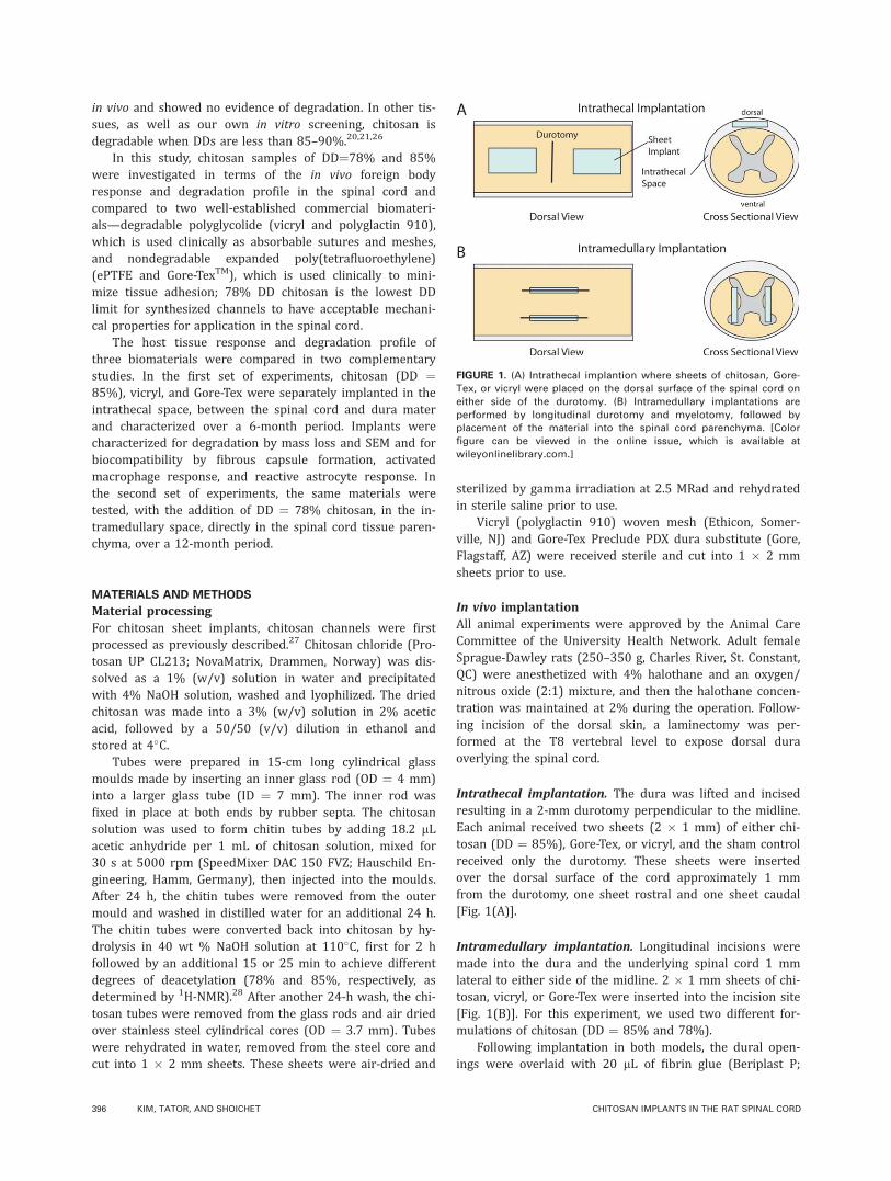
Intrathecal implantation. The dura was lifted and incisedresulting in a 2-mm durotomy perpendicular to the midline.Each animal received two sheets (2 � 1 mm) of either chi-tosan (DD ¼ 85%), Gore-Tex, or vicryl, and the sham controlreceived only the durotomy. These sheets were insertedover the dorsal surface of the cord approximately 1 mmfrom the durotomy, one sheet rostral and one sheet caudal[Fig. 1(A)].
Intramedullary implantation. Longitudinal incisions weremade into the dura and the underlying spinal cord 1 mmlateral to either side of the midline. 2 � 1 mm sheets of chi-tosan, vicryl, or Gore-Tex were inserted into the incision site[Fig. 1(B)]. For this experiment, we used two different for-mulations of chitosan (DD ¼ 85% and 78%).
Following implantation in both models, the dural open-ings were overlaid with 20 lL of fibrin glue (Beriplast P;
FIGURE 1. (A) Intrathecal implantion where sheets of chitosan, Gore-
Tex, or vicryl were placed on the dorsal surface of the spinal cord on
either side of the durotomy. (B) Intramedullary implantations are
performed by longitudinal durotomy and myelotomy, followed by
placement of the material into the spinal cord parenchyma. [Color
figure can be viewed in the online issue, which is available at
wileyonlinelibrary.com.]
396 KIM, TATOR, AND SHOICHET CHITOSAN IMPLANTS IN THE RAT SPINAL CORD
CSL Behring, King of Prussia, PA) and the overlying muscleand skin were closed with vicryl sutures and metal clips,respectively. Rats were given buprinorphrine post-surgery,and every 8–12 h for the next 48 h. In the intrathecal im-plantation study, animals were kept for either 1 month or 6months (n ¼ 4 per group per timepoint). The animalsreceiving intramedullary implants were kept for 1, 6, or 12months (n ¼ 3 per group per timepoint).
Tissue preparationAt the specified times after implantation, rats were transcar-dially perfused with neutral buffered formalin as previouslydescribed,29 and the entire segment of the spinal cord adja-cent to or containing the implanted materials was removed.A 1-cm portion of the spinal cord encompassing the implan-tation site (i.e., the caudal sheet in the intrathecal modeland both sheets in the intramedullary model) was harvestedand post-fixed for up to 1 week in formalin followed by par-affin embedding.
For the intrathecally implanted animals, the rostralimplanted sheet was removed, washed in saline, and driedfor explant analysis as indicated below. Sections of the spi-nal cord were cut at 8-lm thickness and mounted on Super-frosted Plus slides (Fisher Scientific, Markham, ON). The spi-nal cords were sectioned parasagittally or as cross-sectionsfor the intrathecal and intramedullary studies, respectively.
Explant analysisFor the intrathecal experiment, the rostral sheet sampleswere removed at 1 and 6 months and analyzed as follows.For chitosan and Gore-Tex, the fibrous capsules could beeasily separated from the implant and removed. Due to theinterwoven nature of vicryl, it was not possible to separatethe fibrous capsule. The explanted materials were washedand air dried. Mass measurements were taken and com-pared with those before implantation. Samples were thengold-sputter coated and imaged by scanning electron mi-croscopy (Hitachi S2500) at an acceleration voltage of 20kV.
Staining of paraffin-embedded tissueEvery sixth section was stained with Luxol fast blue and he-matoxylin and eosin (LFB/H&E) or Masson’s trichrome.LFB/H&E was used to examine general tissue architectureincluding fibrous encapsulation and Masson’s trichrome wasused to stain collagen including the extent of fibrousencapsulation.
Immunohistochemical staining with the following anti-bodies was also performed as previously described:29
mouse anti-glial fibrillary acidic protein (GFAP; 1;200,Chemicon, Temecula, CA) to visualize reactive astrocytes,and mouse anti-rat monocytes/macrophages antibody (ED-1; 1:200, Serotec, Raleigh, NC) to visualize activatedmacrophages.
StatisticsComparisons involving one independent factor (e.g., materialtype) or two independent factors (e.g., time and material
type) were analyzed using one-way and two-way ANOVA,respectively, followed by Bonferroni post-hoc test. P-valuesless than 0.05 were used as the criteria for statistical signifi-cance. Statistical analysis was performed using GraphPadPrism Software. All values are represented as mean 6standard deviation.
RESULTS
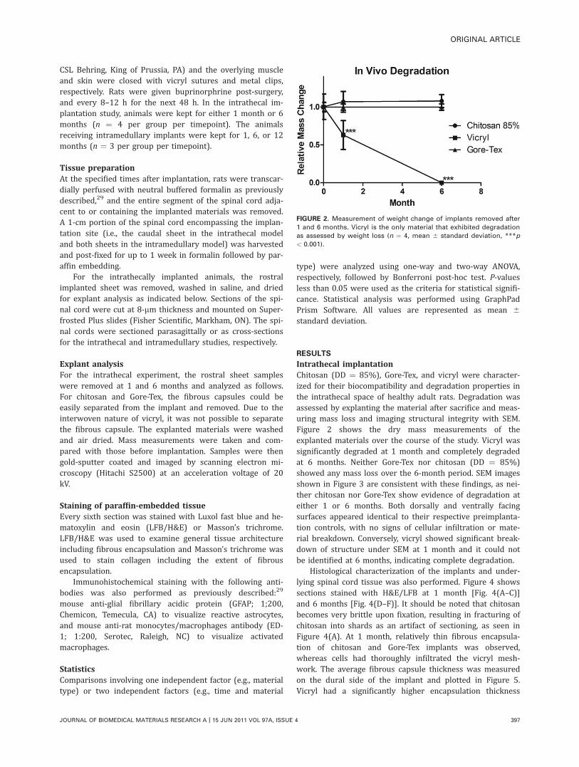
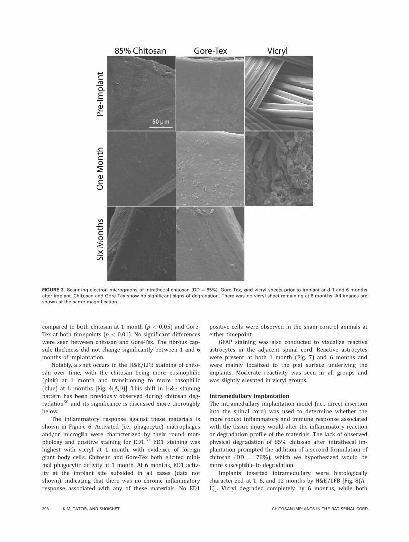
Intrathecal implantationChitosan (DD ¼ 85%), Gore-Tex, and vicryl were character-ized for their biocompatibility and degradation properties inthe intrathecal space of healthy adult rats. Degradation wasassessed by explanting the material after sacrifice and meas-uring mass loss and imaging structural integrity with SEM.Figure 2 shows the dry mass measurements of theexplanted materials over the course of the study. Vicryl wassignificantly degraded at 1 month and completely degradedat 6 months. Neither Gore-Tex nor chitosan (DD ¼ 85%)showed any mass loss over the 6-month period. SEM imagesshown in Figure 3 are consistent with these findings, as nei-ther chitosan nor Gore-Tex show evidence of degradation ateither 1 or 6 months. Both dorsally and ventrally facingsurfaces appeared identical to their respective preimplanta-tion controls, with no signs of cellular infiltration or mate-rial breakdown. Conversely, vicryl showed significant break-down of structure under SEM at 1 month and it could notbe identified at 6 months, indicating complete degradation.
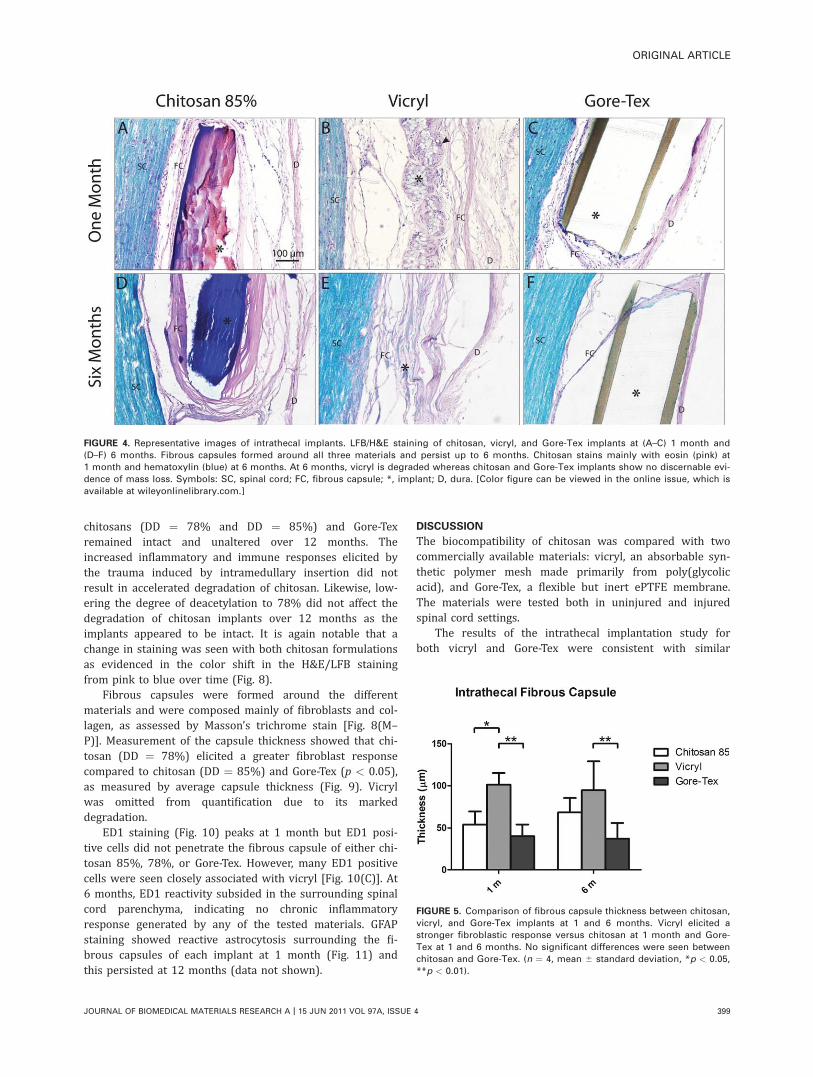
Histological characterization of the implants and under-lying spinal cord tissue was also performed. Figure 4 showssections stained with H&E/LFB at 1 month [Fig. 4(A–C)]and 6 months [Fig. 4(D–F)]. It should be noted that chitosanbecomes very brittle upon fixation, resulting in fracturing ofchitosan into shards as an artifact of sectioning, as seen inFigure 4(A). At 1 month, relatively thin fibrous encapsula-tion of chitosan and Gore-Tex implants was observed,whereas cells had thoroughly infiltrated the vicryl mesh-work. The average fibrous capsule thickness was measuredon the dural side of the implant and plotted in Figure 5.Vicryl had a significantly higher encapsulation thickness
FIGURE 2. Measurement of weight change of implants removed after
1 and 6 months. Vicryl is the only material that exhibited degradation
as assessed by weight loss (n ¼ 4, mean 6 standard deviation, ***p
< 0.001).
ORIGINAL ARTICLE
JOURNAL OF BIOMEDICAL MATERIALS RESEARCH A | 15 JUN 2011 VOL 97A, ISSUE 4 397
compared to both chitosan at 1 month (p < 0.05) and Gore-Tex at both timepoints (p < 0.01). No significant differenceswere seen between chitosan and Gore-Tex. The fibrous cap-sule thickness did not change significantly between 1 and 6months of implantation.
Notably, a shift occurs in the H&E/LFB staining of chito-san over time, with the chitosan being more eosinophilic(pink) at 1 month and transitioning to more basophilic(blue) at 6 months [Fig. 4(A,D)]. This shift in H&E stainingpattern has been previously observed during chitosan deg-radation30 and its significance is discussed more thoroughlybelow.
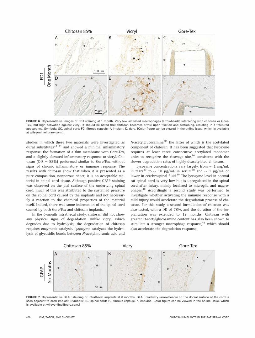
The inflammatory response against these materials isshown in Figure 6. Activated (i.e., phagocytic) macrophagesand/or microglia were characterized by their round mor-phology and positive staining for ED1.31 ED1 staining washighest with vicryl at 1 month, with evidence of foreigngiant body cells. Chitosan and Gore-Tex both elicited mini-mal phagocytic activity at 1 month. At 6 months, ED1 activ-ity at the implant site subsided in all cases (data notshown), indicating that there was no chronic inflammatoryresponse associated with any of these materials. No ED1
positive cells were observed in the sham control animals ateither timepoint.
GFAP staining was also conducted to visualize reactiveastrocytes in the adjacent spinal cord. Reactive astrocyteswere present at both 1 month (Fig. 7) and 6 months andwere mainly localized to the pial surface underlying theimplants. Moderate reactivity was seen in all groups andwas slightly elevated in vicryl groups.
Intramedullary implantationThe intramedullary implantation model (i.e., direct insertioninto the spinal cord) was used to determine whether themore robust inflammatory and immune response associatedwith the tissue injury would alter the inflammatory reactionor degradation profile of the materials. The lack of observedphysical degradation of 85% chitosan after intrathecal im-plantation prompted the addition of a second formulation ofchitosan (DD ¼ 78%), which we hypothesized would bemore susceptible to degradation.
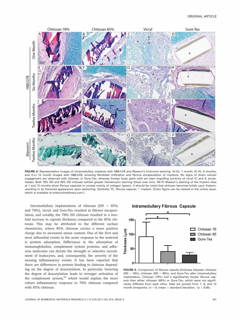
Implants inserted intramedullary were histologicallycharacterized at 1, 6, and 12 months by H&E/LFB [Fig. 8(A–L)]. Vicryl degraded completely by 6 months, while both
FIGURE 3. Scanning electron micrographs of intrathecal chitosan (DD ¼ 85%), Gore-Tex, and vicryl sheets prior to implant and 1 and 6 months
after implant. Chitosan and Gore-Tex show no significant signs of degradation. There was no vicryl sheet remaining at 6 months. All images are
shown at the same magnification.
398 KIM, TATOR, AND SHOICHET CHITOSAN IMPLANTS IN THE RAT SPINAL CORD
chitosans (DD ¼ 78% and DD ¼ 85%) and Gore-Texremained intact and unaltered over 12 months. Theincreased inflammatory and immune responses elicited bythe trauma induced by intramedullary insertion did notresult in accelerated degradation of chitosan. Likewise, low-ering the degree of deacetylation to 78% did not affect thedegradation of chitosan implants over 12 months as theimplants appeared to be intact. It is again notable that achange in staining was seen with both chitosan formulationsas evidenced in the color shift in the H&E/LFB stainingfrom pink to blue over time (Fig. 8).
Fibrous capsules were formed around the differentmaterials and were composed mainly of fibroblasts and col-lagen, as assessed by Masson’s trichrome stain [Fig. 8(M–P)]. Measurement of the capsule thickness showed that chi-tosan (DD ¼ 78%) elicited a greater fibroblast responsecompared to chitosan (DD ¼ 85%) and Gore-Tex (p < 0.05),as measured by average capsule thickness (Fig. 9). Vicrylwas omitted from quantification due to its markeddegradation.
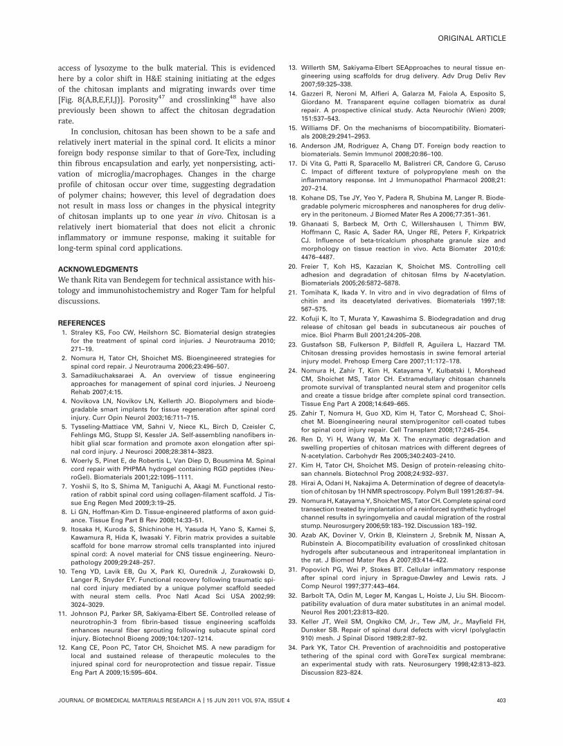
ED1 staining (Fig. 10) peaks at 1 month but ED1 posi-tive cells did not penetrate the fibrous capsule of either chi-tosan 85%, 78%, or Gore-Tex. However, many ED1 positivecells were seen closely associated with vicryl [Fig. 10(C)]. At6 months, ED1 reactivity subsided in the surrounding spinalcord parenchyma, indicating no chronic inflammatoryresponse generated by any of the tested materials. GFAPstaining showed reactive astrocytosis surrounding the fi-brous capsules of each implant at 1 month (Fig. 11) andthis persisted at 12 months (data not shown).
DISCUSSION
The biocompatibility of chitosan was compared with twocommercially available materials: vicryl, an absorbable syn-thetic polymer mesh made primarily from poly(glycolicacid), and Gore-Tex, a flexible but inert ePTFE membrane.The materials were tested both in uninjured and injuredspinal cord settings.
The results of the intrathecal implantation study forboth vicryl and Gore-Tex were consistent with similar
FIGURE 5. Comparison of fibrous capsule thickness between chitosan,
vicryl, and Gore-Tex implants at 1 and 6 months. Vicryl elicited a
stronger fibroblastic response versus chitosan at 1 month and Gore-
Tex at 1 and 6 months. No significant differences were seen between
chitosan and Gore-Tex. (n ¼ 4, mean 6 standard deviation, *p < 0.05,
**p < 0.01).
FIGURE 4. Representative images of intrathecal implants. LFB/H&E staining of chitosan, vicryl, and Gore-Tex implants at (A–C) 1 month and
(D–F) 6 months. Fibrous capsules formed around all three materials and persist up to 6 months. Chitosan stains mainly with eosin (pink) at
1 month and hematoxylin (blue) at 6 months. At 6 months, vicryl is degraded whereas chitosan and Gore-Tex implants show no discernable evi-
dence of mass loss. Symbols: SC, spinal cord; FC, fibrous capsule; *, implant; D, dura. [Color figure can be viewed in the online issue, which is
available at wileyonlinelibrary.com.]
ORIGINAL ARTICLE
JOURNAL OF BIOMEDICAL MATERIALS RESEARCH A | 15 JUN 2011 VOL 97A, ISSUE 4 399
studies in which these two materials were investigated asdural substitutes32–34 and showed a minimal inflammatoryresponse, the formation of a thin membrane with Gore-Tex,and a slightly elevated inflammatory response to vicryl. Chi-tosan (DD ¼ 85%) performed similar to Gore-Tex, withoutsigns of chronic inflammatory or immune response. Theresults with chitosan show that when it is presented as apure composition, nonporous sheet, it is an acceptable ma-terial in spinal cord tissue. Although positive GFAP stainingwas observed on the pial surface of the underlying spinalcord, much of this was attributed to the sustained pressureon the spinal cord caused by the implants and not necessar-ily a reaction to the chemical properties of the materialitself. Indeed, there was some indentation of the spinal cordcaused by both Gore-Tex and chitosan implants.
In the 6-month intrathecal study, chitosan did not showany physical signs of degradation. Unlike vicryl, whichdegrades due to hydrolysis, the degradation of chitosanrequires enzymatic catalysis. Lysozyme catalyzes the hydro-lysis of glycosidic bonds between N-acetylmuramic acid and
N-acetylglucosamine,35 the latter of which is the acetylatedcomponent of chitosan. It has been suggested that lysozymerequires at least three consecutive acetylated monomerunits to recognize the cleavage site,36 consistent with theslower degradation rates of highly deacetylated chitosans.
Lysozyme concentrations vary largely, from � 1 mg/mLin tears37 to � 10 lg/mL in serum38 and � 1 lg/mL orlower in cerebrospinal fluid.39 The lysozyme level in normalrat spinal cord is very low but is upregulated in the spinalcord after injury, mainly localized to microglia and macro-phages.40 Accordingly, a second study was performed toinvestigate whether activating the immune response with amild injury would accelerate the degradation process of chi-tosan. For this study, a second formulation of chitosan wasalso tested, with a DD of 78%, and the duration of the im-plantation was extended to 12 months. Chitosan withgreater D-acetylglucosamine content has also been shown tostimulate a stronger macrophage response,41 which shouldalso accelerate the degradation response.
FIGURE 6. Representative images of ED1 staining at 1 month. Very few activated macrophages (arrowheads) interacting with chitosan or Gore-
Tex, but high activation against vicryl. It should be noted that chitosan becomes brittle upon fixation and sectioning, resulting in a fractured
appearance. Symbols: SC, spinal cord; FC, fibrous capsule; *, implant; D, dura. [Color figure can be viewed in the online issue, which is available
at wileyonlinelibrary.com.]
FIGURE 7. Representative GFAP staining of intrathecal implants at 6 months. GFAP reactivity (arrowheads) on the dorsal surface of the cord is
seen adjacent to each implant. Symbols: SC, spinal cord; FC, fibrous capsule, *, implant. [Color figure can be viewed in the online issue, which
is available at wileyonlinelibrary.com.]
400 KIM, TATOR, AND SHOICHET CHITOSAN IMPLANTS IN THE RAT SPINAL CORD
Intramedullary implantations of chitosan (DD ¼ 85%and 78%), vicryl, and Gore-Tex resulted in fibrous encapsu-lation, and notably, the 78% DD chitosan resulted in a two-fold increase in capsule thickness compared to the 85% chi-tosan. This may be attributed to the different surfacechemistries, where 85% chitosan carries a more positivecharge due to increased amine content. One of the first andmost influential events in the acute response to the materialis protein adsorption. Differences in the adsorption ofimmunoglobulins, complement system proteins, and adhe-sion molecules can dictate the strength or selective recruit-ment of leukocytes, and, consequently, the severity of theensuing inflammatory events. It has been reported thatthere are differences in protein binding to chitosan depend-ing on the degree of deacetylation. In particular, loweringthe degree of deacetylation leads to stronger activation ofthe complement system,42 which would explain the morerobust inflammatory response to 78% chitosan comparedwith 85% chitosan.
FIGURE 8. Representative images of intramedullary implants with H&E/LFB and Masson’s trichrome staining; (A–D), 1 month; (E–H), 6 months,
and (I–L) 12 month images with H&E/LFB, showing fibroblast infiltration and fibrous encapsulation of implants. No signs of direct cellular
engagement are observed with chitosan or Gore-Tex, whereas foreign body giant cells are seen engulfing portions of vicryl (C and G arrow-
heads). Both 78% DD and 85% DD chitosan exhibit greater hematoxylin staining (blue) over time. (M–P) Masson’s staining of the implant sites
at 1 and 12 months show fibrous capsules to consist mainly of collagen (green). It should be noted that chitosan becomes brittle upon fixation,
resulting in its fractured appearance upon sectioning. Symbols: FC, fibrous capsule; *, implant. [Color figure can be viewed in the online issue,
which is available at wileyonlinelibrary.com.]
FIGURE 9. Comparison of fibrous capsule thickness between chitosan
(DD ¼ 78%), chitosan (DD ¼ 85%), and Gore-Tex after intramedullary
implantation. Chitosan (78%) had a significantly thicker fibrous cap-
sule than either chitosan (85%) or Gore-Tex, which were not signifi-
cantly different from each other. Data are pooled from 1, 6, and 12
month timepoints. (n ¼ 6, mean 6 standard deviation, *p < 0.05).
ORIGINAL ARTICLE
JOURNAL OF BIOMEDICAL MATERIALS RESEARCH A | 15 JUN 2011 VOL 97A, ISSUE 4 401
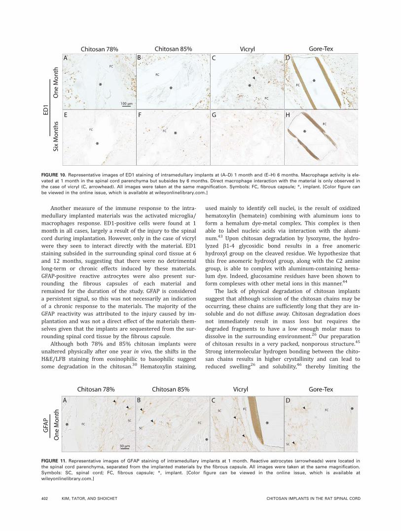
Another measure of the immune response to the intra-medullary implanted materials was the activated microglia/macrophages response. ED1-positive cells were found at 1month in all cases, largely a result of the injury to the spinalcord during implantation. However, only in the case of vicrylwere they seen to interact directly with the material. ED1staining subsided in the surrounding spinal cord tissue at 6and 12 months, suggesting that there were no detrimentallong-term or chronic effects induced by these materials.GFAP-positive reactive astrocytes were also present sur-rounding the fibrous capsules of each material andremained for the duration of the study. GFAP is considereda persistent signal, so this was not necessarily an indicationof a chronic response to the materials. The majority of theGFAP reactivity was attributed to the injury caused by im-plantation and was not a direct effect of the materials them-selves given that the implants are sequestered from the sur-rounding spinal cord tissue by the fibrous capsule.
Although both 78% and 85% chitosan implants wereunaltered physically after one year in vivo, the shifts in theH&E/LFB staining from eosinophilic to basophilic suggestsome degradation in the chitosan.30 Hematoxylin staining,
used mainly to identify cell nuclei, is the result of oxidizedhematoxylin (hematein) combining with aluminum ions toform a hemalum dye-metal complex. This complex is thenable to label nucleic acids via interaction with the alumi-num.43 Upon chitosan degradation by lysozyme, the hydro-lyzed b1-4 glycosidic bond results in a free anomerichydroxyl group on the cleaved residue. We hypothesize thatthis free anomeric hydroxyl group, along with the C2 aminegroup, is able to complex with aluminum-containing hema-lum dye. Indeed, glucosamine residues have been shown toform complexes with other metal ions in this manner.44
The lack of physical degradation of chitosan implantssuggest that although scission of the chitosan chains may beoccurring, these chains are sufficiently long that they are in-soluble and do not diffuse away. Chitosan degradation doesnot immediately result in mass loss but requires thedegraded fragments to have a low enough molar mass todissolve in the surrounding environment.26 Our preparationof chitosan results in a very packed, nonporous structure.45
Strong intermolecular hydrogen bonding between the chito-san chains results in higher crystallinity and can lead toreduced swelling26 and solubility,46 thereby limiting the
FIGURE 10. Representative images of ED1 staining of intramedullary implants at (A–D) 1 month and (E–H) 6 months. Macrophage activity is ele-
vated at 1 month in the spinal cord parenchyma but subsides by 6 months. Direct macrophage interaction with the material is only observed in
the case of vicryl (C, arrowhead). All images were taken at the same magnification. Symbols: FC, fibrous capsule; *, implant. [Color figure can
be viewed in the online issue, which is available at wileyonlinelibrary.com.]
FIGURE 11. Representative images of GFAP staining of intramedullary implants at 1 month. Reactive astrocytes (arrowheads) were located in
the spinal cord parenchyma, separated from the implanted materials by the fibrous capsule. All images were taken at the same magnification.
Symbols: SC, spinal cord; FC, fibrous capsule; *, implant. [Color figure can be viewed in the online issue, which is available at
wileyonlinelibrary.com.]
402 KIM, TATOR, AND SHOICHET CHITOSAN IMPLANTS IN THE RAT SPINAL CORD
access of lysozyme to the bulk material. This is evidencedhere by a color shift in H&E staining initiating at the edgesof the chitosan implants and migrating inwards over time[Fig. 8(A,B,E,F,I,J)]. Porosity47 and crosslinking48 have alsopreviously been shown to affect the chitosan degradationrate.
In conclusion, chitosan has been shown to be a safe andrelatively inert material in the spinal cord. It elicits a minorforeign body response similar to that of Gore-Tex, includingthin fibrous encapsulation and early, yet nonpersisting, acti-vation of microglia/macrophages. Changes in the chargeprofile of chitosan occur over time, suggesting degradationof polymer chains; however, this level of degradation doesnot result in mass loss or changes in the physical integrityof chitosan implants up to one year in vivo. Chitosan is arelatively inert biomaterial that does not elicit a chronicinflammatory or immune response, making it suitable forlong-term spinal cord applications.
ACKNOWLEDGMENTS
We thank Rita van Bendegem for technical assistance with his-tology and immunohistochemistry and Roger Tam for helpfuldiscussions.
REFERENCES1. Straley KS, Foo CW, Heilshorn SC. Biomaterial design strategies
for the treatment of spinal cord injuries. J Neurotrauma 2010;
271–19.
2. Nomura H, Tator CH, Shoichet MS. Bioengineered strategies for
spinal cord repair. J Neurotrauma 2006;23:496–507.
3. Samadikuchaksaraei A. An overview of tissue engineering
approaches for management of spinal cord injuries. J Neuroeng
Rehab 2007;4:15.
4. Novikova LN, Novikov LN, Kellerth JO. Biopolymers and biode-
gradable smart implants for tissue regeneration after spinal cord
injury. Curr Opin Neurol 2003;16:711–715.
5. Tysseling-Mattiace VM, Sahni V, Niece KL, Birch D, Czeisler C,
Fehlings MG, Stupp SI, Kessler JA. Self-assembling nanofibers in-
hibit glial scar formation and promote axon elongation after spi-
nal cord injury. J Neurosci 2008;28:3814–3823.
6. Woerly S, Pinet E, de Robertis L, Van Diep D, Bousmina M. Spinal
cord repair with PHPMA hydrogel containing RGD peptides (Neu-
roGel). Biomaterials 2001;22:1095–1111.
7. Yoshii S, Ito S, Shima M, Taniguchi A, Akagi M. Functional resto-
ration of rabbit spinal cord using collagen-filament scaffold. J Tis-
sue Eng Regen Med 2009;3:19–25.
8. Li GN, Hoffman-Kim D. Tissue-engineered platforms of axon guid-
ance. Tissue Eng Part B Rev 2008;14:33–51.
9. Itosaka H, Kuroda S, Shichinohe H, Yasuda H, Yano S, Kamei S,
Kawamura R, Hida K, Iwasaki Y. Fibrin matrix provides a suitable
scaffold for bone marrow stromal cells transplanted into injured
spinal cord: A novel material for CNS tissue engineering. Neuro-
pathology 2009;29:248–257.
10. Teng YD, Lavik EB, Qu X, Park KI, Ourednik J, Zurakowski D,
Langer R, Snyder EY. Functional recovery following traumatic spi-
nal cord injury mediated by a unique polymer scaffold seeded
with neural stem cells. Proc Natl Acad Sci USA 2002;99:
3024–3029.
11. Johnson PJ, Parker SR, Sakiyama-Elbert SE. Controlled release of
neurotrophin-3 from fibrin-based tissue engineering scaffolds
enhances neural fiber sprouting following subacute spinal cord
injury. Biotechnol Bioeng 2009;104:1207–1214.
12. Kang CE, Poon PC, Tator CH, Shoichet MS. A new paradigm for
local and sustained release of therapeutic molecules to the
injured spinal cord for neuroprotection and tissue repair. Tissue
Eng Part A 2009;15:595–604.
13. Willerth SM, Sakiyama-Elbert SEApproaches to neural tissue en-
gineering using scaffolds for drug delivery. Adv Drug Deliv Rev
2007;59:325–338.
14. Gazzeri R, Neroni M, Alfieri A, Galarza M, Faiola A, Esposito S,
Giordano M. Transparent equine collagen biomatrix as dural
repair. A prospective clinical study. Acta Neurochir (Wien) 2009;
151:537–543.
15. Williams DF. On the mechanisms of biocompatibility. Biomateri-
als 2008;29:2941–2953.
16. Anderson JM, Rodriguez A, Chang DT. Foreign body reaction to
biomaterials. Semin Immunol 2008;20:86–100.
17. Di Vita G, Patti R, Sparacello M, Balistreri CR, Candore G, Caruso
C. Impact of different texture of polypropylene mesh on the
inflammatory response. Int J Immunopathol Pharmacol 2008;21:
207–214.
18. Kohane DS, Tse JY, Yeo Y, Padera R, Shubina M, Langer R. Biode-
gradable polymeric microspheres and nanospheres for drug deliv-
ery in the peritoneum. J Biomed Mater Res A 2006;77:351–361.
19. Ghanaati S, Barbeck M, Orth C, Willershausen I, Thimm BW,
Hoffmann C, Rasic A, Sader RA, Unger RE, Peters F, Kirkpatrick
CJ. Influence of beta-tricalcium phosphate granule size and
morphology on tissue reaction in vivo. Acta Biomater 2010;6:
4476–4487.
20. Freier T, Koh HS, Kazazian K, Shoichet MS. Controlling cell
adhesion and degradation of chitosan films by N-acetylation.
Biomaterials 2005;26:5872–5878.
21. Tomihata K, Ikada Y. In vitro and in vivo degradation of films of
chitin and its deacetylated derivatives. Biomaterials 1997;18:
567–575.
22. Kofuji K, Ito T, Murata Y, Kawashima S. Biodegradation and drug
release of chitosan gel beads in subcutaneous air pouches of
mice. Biol Pharm Bull 2001;24:205–208.
23. Gustafson SB, Fulkerson P, Bildfell R, Aguilera L, Hazzard TM.
Chitosan dressing provides hemostasis in swine femoral arterial
injury model. Prehosp Emerg Care 2007;11:172–178.
24. Nomura H, Zahir T, Kim H, Katayama Y, Kulbatski I, Morshead
CM, Shoichet MS, Tator CH. Extramedullary chitosan channels
promote survival of transplanted neural stem and progenitor cells
and create a tissue bridge after complete spinal cord transection.
Tissue Eng Part A 2008;14:649–665.
25. Zahir T, Nomura H, Guo XD, Kim H, Tator C, Morshead C, Shoi-
chet M. Bioengineering neural stem/progenitor cell-coated tubes
for spinal cord injury repair. Cell Transplant 2008;17:245–254.
26. Ren D, Yi H, Wang W, Ma X. The enzymatic degradation and
swelling properties of chitosan matrices with different degrees of
N-acetylation. Carbohydr Res 2005;340:2403–2410.
27. Kim H, Tator CH, Shoichet MS. Design of protein-releasing chito-
san channels. Biotechnol Prog 2008;24:932–937.
28. Hirai A, Odani H, Nakajima A. Determination of degree of deacetyla-
tion of chitosan by 1HNMR spectroscopy. PolymBull 1991;26:87–94.
29. NomuraH, KatayamaY, ShoichetMS, Tator CH. Complete spinal cord
transection treated by implantation of a reinforced synthetic hydrogel
channel results in syringomyelia and caudal migration of the rostral
stump. Neurosurgery 2006;59:183–192. Discussion 183–192.
30. Azab AK, Doviner V, Orkin B, Kleinstern J, Srebnik M, Nissan A,
Rubinstein A. Biocompatibility evaluation of crosslinked chitosan
hydrogels after subcutaneous and intraperitoneal implantation in
the rat. J Biomed Mater Res A 2007;83:414–422.
31. Popovich PG, Wei P, Stokes BT. Cellular inflammatory response
after spinal cord injury in Sprague-Dawley and Lewis rats. J
Comp Neurol 1997;377:443–464.
32. Barbolt TA, Odin M, Leger M, Kangas L, Hoiste J, Liu SH. Biocom-
patibility evaluation of dura mater substitutes in an animal model.
Neurol Res 2001;23:813–820.
33. Keller JT, Weil SM, Ongkiko CM, Jr., Tew JM, Jr., Mayfield FH,
Dunsker SB. Repair of spinal dural defects with vicryl (polyglactin
910) mesh. J Spinal Disord 1989;2:87–92.
34. Park YK, Tator CH. Prevention of arachnoiditis and postoperative
tethering of the spinal cord with GoreTex surgical membrane:
an experimental study with rats. Neurosurgery 1998;42:813–823.
Discussion 823–824.
ORIGINAL ARTICLE
JOURNAL OF BIOMEDICAL MATERIALS RESEARCH A | 15 JUN 2011 VOL 97A, ISSUE 4 403
35. Shigemasa Y, Saito K, Sashiwa H, Saimoto H. Enzymatic degrada-
tion of chitins and partially deacetylated chitins. Int J Biol Macro-
mol 1994;16:43–49.
36. Varum KM, Holme HK, Izume M, Stokke BT, Smidsrod O. Determi-
nation of enzymatic hydrolysis specificity of partially N-acetylated
chitosans. Biochim Biophys Acta 1996;1291:5–15.
37. Temel A, Kazokoglu H, Taga Y. Tear lysozyme levels in contact
lens wearers. Ann Ophthalmol 1991;23:191–194.
38. Currie GA, Eccles SA. Serum lysozyme as a marker of host resist-
ance. I. Production by macrophages resident in rat sarcomata. Br
J Cancer 1976;33:51–59.
39. Mishra OP, Batra P, Ali Z, Anupurba S, Das BK. Cerebrospinal
fluid lysozyme level for the diagnosis of tuberculous meningitis
in children. J Trop Pediatr 2003;49:13–16.
40. Zhang KH, Xiao HS, Lu PH, Shi J, Li GD, Wang YT, Han S, Zhang
FX, Lu YJ, Zhang X, Xu XM. Differential gene expression after
complete spinal cord transection in adult rats: An analysis
focused on a subchronic post-injury stage. Neuroscience 2004;
128:375–388.
41. Peluso G, Petillo O, Ranieri M, Santin M, Ambrosio L, Calabro D,
Avallone B, Balsamo G. Chitosan-mediated stimulation of macro-
phage function. Biomaterials 1994;15:1215–1220.
42. Benesch J, Tengvall P. Blood protein adsorption onto chitosan.
Biomaterials 2002;23:2561–2568.
43. Bettinger C, Zimmermann HW. New investigations on hematoxy-
lin, hematein, and hematein-aluminium complexes. II. Hematein-
aluminium complexes and hemalum staining. Histochemistry
1991;96:215–228.
44. Bunel S, Ibarra C, Moraga E, Blasko A, Bunton CA. Structures of
complexes of cobalt(Iii) and glucosamine––Applications of
molecular mechanics and NMR spectroscopy. Carbohydr Res
1993;244:1–14.
45. Freier T, Montenegro R, Shan Koh H, Shoichet MS. Chitin-based
tubes for tissue engineering in the nervous system. Biomaterials
2005;26:4624–4632.
46. Sogias IA, Khutoryanskiy VV, Williams AC. Exploring the factors
affecting the solubility of chitosan in water. Macromol Chem Phys
2010;211:426–433.
47. Cunha-Reis C, TuzlaKoglu K, Baas E, Yang Y, El Haj A, Reis RL.
Influence of porosity and fibre diameter on the degradation of
chitosan fibre-mesh scaffolds and cell adhesion. J Mater Sci
Mater Med 2007;18:195–200.
48. Mi FL, Tan YC, Liang HF, Sung HW. In vivo biocompatibility and
degradability of a novel injectable-chitosan-based implant. Bioma-
terials 2002;23:181–191.
404 KIM, TATOR, AND SHOICHET CHITOSAN IMPLANTS IN THE RAT SPINAL CORD