Embed Size (px)
Citation preview
Chemical and Spectroscopic Aspects of Polymer Ablation: Special Featuresand Novel Directions
Thomas Lippert*,† and J. Thomas Dickinson‡
Paul Scherrer Institut, 5232 Villigen PSI, Switzerland, and Washington State University, Pullman, Washington 99164-2814 USA
Received August 12, 2002
Contents1 Introduction 453
1.1 History 4531.2 Summary of Ablation Mechanisms 4541.3 Summary of Theoretical Models 455
2 Studied Polymers 4562.1 Doped Polymers 456
2.1.1 Photochemical Active PolyaromaticCompounds, as Fluorescence Probes
456
2.1.2 Photostable Organic Compounds andSimilar Dopants
457
2.2 Neat Polymers 4582.2.1 Designed Polymers Based on
Commercial Polymers458
2.2.2 Novel Designed Polymers for LaserAblation at 308 nm
459
2.2.3 Detailed Analysis of a Selected DesignedPolymer (Triazene-Polymer)
459
2.2.4 Commercial Polymers 4642.3 Summary of Miscellaneous Polymer Work 467
3 Selected Experimental Tools 4683.1 Single Pulse Ablation with Various Pulse
Lengths468
3.2 Mass Spectrometry 4683.2.1 Review of Recent Experimental Work 4683.2.2 Analysis of Neutral and Ionic Emissions 469
4 Structure Modification 4724.1 Physical Surface Modification: Nano- to
Microstructures472
4.2 Chemical Surface Modification 4745 Radiation Sources 475
5.1 Low Intensity (Lamp) Sources 4755.2 Continuous-Wave UV Lasers 4755.3 Ultrafast Lasers 476
5.3.1 Femtosecond Lasers 4765.3.2 Picosecond Lasers 478
5.4 Vacuum-Ultraviolet (VUV) Lasers 4785.5 Synchrotron Radiation (SR) 4795.6 Mid-IR Irradiation 4795.7 Visible Light and High Repetition Rate
Ablation480
6 Outlook 4817 Acknowledgments 4818 References 481
1 Introduction
1.1 HistoryIn 1982, the first reports of laser ablation of
polymers were issued almost simultaneously by Y.Kawamura et al.1 and R. Srinivasan et al.2 Srini-vasan went on to become a leader in the field ofpolymer ablation. Srinivasan probably also coined theterms laser ablation and ablative photodecomposi-tion, now in common use. The onset of materialremoval by laser ablation characteristically occurs ata well-defined laser fluence (energy per unit area).As the fluence is raised above this threshold, theablation rate increases. The threshold fluence (F0 orFth) is material and laser wavelength dependentand can vary from tens of mJ cm-2 to more than1 J cm-2. The discovery of laser ablation of poly-mers sparked research in this field in many groupsaround the world. Many aspects of polymer ablation,and laser processing in general, are reviewed byBauerle.3 Today, commercial applications of poly-mer laser ablation include the preparation of via-holes in polyimide for multichip modules at IBM4
and the production of inkjet printer nozzles (alsopolyimide).5
Considerable progress has been made in under-standing polymer ablation since the last series ofreviews a decade ago.6-8 New developments in poly-mer ablation include the application of femtosecondlaser pulses, vacuum ultraviolet lasers (VUV), andfree electron lasers (FEL). Some of these techniqueshave a great potential for the development of newapplications and research tools. Much of this progressis discussed in this and other articles appearing inthis special issue of Chemical Reviews. Currentresearch on polymer ablation may be divided into twoareas:
(i) Applications of laser ablation, novel materials,and techniques.
(ii) Studies of ablation mechanisms (databasedmodeling).
The first area will be discussed in detail in thisarticle, while the mechanistic aspects, especially thetheoretical part, are discussed in other articles in thisspecial issue.
Many experimental methods and experimentalpolymers have been designed with a view towardimproving our understanding of ablation mecha-nisms. It is often impossible to completely separateexperiments designed to illuminate ablation mech-
† Paul Scherrer Institut.‡ Washington State University.
453Chem. Rev. 2003, 103, 453−485
10.1021/cr010460q CCC: $44.00 © 2003 American Chemical SocietyPublished on Web 01/04/2003
anisms from other experimental studies. A shortsummary of the various models for ablation mecha-nisms is given below. In many cases, experimentaldetails and quantitative models are discussed inother articles in this issue.
1.2 Summary of Ablation Mechanisms
Polymer ablation is typically described in terms ofthermal, photothermal, or photochemical models,either individually or combined.6-8
From an application standpoint, photochemicalprocesses are most desirable. Photochemical decom-position is intrinsically capable of higher spatialresolution because thermal damage to the surround-ing material is minimal. Polymers design for photo-chemical interactions could take advantage of ourexperience with the design of photoresists, where thephotochemical properties of various chromophoresdominate the interaction. Polymers designed to de-compose mainly to gaseous products would reduce oreliminate the problem of redeposited material (de-bris). Incomplete removal of polymer debris requiresadditional cleaning steps and mitigates one of the bigadvantages of laser ablation.
Polymer ablation mechanisms have been debatedfor more than two decades. As a result, the originalstrict separation of the models (photochemical modelsby chemists and thermal/photothermal models byphysicists) is slowly blurring. It is generally agreedthat the energy of nanosecond laser pulses is initiallytransformed into electronic excitations. However,subsequent energy transfer can take a variety offorms. Understanding this energy transfer has beencomplicated by the limited range of experimentaldata accounted for by any given model. Comprehen-sive models that incorporate data acquired by severalexperimental techniques simply do not exist. Anotherproblem has been the wide variety of empiricaldefinitions for ablation phenomena, especially nearthe threshold fluence. Experimental facts that mustbe explained by such models include:
(i) Ablation depths as measured by profilometry(optical interferometry, stylus profilometry,9 atomicforce microscopy10) show sudden increases at thethreshold fluence. This observation is supported byreflectivity11 and acoustic measurements.12 Experi-mental comparisons are complicated by the use ofsingle laser pulses in some threshold measurementsand multiple pulses in others. The resulting thresh-olds are not necessarily equivalent.
(ii) Ablation rates calculated from mass loss mea-surements using quartz crystal microbalances (QCM)13
or mass spectrometry14 reveal a so-called Arrheniustail13 (linear fluence dependence at low fluences, withmuch stronger fluence dependencies at still higherfluences).
(iii) Ablation can change the chemical and/orphysical (morphological) properties of the remainingpolymer, e.g., altered optical absorption,15-17 modifiedchemical composition,18-20 and in the case of polyim-ides, increased electrical conductivity.21
We note that at sufficiently high fluences, theablation rates of most polymers are similar22 beinglimited by screening of the radiation by the ablatedproducts23,24 and the associated plasma.25 Thus, highfluence measurements of ablation rate seldom yieldmuch information on the underlying ablation mech-anisms.
Thomas Lippert was born in Germany where he studied chemistry at theUniversity of Bayreuth. He received his Diploma in Ecological Chemistry(in 1990) and his Ph.D. in Physical Chemistry (in 1993) under ProfessorAlexander Wokaun. He did postdoctoral studies for Wacker Chemistry(Burghausen, Germany) and went then as a STA/Alexander von Humboldtpostdoctoral fellow to Tsukuba, Japan (in 1994), where he worked at theNational Institute of Materials and Chemical Research with Dr. Akira Yabe.He moved then as postdoctoral fellow (in 1995) to Los Alamos NationalLaboratory (Los Alamos, NM) where he later became a Director’sPostdoctoral Fellow and Technical Staff Member. He then joined the PaulScherrer Institut (Villigen, Switzerland) as Senior Scientist (in 1999), andis currently the head of a research group. He received his Habilitation atthe ETH Zuerich in Physical Chemistry (in 2002), where he is also afaculty member. His work has been focused on the design of novelpolymers, ultrafast spectroscopy, thin film deposition, and microstructuring/surface modification. Lippert is author or coauthor of over 90 technicalarticles. He has presented more than 30 invited talks on areas of laserinteractions with materials.
J. Thomas Dickinson received his B.A. degree in physics from WesternMichigan University in 1963, and his Ph.D. degree in chemical physicsfrom the University of Michigan in 1968. He went directly to WashingtonState University where he is now the Paul A. Anderson Professor ofPhysics. Dickinson’s research has been in the general areas of materialsphysics, materials chemistry, and surface science. Recent research hasfocused on: (a) laser-ablation and laser desorption mechanisms ininorganic dielectric crystals and polymers; (b) the applications of lasersin materials and chemical analysis; and (c) use of scanning tunnelingmicroscopy and atomic force microscopy to study laser surface modifica-tion, nano-tribology and deformation physics. Dickinson is author orcoauthor of over 270 technical articles. He has presented more than 200invited talks on areas of materials physics and chemistry over the last 15years, including 15 Gordon Conference talks.
454 Chemical Reviews, 2003, Vol. 103, No. 2 Lippert and Dickinson
1.3 Summary of Theoretical ModelsThe various ablation models incorporate one or
more of the above-mentioned ablation mechanisms:Photochemical models: Electronic excitation results
in direct bond breaking;7,26-29 excited-state absorptionmay be included (two-level model).30-32
Photothermal models:23,33-36 Electronic excitationsthermalize on picosecond-time scales, yielding ther-mally broken bonds.
Photophysical models: Both thermal and nonther-mal processes are important. The models may incor-porate two independent channels of bond break-ing,24,37 or employ different bond breaking energiesfor ground state and electronically excited chro-mophores.38,39 These models are most important forshort pulses (ps and fs).40
Photochemical and thermal models can be furthercategorized as volume and surface models. Surfacemodels consider only material within several mono-layers of the surface. As a result, the velocity of theinterface between the gaseous and condensed phasedepends explicitly on the surface temperature or laserintensity. In volume models, ablation processes in thebulk of the material are explicitly treated. In com-bination with the assumed ablation mechanism(s),surface and volume models typically have the follow-ing properties:
Photochemical surface models41 typically involvelonger interaction times and/or higher laser fluences.
Photochemical volume models7,8,26-28 predict sharpablation thresholds and ablation depths that dependlogarithmically on the number of laser pulses. Linearincreases in the ablation depth can result frommodels that account for the motion of the ablationfront and neglect screening by ablation products.These models do not explain the Arrhenius tailsobserved in mass loss measurements.
Thermal surface models23,24,36,42 (developed mainlyfor metal ablation43) account for smooth Arrheniustails, due to the Arrhenius dependence of the reces-sion velocity on temperature. These models do notaccount for sharp ablation thresholds, as observedin polymers.
Photothermal volume models are often oversimpli-fied,13,35 because it is often convenient to ignore themotion of the gas-solid interface,34 which results inunrealistically high temperatures. These modelsoften fail to account for Arrhenius tails.
Newer models combine various features of thesemodels. For instance, a photochemical volume modeland a thermal surface model have been combined toconstruct a volume photothermal model of Arnold etal.44 This model treats ablation in terms of photo-thermal bond breaking within the bulk material (afirst-order chemical reaction with Arrhenius temper-ature dependence). Ablation begins when the densityof broken bonds at the surface reaches a criticalvalue. These models can account for sharp ablationthresholds and Arrhenius tails, and are described indetail by Bityurin et al. in this issue. To date, thisvolume photothermal model had been applied topolyimides only, and does not account for the pos-sibility of photochemical decomposition. This model,like other thermal models, employs many material
parameters. Several of these parameters are obtainedfrom fitting to data. One challenge has been thetemperature dependence of these material param-eters (e.g., thermal conductivity or specific heat).Even when data are available as a function oftemperature, the temperature range is often quitelimited (up to a few hundred K), and often acquiredat relatively slow heating rates (up to several K persecond). For laser ablation modeling, these data areextrapolated to temperatures of up to several thou-sand K and heating rates up to 1010 K s-1.
Photothermal models typically have difficulty ac-counting for the way the ablation products changewith laser wavelength. As emphasized by Srinivasan:45 the products generated by ablation with CO2-lasersare very different from the products generated byexcimer laser ablation (in the UV). Thermal effectsalone cannot account for this difference, suggestingthat photoelectronic processes play an important roleunder UV laser irradiation.
Laser absorption in the UV is often dominated byphotoelectronic excitations. In doped polymer sys-tems, one can provide strongly absorbing chro-mophores that are responsible for the bulk of laserabsorption. Subsequent thermalization of this energycan then decompose the matrix material by thermalprocesses. This is especially important for polymerscontaining polyaromatic dopants irradiated at wave-lengths greater than or about 248 nm. Time-resolvedabsorption/emission measurements46,47 and TOF-MSdata48 indicate the presence of cyclic multiphotonabsorption with up to 10 photons.49 The photonenergy is transferred from the highly excited poly-aromatic dopant molecules to the polymer matrix viarapid internal conversion. The associated heatingresults in the thermal decomposition of the polymer.Time-dependent absorption measurements suggestthat excited triplet states in the dopants play a keyrole in the process, due to their long lifetimes.
The multiphoton absorption cycle was confirmedby comparing the temporal profile of the fluorescenceof anthracene-doped polystyrene films with compu-tational results based on the cyclic process.50 In thecomputational studies, the ground state, first excitedsinglet state, and lowest triplet state were included.The calculated temperature rise during the laserpulse depends nonlinearly on the laser intensity.Rapid internal conversion within the triplet manifoldis the most effective mechanism for depositing heatat the irradiated surface.
Progress in computational hardware and softwarehas allowed the development of entirely differentkinds of models for laser ablation, including molec-ular dynamics (MD) calculations.51-53 To gain amicroscopic view of laser ablation, the breathingsphere model was developed. Each molecule (orappropriate group of atoms) is typically representedby a single particle with no true translational degreesof freedom, but an approximate internal degree offreedom.54,55 This internal (breathing) mode allowsone to describe a realistic conversion of internalelectronic energy (due to laser absorption) to trans-lational motion. Using molecules (are larger groupsof atoms) rather than the atoms themselves, the
Chemical and Spectroscopic Aspects of Polymer Ablation Chemical Reviews, 2003, Vol. 103, No. 2 455
system size can be large enough to describe thecollective dynamics leading to laser ablation anddamage. Similarly, neglecting the high-frequencyatomic vibrations allows one to use longer time-stepsin the numerical integration and model the effect ofrealistic laser pulses.56 Molecular dynamics modelsyield a microscopic view of ablation,54-57 includingproperties of the ejected material: the velocity dis-tribution of matrix and analyte molecules in MAL-DI,58,59 the ejection of clusters,56-61 and their de-pendence on the irradiation conditions (laser flu-ence,54,55,57,60 pulse duration,56 and initial sampletemperature61). One effect that cannot be directlysimulated in polymers (possible for metals and ul-trashort pulses62) within the breathing sphere modelis the propagation of laser induced pressure wavesfrom the absorption region deeper into the bulk ofthe irradiated sample. This can be remedied bycombining molecular dynamics models of molecule-scale processes with the continuum finite elementmodels63 of the long-range propagation of waves andtheir interaction with other regions of a large system.Long-range wave propagation can spall material fromthe free surface at the back of such samples,64 forinstance, as the wave of compression produced bylaser absorption reflects from the back free surface.Spallation results when the dynamic tensile strengthof the material is exceeded by the (tensile) pressureof the reflected wave, which may also result inmaterial removal from the front surface of samples.65
The plume development in molecular dynamicssimulations of the bulk is usually only followed for afew nanoseconds after the laser pulse, which is notenough to compare the data with various experimen-tal techniques (such as MALDI, TOF-MS, shadowg-raphy, interferometry or for PLD). Long-term plumeexpansion can be simulated by extending the bulkmodels with direct simulation Monte Carlo tech-niques, which have been recently applied to systemsrelevant to MALDI.66 Details of molecular dynamicsmodels are discussed by Zhigilei et al. in this issue.
2 Studied Polymers
Two major kinds of polymers have been studied:doped polymers and neat polymers.
2.1 Doped PolymersThe ablation of doped polymers was reviewed in
1997 by Lippert et al.67 and the ablation process wasclassified according to the absorption properties ofthe polymer-dopant system. These systems rangefrom polymer blends, to systems where only thedopant or only the polymer are absorbing. Theauthors suggested a qualitative scheme where abla-tion results from a complex mixture of processesoriginating from the dopants or the matrix. Depend-ing on the properties of the dopant, different ablationpathways are dominant.
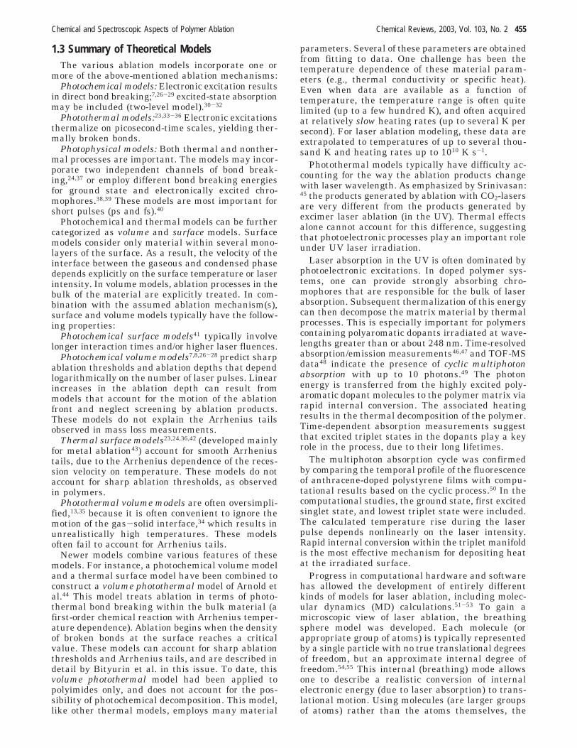
The ablation behavior of doped polymer systemswith absorbing dopants depends strongly on whetherabsorption actually decomposes the dopant molecule.Photolabile dopants that decompose to gaseous prod-ucts typically produce pronounced surface swelling
at low fluences (shown in Figure 1 a). Very highablation rates (up to 80 µm per pulse) can be achievedat high fluences,68 but always with pronounced signsof surface melting (shown in Figure 1b). In contrast,photostable dopants (e.g., polyaromatic compounds)produce much less surface swelling and lower abla-tion rates. As discussed above, the behavior of pho-tostable dopants can be understood in terms of cyclic-multiphoton absorption.
Recent studies of photochemically active, photo-labile, or photostable dopants include the following.
2.1.1 Photochemical Active Polyaromatic Compounds, asFluorescence Probes
These compounds are typically substituted naph-thalenes (i.e., Br and I substituted), which do notexhibit pronounced fluorescence. In contrast, thephotoproduct naphthalene exhibits a very pro-nounced emission (1B3u f 1A1g). This difference allowsone to probe the photochemical modification of thedopant by time-dependent fluorescence measure-ments. Georgiou et al. performed such measurementsusing polymethyl-methacrylate (PMMA) and van derWaals films as matrices69-72 (discussed in detail inthis issue) at two wavelengths (193 and 248 nm)73
and pulse lengths (30 ns and 500 fs).74-76 As shownin Figure 2, the onset of increased photoproduct yieldat 248 nm (i.e., naphthalene and naphthalene dimers)coincides with the onset of significant surface swell-
Figure 1. (a) Surface swelling and bubble formationduring ablation of PMMA doped with low dopant concen-tration (0.25 wt% of a dialkyl-aryl triazene-compound).Irradiation at 308 nm with single pulses and increasingfluence (from left to right). (b) PMMA doped with 2 wt% ofthe dopant and irradiated with 2 pulses with 5.8 J cm-2.
456 Chemical Reviews, 2003, Vol. 103, No. 2 Lippert and Dickinson
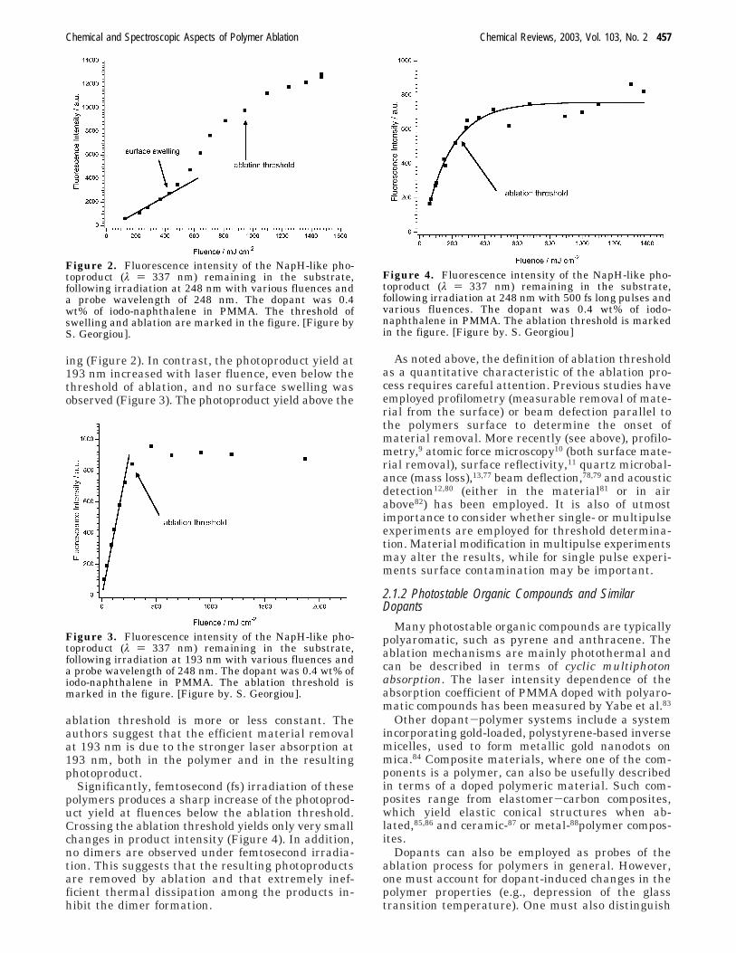
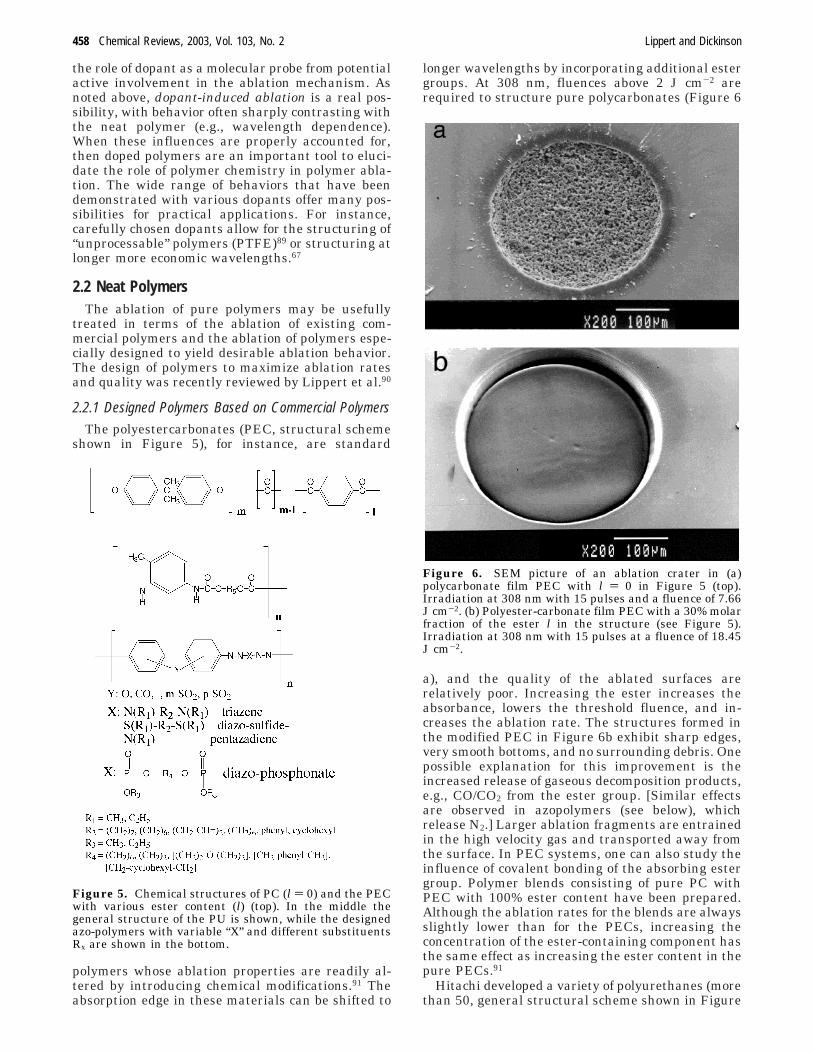
ing (Figure 2). In contrast, the photoproduct yield at193 nm increased with laser fluence, even below thethreshold of ablation, and no surface swelling wasobserved (Figure 3). The photoproduct yield above the
ablation threshold is more or less constant. Theauthors suggest that the efficient material removalat 193 nm is due to the stronger laser absorption at193 nm, both in the polymer and in the resultingphotoproduct.
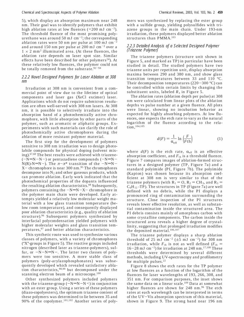
Significantly, femtosecond (fs) irradiation of thesepolymers produces a sharp increase of the photoprod-uct yield at fluences below the ablation threshold.Crossing the ablation threshold yields only very smallchanges in product intensity (Figure 4). In addition,no dimers are observed under femtosecond irradia-tion. This suggests that the resulting photoproductsare removed by ablation and that extremely inef-ficient thermal dissipation among the products in-hibit the dimer formation.
As noted above, the definition of ablation thresholdas a quantitative characteristic of the ablation pro-cess requires careful attention. Previous studies haveemployed profilometry (measurable removal of mate-rial from the surface) or beam defection parallel tothe polymers surface to determine the onset ofmaterial removal. More recently (see above), profilo-metry,9 atomic force microscopy10 (both surface mate-rial removal), surface reflectivity,11 quartz microbal-ance (mass loss),13,77 beam deflection,78,79 and acousticdetection12,80 (either in the material81 or in airabove82) has been employed. It is also of utmostimportance to consider whether single- or multipulseexperiments are employed for threshold determina-tion. Material modification in multipulse experimentsmay alter the results, while for single pulse experi-ments surface contamination may be important.
2.1.2 Photostable Organic Compounds and SimilarDopants
Many photostable organic compounds are typicallypolyaromatic, such as pyrene and anthracene. Theablation mechanisms are mainly photothermal andcan be described in terms of cyclic multiphotonabsorption. The laser intensity dependence of theabsorption coefficient of PMMA doped with polyaro-matic compounds has been measured by Yabe et al.83
Other dopant-polymer systems include a systemincorporating gold-loaded, polystyrene-based inversemicelles, used to form metallic gold nanodots onmica.84 Composite materials, where one of the com-ponents is a polymer, can also be usefully describedin terms of a doped polymeric material. Such com-posites range from elastomer-carbon composites,which yield elastic conical structures when ab-lated,85,86 and ceramic-87 or metal-88polymer compos-ites.
Dopants can also be employed as probes of theablation process for polymers in general. However,one must account for dopant-induced changes in thepolymer properties (e.g., depression of the glasstransition temperature). One must also distinguish
Figure 2. Fluorescence intensity of the NapH-like pho-toproduct (λ ) 337 nm) remaining in the substrate,following irradiation at 248 nm with various fluences anda probe wavelength of 248 nm. The dopant was 0.4wt% of iodo-naphthalene in PMMA. The threshold ofswelling and ablation are marked in the figure. [Figure byS. Georgiou].
Figure 3. Fluorescence intensity of the NapH-like pho-toproduct (λ ) 337 nm) remaining in the substrate,following irradiation at 193 nm with various fluences anda probe wavelength of 248 nm. The dopant was 0.4 wt% ofiodo-naphthalene in PMMA. The ablation threshold ismarked in the figure. [Figure by. S. Georgiou].
Figure 4. Fluorescence intensity of the NapH-like pho-toproduct (λ ) 337 nm) remaining in the substrate,following irradiation at 248 nm with 500 fs long pulses andvarious fluences. The dopant was 0.4 wt% of iodo-naphthalene in PMMA. The ablation threshold is markedin the figure. [Figure by. S. Georgiou]
Chemical and Spectroscopic Aspects of Polymer Ablation Chemical Reviews, 2003, Vol. 103, No. 2 457
the role of dopant as a molecular probe from potentialactive involvement in the ablation mechanism. Asnoted above, dopant-induced ablation is a real pos-sibility, with behavior often sharply contrasting withthe neat polymer (e.g., wavelength dependence).When these influences are properly accounted for,then doped polymers are an important tool to eluci-date the role of polymer chemistry in polymer abla-tion. The wide range of behaviors that have beendemonstrated with various dopants offer many pos-sibilities for practical applications. For instance,carefully chosen dopants allow for the structuring of“unprocessable” polymers (PTFE)89 or structuring atlonger more economic wavelengths.67
2.2 Neat PolymersThe ablation of pure polymers may be usefully
treated in terms of the ablation of existing com-mercial polymers and the ablation of polymers espe-cially designed to yield desirable ablation behavior.The design of polymers to maximize ablation ratesand quality was recently reviewed by Lippert et al.90
2.2.1 Designed Polymers Based on Commercial PolymersThe polyestercarbonates (PEC, structural scheme
shown in Figure 5), for instance, are standard
polymers whose ablation properties are readily al-tered by introducing chemical modifications.91 Theabsorption edge in these materials can be shifted to
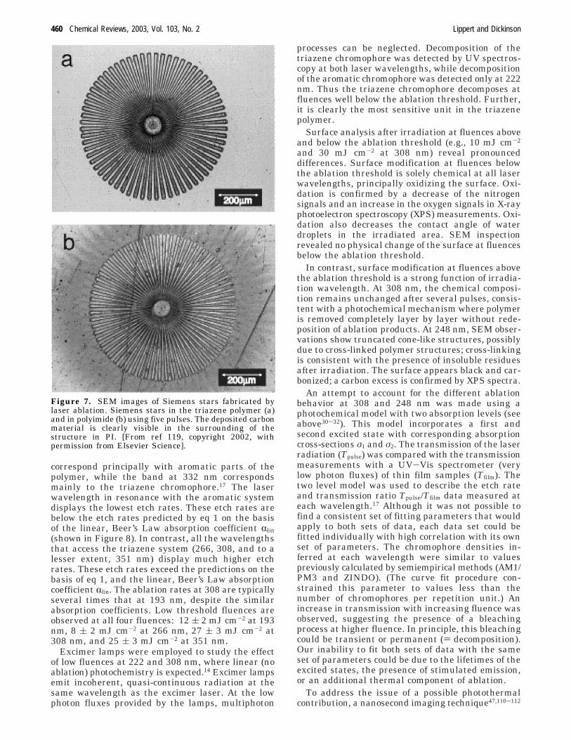
longer wavelengths by incorporating additional estergroups. At 308 nm, fluences above 2 J cm-2 arerequired to structure pure polycarbonates (Figure 6
a), and the quality of the ablated surfaces arerelatively poor. Increasing the ester increases theabsorbance, lowers the threshold fluence, and in-creases the ablation rate. The structures formed inthe modified PEC in Figure 6b exhibit sharp edges,very smooth bottoms, and no surrounding debris. Onepossible explanation for this improvement is theincreased release of gaseous decomposition products,e.g., CO/CO2 from the ester group. [Similar effectsare observed in azopolymers (see below), whichrelease N2.] Larger ablation fragments are entrainedin the high velocity gas and transported away fromthe surface. In PEC systems, one can also study theinfluence of covalent bonding of the absorbing estergroup. Polymer blends consisting of pure PC withPEC with 100% ester content have been prepared.Although the ablation rates for the blends are alwaysslightly lower than for the PECs, increasing theconcentration of the ester-containing component hasthe same effect as increasing the ester content in thepure PECs.91
Hitachi developed a variety of polyurethanes (morethan 50, general structural scheme shown in Figure
Figure 5. Chemical structures of PC (l ) 0) and the PECwith various ester content (l) (top). In the middle thegeneral structure of the PU is shown, while the designedazo-polymers with variable “X” and different substituentsRx are shown in the bottom.
Figure 6. SEM picture of an ablation crater in (a)polycarbonate film PEC with l ) 0 in Figure 5 (top).Irradiation at 308 nm with 15 pulses and a fluence of 7.66J cm-2. (b) Polyester-carbonate film PEC with a 30% molarfraction of the ester l in the structure (see Figure 5).Irradiation at 308 nm with 15 pulses at a fluence of 18.45J cm-2.
458 Chemical Reviews, 2003, Vol. 103, No. 2 Lippert and Dickinson
5), which display an absorption maximum near 248nm. Their goal was to identify polymers that exhibithigh ablation rates at low fluences (<200 mJ cm-2).The threshold fluence of the most promising poly-urethane was around 50 mJ cm-2; the correspondingablation rates were 50 nm per pulse at 100 mJ cm-2
and around 150 nm per pulse at 200 mJ cm-2 over a1 × 2 mm2 illuminated area. (At these fluences, theablation rate depends on laser spot size. Similareffects have been described for other polymers24). Atthese relatively low fluences, the polymer could notbe totally removed from the substrate.92-94
2.2.2 Novel Designed Polymers for Laser Ablation at 308nm
Irradiation at 308 nm is convenient from a com-mercial point of view due to the lifetime of opticalcomponents and laser gas (XeCl excimer) laser.Applications which do not require submicron resolu-tion are often well-served with 308 nm lasers. At 308nm, it is possible to selectively irradiate into theabsorption band of a photochemically active chro-mophore, with little absorption by other parts of thepolymer, such as aromatic or aliphatic groups. Ex-periments with such materials can clarify the role ofphotochemically active chromophores during theablation of more resistant polymer material.
The first step for the development of polymerssensitive to 308 nm irradiation was to design photo-labile compounds for physical doping (solvent mix-ing).67,68 The best results were achieved with triazene-(-NdN-N-) or pentazadiene compounds (-NdN-N(R)-NdN-). The π-π* transition of the -NdN-X- chromophore is near 300 nm.95 The chromophoresdecompose into N2 and other gaseous products, whichcan promote ablation. Early work indicated that thephotochemical properties of the dopants influencedthe resulting ablation characteristics.68 Subsequently,polymers containing the -NdN-X- chromophore inthe polymer main chain were developed. Early at-tempts yielded a relatively low molecular weight ma-terial with a low glass transition temperature (be-low room temperature), and consequently displayedpoor ablation characteristics (e.g., quality of ablationstructure).96 Subsequent polymers synthesized byinterfacial polycondensation yielded polymers withhigher molecular weights and glass transition tem-peratures,97 and better ablation characteristics.
This synthetic route was used to synthesize variousclasses of polymers, with a variety of chromophores(“X”-groups in Figure 5). The reactive groups includednitrogen (described later as triazene-polymers), sul-fur, or -N-NdN-. The latter two classes of poly-mers were too sensitive. A more stable class ofpolymers (poly-arylazophosphonates) was subse-quently developed which revealed satisfactory abla-tion characteristics,98,99 but decomposed under thescanning electron beam of a microscope.100
Other synthesized polymers included polymerswith the triazene-group (-NdN-N<) in conjunctionwith an ester group. Using a series of these polymers(diazo-copolyesters), the optimum triazene content ofthese polymers was determined to lie between 35 and90% of the copolymer.101,102 Another series of poly-
mers was synthesized by replacing the ester groupwith a sulfide group, yielding polysulfides with tri-azene groups in the main chain. Under 193-nmirradiation, these polymers displayed better ablationstructures than PMMA.103
2.2.3 Detailed Analysis of a Selected Designed Polymer(Triazene Polymer)
The triazene polymers (structure unit shown inFigure 5, and marked as TP) in particular have beenstudied in detail. The studied polymers have twotriazene units per repetition unit, display absorptionmaxima between 290 and 380 nm, and show glasstransition temperatures between 33 and 110 °C.Their decomposition temperatures (220-300 °C) canbe controlled within certain limits by changing thesubstituent units, labeled Rx in Figure 5.
The ablation rates (ablation depth per pulse) at 308nm were calculated from linear plots of the ablationdepths vs pulse number at a given fluence. All plotswere linear, showing no incubation behavior asexpected for highly absorbing polymers. At low flu-ences, one expects the etch rate to vary as the naturallogarithm of the fluence according to the rela-tion,104,105
where d(F) is the etch rate, Reff is an effectiveabsorption coefficient, and Fth is a threshold fluence.Figure 7 compares images of ablation-formed struc-tures in a designed polymer (TP, Figure 7a) and astandard polyimide (PI, Figure 7b). The polyimide(Kapton) was chosen because its absorption coef-ficient at 308 nm is very similar to that of thetriazene polymers (with R1 ) O, R2 ) CH3, and R3 )C6H12 -TP). The structures in TP (Figure 7a) are welldefined with no debris, while the PI displays apronounced ring of contamination surrounding thestructure. Close inspection of the PI structuresreveals lower effective resolution, as well as substan-tial contamination inside the structured area. ThePI debris consists mainly of amorphous carbon withsome crystalline components. The carbon inside thestructured area reveals a higher degree of crystal-linity, suggesting that prolonged irradiation modifiesthe deposited material.106,107
The triazene polymer displays a sharp ablationthreshold of 25 mJ cm-2 ((5 mJ cm-2) for 308 nmirradiation, while Fth is not as well defined (Fth )16-28 mJ cm-2) for irradiation at 248 nm.17,108 Thesethresholds were determined by several differentmethods, including UV-spectroscopy and profilometryfor multiple pulses.22
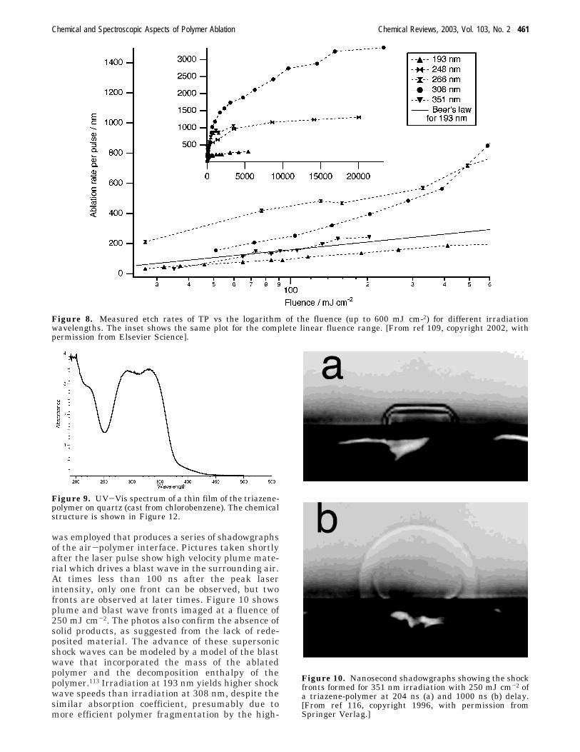
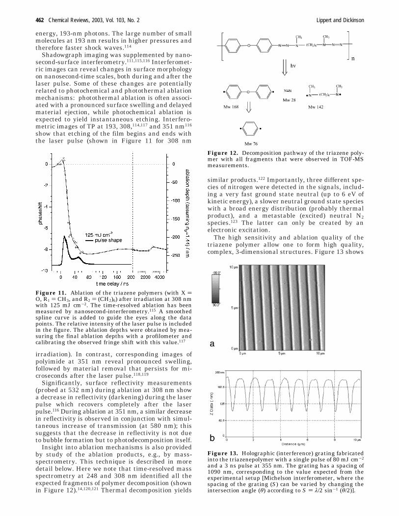
Figure 8 shows the etch rates for the TP polymerat low fluences as a function of the logarithm of thefluences for laser wavelengths of 193, 266, 308, and351 nm. For comparison purposes, the inset showsthe same data on a linear scale.109 Data at somewhathigher fluences are shown for 248 nm.96 The etchrates at each wavelength can be interpreted in termsof the UV-Vis absorption spectrum of this material,shown in Figure 9. The strong band near 196 nm
d(F) ) 1Reff
ln ( FFth) (1)
Chemical and Spectroscopic Aspects of Polymer Ablation Chemical Reviews, 2003, Vol. 103, No. 2 459
correspond principally with aromatic parts of thepolymer, while the band at 332 nm correspondsmainly to the triazene chromophore.17 The laserwavelength in resonance with the aromatic systemdisplays the lowest etch rates. These etch rates arebelow the etch rates predicted by eq 1 on the basisof the linear, Beer’s Law absorption coefficient Rlin(shown in Figure 8). In contrast, all the wavelengthsthat access the triazene system (266, 308, and to alesser extent, 351 nm) display much higher etchrates. These etch rates exceed the predictions on thebasis of eq 1, and the linear, Beer’s Law absorptioncoefficient Rlin. The ablation rates at 308 are typicallyseveral times that at 193 nm, despite the similarabsorption coefficients. Low threshold fluences areobserved at all four fluences: 12 ( 2 mJ cm-2 at 193nm, 8 ( 2 mJ cm-2 at 266 nm, 27 ( 3 mJ cm-2 at308 nm, and 25 ( 3 mJ cm-2 at 351 nm.
Excimer lamps were employed to study the effectof low fluences at 222 and 308 nm, where linear (noablation) photochemistry is expected.14 Excimer lampsemit incoherent, quasi-continuous radiation at thesame wavelength as the excimer laser. At the lowphoton fluxes provided by the lamps, multiphoton
processes can be neglected. Decomposition of thetriazene chromophore was detected by UV spectros-copy at both laser wavelengths, while decompositionof the aromatic chromophore was detected only at 222nm. Thus the triazene chromophore decomposes atfluences well below the ablation threshold. Further,it is clearly the most sensitive unit in the triazenepolymer.
Surface analysis after irradiation at fluences aboveand below the ablation threshold (e.g., 10 mJ cm-2
and 30 mJ cm-2 at 308 nm) reveal pronounceddifferences. Surface modification at fluences belowthe ablation threshold is solely chemical at all laserwavelengths, principally oxidizing the surface. Oxi-dation is confirmed by a decrease of the nitrogensignals and an increase in the oxygen signals in X-rayphotoelectron spectroscopy (XPS) measurements. Oxi-dation also decreases the contact angle of waterdroplets in the irradiated area. SEM inspectionrevealed no physical change of the surface at fluencesbelow the ablation threshold.
In contrast, surface modification at fluences abovethe ablation threshold is a strong function of irradia-tion wavelength. At 308 nm, the chemical composi-tion remains unchanged after several pulses, consis-tent with a photochemical mechanism where polymeris removed completely layer by layer without rede-position of ablation products. At 248 nm, SEM obser-vations show truncated cone-like structures, possiblydue to cross-linked polymer structures; cross-linkingis consistent with the presence of insoluble residuesafter irradiation. The surface appears black and car-bonized; a carbon excess is confirmed by XPS spectra.
An attempt to account for the different ablationbehavior at 308 and 248 nm was made using aphotochemical model with two absorption levels (seeabove30-32). This model incorporates a first andsecond excited state with corresponding absorptioncross-sections σ1 and σ2. The transmission of the laserradiation (Tpulse) was compared with the transmissionmeasurements with a UV-Vis spectrometer (verylow photon fluxes) of thin film samples (Tfilm). Thetwo level model was used to describe the etch rateand transmission ratio Tpulse/Tfilm data measured ateach wavelength.17 Although it was not possible tofind a consistent set of fitting parameters that wouldapply to both sets of data, each data set could befitted individually with high correlation with its ownset of parameters. The chromophore densities in-ferred at each wavelength were similar to valuespreviously calculated by semiempirical methods (AM1/PM3 and ZINDO). (The curve fit procedure con-strained this parameter to values less than thenumber of chromophores per repetition unit.) Anincrease in transmission with increasing fluence wasobserved, suggesting the presence of a bleachingprocess at higher fluence. In principle, this bleachingcould be transient or permanent () decomposition).Our inability to fit both sets of data with the sameset of parameters could be due to the lifetimes of theexcited states, the presence of stimulated emission,or an additional thermal component of ablation.
To address the issue of a possible photothermalcontribution, a nanosecond imaging technique47,110-112
Figure 7. SEM images of Siemens stars fabricated bylaser ablation. Siemens stars in the triazene polymer (a)and in polyimide (b) using five pulses. The deposited carbonmaterial is clearly visible in the surrounding of thestructure in PI. [From ref 119, copyright 2002, withpermission from Elsevier Science].
460 Chemical Reviews, 2003, Vol. 103, No. 2 Lippert and Dickinson
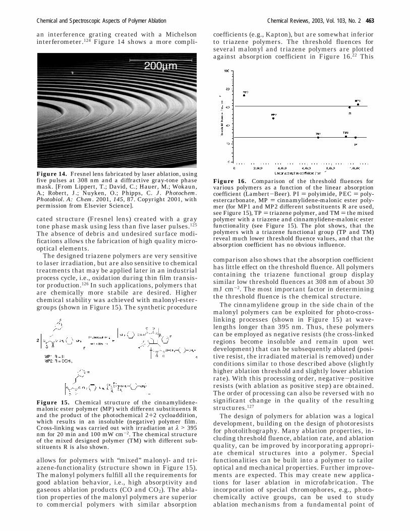
was employed that produces a series of shadowgraphsof the air-polymer interface. Pictures taken shortlyafter the laser pulse show high velocity plume mate-rial which drives a blast wave in the surrounding air.At times less than 100 ns after the peak laserintensity, only one front can be observed, but twofronts are observed at later times. Figure 10 showsplume and blast wave fronts imaged at a fluence of250 mJ cm-2. The photos also confirm the absence ofsolid products, as suggested from the lack of rede-posited material. The advance of these supersonicshock waves can be modeled by a model of the blastwave that incorporated the mass of the ablatedpolymer and the decomposition enthalpy of thepolymer.113 Irradiation at 193 nm yields higher shockwave speeds than irradiation at 308 nm, despite thesimilar absorption coefficient, presumably due tomore efficient polymer fragmentation by the high-
Figure 8. Measured etch rates of TP vs the logarithm of the fluence (up to 600 mJ cm-2) for different irradiationwavelengths. The inset shows the same plot for the complete linear fluence range. [From ref 109, copyright 2002, withpermission from Elsevier Science].
Figure 9. UV-Vis spectrum of a thin film of the triazene-polymer on quartz (cast from chlorobenzene). The chemicalstructure is shown in Figure 12.
Figure 10. Nanosecond shadowgraphs showing the shockfronts formed for 351 nm irradiation with 250 mJ cm-2 ofa triazene-polymer at 204 ns (a) and 1000 ns (b) delay.[From ref 116, copyright 1996, with permission fromSpringer Verlag.]
Chemical and Spectroscopic Aspects of Polymer Ablation Chemical Reviews, 2003, Vol. 103, No. 2 461
energy, 193-nm photons. The large number of smallmolecules at 193 nm results in higher pressures andtherefore faster shock waves.114
Shadowgraph imaging was supplemented by nano-second-surface interferometry.111,115,116 Interferomet-ric images can reveal changes in surface morphologyon nanosecond-time scales, both during and after thelaser pulse. Some of these changes are potentiallyrelated to photochemical and photothermal ablationmechanisms: photothermal ablation is often associ-ated with a pronounced surface swelling and delayedmaterial ejection, while photochemical ablation isexpected to yield instantaneous etching. Interfero-metric images of TP at 193, 308,114,117 and 351 nm116
show that etching of the film begins and ends withthe laser pulse (shown in Figure 11 for 308 nm
irradiation). In contrast, corresponding images ofpolyimide at 351 nm reveal pronounced swelling,followed by material removal that persists for mi-croseconds after the laser pulse.118,119
Significantly, surface reflectivity measurements(probed at 532 nm) during ablation at 308 nm showa decrease in reflectivity (darkening) during the laserpulse which recovers completely after the laserpulse.116 During ablation at 351 nm, a similar decreasein reflectivity is observed in conjunction with simul-taneous increase of transmission (at 580 nm); thissuggests that the decrease in reflectivity is not dueto bubble formation but to photodecomposition itself.
Insight into ablation mechanisms is also providedby study of the ablation products, e.g., by mass-spectrometry. This technique is described in moredetail below. Here we note that time-resolved massspectrometry at 248 and 308 nm identified all theexpected fragments of polymer decomposition (shownin Figure 12).14,120,121 Thermal decomposition yields
similar products.122 Importantly, three different spe-cies of nitrogen were detected in the signals, includ-ing a very fast ground state neutral (up to 6 eV ofkinetic energy), a slower neutral ground state specieswith a broad energy distribution (probably thermalproduct), and a metastable (excited) neutral N2species.123 The latter can only be created by anelectronic excitation.
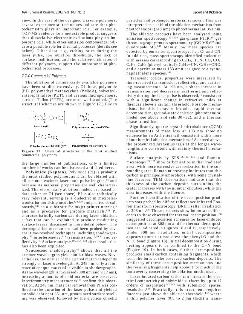
The high sensitivity and ablation quality of thetriazene polymer allow one to form high quality,complex, 3-dimensional structures. Figure 13 shows
Figure 11. Ablation of the triazene polymers (with X )O, R1 ) CH3, and R2 ) (CH2)6) after irradiation at 308 nmwith 125 mJ cm-2. The time-resolved ablation has beenmeasured by nanosecond-interferometry.115 A smoothedspline curve is added to guide the eyes along the datapoints. The relative intensity of the laser pulse is includedin the figure. The ablation depths were obtained by mea-suring the final ablation depths with a profilometer andcalibrating the observed fringe shift with this value.117
Figure 12. Decomposition pathway of the triazene poly-mer with all fragments that were observed in TOF-MSmeasurements.
Figure 13. Holographic (interference) grating fabricatedinto the triazenepolymer with a single pulse of 80 mJ cm-2
and a 3 ns pulse at 355 nm. The grating has a spacing of1090 nm, corresponding to the value expected from theexperimental setup [Michelson interferometer, where thespacing of the grating (S) can be varied by changing theintersection angle (θ) according to S ) λ/2 sin-1 (θ/2)].
462 Chemical Reviews, 2003, Vol. 103, No. 2 Lippert and Dickinson
an interference grating created with a Michelsoninterferometer.124 Figure 14 shows a more compli-
cated structure (Fresnel lens) created with a graytone phase mask using less than five laser pulses.125
The absence of debris and undesired surface modi-fications allows the fabrication of high quality micro-optical elements.
The designed triazene polymers are very sensitiveto laser irradiation, but are also sensitive to chemicaltreatments that may be applied later in an industrialprocess cycle, i.e., oxidation during thin film transis-tor production.126 In such applications, polymers thatare chemically more stabile are desired. Higherchemical stability was achieved with malonyl-ester-groups (shown in Figure 15). The synthetic procedure
allows for polymers with “mixed” malonyl- and tri-azene-functionality (structure shown in Figure 15).The malonyl polymers fulfill all the requirements forgood ablation behavior, i.e., high absorptivity andgaseous ablation products (CO and CO2). The abla-tion properties of the malonyl polymers are superiorto commercial polymers with similar absorption
coefficients (e.g., Kapton), but are somewhat inferiorto triazene polymers. The threshold fluences forseveral malonyl and triazene polymers are plottedagainst absorption coefficient in Figure 16.22 This
comparison also shows that the absorption coefficienthas little effect on the threshold fluence. All polymerscontaining the triazene functional group displaysimilar low threshold fluences at 308 nm of about 30mJ cm-2. The most important factor in determiningthe threshold fluence is the chemical structure.
The cinnamylidene group in the side chain of themalonyl polymers can be exploited for photo-cross-linking processes (shown in Figure 15) at wave-lengths longer than 395 nm. Thus, these polymerscan be employed as negative resists (the cross-linkedregions become insoluble and remain upon wetdevelopment) that can be subsequently ablated (posi-tive resist, the irradiated material is removed) underconditions similar to those described above (slightlyhigher ablation threshold and slightly lower ablationrate). With this processing order, negative-positiveresists (with ablation as positive step) are obtained.The order of processing can also be reversed with nosignificant change in the quality of the resultingstructures.127
The design of polymers for ablation was a logicaldevelopment, building on the design of photoresistsfor photolithography. Many ablation properties, in-cluding threshold fluence, ablation rate, and ablationquality, can be improved by incorporating appropri-ate chemical structures into a polymer. Specialfunctionalities can be built into a polymer to tailoroptical and mechanical properties. Further improve-ments are expected. This may create new applica-tions for laser ablation in microfabrication. Theincorporation of special chromophores, e.g., photo-chemically active groups, can be used to studyablation mechanisms from a fundamental point of
Figure 16. Comparison of the threshold fluences forvarious polymers as a function of the linear absorptioncoefficient (Lambert-Beer). PI ) polyimide, PEC ) poly-estercarbonate, MP ) cinnamylidene-malonic ester poly-mer (for MP1 and MP2 different substituents R are used,see Figure 15), TP ) triazene polymer, and TM ) the mixedpolymer with a triazene and cinnamylidene-malonic esterfunctionality (see Figure 15). The plot shows, that thepolymers with a triazene functional group (TP and TM)reveal much lower threshold fluence values, and that theabsorption coefficient has no obvious influence.
Figure 14. Fresnel lens fabricated by laser ablation, usingfive pulses at 308 nm and a diffractive gray-tone phasemask. [From Lippert, T.; David, C.; Hauer, M.; Wokaun,A.; Robert, J.; Nuyken, O.; Phipps, C. J. Photochem.Photobiol. A: Chem. 2001, 145, 87. Copyright 2001, withpermission from Elsevier Science].
Figure 15. Chemical structure of the cinnamylidene-malonic ester polymer (MP) with different substituents Rand the product of the photochemical 2+2 cycloaddition,which results in an insoluble (negative) polymer film.Cross-linking was carried out with irradiation at λ > 395nm for 20 min and 100 mW cm-2. The chemical structureof the mixed designed polymer (TM) with different sub-stituents R is also shown.
Chemical and Spectroscopic Aspects of Polymer Ablation Chemical Reviews, 2003, Vol. 103, No. 2 463
view. In the case of the designed triazene polymers,several experimental techniques indicate that pho-tochemistry plays an important role. For example,TOF-MS evidence for a metastable product suggeststhat dissociative electronic excitations play an im-portant role, while other emission components indi-cate a possible role for thermal processes (details seebelow). Other data, e.g., etching rates during thelaser pulse, low ablation thresholds, the lack ofsurface modification, and the relative etch rates ofdifferent polymers, support the importance of pho-tochemical processes.
2.2.4 Commercial PolymersThe ablation of commercially available polymers
have been studied extensively. Of these, polyimide(PI), poly-methyl methacrylate (PMMA), polyethyl-enterephthalate (PET), and various fluoropolymers,such as Teflon (PTFE), are most well studied. (Thestructural schemes are shown in Figure 17.) Due to
the large number of publications, only a limitednumber of works can be discussed and cited here.
Polyimide (Kapton). Polyimide (PI) is probablythe most studied polymer, as it can be ablated withall common excimer lasers and pulse lengths,44 andbecause its material properties are well character-ized. Therefore, many ablation models are based ondata taken on PI (see above). PI is also technicallyvery relevant, serving as a dielectric in microelec-tronics for multichip modules128,129 and printed circuitboards,130 as a substrate for inkjet printer nozzles,5and as a precursor for graphite materials.131 PIcharacteristically carbonizes during laser ablation,a fact that can be exploited to produce conductingsurface layers (discussed in more detail below). Thedecomposition mechanism had been probed by sev-eral time-resolved techniques, including shadowgra-phy,45 interferometry,118 transmission,15,29,30 and re-flectivity.15 Surface analysis106,132-138 after irradiationhas also been exploited.
Nanosecond shadowgraphy45 shows that all theexcimer wavelengths yield similar blast waves. Nev-ertheless, the nature of the ejected material dependsstrongly on laser wavelength. At 248 nm, only a fainttrace of opaque material is visible in shadowgraphs.As the wavelength is increased (308 nm and 9.17 µm),increasing amounts of solid material are observed.Interferometry measurements118 confirm this obser-vation. At 248 nm, material removal from PI was con-fined to the duration of the laser pulse and yieldedno solid debris; at 351 nm, pronounced surface swell-ing was observed, followed by the ejection of solid
particles and prolonged material removal. This wasinterpreted as a shift of the ablation mechanism fromphotochemical (248 nm) to photothermal at 351 nm.
The ablation products have been analyzed usingemission spectroscopy,137,139 gas-phase FTIR,18 gaschromatography-mass spectrometry (GC-MS)140 andquadrupole MS.141 Mainly low mass species aredetected by emission spectroscopy, i.e., C2 and CN.In addition, mass spectroscopy identified moleculeswith masses corresponding to C2H2, HCN, CO, CO2,C4H2, C6H2 (phenyl radical), C6H5-CN, C6H5-CNO,and a species at mass 153 amu assigned to a cyano-naphathalene species.141
Transient optical properties were measured bytime-resolved transmission, reflectivity, and scatter-ing measurements. At 193 nm, a sharp increase intransmission and decrease in scattering and reflec-tivity during the laser pulse is observed,15 consistentwith a significant change in refractive index atfluences above a certain threshold. Possible mecha-nisms for this behavior include: rapid thermaldecomposition, ground-state depletion (photochemicalmodel; see above and refs 30-32), and a thermalphase transition.
Significantly, quartz crystal microbalance (QCM)measurements of mass loss at 193 nm show noevidence for an Arrhenius tail, consistent with a morephotochemical ablation mechanism.13 As noted above,the pronounced Arrhenius tails at the longer wave-lengths are consistent with mainly thermal mecha-nisms.
Surface analysis by XPS106,132-136 and Raman-microscopy106,107 show carbonization in the irradiatedarea, with more extensive carbonization in the sur-rounding area. Raman microscopy indicates that thiscarbon is principally amorphous, with some crystal-line features. TEM observations indicate that thethickness of the carbon deposits surrounding thecrater increases with the number of pulses, while thearea increases with the fluence.
Further identification of the decomposition prod-ucts was probed by diffuse reflectance infrared Fou-rier transform spectroscopy (DRIFT) after irradiationat 308 nm.137 These products show important differ-ences to those observed for thermal decomposition.138
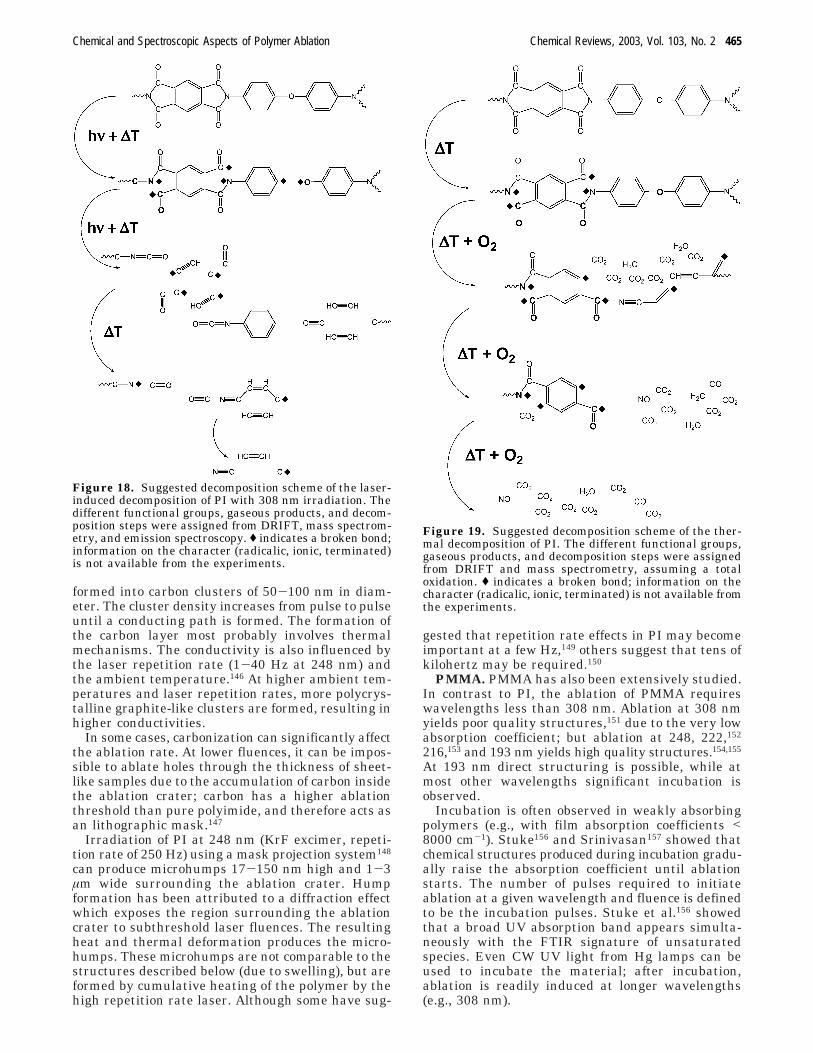
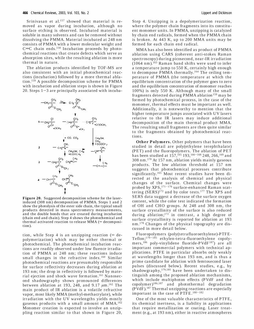
Suggested decomposition schemes for laser-induceddecomposition at 308 nm and for thermal decomposi-tion are indicated in Figures 18 and 19, respectively.Under 308 nm irradiation, initial decompositionappears to occur at two sites: the phenyl-O and theN-C bond (Figure 18). Initial decomposition duringheating appears to be confined to the C-N bond(Figure 19). In both cases, further decompositionproduces small carbon containing fragments, whichform the bulk of the observed carbon deposits. Thesimilarity of these decomposition mechanisms andthe resulting fragments help account for much of thecontroversy concerning the ablation mechanism.
Laser-induced carbonization can increase the elec-trical conductivity of polyimide surfaces by up to 17orders of magnitude142,143 with submicron spatialresolution.144 Practically, this treatment requiresfluences just above the ablation threshold,145 wherea thin polymer layer (0.5 to 2 µm thick) is trans-
Figure 17. Chemical structures of the most studiedcommercial polymers.
464 Chemical Reviews, 2003, Vol. 103, No. 2 Lippert and Dickinson
formed into carbon clusters of 50-100 nm in diam-eter. The cluster density increases from pulse to pulseuntil a conducting path is formed. The formation ofthe carbon layer most probably involves thermalmechanisms. The conductivity is also influenced bythe laser repetition rate (1-40 Hz at 248 nm) andthe ambient temperature.146 At higher ambient tem-peratures and laser repetition rates, more polycrys-talline graphite-like clusters are formed, resulting inhigher conductivities.
In some cases, carbonization can significantly affectthe ablation rate. At lower fluences, it can be impos-sible to ablate holes through the thickness of sheet-like samples due to the accumulation of carbon insidethe ablation crater; carbon has a higher ablationthreshold than pure polyimide, and therefore acts asan lithographic mask.147
Irradiation of PI at 248 nm (KrF excimer, repeti-tion rate of 250 Hz) using a mask projection system148
can produce microhumps 17-150 nm high and 1-3µm wide surrounding the ablation crater. Humpformation has been attributed to a diffraction effectwhich exposes the region surrounding the ablationcrater to subthreshold laser fluences. The resultingheat and thermal deformation produces the micro-humps. These microhumps are not comparable to thestructures described below (due to swelling), but areformed by cumulative heating of the polymer by thehigh repetition rate laser. Although some have sug-
gested that repetition rate effects in PI may becomeimportant at a few Hz,149 others suggest that tens ofkilohertz may be required.150
PMMA. PMMA has also been extensively studied.In contrast to PI, the ablation of PMMA requireswavelengths less than 308 nm. Ablation at 308 nmyields poor quality structures,151 due to the very lowabsorption coefficient; but ablation at 248, 222,152
216,153 and 193 nm yields high quality structures.154,155
At 193 nm direct structuring is possible, while atmost other wavelengths significant incubation isobserved.
Incubation is often observed in weakly absorbingpolymers (e.g., with film absorption coefficients <8000 cm-1). Stuke156 and Srinivasan157 showed thatchemical structures produced during incubation gradu-ally raise the absorption coefficient until ablationstarts. The number of pulses required to initiateablation at a given wavelength and fluence is definedto be the incubation pulses. Stuke et al.156 showedthat a broad UV absorption band appears simulta-neously with the FTIR signature of unsaturatedspecies. Even CW UV light from Hg lamps can beused to incubate the material; after incubation,ablation is readily induced at longer wavelengths(e.g., 308 nm).
Figure 18. Suggested decomposition scheme of the laser-induced decomposition of PI with 308 nm irradiation. Thedifferent functional groups, gaseous products, and decom-position steps were assigned from DRIFT, mass spectrom-etry, and emission spectroscopy. ( indicates a broken bond;information on the character (radicalic, ionic, terminated)is not available from the experiments.
Figure 19. Suggested decomposition scheme of the ther-mal decomposition of PI. The different functional groups,gaseous products, and decomposition steps were assignedfrom DRIFT and mass spectrometry, assuming a totaloxidation. ( indicates a broken bond; information on thecharacter (radicalic, ionic, terminated) is not available fromthe experiments.
Chemical and Spectroscopic Aspects of Polymer Ablation Chemical Reviews, 2003, Vol. 103, No. 2 465
Srinivasan et al.157 showed that material is re-moved as vapor during incubation, although nosurface etching is observed. Incubated material issoluble in many solvents and can be removed withoutdissolving the PMMA. Material incubated at 248 nmconsists of PMMA with a lower molecular weight andCdC chain ends.158 Incubation proceeds by photo-chemical reactions that create defects which serve asabsorption sites, while the resulting ablation is morethermal in nature.
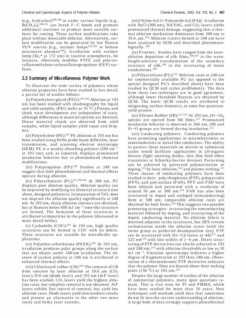
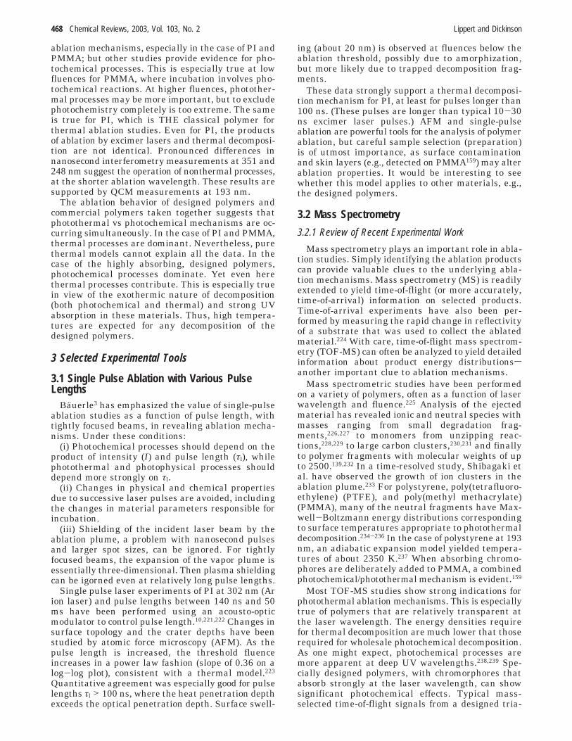
The ablation products identified by TOF-MS arealso consistent with an initial photochemical reac-tions (incubation) followed by a more thermal abla-tion.159 A possible decomposition scheme for PMMAwith incubation and ablation steps is shown in Figure20. Steps 1-3 are principally associated with incuba-
tion, while Step 4 is an unzipping reaction () de-polymerization) which may be either thermal orphotochemical. The photochemical incubation reac-tions are readily observed under low fluence irradia-tion of PMMA at 248 nm; these reactions inducesmall changes in the refractive index.160 Similarphotochemical reactions are presumably responsiblefor surface reflectivity decreases during ablation at193 nm; the drop in reflectivity is followed by mate-rial ejection and shock wave formation.161 Nanosec-ond shadowgraphy shows pronounced differencesbetween ablation at 193, 248, and 9.17 µm.162 Themain product of IR ablation is a volatile refractivevapor, most likely MMA (methyl-methacrylate), whileirradiation with the UV wavelengths yields mostlygaseous products with a small amount of MMA.162
Monomer creation is expected to involve an unzip-pling reaction similar to that shown in Figure 20,
Step 4. Unzipping is a depolymerization reaction,where the polymer chain fragments into its constitu-ent monomer units. In PMMA, unzipping is catalyzedby chain end radicals, formed when the PMMA chainis broken. At 443 K, up to 200 MMA units may beformed for each chain end radical.
MMA has also been identified as product of PMMAablation using CARS (coherent anti-stokes Ramanspectroscopy) during picosecond, near-IR irradiation(1064 nm).163 Raman band shifts were used to infera temperature jump to 550 K, certainly high enoughto decompose PMMA thermally.164 The ceiling tem-perature of PMMA (the temperature at which theequilibrium concentration of the polymer goes to zeroand the equilibrium concentration of monomer reaches100%) is only 550 K. Although many of the smallfragments detected during PMMA ablation159 may beformed by photochemical process, in the case of themonomer, thermal effects must be important as well.Additionally, it is noteworthy to mention that thehigher temperature jumps associated with UV lasersrelative to the IR lasers may induce additionaldecomposition of the main thermal product MMA.The resulting small fragments are then quite similarto the fragments obtained by photochemical reac-tions.
Other Polymers. Other polymers that have beenstudied in detail are poly(ethylene terephthalate)(PET) and the fluoropolymers. The ablation of PEThas been studied at 157,165 193,166-168 248, 266,169 and308 nm.170 At 157 nm, ablation yields mainly gaseousproducts. The low ablation threshold at 157 nmsuggests that photochemical processes contributesignificantly.165 Most recent studies have been di-rected at the analysis of chemical and physicalchanges of the surface. Chemical changes wereprobed by XPS,171-176 surface-enhanced Raman scat-tering (SERS)172 and by color tests.177 The XPS andSERS data suggest a decrease of the surface oxygencontent, while the color test indicated the formationof OH and CHO groups. At 248 and 308 nm, thesurface crystallinity of the surface is also reducedduring ablation;172 in contrast, a high degree ofsurface crystallinity is reported for ablation at 193nm.178 Changes of the physical topography are dis-cussed in more detail below.
Fluoropolymers (poly(tetrafluoroethylene)-PTFE-Teflon;179-185 ethylen-tetra-fluoroethylene copoly-mers,186 poly-vinylidene fluoride-PVdF187) are allimportant commercial polymers with technical ap-plications. PTFE in particular absorbs only weaklyat wavelengths longer than 193 nm, and is thus aprime candidate for ablation with femtosecond laserpulses (discussed below). Recent studies, e.g., byshadowgraphy,179,185 have been undertaken to dis-tinguish among the proposed ablation mechanisms,which include multiphoton effects (PVdF and thecopolymer)186,187 and photothermal degradation(PVdF).187 Thermal unzipping reactions are especiallyimportant in the case of PTFE.180-182
One of the most valuable characteristics of PTFE,its chemical inertness, is a liability in applicationsthat require metallization or coating. Laser treat-ment (e.g., at 193 nm), either in reactive atmospheres
Figure 20. Suggested decomposition scheme for the laser-induced (308 nm) decomposition of PMMA. Steps 1 and 2show the photolysis of the ester side chain, the typical smallproducts detected in mass spectrometry measurements,and the double bonds that are created during incubation(chain end and chain). Step 4 shows the photochemical andthermal activated reaction to release MMA () decomposi-tion).
466 Chemical Reviews, 2003, Vol. 103, No. 2 Lippert and Dickinson
(e.g., hydrazine)188,189 or under various liquids [e.g.,B(CH3)3],190,191 can break F-C bonds and promoteadditional reactions to produce more suitable sur-faces for coating. These surface modifications takeplace without detectable ablation. Alternatively, sur-face modification can be generated by low fluenceVUV sources (e.g., excimer lamps192,193 or heliummicrowave plasmas194). Irradiation with excimerlamp (Xe2* at 172 nm) in reactive atmospheres, forinstance, effectively modifies PTFE and poly-tet-rafluoroethylene-co-hexafluoropropylene (FEP) sur-faces.
2.3 Summary of Miscellaneous Polymer WorkTo illustrate the wide variety of polymers whose
ablation properties have been studied in less detail,a partial list of studies follows:
(i) Polyethylene-glycol (PEG):195,196 Ablation at 193nm has been studied with shadowgraphy for liquidand solid samples. (PEG melts at 35 °C.) Plasma andshock wave development are independent of state,although differences in material ejection are detected.Dense material clouds are observed from solidsamples, while liquid samples yield vapor and drop-lets.
(ii) Polyethylene (PE):197 PE ablation at 193 nm hasbeen studied using HeNe probe beam deflection, lasertransmission, and scanning electron microscopy(SEM). PE is a weakly absorbing polymer (500 cm-1
at 193 nm), and, as expected, displays pronouncedincubation behavior due to photoinduced chemicalmodifications.
(iii) Polypropylene (PP):198 Studies at 248 nmsuggest that both photochemical and thermal effectsoperate during ablation.
(iv) Polycarbonates (PC):91,199,200 At 308 nm, PCdisplays poor ablation quality. Ablation quality canbe improved by modifying its chemical structure (seeabove, designed polymers). Incorporation of TiO2 doesnot improve the ablation quality significantly at 248nm. At 193 nm, sharp ablation contours are obtained,but at fluences below 600 mJ cm-2 cone-like structureare formed. The formation of these structures isattributed to impurities in the polymer (discussed inmore detail below).
(v) Cycloolefin (COC):201 At 193 nm, high qualitystructures can be formed in COC with no debris.These structures are suitable for microfluidic ap-plications.
(vi) Polyether-etherketone (PEEK):202 At 193 nm,irradiation produces polar groups along the surfacethat are absent under 248-nm irradiation. The ab-sence of surface polarity at 248 nm is attributed toenhanced thermal effects.
(vii) Chlorinated rubber (CR):203 The removal of CRfrom concrete by laser ablation at 10.6 µm (CO2laser), 810 nm (diode laser), and 193 nm (ArF laser)has been studied. CO2 lasers yield the highest abla-tion rates, but complete removal is not obtained. ArFlasers exhibit fine control of removal, but yield lowablation rates. Diode lasers yield intermediate resultsand present an alternative to the other two morecostly and bulky laser systems.
(viii) Nylon-6,6 [) Polyamide-6,6 (PA)]: Irradiationwith XeCl (308 nm), Nd:YAG, and CO2 lasers yieldspronounced thermal damage, suggesting that a ther-mal ablation mechanism dominates from 308 nm to10.6. µm.204 Ablation craters formed at 248 nm havebeen analyzed by SEM and described phenomeno-logically. 205
(ix) Proteins: Studies have ranged from the laser-ablation deposition of silk films,206,207 to the wave-length-selective transformation of the secondarystructure of silk,208 to the structuring of insectexoskeletons.209
(x) Polyurethane (PU):210 Ablation rates at 248 nmfor commercially available PU (as opposed to thespecial designed PU’s described above) have beenstudied by QCM and stylus profilometry. The datafrom these two techniques are in good agreement,although lower threshold fluences are obtained byQCM. The lower QCM results are attributed tooutgassing, surface chemistry, or some low quantum-yield process.
(xi) Silicone Rubber (SR):211,212 At 193 nm, (Si-O)nspecies are ejected from SR films.211 Pronouncedincubation behavior is observed at 266 nm; OH andSi-O groups are formed during incubation.212
(xii) Conducting polymers: Conducting polymershave promising applications in microelectronics assemiconductors or metal-like conductors. The abilityto pattern these materials on micron or submicronscales would facilitate applications in all-organicdevices (light emitting diodes, thin film field effecttransistors or Schottky-barrier devices). Patterningcan be achieved by patterned polymerization ordoping,213 by laser ablation,214 and plasma etching.215
Three classes of conducting polymers have beenstudied to date: poly-thiophenes (PTP), polypyrroles(PPY), and poly-aniline (PAN). PPY and PAN havebeen ablated and patterned with a resolution ofaround 50 µm at 308 nm.214 PAN has also beenstructured in doped and undoped (nonconducting)form at 308 nm; comparable ablation rates areobtained for both forms.216 This suggests two possibleprocessing strategies: structuring the nonconductingmaterial followed by doping, and structuring of thedoped, conducting material. No ablation debris isdetected adjacent to the structures, but XPS revealscarbonization inside the ablation crater (with theimine group as preferred decomposition site). PTPcan be structured with He-Cd lasers at 442217 and325 nm218 with line widths of 1-6 µm. Direct struc-turing of PTP derivatives can also be achieved at 193and 248 nm,219 with ablation thresholds as low as 30mJ cm-2. Emission spectroscopy indicates a higherdegree of fragmentation at 193 than 248 nm. Obser-vation of a thermochromic PTP derivative indicatesthat the polymer films are heated above their meltingpoint (136 °C) at 193 nm.220
Despite the large number of studies of the ablationof commercial polymers, many open questions re-main. This is true even for PI and PMMA, whichhave been studied for more than 20 years. Newtechniques and methods yield data that sometimesdo not fit into the current understanding of ablation.A large body of data strongly supports photothermal
Chemical and Spectroscopic Aspects of Polymer Ablation Chemical Reviews, 2003, Vol. 103, No. 2 467
ablation mechanisms, especially in the case of PI andPMMA; but other studies provide evidence for pho-tochemical processes. This is especially true at lowfluences for PMMA, where incubation involves pho-tochemical reactions. At higher fluences, photother-mal processes may be more important, but to excludephotochemistry completely is too extreme. The sameis true for PI, which is THE classical polymer forthermal ablation studies. Even for PI, the productsof ablation by excimer lasers and thermal decomposi-tion are not identical. Pronounced differences innanosecond interferometry measurements at 351 and248 nm suggest the operation of nonthermal processes,at the shorter ablation wavelength. These results aresupported by QCM measurements at 193 nm.
The ablation behavior of designed polymers andcommercial polymers taken together suggests thatphotothermal vs photochemical mechanisms are oc-curring simultaneously. In the case of PI and PMMA,thermal processes are dominant. Nevertheless, purethermal models cannot explain all the data. In thecase of the highly absorbing, designed polymers,photochemical processes dominate. Yet even herethermal processes contribute. This is especially truein view of the exothermic nature of decomposition(both photochemical and thermal) and strong UVabsorption in these materials. Thus, high tempera-tures are expected for any decomposition of thedesigned polymers.
3 Selected Experimental Tools
3.1 Single Pulse Ablation with Various PulseLengths
Bauerle3 has emphasized the value of single-pulseablation studies as a function of pulse length, withtightly focused beams, in revealing ablation mecha-nisms. Under these conditions:
(i) Photochemical processes should depend on theproduct of intensity (I) and pulse length (τl), whilephotothermal and photophysical processes shoulddepend more strongly on τl.
(ii) Changes in physical and chemical propertiesdue to successive laser pulses are avoided, includingthe changes in material parameters responsible forincubation.
(iii) Shielding of the incident laser beam by theablation plume, a problem with nanosecond pulsesand larger spot sizes, can be ignored. For tightlyfocused beams, the expansion of the vapor plume isessentially three-dimensional. Then plasma shieldingcan be igorned even at relatively long pulse lengths.
Single pulse laser experiments of PI at 302 nm (Arion laser) and pulse lengths between 140 ns and 50ms have been performed using an acousto-opticmodulator to control pulse length.10,221,222 Changes insurface topology and the crater depths have beenstudied by atomic force microscopy (AFM). As thepulse length is increased, the threshold fluenceincreases in a power law fashion (slope of 0.36 on alog-log plot), consistent with a thermal model.223
Quantitative agreement was especially good for pulselengths τl > 100 ns, where the heat penetration depthexceeds the optical penetration depth. Surface swell-
ing (about 20 nm) is observed at fluences below theablation threshold, possibly due to amorphization,but more likely due to trapped decomposition frag-ments.
These data strongly support a thermal decomposi-tion mechanism for PI, at least for pulses longer than100 ns. (These pulses are longer than typical 10-30ns excimer laser pulses.) AFM and single-pulseablation are powerful tools for the analysis of polymerablation, but careful sample selection (preparation)is of utmost importance, as surface contaminationand skin layers (e.g., detected on PMMA159) may alterablation properties. It would be interesting to seewhether this model applies to other materials, e.g.,the designed polymers.
3.2 Mass Spectrometry
3.2.1 Review of Recent Experimental Work
Mass spectrometry plays an important role in abla-tion studies. Simply identifying the ablation productscan provide valuable clues to the underlying abla-tion mechanisms. Mass spectrometry (MS) is readilyextended to yield time-of-flight (or more accurately,time-of-arrival) information on selected products.Time-of-arrival experiments have also been per-formed by measuring the rapid change in reflectivityof a substrate that was used to collect the ablatedmaterial.224 With care, time-of-flight mass spectrom-etry (TOF-MS) can often be analyzed to yield detailedinformation about product energy distributionssanother important clue to ablation mechanisms.
Mass spectrometric studies have been performedon a variety of polymers, often as a function of laserwavelength and fluence.225 Analysis of the ejectedmaterial has revealed ionic and neutral species withmasses ranging from small degradation frag-ments,226,227 to monomers from unzipping reac-tions,228,229 to large carbon clusters,230,231 and finallyto polymer fragments with molecular weights of upto 2500.139,232 In a time-resolved study, Shibagaki etal. have observed the growth of ion clusters in theablation plume.233 For polystyrene, poly(tetrafluoro-ethylene) (PTFE), and poly(methyl methacrylate)(PMMA), many of the neutral fragments have Max-well-Boltzmann energy distributions correspondingto surface temperatures appropriate to photothermaldecomposition.234-236 In the case of polystyrene at 193nm, an adiabatic expansion model yielded tempera-tures of about 2350 K.237 When absorbing chromo-phores are deliberately added to PMMA, a combinedphotochemical/photothermal mechanism is evident.159
Most TOF-MS studies show strong indications forphotothermal ablation mechanisms. This is especiallytrue of polymers that are relatively transparent atthe laser wavelength. The energy densities requirefor thermal decomposition are much lower that thoserequired for wholesale photochemical decomposition.As one might expect, photochemical processes aremore apparent at deep UV wavelengths.238,239 Spe-cially designed polymers, with chromorphores thatabsorb strongly at the laser wavelength, can showsignificant photochemical effects. Typical mass-selected time-of-flight signals from a designed tria-
468 Chemical Reviews, 2003, Vol. 103, No. 2 Lippert and Dickinson
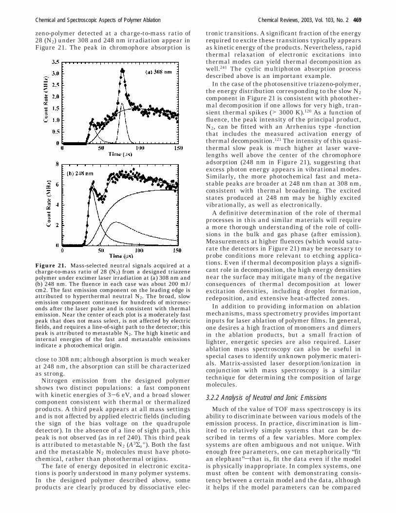
zeno-polymer detected at a charge-to-mass ratio of28 (N2) under 308 and 248 nm irradiation appear inFigure 21. The peak in chromophore absorption is
close to 308 nm; although absorption is much weakerat 248 nm, the absorption can still be characterizedas strong.
Nitrogen emission from the designed polymershows two distinct populations: a fast componentwith kinetic energies of 3-6 eV, and a broad slowercomponent consistent with thermal or thermalizedproducts. A third peak appears at all mass settingsand is not affected by applied electric fields (includingthe sign of the bias voltage on the quadrupoledetector). In the absence of a line of sight path, thispeak is not observed (as in ref 240). This third peakis attributed to metastable N2 (A3Σu
+). Both the fastand the metastable N2 molecules must have photo-chemical, rather than photothermal origins.
The fate of energy deposited in electronic excita-tions is poorly understood in many polymer systems.In the designed polymer described above, someproducts are clearly produced by dissociative elec-
tronic transitions. A significant fraction of the energyrequired to excite these transitions typically appearsas kinetic energy of the products. Nevertheless, rapidthermal relaxation of electronic excitations intothermal modes can yield thermal decomposition aswell.241 The cyclic multiphoton absorption processdescribed above is an important example.
In the case of the photosensitive triazeno-polymer,the energy distribution corresponding to the slow N2component in Figure 21 is consistent with photother-mal decomposition if one allows for very high, tran-sient thermal spikes (> 3000 K).120 As a function offluence, the peak intensity of the principal product,N2, can be fitted with an Arrhenius type -functionthat includes the measured activation energy ofthermal decomposition.121 The intensity of this quasi-thermal slow peak is much higher at laser wave-lengths well above the center of the chromophoreadsorption (248 nm in Figure 21), suggesting thatexcess photon energy appears in vibrational modes.Similarly, the more photochemical fast and meta-stable peaks are broader at 248 nm than at 308 nm,consistent with thermal broadening. The excitedstates produced at 248 nm may be highly excitedvibrationally, as well as electronically.
A definitive determination of the role of thermalprocesses in this and similar materials will requirea more thorough understanding of the role of colli-sions in the bulk and gas phase (after emission).Measurements at higher fluences (which would satu-rate the detectors in Figure 21) may be necessary toprobe conditions more relevant to etching applica-tions. Even if thermal decomposition plays a signifi-cant role in decomposition, the high energy densitiesnear the surface may mitigate many of the negativeconsequences of thermal decomposition at lowerexcitation densities, including droplet formation,redeposition, and extensive heat-affected zones.
In addition to providing information on ablationmechanisms, mass spectrometry provides importantinputs for laser ablation of polymer films. In general,one desires a high fraction of monomers and dimersin the ablation products, but a small fraction oflighter, energetic species are also required. Laserablation mass spectroscopy can also be useful inspecial cases to identify unknown polymeric materi-als. Matrix-assisted laser desorption/ionization inconjunction with mass spectroscopy is a similartechnique for determining the composition of largemolecules.
3.2.2 Analysis of Neutral and Ionic Emissions
Much of the value of TOF mass spectroscopy is itsability to discriminate between various models of theemission process. In practice, discrimination is lim-ited to relatively simple systems that can be de-scribed in terms of a few variables. More complexsystems are often ambiguous and not unique. Withenough free parameters, one can metaphorically “fitan elephant”sthat is, fit the data even if the modelis physically inappropriate. In complex systems, onemust often be content with demonstrating consis-tency between a certain model and the data, althoughit helps if the model parameters can be compared
Figure 21. Mass-selected neutral signals acquired at acharge-to-mass ratio of 28 (N2) from a designed triazenepolymer under excimer laser irradiation at (a) 308 nm and(b) 248 nm. The fluence in each case was about 200 mJ/cm2. The fast emission component on the leading edge isattributed to hyperthermal neutral N2. The broad, slowemission component continues for hundreds of microsec-onds after the laser pulse and is consistent with thermalemission. Near the center of each plot is a moderately fastpeak that does not mass select, is not affected by electricfields, and requires a line-of-sight path to the detector; thispeak is attributed to metastable N2. The high kinetic andinternal energies of the fast and metastable emissionsindicate a photochemical origin.
Chemical and Spectroscopic Aspects of Polymer Ablation Chemical Reviews, 2003, Vol. 103, No. 2 469
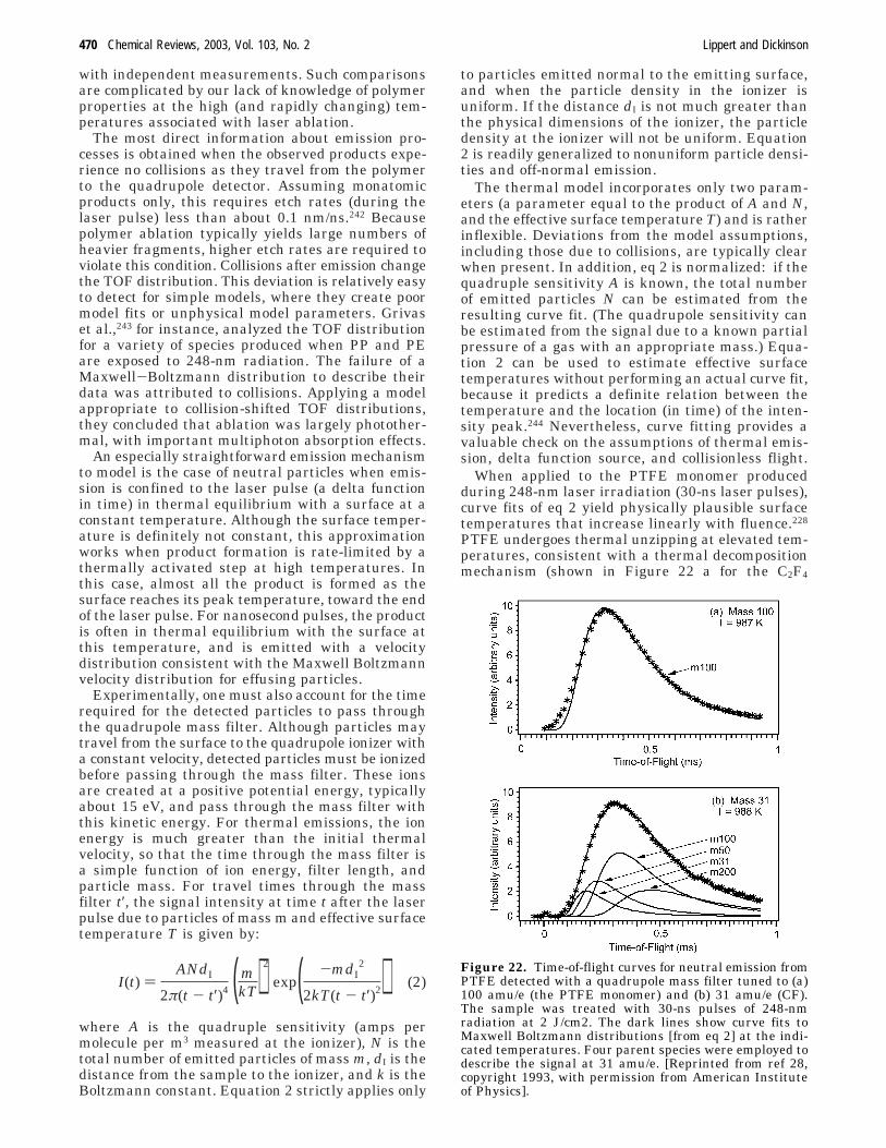
with independent measurements. Such comparisonsare complicated by our lack of knowledge of polymerproperties at the high (and rapidly changing) tem-peratures associated with laser ablation.
The most direct information about emission pro-cesses is obtained when the observed products expe-rience no collisions as they travel from the polymerto the quadrupole detector. Assuming monatomicproducts only, this requires etch rates (during thelaser pulse) less than about 0.1 nm/ns.242 Becausepolymer ablation typically yields large numbers ofheavier fragments, higher etch rates are required toviolate this condition. Collisions after emission changethe TOF distribution. This deviation is relatively easyto detect for simple models, where they create poormodel fits or unphysical model parameters. Grivaset al.,243 for instance, analyzed the TOF distributionfor a variety of species produced when PP and PEare exposed to 248-nm radiation. The failure of aMaxwell-Boltzmann distribution to describe theirdata was attributed to collisions. Applying a modelappropriate to collision-shifted TOF distributions,they concluded that ablation was largely photother-mal, with important multiphoton absorption effects.
An especially straightforward emission mechanismto model is the case of neutral particles when emis-sion is confined to the laser pulse (a delta functionin time) in thermal equilibrium with a surface at aconstant temperature. Although the surface temper-ature is definitely not constant, this approximationworks when product formation is rate-limited by athermally activated step at high temperatures. Inthis case, almost all the product is formed as thesurface reaches its peak temperature, toward the endof the laser pulse. For nanosecond pulses, the productis often in thermal equilibrium with the surface atthis temperature, and is emitted with a velocitydistribution consistent with the Maxwell Boltzmannvelocity distribution for effusing particles.
Experimentally, one must also account for the timerequired for the detected particles to pass throughthe quadrupole mass filter. Although particles maytravel from the surface to the quadrupole ionizer witha constant velocity, detected particles must be ionizedbefore passing through the mass filter. These ionsare created at a positive potential energy, typicallyabout 15 eV, and pass through the mass filter withthis kinetic energy. For thermal emissions, the ionenergy is much greater than the initial thermalvelocity, so that the time through the mass filter isa simple function of ion energy, filter length, andparticle mass. For travel times through the massfilter t′, the signal intensity at time t after the laserpulse due to particles of mass m and effective surfacetemperature T is given by:
where A is the quadruple sensitivity (amps permolecule per m3 measured at the ionizer), N is thetotal number of emitted particles of mass m, dI is thedistance from the sample to the ionizer, and k is theBoltzmann constant. Equation 2 strictly applies only
to particles emitted normal to the emitting surface,and when the particle density in the ionizer isuniform. If the distance dI is not much greater thanthe physical dimensions of the ionizer, the particledensity at the ionizer will not be uniform. Equation2 is readily generalized to nonuniform particle densi-ties and off-normal emission.
The thermal model incorporates only two param-eters (a parameter equal to the product of A and N,and the effective surface temperature T) and is ratherinflexible. Deviations from the model assumptions,including those due to collisions, are typically clearwhen present. In addition, eq 2 is normalized: if thequadruple sensitivity A is known, the total numberof emitted particles N can be estimated from theresulting curve fit. (The quadrupole sensitivity canbe estimated from the signal due to a known partialpressure of a gas with an appropriate mass.) Equa-tion 2 can be used to estimate effective surfacetemperatures without performing an actual curve fit,because it predicts a definite relation between thetemperature and the location (in time) of the inten-sity peak.244 Nevertheless, curve fitting provides avaluable check on the assumptions of thermal emis-sion, delta function source, and collisionless flight.
When applied to the PTFE monomer producedduring 248-nm laser irradiation (30-ns laser pulses),curve fits of eq 2 yield physically plausible surfacetemperatures that increase linearly with fluence.228
PTFE undergoes thermal unzipping at elevated tem-peratures, consistent with a thermal decompositionmechanism (shown in Figure 22 a for the C2F4
I(t) )ANdI
2π(t - t′)4 ( mkT)2
exp( -mdI2
2kT(t - t′)2) (2)Figure 22. Time-of-flight curves for neutral emission fromPTFE detected with a quadrupole mass filter tuned to (a)100 amu/e (the PTFE monomer) and (b) 31 amu/e (CF).The sample was treated with 30-ns pulses of 248-nmradiation at 2 J/cm2. The dark lines show curve fits toMaxwell Boltzmann distributions [from eq 2] at the indi-cated temperatures. Four parent species were employed todescribe the signal at 31 amu/e. [Reprinted from ref 28,copyright 1993, with permission from American Instituteof Physics].
470 Chemical Reviews, 2003, Vol. 103, No. 2 Lippert and Dickinson
monomer). At lower masses, the TOF mass spectraare complicated by fragmentation in the quadrupoleionizer. For instance, most of the signal at a charge-to-mass ratio of 31 amu/e (CF) from PTFE is actuallydue to CF fragments generated in the ionizer fromthe dimer, the monomer, and CF2. These heavier,parent species travel more slowly than CF, andsubstantially broaden the TOF signal observed at 31amu/e. To derive surface temperatures from the CFsignal, one must account for the CF generated asheavier parent species move through the ionizer.When this is done, good curve fits are obtained withsurface temperatures that agree well with temper-atures based on the TOF of the monomer, wherecontributions due to fragmentation are not significant(shown in Figure 22b). If possible, surface tempera-tures and model testing should be based on TOFsignals at masses that can unambiguously be at-tributed to a single species. As noted above, complexmodels are often not unique and easily misinter-preted.
For low numbers of collisions (less than four or fiveper particle), collisions tend to sharpen the velocitydistribution, move the peak to shorter time-of-flights,and make the distribution more forward directed(normal to the polymer surface).245 When a varietyof particles are emitted simultaneously, collisionstend to slow the lighter particles and accelerate theheavy onessboth light and heavy particles tend todisplay similar peak time-of-flights. With somewhatlarger numbers of collisions, the velocity distributioncan be modeled in terms of an unsteady, adiabaticexpansion into vacuum.
Neutral molecules produced by photochemical pro-cesses can be ejected from the surface with hyper-thermal energies. As the physical TOF distributionof these particles is not known, an empirical TOFdistribution based on a Gaussian energy distributionin one dimension is useful. This distribution often fitsthe data adequately, and has the advantage of beingeasily interpreted. Since the observed signal is pro-portional to a density, we form a one-dimensionaldensity by transforming the Gaussian energy distri-bution, dN/dx ) (dN/dE)(dE/dx), or
where E0 is the most probable kinetic energy, and σis the standard deviation. For low energy particles,t′ is well approximated by the time required for aparticle to pass through the mass filter with a kineticenergy equal to the potential energy at which the ionwas created in the ionizer (as above). Hyperthermalenergies can be a significant fraction of this energy,in which case t′ can calculated by equating theparticle kinetic energy through the mass filter withthe sum of its initial kinetic energy and the ionenergy setting.
Ions are detected without an ionization step by afundamentally different process. Whereas the signalintensity due to neutral particles is proportional to
the particle density in the quadrupole ionizer (par-ticles per unit volume), the signal intensity due toions is proportional to the flux of ions striking thedetector (particles per unit time). This differenceaffects the expected time behavior of the signal.Another difference between ion and neutral detectionis that ion detection usually requires a clear line-of-sight path from the laser spot, through any aperturesin the quadrupole optics, to the quadrupole detector.This requires careful alignment of the quadrupolemass filter. It is often reasonable to expect ionintensities and velocities to vary strongly with theangle at which they are emitted relative to thesurface normal. In most work, emission normal to thesurface is reported.
Analysis of ion velocity distributions is greatlysimplified if the sample is kept at ground potential,and electric fields between the sample and thedetector minimized. This generally requires that theelectron optics associated with the ionizer be removedand/or grounded. If the axis of the mass filter is wellaligned with the laser spot, the electric fields in themass filter are predominately perpendicular to theion flight path and have little effect on the ionvelocities. In this case, ions should move from thesample to the quadrupole detector a constant velocity,and the ion velocity can be determined from the TOF.
As with high-energy neutral particles, a one-dimensional Gaussian energy distribution often pro-vides a good, albeit empirical, description of the TOFsignal. Because the detector output is proportionalto the particle flux, the signal as a function of timeis proportional to dN/dt ) (dN/dE)(dE/dt).
where dD is the distance between the sample all theway to the quadrupole detector.
Campbell et al.246,247 has analyzed the ionic prod-ucts (both + and -) generated during ablation of PIand PS during exposure to 14 ns 308 nm excimerradiation using a reflectron TOF unit. Large masses(up to 900 amu) were observed and very high plumevelocities (between 1400 and 10 000 m/s), attributedto electrostatic effects.
Although less common, metastable particles withsufficient internal energy (> 5 eV) can be detectedwithout an ionization step by many charged particledetectors. In this case, the detector output reflectsthe particle flux, as in the case of ions, and eq 4 canbe used to describe the results. In principle, thevelocity distribution of metastables is not affected bythe electric fields of the ionizer optics. However,strong electric fields can de-excite metastables. Slowerparticles spend more time in the strong-field regionand can be preferentially de-excited, which effectivelyshifts the apparent TOF distribution to shorter times.This effect can be probed by changing the masssetting of the mass filter, which changes the internalfields.
Fitting eqs 2-4 to experimental data can oftenprovide important information about the observed
dNdx
)N0 mdI
x2πσ(t - t′)2exp[-(E0 -
mdI2
2(t - t′)2)2σ2 ] (3)
dNdx
)N0 mdD
2
x2πσt3exp[-(E0 -
mdD2
2t2 )2σ2 ] (4)
Chemical and Spectroscopic Aspects of Polymer Ablation Chemical Reviews, 2003, Vol. 103, No. 2 471
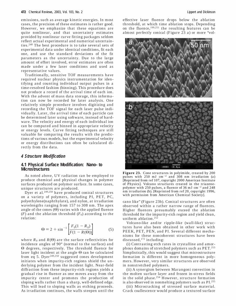
emissions, such as average kinetic energies. In mostcases, the precision of these estimates is rather good.However, we emphasize that these equations arequite nonlinear, and that uncertainty estimatesprovided by nonlinear curve fitting packages seldomreflect actual experimental and numerical uncertain-ties.248 The best procedure is to take several sets ofexperimental data under identical conditions, fit eachone, and use the standard deviations of the fitparameters as the uncertainty. Due to the largeamount of effort involved, error estimates are oftenmade under a few laser conditions and used asrepresentative values.
Traditionally, sensitive TOF measurements haverequired nuclear physics instrumentation for iden-tifying and counting individual output pulses in atime-resolved fashion (binning). This procedure doesnot produce a record of the arrival time of each ion.With the advent of mass data storage, this informa-tion can now be recorded for later analysis. Onerelatively simple procedure involves digitizing andrecording the TOF signal for each laser pulse indi-vidually. Later, the arrival time of each particle canbe determined later using software, instead of hard-ware. The velocity and energy of each individual ioncan be computed and binned in appropriate velocityor energy levels. Curve fitting techniques are stillvaluable for comparing the results with the predic-tions of various models, but the experimental velocityor energy distributions can often be calculated di-rectly from the data.
4 Structure Modification
4.1 Physical Surface Modification: Nano- toMicrostructures
As noted above, UV radiation can be employed toproduce chemical and physical changes in polymersurfaces produced on polymer surface. In some cases,unique structures are produced.
Dyer et al.249,250 have produced conical structureson a variety of polymers, including PI, PET, PC,polyethylene(naphthylate), and nylon, at irradiationwavelengths ranging from 157 to 308 nm. The apexangle of the cones (Θ) varies with the applied fluence(F) and the ablation threshold (F0) according to therelation:
where R0 and R(Θ) are the surface reflectivities forincidence angles of 90° (normal to the surface) andΘ degrees, respectively. The threshold fluence forlaser light incident at the angle Θ can be calculatedfrom eq 5. Dyer249,250 suggested cones developmentinitiates when impurity-rich regions shield the un-derlying polymer from the incident light. Near-fielddiffraction from these impurity-rich regions yields agradual rise in fluence as one moves away from theimpurity center and promotes the production ofsloping walls rather than a sharp, well-defined edge.This will lead to sloping walls as etching proceeds.As irradiation continues, the walls steepen until the
effective laser fluence drops below the ablationthreshold, at which time ablation stops. Dependingon the fluence,199,251 the resulting features can bealmost perfectly conical (Figure 23 a) or more “vol-
cano like” (Figure 23b). Conical structures are oftenobserved within a rather narrow range of fluences.Higher fluences presumably exceed the ablationthreshold for the impurity-rich region and yield clean,uniform ablation.252
Volcano-like and/or ripple-like (wall-like) struc-tures have also been obtained in other work withPEEK, PET, PEN, and PI. Several different mecha-nisms for these noncoherent structures have beendiscussed,172 including:
(i) Contrasting etch rates in crystalline and amor-phous domains of stretched polymers such as PET.253
Simplistically, this would suggest that microstructureformation is different in more homogeneous poly-mers. However, very similar structures are observedin nonstretched polymers.
(ii) A synergism between Marangoni convection inthe molten surface layer and frozen in-stress fieldsin the solid phase.254 However, structure formationis also observed in nonmelting polymers such as PI.255
(iii) Microcracking of stressed surface material.Crack coallescence would produce a textured surface
Θ ) 2 + sin-1 [ F0(1 - R0)
F(1 - R(Θ))] (5)
Figure 23. Cone structures in polyimide, created by 200pulses with 250 mJ cm-2 and 308 nm irradiation (a)[Reprinted from ref 107, copyright 2000 American Instituteof Physics]. Volcano structures created in the triazene-polymer with 250 pulses, a fluence of 36 mJ cm-2 and 248nm irradiation (b). [Reprinted from ref 20, copyright 1996,with permission from American Chemical Society].
472 Chemical Reviews, 2003, Vol. 103, No. 2 Lippert and Dickinson
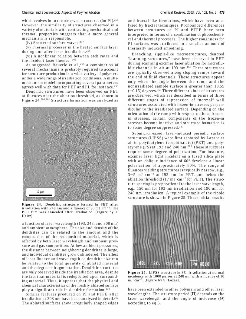
which evolves in to the observed structures (for PI).256
However, the similarity of structures observed in avariety of materials with contrasting mechanical andthermal properties suggests that a more generalmechanism is responsible.
(iv) Scattered surface waves.257
(v) Thermal processes in the heated surface layerduring and after laser irradiation.258
(vi) A nonlinear relation between etch rates andthe incident laser fluence. 169
As suggested Bauerle et al.,255 a combination ofseveral mechanisms is probably required to accountfor structure production in a wide variety of polymersunder a wide range of irradiation conditions. A multi-mechanism model incorporating several parametersagrees well with data for PET and PI, for instance.259
Dendritic structures have been observed on PETat fluences near the ablation threshold, as shown inFigure 24.260,261 Structure formation was analyzed as
a function of laser wavelength (193, 248, and 308 nm)and ambient atmosphere. The size and density of thedendrites can be related to the amount and thecomposition of the redeposited material, which isaffected by both laser wavelength and ambient pres-sure and gas composition. At low ambient pressures,the distance between neighboring dendrites is large,and individual dendrites grow unhindered. The effectof laser fluence and wavelength on dendrite size canbe related to the total quantity of ablated materialand the degree of fragmentation. Dendritic structuresare only observed inside the irradiation area, despitethe fact that material is redeposited upon surround-ing material. Thus, it appears that the physical andchemical characteristics of the freshly ablated surfaceplay a significant role in dendrite formation.170
Similar features produced on PI and PTFE afterirradiation at 308 nm have been analyzed in detail.262
The ablated surfaces show irregularly shaped edges
and fractal-like formations, which have been ana-lyzed by fractal techniques. Pronounced differencesbetween structures on PI and PTFE have beeninterpreted in terms of a combination of photochemi-cal and thermal processes. The higher roughness forPI surfaces was attributed to a smaller amount ofthermally induced smoothing.
Branching, ripple-like microstructures, denoted“scanning structures,” have been observed in PETduring scanning excimer laser ablation for microflu-idic channels in air at 193 nm.168 These structuresare typically observed along sloping ramps towardthe end of fluid channels. These structures appearonly when the angle between the ramp and thenonirradiated sample surface is greater than 10.55((0.15) degrees.166 Three different kinds of structuresare observed, which are described in terms of threedifferent stages of suppression of “normal” wallstructures associated with frozen-in stresses perpen-dicular to the irradiated surface. Depending on theorientation of the ramp with respect to these frozen-in stresses, certain components of the frozen-instresses become inactive and structure formation isto some degree suppressed.167
Submicron-sized, laser-induced periodic surfacestructures (LIPSS) were first reported by Lazare etal. in poly(butylene terephthalate) (PET) and poly-styrene (PS) at 193 and 248 nm.263 These structuresrequire some degree of polarization. For instance,excimer laser light incident on a fused silica platewith an oblique incidence of 60° develops a linearpolarization of approximately 80%. The range offluences yielding structures is typically narrow, e.g.,3-5 mJ cm-2 at 193 nm for PET, and below theablation threshold (17 mJ cm-2 for PET). The struc-ture spacing is proporational to the laser wavelength,e.g., 150 nm for 193 nm irradiation and 190 nm for248 nm irradiation. A typical example of the ripplestructure is shown in Figure 25. These initial results
have been extended to other polymers and other laserwavelengths. The structure period (Π) depends on thelaser wavelength and the angle of incidence (Θ)according to eq 6.
Figure 24. Dendritic structure formed in PET afterirradiation with 248 nm and a fluence of 30 mJ cm-2. ThePET film was annealed after irradiation. [Figure by J.Heitz]
Figure 25. LIPSS structure in PC. Irradiation at normalincidence with 1000 pulses at 248 nm with a fluence of 10mJ cm-2. [Figure by S. Lazare].
Chemical and Spectroscopic Aspects of Polymer Ablation Chemical Reviews, 2003, Vol. 103, No. 2 473
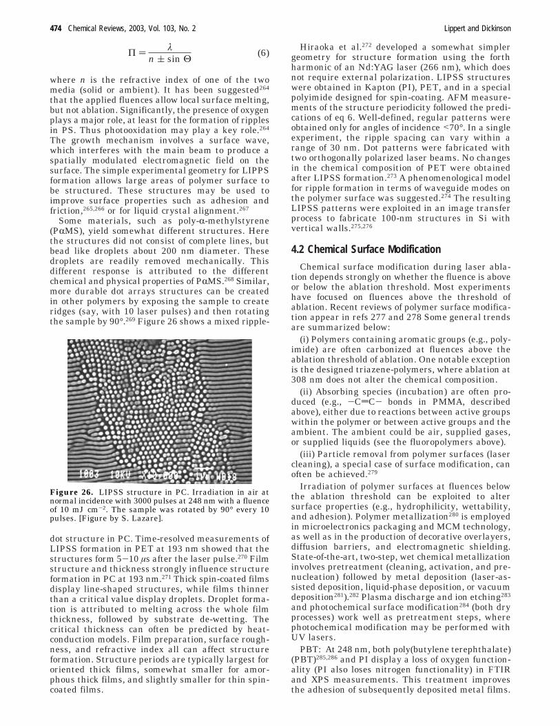
where n is the refractive index of one of the twomedia (solid or ambient). It has been suggested264
that the applied fluences allow local surface melting,but not ablation. Significantly, the presence of oxygenplays a major role, at least for the formation of ripplesin PS. Thus photooxidation may play a key role.264
The growth mechanism involves a surface wave,which interferes with the main beam to produce aspatially modulated electromagnetic field on thesurface. The simple experimental geometry for LIPPSformation allows large areas of polymer surface tobe structured. These structures may be used toimprove surface properties such as adhesion andfriction,265,266 or for liquid crystal alignment.267
Some materials, such as poly-R-methylstyrene(PRMS), yield somewhat different structures. Herethe structures did not consist of complete lines, butbead like droplets about 200 nm diameter. Thesedroplets are readily removed mechanically. Thisdifferent response is attributed to the differentchemical and physical properties of PRMS.268 Similar,more durable dot arrays structures can be createdin other polymers by exposing the sample to createridges (say, with 10 laser pulses) and then rotatingthe sample by 90°.269 Figure 26 shows a mixed ripple-
dot structure in PC. Time-resolved measurements ofLIPSS formation in PET at 193 nm showed that thestructures form 5-10 µs after the laser pulse.270 Filmstructure and thickness strongly influence structureformation in PC at 193 nm.271 Thick spin-coated filmsdisplay line-shaped structures, while films thinnerthan a critical value display droplets. Droplet forma-tion is attributed to melting across the whole filmthickness, followed by substrate de-wetting. Thecritical thickness can often be predicted by heat-conduction models. Film preparation, surface rough-ness, and refractive index all can affect structureformation. Structure periods are typically largest fororiented thick films, somewhat smaller for amor-phous thick films, and slightly smaller for thin spin-coated films.
Hiraoka et al.272 developed a somewhat simplergeometry for structure formation using the forthharmonic of an Nd:YAG laser (266 nm), which doesnot require external polarization. LIPSS structureswere obtained in Kapton (PI), PET, and in a specialpolyimide designed for spin-coating. AFM measure-ments of the structure periodicity followed the predi-cations of eq 6. Well-defined, regular patterns wereobtained only for angles of incidence <70°. In a singleexperiment, the ripple spacing can vary within arange of 30 nm. Dot patterns were fabricated withtwo orthogonally polarized laser beams. No changesin the chemical composition of PET were obtainedafter LIPSS formation.273 A phenomenological modelfor ripple formation in terms of waveguide modes onthe polymer surface was suggested.274 The resultingLIPSS patterns were exploited in an image transferprocess to fabricate 100-nm structures in Si withvertical walls.275,276
4.2 Chemical Surface Modification
Chemical surface modification during laser abla-tion depends strongly on whether the fluence is aboveor below the ablation threshold. Most experimentshave focused on fluences above the threshold ofablation. Recent reviews of polymer surface modifica-tion appear in refs 277 and 278 Some general trendsare summarized below:
(i) Polymers containing aromatic groups (e.g., poly-imide) are often carbonized at fluences above theablation threshold of ablation. One notable exceptionis the designed triazene-polymers, where ablation at308 nm does not alter the chemical composition.
(ii) Absorbing species (incubation) are often pro-duced (e.g., -CdC- bonds in PMMA, describedabove), either due to reactions between active groupswithin the polymer or between active groups and theambient. The ambient could be air, supplied gases,or supplied liquids (see the fluoropolymers above).
(iii) Particle removal from polymer surfaces (lasercleaning), a special case of surface modification, canoften be achieved.279
Irradiation of polymer surfaces at fluences belowthe ablation threshold can be exploited to altersurface properties (e.g., hydrophilicity, wettability,and adhesion). Polymer metallization280 is employedin microelectronics packaging and MCM technology,as well as in the production of decorative overlayers,diffusion barriers, and electromagnetic shielding.State-of-the-art, two-step, wet chemical metallizationinvolves pretreatment (cleaning, activation, and pre-nucleation) followed by metal deposition (laser-as-sisted deposition, liquid-phase deposition, or vacuumdeposition281).282 Plasma discharge and ion etching283
and photochemical surface modification284 (both dryprocesses) work well as pretreatment steps, wherephotochemical modification may be performed withUV lasers.
PBT: At 248 nm, both poly(butylene terephthalate)(PBT)285,286 and PI display a loss of oxygen function-ality (PI also loses nitrogen functionality) in FTIRand XPS measurements. This treatment improvesthe adhesion of subsequently deposited metal films.
Π ) λn ( sin Θ
(6)
Figure 26. LIPSS structure in PC. Irradiation in air atnormal incidence with 3000 pulses at 248 nm with a fluenceof 10 mJ cm-2. The sample was rotated by 90° every 10pulses. [Figure by S. Lazare].
474 Chemical Reviews, 2003, Vol. 103, No. 2 Lippert and Dickinson
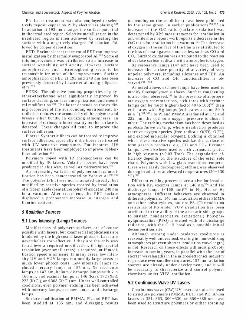
PI: Laser treatment was also employed to selec-tively deposit copper on PI by electroless plating.287
Irradiation at 193 nm changes the surface potentialin the irradiated region. Selective metallization in theirradiated region is then achieved by treating thesurface with a negatively charged Pd-solution, fol-lowed by copper deposition.
PET: Excimer laser treatment of PET can improvemetallization by thermally evaporated Al.288 Some ofthis improvement was attributed to an increase insurface wettability and acidity. However, surfaceamorphization and microroughening seem to beresponsible for most of the improvement. Surfaceamorphization of PET at 193 and 248 nm has beenpreviously detected by Lazare et al. using ellipsom-etry.289
PEEK: The adhesive bonding properties of poly-ether-etherketone were significantly improved bysurface cleaning, surface amorphization, and chemi-cal modification.290 The latter depends on the oxidiz-ing properties of the surrounding environment. Ir-radiation reduces the aromaticity of the polymer andbreaks ether bonds. In oxidizing atmospheres, anincrease of carboxylic functionality on the surface isdetected. These changes all tend to improve thesurface adhesion.
Fibers: Synthetic fibers can be treated to improvesurface adhesion, especially if the fibers are treatedwith UV sensitive compounds. For instance, UVtreatments have been employed to improve rubber-fiber adhesion.291
Polymers doped with IR chromophores can bemodified by IR lasers. Volatile species have beenproduced in this way, as well as microcavities.292
An interesting variation of polymer surface modi-fication has been demonstrated by Yabe et al.293,294
The polymer (PET) was not irradiated directly, butmodified by reactive species created by irradiationof a frozen azide (pentafluorophenyl azide) at 248 nmin a vacuum. After treatment, the PET surfacedisplayed a pronounced increase in nitrogen andfluorine content.
5 Radiation Sources
5.1 Low Intensity (Lamp) SourcesModifications of polymers surfaces are of course
possible with lasers, but commercial applications arehindered by the high cost of laser photons. Laser arenevertheless cost-effective if they are the only wayto achieve a required modification, if high spatialresolution (over small areas) is required, or if modi-fication speed is an issue. In many cases, low inten-sity UV and VUV lamps can modify large areas atmuch lower photon costs. Low intensity lamps in-cluded mercury lamps at 185 nm, Xe resonancelamps at 147 nm, helium discharge lamps with λ <160 nm, and excimer lamps at 146 (Kr2), 172 (Xe2),222 (KrCl), and 308 (XeCl) nm. Under well-controlledconditions, even polymer etching has been achievedwith mercury lamps, excimer lamps, and dischargelamps.
Surface modification of PMMA, PI, and PET hasbeen studied at 185 nm, and diverging results
(depending on the conditions) have been publishedby the same group. In earlier publications176,295 anincrease of the O/C ratio (surface oxidation) wasdetermined by XPS measurements for irradiation inair, while more recent work reports a decrease in theO/C ratio for irradiation in a vacuum.171 The decreaseof oxygen in the surface of the film was attributed tothe loss of small gaseous molecules, such as CO andCO2. Surface oxidation was attributed to the reactionof surface carbon radicals with atmospheric oxygen.
Xe resonance lamps (147 nm) have been used toincrease the surface polarity of a variety of veryunpolar polymers, including siloxanes and FEP. Anincrease of CO and OH functionalities is ob-served.296-298
As noted above, excimer lamps have been used tomodify fluoropolymer surfaces. Surface rougheningis also often observed.299 In the presence of appropri-ate oxygen concentrations, etch rates with excimerlamps can be much higher (factor 40 to 100)300 thanetch rates with Hg lamps (etch rates around 50 Åmin-1).301,302 For PI and PMMA irradiated at 172 and222 nm, the optimum oxygen pressure is about 1mbar. The etching mechanism has been described asphotooxidative etching, where irradiation producesreactive oxygen species (free radicals O(1D), O(3P),and excited molecular oxygen). Etching is obtainedwhen these reactive species oxidize the surface toform gaseous products, e.g., CO and CO2. Excimerlamps have also been used to etch various acrylatesin high vacuum (<0.03 Torr). The degradation ef-ficiency depends on the structure of the ester sidechain. Polymers with low glass transition tempera-tures were easily decomposed; etching was observedduring irradiation at elevated temperatures (50-130°C).303
Different etching processes are active for irradia-tion with Kr2 excimer lamps at 146 nm304 and Hedischarge lamps (<160 nm)305 in N2, He, or H2atmospheres. Different behaviors are observed indifferent polymers: 146 nm irradiation etches PMMAand other polyacrylates, but not PS. (The radiationresistance of PS under VUV irradiation has beenattributed to the ability of the aromatic side groupsto sustain nondissociative excitations.) Poly-phe-nylquinoxaline (PPQ) is etched with He dischargeradiation, with the C-H bond as a possible initialdecomposition site.
Although etching under oxidative conditions isreasonably well understood, etching in non-oxidizingatmospheres (at even shorter irradiation wavelength)is not. Research on these effects will most probablyincrease in coming years, in parallel with the use ofshorter wavelengths in the microelectronics industryto produce ever-smaller structures. 157 nm radiationsources are already under development, and it willbe necessary to characterize and control polymerchemistry under VUV irradiation.
5.2 Continuous-Wave UV LasersContinuous wave (CW) UV lasers can also be used
to structure polymers (PMMA, PET, and PI). Ar-ionlasers at 351, 363, 300-330, or 350-380 nm havebeen used to structure polymers by either scanning
Chemical and Spectroscopic Aspects of Polymer Ablation Chemical Reviews, 2003, Vol. 103, No. 2 475
the beam across the surface to be processed (transittime over beam diameter 1-1000 µs) or by mechani-cally chopping the beam (10-400 µs pulses). In thecase of PI, the fluences required for etching aresimilar to the fluences employed with excimer lasers,but typical etch depths per joule are about 100 foldless.306 Etching is observed only when the laser beamis translated across the sample. Irradiation at asingle position merely blackens and swells the sur-face. No acoustic report (audible snap) is heard, incontrast to ablation with pulsed lasers. Nevertheless,only a very small fraction of the material is removedas debris. By chopping the laser beam (350 µs pulses)holes can be drilled in PI. Although the walls of thehole are heavily carbonized, no signs of debris areobserved.307 Under irradiation with very long (mil-lisecond) pulses, a chemical transformation yieldscarbon, similar to glassy carbon.18
PET behaves differently.308 PET is five times moresensitive than PI and PMMA to etching, with etchrates that increase with pulse width. Pulse widthslonger than 200 µs damage the surrounding material.A significant fraction of removed material (about20%) is ejected as molten droplets and depositedalong the edge of the structure. Etching is attributedto largely thermal processes.
PMMA has been studied in its pure form or dopedwith a UV chromophore (Tinuvin) to increase absorp-tion at the laser wavelength. Doped PMMA could beetched by scanning the laser beam, with an optimumscan rate (transit time over the beam diameter) ofabout 50 µs.309 As in the case of PI, no acoustic reportor ablative material ejection was observed. Thisprocess operates at power densities about 50 timeslower than those in “normal” laser ablation, and wasdenoted photokinetic etching.
At the 351 and 363 nm emission lines of the Ar-ion laser, CW irradiation of neat PMMA (transparentat both wavelengths)310 produces an effect similar toincubation. The incubation time decreases with in-creasing laser intensity or sample temperature. Atsufficiently high intensities, the incubation timedepends only on the sample temperature. This be-havior can be described in terms of an incubationprocess followed by a volume explosion.311
Structuring with these long pulses, or better, byscanning the cw-laser beam, apparently involvesdifferent processes than structuring with excimerlasers. As suggested by Bauerle (see above), theseprocesses are probably thermal for the most part.
5.3 Ultrafast LasersWith the development of chirped pulse amplifica-
tion (CPA), IR solid state (Ti:sapphire) lasers havebecome compact, extremely high-brightness sources.312
Material processing with femtosecond lasers is al-ready an established technique. Ablation with fem-tosecond pulses has unique advantages, including anegligible heat-affected zone, lower ablation thresh-old, absence of plasma shielding, and the ability tostructure “transparent” materials. In addition topolymers (discussed here), these lasers are used toprocess metals, glasses, ionic crystals, ceramics, andsemiconductors. Laser-induced breakdown in dielec-
trics has been described in terms of producing acritical electron density in the conduction band,313
either by avalanche ionization or multiphoton ioniza-tion.314 The rapid transfer of laser technology fromexperimental systems to industrial environments (asin mask repair315,316) has been astonishing. We havecome a long way since the first reports of femtosecondlaser ablation of polymers in 1987 by Srinivasan etal.317 and Stuke et al.318
5.3.1 Femtosecond Lasers
Srinivasan showed,317 that it is possible to producehigh quality structures in PMMA with 160 fs pulsesat 308 nm, whereas nanosecond pulses at the samewavelength only “damage” the surface. Stuke’s318
work on PMMA with 300 fs, 248-nm laser pulsesshowed that femtosecond pulses have a lower thresh-old for structuring by a factor of 5 relative tonanosecond pulses, and produce much better struc-tures. Femtosecond pulses also produce high qualitystructures in Teflon,319 with no signs of the thermaldegradation associated with nanosecond pulses.320
Removal rates up to 1 µm per pulse can be achieved.Time-resolved studies321 with a pair of time-delayed(-200 to 200 ps) 500 fs pulses at 248 m showed thatthe ablation rate is very sensitive to the time delay.When the combined fluence is close to the ablationthreshold, significant changes in the ablation rate ofPMMA and Teflon are observed for delays < 3ps. Forthe absorbed energy density to exceed the thresholdfluence, the two pulses must overlap sufficiently intime. Thus, multiphoton absorption dominates theablation process in PMMA and Teflon. In the case ofPI,322 excited state absorption actually decreases theablation rate for delay times below 30 ps.
The first ablation experiments for medical applica-tions using femtosecond pulses were published in1994 using a femtosecond dye laser at 615 nm.323
Femtosecond lasers produced heat-affected zones(HAZ) less than 0.5 µm thick, while comparablenanosecond lasers (at 600 nm) produced severethermal damage.
Chirped-pulse Ti:sapphire systems were first ap-plied to polymers in 1994.324 The ablation rates forTeflon, PI, and FEP were adequately described by atwo-level absorption model with the chromophoredensity as a free parameter.32 In the case of FEP,raising the pulse length from 170 fs to 12 ps shows apronounced degradation of ablation quality at about500 fs.325 The ablation threshold in this fluence rangeis inversely proportional to the square root of thepulse duration. Femtosecond laser ablation can beused for high aspect ratio micro-machining of poly-mers (PMMA, PC, and PTFE). The aspect ratio isnormally limited to about 10; deeper structures areoften tapered and sometimes even bend.326
Studies of Nylon 6-6 and PMMA indicate that theablation rates for 0.8-ps pulses at 595 nm (dye laser)are lower than the rates for 100-ps pulses at 532nm.327 Subsequent work with nanosecond pulsesshowed that the highest ablation rates are achievedwith picosecond pulses.328
Kautek et al.329-332 have studied the ablation of PI,PC, PET, and PMMA due to 150-fs pulses at 800 nm.
476 Chemical Reviews, 2003, Vol. 103, No. 2 Lippert and Dickinson
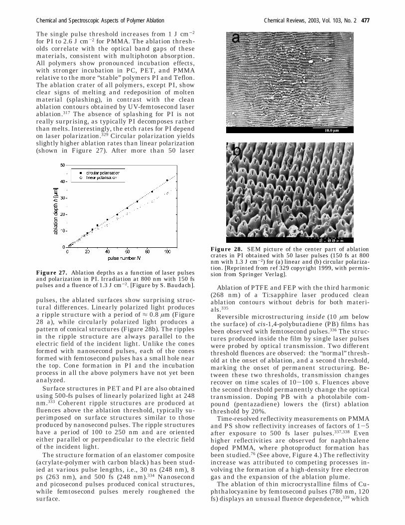
The single pulse threshold increases from 1 J cm-2
for PI to 2.6 J cm-2 for PMMA. The ablation thresh-olds correlate with the optical band gaps of thesematerials, consistent with multiphoton absorption.All polymers show pronounced incubation effects,with stronger incubation in PC, PET, and PMMArelative to the more “stable” polymers PI and Teflon.The ablation crater of all polymers, except PI, showclear signs of melting and redeposition of moltenmaterial (splashing), in contrast with the cleanablation contours obtained by UV-femtosecond laserablation.317 The absence of splashing for PI is notreally surprising, as typically PI decomposes ratherthan melts. Interestingly, the etch rates for PI dependon laser polarization.329 Circular polarization yieldsslightly higher ablation rates than linear polarization(shown in Figure 27). After more than 50 laser
pulses, the ablated surfaces show surprising struc-tural differences. Linearly polarized light producesa ripple structure with a period of ≈ 0.8 µm (Figure28 a), while circularly polarized light produces apattern of conical structures (Figure 28b). The ripplesin the ripple structure are always parallel to theelectric field of the incident light. Unlike the conesformed with nanosecond pulses, each of the conesformed with femtosecond pulses has a small hole nearthe top. Cone formation in PI and the incubationprocess in all the above polymers have not yet beenanalyzed.
Surface structures in PET and PI are also obtainedusing 500-fs pulses of linearly polarized light at 248nm.333 Coherent ripple structures are produced atfluences above the ablation threshold, typically su-perimposed on surface structures similar to thoseproduced by nanosecond pulses. The ripple structureshave a period of 100 to 250 nm and are orientedeither parallel or perpendicular to the electric fieldof the incident light.
The structure formation of an elastomer composite(acrylate-polymer with carbon black) has been stud-ied at various pulse lengths, i.e., 30 ns (248 nm), 8ps (263 nm), and 500 fs (248 nm).334 Nanosecondand picosecond pulses produced conical structures,while femtosecond pulses merely roughened thesurface.
Ablation of PTFE and FEP with the third harmonic(268 nm) of a Ti:sapphire laser produced cleanablation contours without debris for both materi-als.335
Reversible microstructuring inside (10 µm belowthe surface) of cis-1,4-polybutadiene (PB) films hasbeen observed with femtosecond pulses.336 The struc-tures produced inside the film by single laser pulseswere probed by optical transmission. Two differentthreshold fluences are observed: the “normal” thresh-old at the onset of ablation, and a second threshold,marking the onset of permanent structuring. Be-tween these two thresholds, transmission changesrecover on time scales of 10-100 s. Fluences abovethe second threshold permanently change the opticaltransmission. Doping PB with a photolabile com-pound (pentazadiene) lowers the (first) ablationthreshold by 20%.
Time-resolved reflectivity measurements on PMMAand PS show reflectivity increases of factors of 1-5after exposure to 500 fs laser pulses.337,338 Evenhigher reflectivities are observed for naphthalenedoped PMMA, where photoproduct formation hasbeen studied.76 (See above, Figure 4.) The reflectivityincrease was attributed to competing processes in-volving the formation of a high-density free electrongas and the expansion of the ablation plume.
The ablation of thin microcrystalline films of Cu-phthalocyanine by femtosecond pulses (780 nm, 120fs) displays an unusual fluence dependence,339 which
Figure 27. Ablation depths as a function of laser pulsesand polarization in PI. Irradiation at 800 nm with 150 fspulses and a fluence of 1.3 J cm-2. [Figure by S. Baudach].
Figure 28. SEM picture of the center part of ablationcrates in PI obtained with 50 laser pulses (150 fs at 800nm with 1.3 J cm-2) for (a) linear and (b) circular polariza-tion. [Reprinted from ref 329 copyright 1999, with permis-sion from Springer Verlag].
Chemical and Spectroscopic Aspects of Polymer Ablation Chemical Reviews, 2003, Vol. 103, No. 2 477
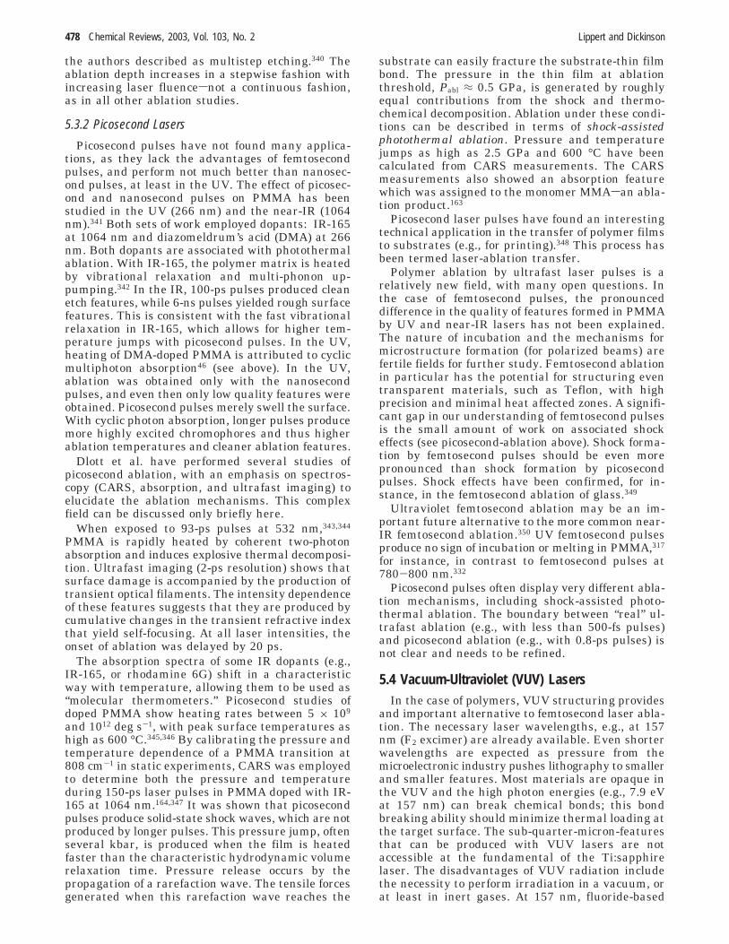
the authors described as multistep etching.340 Theablation depth increases in a stepwise fashion withincreasing laser fluencesnot a continuous fashion,as in all other ablation studies.
5.3.2 Picosecond Lasers
Picosecond pulses have not found many applica-tions, as they lack the advantages of femtosecondpulses, and perform not much better than nanosec-ond pulses, at least in the UV. The effect of picosec-ond and nanosecond pulses on PMMA has beenstudied in the UV (266 nm) and the near-IR (1064nm).341 Both sets of work employed dopants: IR-165at 1064 nm and diazomeldrum’s acid (DMA) at 266nm. Both dopants are associated with photothermalablation. With IR-165, the polymer matrix is heatedby vibrational relaxation and multi-phonon up-pumping.342 In the IR, 100-ps pulses produced cleanetch features, while 6-ns pulses yielded rough surfacefeatures. This is consistent with the fast vibrationalrelaxation in IR-165, which allows for higher tem-perature jumps with picosecond pulses. In the UV,heating of DMA-doped PMMA is attributed to cyclicmultiphoton absorption46 (see above). In the UV,ablation was obtained only with the nanosecondpulses, and even then only low quality features wereobtained. Picosecond pulses merely swell the surface.With cyclic photon absorption, longer pulses producemore highly excited chromophores and thus higherablation temperatures and cleaner ablation features.
Dlott et al. have performed several studies ofpicosecond ablation, with an emphasis on spectros-copy (CARS, absorption, and ultrafast imaging) toelucidate the ablation mechanisms. This complexfield can be discussed only briefly here.
When exposed to 93-ps pulses at 532 nm,343,344
PMMA is rapidly heated by coherent two-photonabsorption and induces explosive thermal decomposi-tion. Ultrafast imaging (2-ps resolution) shows thatsurface damage is accompanied by the production oftransient optical filaments. The intensity dependenceof these features suggests that they are produced bycumulative changes in the transient refractive indexthat yield self-focusing. At all laser intensities, theonset of ablation was delayed by 20 ps.
The absorption spectra of some IR dopants (e.g.,IR-165, or rhodamine 6G) shift in a characteristicway with temperature, allowing them to be used as“molecular thermometers.” Picosecond studies ofdoped PMMA show heating rates between 5 × 10⁹
and 1012 deg s-1, with peak surface temperatures ashigh as 600 °C.345,346 By calibrating the pressure andtemperature dependence of a PMMA transition at808 cm-1 in static experiments, CARS was employedto determine both the pressure and temperatureduring 150-ps laser pulses in PMMA doped with IR-165 at 1064 nm.164,347 It was shown that picosecondpulses produce solid-state shock waves, which are notproduced by longer pulses. This pressure jump, oftenseveral kbar, is produced when the film is heatedfaster than the characteristic hydrodynamic volumerelaxation time. Pressure release occurs by thepropagation of a rarefaction wave. The tensile forcesgenerated when this rarefaction wave reaches the
substrate can easily fracture the substrate-thin filmbond. The pressure in the thin film at ablationthreshold, Pabl ≈ 0.5 GPa, is generated by roughlyequal contributions from the shock and thermo-chemical decomposition. Ablation under these condi-tions can be described in terms of shock-assistedphotothermal ablation. Pressure and temperaturejumps as high as 2.5 GPa and 600 °C have beencalculated from CARS measurements. The CARSmeasurements also showed an absorption featurewhich was assigned to the monomer MMAsan abla-tion product.163
Picosecond laser pulses have found an interestingtechnical application in the transfer of polymer filmsto substrates (e.g., for printing).348 This process hasbeen termed laser-ablation transfer.
Polymer ablation by ultrafast laser pulses is arelatively new field, with many open questions. Inthe case of femtosecond pulses, the pronounceddifference in the quality of features formed in PMMAby UV and near-IR lasers has not been explained.The nature of incubation and the mechanisms formicrostructure formation (for polarized beams) arefertile fields for further study. Femtosecond ablationin particular has the potential for structuring eventransparent materials, such as Teflon, with highprecision and minimal heat affected zones. A signifi-cant gap in our understanding of femtosecond pulsesis the small amount of work on associated shockeffects (see picosecond-ablation above). Shock forma-tion by femtosecond pulses should be even morepronounced than shock formation by picosecondpulses. Shock effects have been confirmed, for in-stance, in the femtosecond ablation of glass.349
Ultraviolet femtosecond ablation may be an im-portant future alternative to the more common near-IR femtosecond ablation.350 UV femtosecond pulsesproduce no sign of incubation or melting in PMMA,317
for instance, in contrast to femtosecond pulses at780-800 nm.332
Picosecond pulses often display very different abla-tion mechanisms, including shock-assisted photo-thermal ablation. The boundary between “real” ul-trafast ablation (e.g., with less than 500-fs pulses)and picosecond ablation (e.g., with 0.8-ps pulses) isnot clear and needs to be refined.
5.4 Vacuum-Ultraviolet (VUV) LasersIn the case of polymers, VUV structuring provides
and important alternative to femtosecond laser abla-tion. The necessary laser wavelengths, e.g., at 157nm (F2 excimer) are already available. Even shorterwavelengths are expected as pressure from themicroelectronic industry pushes lithography to smallerand smaller features. Most materials are opaque inthe VUV and the high photon energies (e.g., 7.9 eVat 157 nm) can break chemical bonds; this bondbreaking ability should minimize thermal loading atthe target surface. The sub-quarter-micron-featuresthat can be produced with VUV lasers are notaccessible at the fundamental of the Ti:sapphirelaser. The disadvantages of VUV radiation includethe necessity to perform irradiation in a vacuum, orat least in inert gases. At 157 nm, fluoride-based
478 Chemical Reviews, 2003, Vol. 103, No. 2 Lippert and Dickinson
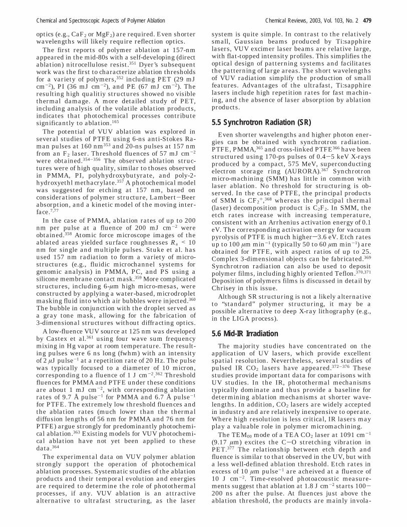
optics (e.g., CaF2 or MgF2) are required. Even shorterwavelengths will likely require reflection optics.
The first reports of polymer ablation at 157-nmappeared in the mid-80s with a self-developing (directablation) nitrocellulose resist.351 Dyer’s subsequentwork was the first to characterize ablation thresholdsfor a variety of polymers,352 including PET (29 mJcm-2), PI (36 mJ cm-2), and PE (67 mJ cm-2). Theresulting high qualitiy structures showed no visiblethermal damage. A more detailed study of PET,including analysis of the volatile ablation products,indicates that photochemical processes contributesignificantly to ablation.165
The potential of VUV ablation was explored inseveral studies of PTFE using 6-ns anti-Stokes Ra-man pulses at 160 nm353 and 20-ns pulses at 157 nmfrom an F2 laser. Threshold fluences of 57 mJ cm-2
were obtained.354-356 The observed ablation struc-tures were of high quality, similar to thoses observedin PMMA, PI, polyhydroxybutyrate, and poly-2-hydroxyethl methacrylate.357 A photochemical modelwas suggested for etching at 157 nm, based onconsiderations of polymer structure, Lambert-Beerabsorption, and a kinetic model of the moving inter-face.7,77
In the case of PMMA, ablation rates of up to 200nm per pulse at a fluence of 200 mJ cm-2 wereobtained.358 Atomic force microscope images of theablated areas yielded surface roughnesses Ra < 10nm for single and multiple pulses. Stuke et al. hasused 157 nm radiation to form a variety of micro-structures (e.g., fluidic microchannel systems forgenomic analysis) in PMMA, PC, and PS using asilicone membrane contact mask.359 More complicatedstructures, including 6-µm high micro-mesas, wereconstructed by applying a water-based, microdropletmasking fluid into which air bubbles were injected.360
The bubble in conjunction with the droplet served asa gray tone mask, allowing for the fabrication of3-dimensional structures without diffracting optics.
A low-fluence VUV source at 125 nm was developedby Castex et al.361 using four wave sum frequencymixing in Hg vapor at room temperature. The result-ing pulses were 6 ns long (fwhm) with an intensityof 2 µJ pulse-1 at a repetition rate of 20 Hz. The pulsewas typically focused to a diameter of 10 micron,corresponding to a fluence of 1 J cm-2.362 Thresholdfluences for PMMA and PTFE under these conditionsare about 1 mJ cm-2, with corresponding ablationrates of 9.7 Å pulse-1 for PMMA and 6.7 Å pulse-1
for PTFE. The extremely low threshold fluences andthe ablation rates (much lower than the thermaldiffusion lengths of 56 nm for PMMA and 76 nm forPTFE) argue strongly for predominantly photochemi-cal ablation.363 Existing models for VUV photochemi-cal ablation have not yet been applied to thesedata.364
The experimental data on VUV polymer ablationstrongly support the operation of photochemicalablation processes. Systematic studies of the ablationproducts and their temporal evolution and energiesare required to determine the role of photothermalprocesses, if any. VUV ablation is an attractivealternative to ultrafast structuring, as the laser
system is quite simple. In contrast to the relativelysmall, Gaussian beams produced by Ti:sapphirelasers, VUV excimer laser beams are relative large,with flat-topped intensity profiles. This simplifies theoptical design of patterning systems and facilitatesthe patterning of large areas. The short wavelengthsof VUV radiation simplify the production of smallfeatures. Advantages of the ultrafast, Ti:sapphirelasers include high repetition rates for fast machin-ing, and the absence of laser absorption by ablationproducts.
5.5 Synchrotron Radiation (SR)
Even shorter wavelengths and higher photon ener-gies can be obtained with synchrotron radiation.PTFE, PMMA,365 and cross-linked PTFE366 have beenstructured using 170-ps pulses of 0.4-5 keV X-raysproduced by a compact, 575 MeV, superconductingelectron storage ring (AURORA).367 Synchrotronmicro-machining (SMM) has little in common withlaser ablation. No threshold for structuring is ob-served. In the case of PTFE, the principal productsof SMM is CF2
+,368 whereas the principal thermal(laser) decomposition product is C2F2. In SMM, theetch rates increase with increasing temperature,consistent with an Arrhenius activation energy of 0.1eV. The corresponding activation energy for vacuumpyrolysis of PTFE is much highers3.6 eV. Etch ratesup to 100 µm min-1 (typically 50 to 60 µm min-1) areobtained for PTFE, with aspect ratios of up to 25.Complex 3-dimensional objects can be fabricated.369
Synchrotron radiation can also be used to depositpolymer films, including highly oriented Teflon.370,371
Deposition of polymers films is discussed in detail byChrisey in this issue.
Although SR structuring is not a likely alternativeto “standard” polymer structuring, it may be apossible alternative to deep X-ray lithography (e.g.,in the LIGA process).
5.6 Mid-IR Irradiation
The majority studies have concentrated on theapplication of UV lasers, which provide excellentspatial resolution. Nevertheless, several studies ofpulsed IR CO2 lasers have appeared.372-376 Thesestudies provide important data for comparisons withUV studies. In the IR, photothermal mechanismstypically dominate and thus provide a baseline fordetermining ablation mechanisms at shorter wave-lengths. In addition, CO2 lasers are widely acceptedin industry and are relatively inexpensive to operate.Where high resolution is less critical, IR lasers mayplay a valuable role in polymer micromachining.
The TEM00 mode of a TEA CO2 laser at 1091 cm-1
(9.17 µm) excites the C-O stretching vibration inPET.377 The relationship between etch depth andfluence is similar to that observed in the UV, but witha less well-defined ablation threshold. Etch rates inexcess of 10 µm pulse-1 are acheived at a fluence of10 J cm-2. Time-resolved photoacoustic measure-ments suggest that ablation at 1.8 J cm-2 starts 100-200 ns after the pulse. At fluences just above theablation threshold, the products are mainly invola-
Chemical and Spectroscopic Aspects of Polymer Ablation Chemical Reviews, 2003, Vol. 103, No. 2 479
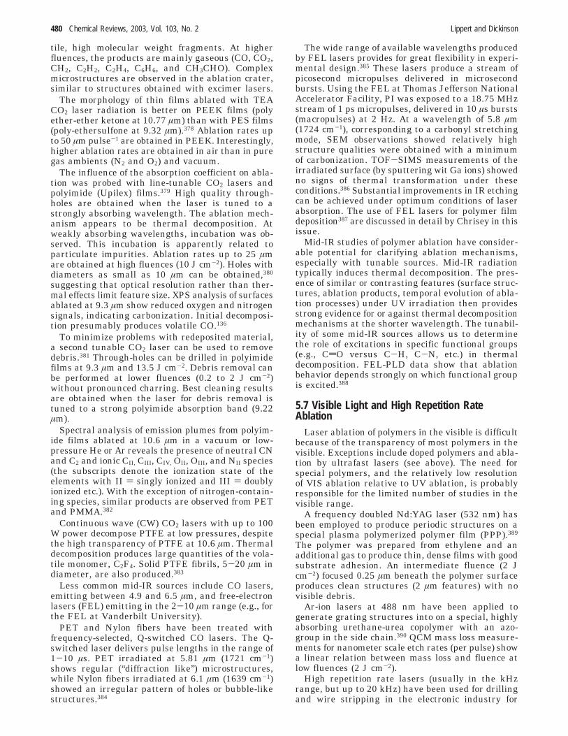
tile, high molecular weight fragments. At higherfluences, the products are mainly gaseous (CO, CO2,CH2, C2H2, C2H4, C6H6, and CH3CHO). Complexmicrostructures are observed in the ablation crater,similar to structures obtained with excimer lasers.
The morphology of thin films ablated with TEACO2 laser radiation is better on PEEK films (polyether-ether ketone at 10.77 µm) than with PES films(poly-ethersulfone at 9.32 µm).378 Ablation rates upto 50 µm pulse-1 are obtained in PEEK. Interestingly,higher ablation rates are obtained in air than in puregas ambients (N2 and O2) and vacuum.
The influence of the absorption coefficient on abla-tion was probed with line-tunable CO2 lasers andpolyimide (Upilex) films.379 High quality through-holes are obtained when the laser is tuned to astrongly absorbing wavelength. The ablation mech-anism appears to be thermal decomposition. Atweakly absorbing wavelengths, incubation was ob-served. This incubation is apparently related toparticulate impurities. Ablation rates up to 25 µmare obtained at high fluences (10 J cm-2). Holes withdiameters as small as 10 µm can be obtained,380
suggesting that optical resolution rather than ther-mal effects limit feature size. XPS analysis of surfacesablated at 9.3 µm show reduced oxygen and nitrogensignals, indicating carbonization. Initial decomposi-tion presumably produces volatile CO.136
To minimize problems with redeposited material,a second tunable CO2 laser can be used to removedebris.381 Through-holes can be drilled in polyimidefilms at 9.3 µm and 13.5 J cm-2. Debris removal canbe performed at lower fluences (0.2 to 2 J cm-2)without pronounced charring. Best cleaning resultsare obtained when the laser for debris removal istuned to a strong polyimide absorption band (9.22µm).
Spectral analysis of emission plumes from polyim-ide films ablated at 10.6 µm in a vacuum or low-pressure He or Ar reveals the presence of neutral CNand C2 and ionic CII, CIII, CIV, OII, OIII, and NII species(the subscripts denote the ionization state of theelements with II ) singly ionized and III ) doublyionized etc.). With the exception of nitrogen-contain-ing species, similar products are observed from PETand PMMA.382
Continuous wave (CW) CO2 lasers with up to 100W power decompose PTFE at low pressures, despitethe high transparency of PTFE at 10.6 µm. Thermaldecomposition produces large quantities of the vola-tile monomer, C2F4. Solid PTFE fibrils, 5-20 µm indiameter, are also produced.383
Less common mid-IR sources include CO lasers,emitting between 4.9 and 6.5 µm, and free-electronlasers (FEL) emitting in the 2-10 µm range (e.g., forthe FEL at Vanderbilt University).
PET and Nylon fibers have been treated withfrequency-selected, Q-switched CO lasers. The Q-switched laser delivers pulse lengths in the range of1-10 µs. PET irradiated at 5.81 µm (1721 cm-1)shows regular (“diffraction like”) microstructures,while Nylon fibers irradiated at 6.1 µm (1639 cm-1)showed an irregular pattern of holes or bubble-likestructures.384
The wide range of available wavelengths producedby FEL lasers provides for great flexibility in experi-mental design.385 These lasers produce a stream ofpicosecond micropulses delivered in microsecondbursts. Using the FEL at Thomas Jefferson NationalAccelerator Facility, PI was exposed to a 18.75 MHzstream of 1 ps micropulses, delivered in 10 µs bursts(macropulses) at 2 Hz. At a wavelength of 5.8 µm(1724 cm-1), corresponding to a carbonyl stretchingmode, SEM observations showed relatively highstructure qualities were obtained with a minimumof carbonization. TOF-SIMS measurements of theirradiated surface (by sputtering wit Ga ions) showedno signs of thermal transformation under theseconditions.386 Substantial improvements in IR etchingcan be achieved under optimum conditions of laserabsorption. The use of FEL lasers for polymer filmdeposition387 are discussed in detail by Chrisey in thisissue.
Mid-IR studies of polymer ablation have consider-able potential for clarifying ablation mechanisms,especially with tunable sources. Mid-IR radiationtypically induces thermal decomposition. The pres-ence of similar or contrasting features (surface struc-tures, ablation products, temporal evolution of abla-tion processes) under UV irradiation then providesstrong evidence for or against thermal decompositionmechanisms at the shorter wavelength. The tunabil-ity of some mid-IR sources allows us to determinethe role of excitations in specific functional groups(e.g., CdO versus C-H, C-N, etc.) in thermaldecomposition. FEL-PLD data show that ablationbehavior depends strongly on which functional groupis excited.388
5.7 Visible Light and High Repetition RateAblation
Laser ablation of polymers in the visible is difficultbecause of the transparency of most polymers in thevisible. Exceptions include doped polymers and abla-tion by ultrafast lasers (see above). The need forspecial polymers, and the relatively low resolutionof VIS ablation relative to UV ablation, is probablyresponsible for the limited number of studies in thevisible range.
A frequency doubled Nd:YAG laser (532 nm) hasbeen employed to produce periodic structures on aspecial plasma polymerized polymer film (PPP).389
The polymer was prepared from ethylene and anadditional gas to produce thin, dense films with goodsubstrate adhesion. An intermediate fluence (2 Jcm-2) focused 0.25 µm beneath the polymer surfaceproduces clean structures (2 µm features) with novisible debris.
Ar-ion lasers at 488 nm have been applied togenerate grating structures into on a special, highlyabsorbing urethane-urea copolymer with an azo-group in the side chain.390 QCM mass loss measure-ments for nanometer scale etch rates (per pulse) showa linear relation between mass loss and fluence atlow fluences (2 J cm-2).
High repetition rate lasers (usually in the kHzrange, but up to 20 kHz) have been used for drillingand wire stripping in the electronic industry for
480 Chemical Reviews, 2003, Vol. 103, No. 2 Lippert and Dickinson
several years. Commercial UV laser systems specif-ically designed for PCB drilling became availablearound 1995.391,392 Currently available systems em-ploy 266 and 355 nm radiation. The laser beam isgenerally highly focused, yielding fluences up to 50J cm-2 at 266 nm393 or up to 200 J cm-2 at 355 nm.394
These high fluences, combined with high repetitionrates, allow structuring (drilling, wire removal andmicromachining) of Teflon, PI, resins, copper, glass,ceramics, metals, and other materials. Ablation ratesare much higher at these high repetition rates.Ablation depends on pulse length, which is typicallyin the nanosecond range (e.g., from 24 to 36 ns).
The influence of pulse repetition rate on the abla-tion of PI and a glycol-modified PET (PETG) has beenstudied in more detail at 255 nm (frequency doubledcopper vapor laser).395,396 At repetition rates between0.7 and 17 kHz, the etch rate increases graduallywith increasing repetition rate at a fluence of 0.59 Jcm-2. No attenuation of the ablation laser beam byejected material is observed. After a large numberof pulses interact with the sample, the etch ratesincrease substantially with pulse repetition rate. Theincrease in etch rate is attributed to cumulativeheating of the sample and subsequent pyrolyticdecomposition.
The ablation rate and surface composition of PI(Upilex) has been studied as a function of the repeti-tion rate (5 to 20 kHz) using a Nd:YAG laser at 355nm. Blind holes were obtained at 5 kHz, whilethrough holes were produced at 20 kHz. XPS analysisafter irradiation revealed an increase of the carboncontent with increasing repetition rate, while therelative oxygen and nitrogen content were reduced.397
High repetition rate lasers are now commonlyemployed in material processing, largely because theablation products do not interfere with laser absorp-tion even for nanosecond pulses. The increase inablation rate due to cumulative heating can be useful,especially if the structure quality remains high. Adetailed understanding of the onset of these cumula-tive effects and their exact nature is still lacking.(Compare, for instance, refs 149 and 150).
6 Outlook
The laser ablation of polymers is an establishedtechnique in the electronic industry. The large num-ber of studies published annually indicate that thisis still an attractive area of research. Discussions ofablation mechanism are ongoing and will continue.The development of polymers designed specifically forlaser ablation is a unique tool for probing ablationmechanisms as well as for improving ablation prop-erties. New commercial applications will requireimproved ablation rates and control of undesirablesurface effects, such as debris.
The complexity of interactions between polymersand laser photons are illustrated by the variousprocesses associated with different irradiation condi-tions:
(i) Photothermal-photochemical laser ablation un-der excimer laser irradiation.
(ii) Dopant-induced laser ablation.
(iii) Photooxidative etching with lamps in an oxi-dizing atmosphere.
(iv) VUV etching in the absence of oxidizing condi-tions.
(v) Photokinetic etching with CW UV lasers.(vi) Ultrafast laser ablation, affected by pulse
length, wavelength, and possibly shock waves.(vii) Shock assisted photothermal ablation on pi-
cosecond time scales.(viii) VUV laser ablation: purely photochemical?(ix) Synchrotron structuring.(x) Mid-IR ablation, the influence of exciting vari-
ous functional groups.The ongoing maturation of laser techniques will
increase the number of applications of laser ablation.In the past decade, we have seen the development ofseveral exciting laser ablation tools, including fem-tosecond lasers, VUV lasers, free electron lasers, andhigh repetition-rate lasers. All these new techniquesare applied in ongoing research in conjunction witha variety of analytical techniques. Femtosecondlasers and VUV lasers in particular are expected tolead to important industrial applications.
7 AcknowledgmentsThis work was supported by the Swiss National
Science Foundation and by the US Department ofEnergy under Grant DE-FG03-98ER14864. We thankSteven Langford, WSU, for helpful comments on thismanuscript and Macarena Montenegro, PSI, for thehelp with the literature database.
8 References(1) Kawamura, Y.; Toyoda, K.; Namba, S. Appl. Phys. Lett. 1982,
40, 374.(2) Srinivasan, R.; Mayne-Banton, S. Appl. Phys. Lett. 1982, 41, 576.(3) Bauerle, D. Laser Processing and Chemistry; 3rd ed.; Springer-
Verlag: Berlin, 2000.(4) Patel, R. S.; Wassick, T. A. Proc. SPIE-Int. Soc. Opt. Eng. 1997,
2991, 217.(5) Aoki, H. U.S. Patent 5736999, 1998.(6) Dyer, P. E. Photochemical Processing of Materials; Academic
Press: London, 1992; p 359.(7) Lazare, S.; Granier, V. Laser Chem. 1989, 10, 25.(8) Srinivasan, R.; Braren, B. Chem. Rev. 1989, 89, 1303.(9) Babu, S. V.; D’Couto, G. C.; Egitto, F. D. J. Appl. Phys. 1992,
692.(10) Himmelbauer, M.; Arenholz, E.; Bauerle, D. Appl. Phys. A 1996,
63, 87.(11) Paraskevopoulos, G.; Singleton, D. L.; Irwin, R. S.; Taylor, R. S.
J. Appl. Phys. 1987, 70, 1938.(12) Taylor, R. S.; Singleton, D. L.; Paraskevopoulos, G. Appl. Phys.
Lett. 1987, 50, 1779.(13) Kuper, S.; Brannon, J.; Brannon, K. Appl. Phys. A 1993, 56, 43.(14) Lippert, T.; David, C.; Dickinson, J. T.; Hauer, M.; Kogelschatz,
U.; Langford, S. C.; Nuyken, O.; Phipps, C.; Robert, J.; Wokaun,A. J. Photochem. Photobiol. A Chem. 2001, 145, 145.
(15) Ball, Z.; Hopp, B.; Csete, M.; Ignacz, F.; Racz, B.; Sauerbrey, R.;Szabo, G. Appl. Phys. A 1995, 61, 547.
(16) Ball, Z.; Hopp, B.; Csete, M.; Ignacz, F.; Racz, B.; Szabo, G.;Sauerbrey, R. Appl. Phys. A 1995, 61, 575.
(17) Lippert, T.; Bennett, L. S.; Nakamura, T.; Niino, H.; Ouchi, A.;Yabe, A. Appl. Phys. A 1996, 63, 257.
(18) Srinivasan, R.; Hall, R. R.; Loehle, W. D.; Wilson, W. D.; Allbee,D. C. J. Appl. Phys. 1995, 78, 4881.
(19) Lippert, T.; Nakamura, T.; Niino, H.; Yabe, A. Macromolecules1996, 29, 6301.
(20) Lu, J.; Deshpande, S. V.; Gulari, E.; Kanicki, J.; Warren, W. L.J. Appl. Phys. 1996, 80, 5028.
(21) Schumann, M.; Sauerbrey, R.; Smayling, M. Appl. Phys. Lett.1991, 58, 428.
(22) Wei, J.; Hoogen, N.; Lippert, T.; Nuyken, O.; Wokaun, A. J. Phys.Chem. B 2001, 105, 1267.
Chemical and Spectroscopic Aspects of Polymer Ablation Chemical Reviews, 2003, Vol. 103, No. 2 481
(23) Arnold, N.; Luk’yanchuk, B.; Bityurin, N. Appl. Surf. Sci. 1998,127, 184.
(24) Schmidt, H.; Ihlemann, J.; Wolff-Rottke, B.; Luther, K.; Troe,J. J. Appl. Phys. 1998, 83, 5458.
(25) Lazare, S.; Granier, V. Chem. Phys. Lett. 1990, 168, 593.(26) Deutsch, T. F.; Geis, M. W. J. Appl. Phys. 1983, 54, 7201.(27) Andrew, J. E.; Dyer, P. E.; Foster, D.; Key, P. H. Appl. Phys.
Lett. 1983, 43, 717.(28) Sutcliffe, E.; Srinivasan, R. J. Appl. Phys. 1986, 60, 3315.(29) Mahan, G. D.; Cole, H. S.; Liu, Y. S.; Philipp, H. R. Appl. Phys.
Lett. 1988, 53, 2377.(30) Pettit, G. H.; Sauerbrey, R. Appl. Phys. Lett. 1991, 58, 793.(31) Pettit, G. H.; Ediger, M. N.; Hahn, D. W.; Brinson, B. E.;
Sauerbrey, R. Appl. Phys. A 1994, 58, 573.(32) Pettit, G. H.; Sauerbrey, R. Appl. Phys. A 1993, 56, 51.(33) Cain, S. R.; Burns, F. C.; Otis, C. E. J. Appl. Phys. 1992, 71,
4107.(34) Cain, S. J. Phys. Chem. 1993, 97, 7572.(35) D’Couto, G. C.; Babu, S. V. J. Appl. Phys. 1994, 75, 3052.(36) Luk’yanchuk, B.; Bityurin, N.; Himmelbauer, M.; Arnold, N.
Nucl. Instrum. Methods Phys. Res. B 1997, 122, 347.(37) Srinivasan, V.; Smrtic, M. A.; Babu, S. V. J. Appl. Phys. 1986,
59, 3861.(38) Luk’yanchuk, B.; Bityurin, N.; Anisimov, S.; Bauerle, D. Appl.
Phys. A 1993, 57, 367.(39) Luk’yanchuk, B.; Bityurin, N.; Anisimov, S.; Arnold, N.; Bauerle,
D. Appl. Phys. A 1996, 62, 397.(40) Bityurin, N.; Malyshev, A.; Luk’yanchuk, B.; Anisimov, S.;
Bauerle, D. Proc. SPIE, 1996; p 103.(41) Bityurin, N. Appl. Surf. Sci. 1999, 138-139, 354.(42) Treyz, G. V.; Scarmozzoni, R.; Osgood Jr., R. M. Appl. Phys. Lett.
1989, 55, 346.(43) Anisimov, S. I.; Khokhlov, V. A. Instabilities in Laser-Matter
Interaction; CRC-Press: Boca Raton, 1995.(44) Arnold, N.; Bityurin, N. Appl. Phys. A 1999, 68, 615.(45) Srinivasan, R. Appl. Phys. A 1993, 56, 417.(46) Fujiwara, H.; Fukumura, H.; Masuhara, H. J. Phys. Chem. 1995,
99, 11844.(47) Fukumura, H.; Takahashi, E.-i.; Masuhara, H. J. Phys. Chem.
1995, 99, 750.(48) Fukumura, H.; Mibuka, N.; Eura, S.; Masuhara, H.; Nishi, N.
J. Phys. Chem. 1993, 97, 13761.(49) Fujiwara, H.; Hayashi, T.; Fukumura, H.; Masuhara, H. Appl.
Phys. Lett. 1994, 64, 2451.(50) Fukumura, H.; Masuhara, H. Chem. Phys. Lett. 1994, 221, 373.(51) Wu, X.; Sadeghi, M.; Vertes, A. J. Phys. Chem. B 1998, 102, 4770.(52) Dutkiewicz, L.; Johnson, R. E.; Vertes, A.; Pedrys, R. J. Phys.
Chem. A 1999, 103, 2925.(53) Chang, T.-C.; Dlott, D. J. Phys. Chem. 1989, 90, 3590.(54) Zhigilei, L. V.; Kodali, P. B. S.; Garrison, B. J. J. Phys. Chem. B
1997, 101, 2028.(55) Zhigilei, L. V.; Kodali, P. B. S.; Garrison, B. J. J. Phys. Chem. B
1998, 102, 2845.(56) Zhigilei, L. V.; Garrison, B. J. J. Appl. Phys. 2000, 88, 1281.(57) Zhigilei, L. V.; Kodali, P. B. S.; Garrison, B. J. Chem. Phys. Lett.
1997, 276, 269.(58) Zhigilei, L. V.; Garrison, B. J. Appl. Phys. Lett. 1997, 71, 551.(59) Zhigilei, L. V.; Garrison, B. J. Rapid Commun. Mass Spectrom.
1998, 12, 1273.(60) Zhigilei, L. V.; Garrison, B. J. Appl. Phys. Lett. 1999, 74, 1341.(61) Zhigilei, L. V.; Garrison, B. J. Appl. Phys. A 1999, 69, S75.(62) Afanasov, P. A.; Nedialkov, N. N.; Imamova, S. E.; Ruf, A.; Hugel,
H.; Dausinger, F.; Berger, P. Appl. Surf. Sci. 2002, 186, 369.(63) Smirnova, J. A.; Zhigilei, L. V.; Garrison, B. J. Comput. Phys.
Commun. 1999, 118, 11.(64) Dekel, E.; Eliezer, S.; Henis, Z.; Moshe, E.; Ludmirsky, A.;
Goldberg, I. B. J. Appl. Phys. 1998, 84, 4851.(65) Lippert, T.; Wokaun, A.; Stebani, J.; Nuyken, O.; Ihlemann, J.
J. Phys. Chem. 1993, 97, 12297.(66) Zeifman, M.; Garrison, B. J.; Zhigilei, L. V. J. Appl. Phys. 2002,
92, 2181.(67) Lippert, T.; Yabe, A.; Wokaun, A. Adv. Mater. 1997, 9, 105.(68) Lippert, T.; Stebani, J.; Ihlemann, J.; Nuyken, O.; Wokaun, A.
Angew. Makromol. Chem. 1993, 213, 127.(69) Koubenakis, A.; Labrakis, J.; Georgiou, S. Chem. Phys. Lett.
2001, 346, 54.(70) Koubenakis, A.; Elmioti, T.; Georgiou, S. Appl. Phys. A 1999,
69, S637.(71) Georgiou, S.; Koubenakis, A.; Labrakis, J.; Lassithotaki, M. J.
Chem. Phys. 1998, 109, 8591.(72) Koubenakis, A.; Labrakis, J.; Georgiou, S. J. Chem. Soc.,
Faraday Trans. 1998, 94, 3427.(73) Atanassiou, A.; Andreou, E.; Fragouli, D.; Anglos, D.; Georgiou,
S.; Fotakis, C. J. Photochem. Photobiol. A: Chem. 2001, 145,229.
(74) Atanassiou, A.; Lassithotaki, M.; Anglos, D.; Georgiou, S.;Fotakis, C. Appl. Surf. Sci. 2000, 154-155, 89.
(75) Atanassiou, A.; Andreou, E.; Anglos, D.; Georgiou, S.; Fotakis,C. Appl. Phys. A 1999, 69, S285.
(76) Lassithotaki, M.; Atanassiou, A.; Anglos, D.; Georgiou, S.;Fotakis, C. Appl. Phys. A 1999, 69, 363.
(77) Lazare, S.; Granier, V. J. Appl. Phys. 1988, 63, 2110.(78) Kukreja, L. M.; Hess, P. Appl. Surf. Sci. 1994, 79-80, 158.(79) Sell, J. A.; Heffelfinger, D.; Ventzek, P. L. G.; Gilgenbach, R. L.
J. Appl. Phys. 1991, 69, 1330.(80) Efthimiopoulos, T.; Kritsotakis, E.; Kiagias, H.; Savvakis, C.;
Bertachas, Y. J. Phys. D: Appl. Phys. 1998, 31, 2648.(81) Zweig, A. D.; Venugopalan, V.; Deutsch, T. F. J. Appl. Phys.
1993, 74, 4181.(82) Diaci, J.; Mozina, J. Appl. Phys. A 1992, 55, 352.(83) Wang, J.; Niino, H.; Yabe, A. Jpn. J. Appl. Phys. 1999, 38, 871.(84) Lengl, G.; Plettl, A.; Ziemann, P.; Spatz, J. P.; Moeller, M. Appl.
Phys. A 2001, 72, 679.(85) Sylvain, J.-F.; Niino, H.; Ono, S.; Nakaoka, S.; Yabe, A. Appl.
Surf. Sci. 1999, 141, 25.(86) Ono, S.; Nakaoka, S.; Wang, J.; Niino, H.; Yabe, A. Appl. Surf.
Sci. 1998, 127-129, 821.(87) Low, D. K. Y.; Schmidt, M. J. J.; Li, L. Appl. Surf. Sci. 2000,
168, 170.(88) Slocombe, A.; Taufik, A.; Li, L. Appl. Surf. Sci. 2000, 168, 17.(89) Davis, C. R.; Snyder, R. W.; Egitto, F. D.; D’Couto, G. C.; Babu,
S. V. J. Appl. Phys. 1994, 76, 3049.(90) Lippert, T.; Kunz, T.; Hahn, C.; Wokaun, A. Recent Res. Devel.
Macromol. Res. 1997, 2, 121.(91) Kunz, T.; Stebani, J.; Ihlemannn, J.; Wokaun, A. Appl. Phys. A
1998, 67, 347.(92) Suzuki, K.; Matsuda, M.; Ogino, T.; Hayashi, T.; Terabayashi,
T.; Amemiya, K. Proc. SPIE, 1997; p 98.(93) Hayashi, N.; Suzuki, K.; Matsuda, M.; Ogino, T.; Tomita, Y. Proc.
SPIE, 1997; p 129.(94) Suzuki, K.; Matsuda, M.; Hayashi, N. Appl. Surf. Sci. 1998, 127-
129, 905.(95) Panitz, J.-C.; Lippert, T.; Stebani, J.; Nuyken, O.; Wokaun, A.
J. Phys. Chem. 1993, 97, 5246.(96) Lippert, T.; Stebani, J.; Ihlemann, J.; Nuyken, O.; Wokaun, A.
Angew. Makromol. Chem. 1993, 206, 97.(97) Stebani, J.; Nuyken, O.; Lippert, T.; Wokaun, A. Makromol
Chem. Rapid Commun. 1993, 14, 365.(98) Nuyken, O.; Nobis, M.; Scherer, C.; Ihlemann, J.; Beinhorn, F.
Macromol. Mater. Eng. 2000, 275, 1.(99) Nuyken, O.; Scherer, C.; Nobis, M. N.; Ihlemann, J. Polym.
Mater. Sci. Eng 1997, 76, 206.(100) Kunz, T.; Hahn, C.; Wokaun, A. Adv. Mater. 1998, 10, 786.(101) Hahn, C.; Kunz, T.; Dahn, U.; Nuyken, O.; Wokaun, A. Appl.
Surf. Sci. 1998, 127-129, 899.(102) Nuyken, O.; Dahn, U.; Wokaun, A.; Kunz, T.; Hahn, C.; Hessel,
V.; Landsiedel, J. Acta Polym. 1998, 49, 427.(103) Nuyken, O.; Dahn, U.; Ehrfeld, W.; Hessel, V.; Hesch, K.;
Landsiedel, J.; Diebel, J. Chem. Mater. 1997, 9, 485.(104) Andrews, J. E.; Dyer, P. E.; D., F.; Key, P. H. Appl. Phys. Lett.
1983, 43, 717.(105) Srinivasan, R.; Braren, B. J. Polym. Sci. 1984, 22, 2601.(106) Lippert, T.; Ortelli, E.; Panitz, J.-C.; Raimondi, F.; Wambach,
J.; Wei, J.; Wokaun, A. Appl Phys. A 1999, 69, S651.(107) Raimondi, F.; Abolhassani, S.; Brutsch, R.; Geiger, F.; Lippert,
T.; Wambach, J.; Wei, J.; Wokaun, A. J. Appl. Phys. 2000, 88,3659.
(108) Lippert, T.; Bennett, L. S.; Nakamura, T.; Niino, H.; Yabe, A.Appl. Surf. Sci. 1996, 96-98, 601.
(109) Lippert, T.; Dickinson, J. T.; Hauer, M.; Kopitkovas, G.; Lang-ford, S. C.; Masuhara, H.; Nuyken, O.; Robert, J.; Salmio, H.;Tada, T.; Tomita, K.; Wokaun, A. Appl. Surf. Sci. 2002, 197-198, 746.
(110) Srinivasan, R.; Braren, B.; Casey, K. G.; Yeh, M. J. Appl. Phys.1990, 67, 1604.
(111) Furutani, H.; Fukumura, H.; Masuhara, H. J. Phys. Chem. 1996,100, 6871.
(112) Kim, H.; Postlewaite, J. C.; Zyung, T.; Dlott, D. D. J. Appl. Phys.1988, 64, 2955.
(113) Bennett, L. S.; Lippert, T.; Furutani, H.; Fukumura, H.; Masu-hara, H. Appl. Phys. A 1996, 63, 327.
(114) Hauer, M.; Funk, D. J.; Lippert, T.; Wokaun, A. Appl. Phys. A,in press.
(115) Furutani, H.; Fukumura, H.; Masuhara, H. Appl. Phys. Lett.1994, 65, 3413.
(116) Furutani, H.; Fukumura, H.; Masuhara, H.; Lippert, T.; Yabe,A. J. Phys. Chem. A 1997, 101, 5742.
(117) Hauer, M.; Funk, D. J.; Lippert, T.; Wokaun, A. Proc. SPIE-Int.Soc. Opt. Eng. 2002, 4760, 259.
(118) Masabuchi, T.; Tada, T.; Nomura, E.; Hatanaka, K.; Fukumura,H.; Masuhara, H. J. Phys. Chem. A 2002, 106, 2180.
(119) Lippert, T.; David, C.; Hauer, M.; Masabuchi, T.; Masuhara, H.;Nomura, E.; Nuyken, O.; Phipps, C.; Robert, J.; Tada, T.; Tomita,K.; Wokaun, A. Appl. Surf. Sci. 2002, 186, 14.
(120) Lippert, T.; Wokaun, A.; Langford, S. C.; Dickinson, J. T. Appl.Phys. A 1999, 69, S655.
(121) Lippert, T.; Langford, S. C.; Wokaun, A.; Georgiou, S.; Dickinson,J. T. J. Appl. Phys. 1999, 86, 7116.
482 Chemical Reviews, 2003, Vol. 103, No. 2 Lippert and Dickinson
(122) Nuyken, O.; Stebani, J.; Lippert, T.; Wokaun, A.; Stasko, A.Macromol. Chem. Phys. 1995, 196, 751.
(123) Langford, S. C.; Dickinson, J. T.; Hauer, M.; Lippert, T.; Wokaun,A., submitted.
(124) Lippert, T.; Gerber, T.; Wokaun, A.; Funk, D. J.; Fukumura, H.;Goto, M. Appl. Phys. Lett. 1999, 75, 1018.
(125) David, C.; Wei, J.; Lippert, T.; Wokaun, A. Microlectr. Eng. 2001,57-58, 453.
(126) Lippert, T.; Wei, J.; Wokaun, A.; Hoogen, N.; Nuyken, O. Appl.Surf. Sci. 2000, 168, 270.
(127) Lippert, T.; Wei, J.; Wokaun, A.; Hoogen, N.; Nuyken, O.Macromol. Mater. Eng. 2000, 283, 140.
(128) Bachman, F. Chemtronics 1989, 4, 149.(129) Lankard, J. R.; Wolbold, G. Appl. Phys. A 1992, 54, 355.(130) Beuhler, A.; Tungare, A.; Savic, J. Circuit World 1998, 24, 36.(131) Inagaki, M.; Takichi, T.; Hishiyama, Y.; Obslei, A. Chemistry
and Physics of Carbon; Marcel Dekker: New York, 1999; p 245.(132) Schnyder, B.; Wambach, J.; Kunz, T.; Hahn, C.; Koetz, R. J.
Electron. Spectrosc. 1999, 105, 113.(133) Wesner, D. A.; Aden, M.; Gottmann, J.; Husmann, A.; Kreutz,
E. W. Fresenius J. Anal. Chem. 1999, 105, 113.(134) Zeng, K. C.; Yung, K. C.; Xie, C. S. Surf. Coat. Technol. 2002,
153, 210.(135) Yung, K. C.; Zeng, D. W.; Yue, T. M. Appl. Surf. Sci. 2001, 173,
193.(136) Zeng, D. W.; Yung, K. C. Appl. Surf. Sci. 2001, 180, 280.(137) Ortelli, E. E.; Geiger, F.; Lippert, T.; Wei, J.; Wokaun, A.
Macromolecules 2000, 33, 5090.(138) Ortelli, E. E.; Geiger, F.; Lippert, T.; Wokaun, A. Appl. Spectrosc.
2001, 55, 412.(139) Srinivasan, R.; Braren, B.; Seeger, D. E.; Dreyfus, R. W.
Macromolecules 1986, 19, 916.(140) Singleton, D. L.; Paraskevopoulos, G.; Irwin, R. S. J. Appl. Phys.
1989, 66, 3324.(141) Lazare, S.; Guan, W.; Drilhole, D. Appl. Surf. Sci. 1996, 96-98,
605.(142) Schumann, M.; Sauerbrey, R.; Smayling, M. C. Appl. Phys. Lett.
1991, 58, 428.(143) Feurer, T.; Sauerbrey, R.; Smayling, M. C.; Story, B. J. Appl.
Phys. A 1993, 56, 275.(144) Philips, H. M.; Wahl, S.; Sauerbrey, R. Appl. Phys. Lett. 1995,
62, 2572.(145) Walther, H. G.; Kitzing, T.; Bozoki, Z.; Liakhou, G. L.; Paloni,
S. J. Appl. Phys. 1999, 85, 7540.(146) Qin, Z. Y.; Du, B. Y.; Zhang, J.; He, T. B.; Qin, L.; Zhang, Y. S.
Appl. Phys. A 2001, 72, 711.(147) Ball, Z.; Feurer, T.; Callahan, D. L.; Sauerbrey, R. Appl. Phys.
A 1996, 62, 203.(148) Gu, J.; Tay, E.; Lim, P. K.; Lim, P. Appl. Phys. A 2002, 74, 487.(149) Reyna, L. G.; Sobehart, J. R. J. Appl. Phys. 1995, 78, 3423.(150) Burns, F. C.; Cain, S. R. J. Phys. D: Appl. Phys. 1996, 29, 1349.(151) Efthimiopoulos, T.; Kiagia, C.; Heliotis, G.; Helidonis, E. Can.
J. Phys. 2000, 78, 509.(152) Costela, A.; Figuera, J. M.; Florido, F.; Garcia-Moreno, I.; Collar,
E. P.; Sastre, R. Appl. Phys. A 1995, 60, 261.(153) Bityurin, N.; Muraviov, S.; Alexandrov, A.; Malyshev, A. Appl.
Surf. Sci. 1997, 109-110, 270.(154) Schmidt, H.; Luther, K.; Troe, J. Appl. Surf. Sci. 1999, 138-
139, 102.(155) Lade, R. J.; Morley, I. W.; May, P. W.; Rosser, K. N.; Ashfold,
M. N. R. Diamond Relat. Mater. 1999, 8, 1654.(156) Kuper, S.; Stuke, M. Appl. Phys. A 1989, 49, 211.(157) Srinivasan, R.; Braren, B.; Casey, K. G.; Yeh, M. J. Appl. Phys.
1990, 68, 1842.(158) Blanchet, G. C.; Cotts, P.; Fincher Jr., C. R. J. Appl. Phys. 2000,
88, 2975.(159) Lippert, T.; Webb, R. L.; Langford, S. C.; Dickinson, J. T. J. Appl.
Phys. 1999, 85, 1838.(160) Baker, A. K.; Dyer, P. E. Appl. Phys. A 1993, 57, 543.(161) Hopp, B.; Csete, M.; Szabo, G.; Bor, Z. Appl. Phys. A 1995, 61,
339.(162) Srinivasan, R. J. Appl. Phys. 1993, 73, 2743.(163) Hare, D. E.; Dlott, D. D. Appl. Phys. Lett. 1994, 64, 715.(164) Hare, D. E.; Franken, J.; Dlott, D. D. J. Appl. Phys. 1995, 77,
5950.(165) Dyer, P. E.; Oldershaw, G. A.; Schudel, D. J. Phys. D: Appl.
Phys. 1992, 25, 323.(166) Wagner, F.; Hoffmann, P. Appl. Surf. Sci. 2000, 154-155, 627.(167) Wagner, F.; Hoffmann, P. Appl. Surf. Sci. 2000, 168, 158.(168) Wagner, F.; Hoffmann, P. Appl. Phys. A 1999, 69, S841.(169) Mansour, N.; Ghaleh, K. J. Appl. Phys. A 2002, 74, 63.(170) Klose, S.; Arenholz, E.; Heitz, J.; Bauerle, D. Appl. Phys. A 1999,
69, S487.(171) Srinivasan, R.; Lazare, S. J. Phys. Chem. 1986, 90, 2124.(172) Lippert, T.; Zimmermann, F.; Wokaun, A. Appl. Spectrosc. 1993,
47, 1931.(173) Chtaib, M.; Roberfroid, E. M.; Novis, Y.; Pireaux, J. J.; Caudano,
R.; Lutgen, P.; Feyder, G. J. Vac. Sci. Technol. 1989, A7, 3233.(174) Kokai, F.; Saito, H.; Fujioka, T. Macromolecules 1990, 23, 674.
(175) Dyer, P. E.; Oldershaw, G. A.; Sidhu, J. J. Phys. Chem. 1991,95, 10004.
(176) Lazare, S.; Hoh, P. D.; Baker, J. M.; Srinivasan, R. J. Am. Chem.Soc. 1984, 106, 4288.
(177) Watanabe, H.; Yamamoto, M. J. Appl. Polym. Sci. 1999, 71, 2027.(178) Rossier, J. S.; Bercier, P.; Scwarz, A.; Loridant, S.; Girault, H.
H. Langmuir 1999, 15, 5173.(179) Huber, N.; Gruber, J.; Arnold, N.; Heitz, J.; Bauerle, D. Euro-
phys. Lett. 2000, 51, 674.(180) Blanchet, G. B.; Fincher Jr., C. R. Appl. Phys. Lett. 1994, 65,
1311.(181) Dickinson, J. T.; Shin, J.-J. Nucl. Instrum. Methods Phys. Res.
B 1994, 91, 672.(182) Blanchet, G. B.; Fincher Jr., C. R. Appl. Phys. Lett. 1996, 68,
929.(183) Haba, B.; Morishige, Y.; Kishida, S. Appl. Phys. A 1995, 60, 27.(184) Skordoulis, C. D.; Makropoulou, M. I.; Bolovinos, A. L.; Serafe-
tinides, A. A. Laser Med. Sci. 1997, 12, 313.(185) Smausz, T.; Kresz, N.; Hopp, B. Appl. Surf. Sci. 2001, 177, 66.(186) Hamada, Y.; Kawanishi, S.; Nishii, M.; Sugimoto, S.; Yamamoto,
T. Jpn. J. Appl. Phys. 1994, 33, 4764.(187) Izumi, Y.; Kawanishi, S.; Hara, S.; Yoshikawa, D.; Yamamoto,
T. Bull. Chem. Soc. Jpn. 1998, 71, 2721.(188) Niino, H.; Okano, H.; Inui, K.; Yabe, A. Appl. Surf. Sci. 1997,
109-110, 259.(189) Niino, H.; Yabe, A. Appl. Phys. Lett. 1993, 63, 3527.(190) Okoshi, M.; Murahara, M.; Toyoda, K. J. Mater. Res. 1992, 7,
1912.(191) Murahara, M.; Toyoda, K. J. Adhes. Sci. Technol. 1995, 9, 1601.(192) Heitz, J.; Niino, H.; Yabe, A. Appl. Phys. Lett. 1996, 68, 2648.(193) Heitz, J.; Niino, H.; Yabe, A. Jpn. J. Appl. Phys. 1996, 35, 4110.(194) Matienzo, L. J.; Zimmernmann, J. A.; Egitto, F. D. J. Vac. Sci.
Technol A 1994, 12, 2662.(195) Hopp, B.; Smausz, T.; Tombacz, E.; Wittmann, T.; Ignacz, F. Opt.
Commun. 2000, 181, 337.(196) Hopp, B.; Smausz, T.; Wittmann, T.; Ignacz, F. Appl. Phys. A
2000, 71, 315.(197) Dyer, P. E.; Karnakis, D. M. Appl. Phys. Lett. 1994, 64, 1344.(198) Feng, Y.; Liu, Z. Q.; Yi, X.-S. Appl. Surf. Sci. 2000, 156, 177.(199) Hopp, B.; Bor, Z.; Homolya, E.; Mihalik, E. Appl. Surf. Sci. 1997,
109-110, 232.(200) Devalckenaere, M.; Jadin, A.; Kolev, K.; Laude, L. D. Nucl.
Instrum. Methods Phys. Res. B 1999, 151, 263.(201) Sabbert, D.; Landsiedel, J.; Bauer, A.-D.; Ehrfeld, W. Appl. Surf.
Sci. 1999, 150, 185.(202) Laurens, P.; Sadras, B.; Decobert, F.; Arefei, F.; Amouroux, J.
Appl. Surf. Sci. 1999, 138-139, 93.(203) Schmidt, M. J. J.; Li, L.; Spencer, J. T.; Key, P. H. Appl. Surf.
Sci. 1999, 138-139, 418.(204) Skordoulis, C. D.; Makropoulou, M.; Serafetinides, A. A. Appl.
Surf. Sci. 1995, 86, 239.(205) Laude, L. D.; Soudant, S.; Beauvois, S.; Renaut, D.; Jadin, A.
Nucl. Instrum. Methods Phys. Res. B 1997, 131, 211.(206) Tsuboi, Y.; Goto, M.; Itaya, A. Chem. Lett. 1998, 521.(207) Tsuboi, Y.; Goto, M.; Itaya, A. J. Appl. Phys. 2001, 89, 7917.(208) Tsuboi, Y.; Ikejiri, T.; Yamada, K.; Itaya, A. Appl. Phys. A 2001,
73, 637.(209) Fuhr, G.; Ronacher, B.; Krahe, R.; Fest, S.; Shirley, S. G.;
Rogaschewski, S. Appl. Phys. A 1999, 68, 379.(210) Kuper, S.; Brannon, J.; Brannon, K. Appl. Phys. A 1993, 57, 255.(211) Okoshi, M.; Kuramatsu, M.; Inoue, N. Jpn. J. Appl. Phys. 2001,
40, L41.(212) Graubner, V.-M.; Jordan, R.; Nuyken, O.; Lippert, T.; Hauer,
M.; Schnyder, B.; Wokaun, A. Appl. Surf. Sci. 2002, 197-198,786.
(213) Baumann, R.; Bargon, J. Appl. Surf. Sci. 1996, 106, 287.(214) Van Dyke, L. S.; Brumlik, C. J.; Martin, C. R.; Yu, Z.; Collins,
G. J. Synth. Metals 1992, 52, 299.(215) Kay, E. Methods and Materials in Microelectronic Technology;
Plenum: New York, 1984.(216) Lippert, T.; Raimondi, F.; Wambach, J.; Wei, J.; Wokaun, A. Appl
Phys. A 1999, 63, S291.(217) Abdou, M. S. A.; Xie, Z. W.; Leung, A. M.; Holdcroft, S. Synth.
Met. 1992, 52, 159.(218) Wong, T. K. S.; Gao, S.; Hu, X.; Liu, H.; Chan, Y. C.; Lam, Y. L.
Mater. Sci. Engin. B 1998, 55, 71.(219) Tsunoda, K.; Ishii, T.; Tezuka, Y.; Yajima, H. J. Photochem.
Photobiol. A: Chem. 1997, 106, 21.(220) Tsunoda, K.; Kakinuma, K.; Yajima, H.; Ishii, T. Appl. Phys.
Lett. 1998, 73, 3381.(221) Himmelbauer, M.; Arenholz, E.; Bauerle, D.; Schilcher, K. Appl.
Phys. A 1996, 63, 337.(222) Bauerle, D.; Himmelbauer, M.; Arenholz, E. J. Photochem.
Photobiol. A: Chem. 1997, 106, 27.(223) Piglmayer, K.; Arenholz, E.; Ortwein, C.; Arnold, N.; Bauerle,
D. Appl. Phys. Lett. 1998, 73, 847.(224) Hansen, S. G.; Robitaille, T. E. J. Appl. Phys. 1988, 64, 2122.(225) Kokai, F.; Koga, Y.; Kakudate, Y.; Kawaguchi, M.; Fujiwara, S.;
Kubota, M.; Fukuda, M. Appl. Phys. A 1994, 59, 299.
Chemical and Spectroscopic Aspects of Polymer Ablation Chemical Reviews, 2003, Vol. 103, No. 2 483
(226) Estler, R. C.; Nogar, N. S. Appl. Phys. Lett. 1986, 49, 1175.(227) Krajnovich, D. J. J. Phys. Chem. A 1997, 101, 2033.(228) Dickinson, J. T.; Shin, J.-J.; Jiang, W.; Norton, M. G. J. Appl.
Phys. 1993, 74, 4729.(229) Blanchet, G. B.; Fincher, J. C. R. Appl. Phys. Lett. 1996, 68, 929.(230) Hansen, S. G. J. Appl. Phys. 1989, 66, 1411.(231) Creasy, W. R.; Brenna, J. T. Chem. Phys. 1988, 126, 453.(232) Larciprete, R.; Stuke, M. Appl. Phys. B 1987, 42, 181.(233) Shibagaki, K.; Takada, N.; Sasaki, K.; K., K. J. Appl. Phys. 2002,
91, 2449.(234) Tsunekawa, M.; Nishio, S.; Sato, H. J. Appl. Phys. 1994, 76,
5598.(235) Tsunekawa, M.; Nishio, S.; Sato, H. Jpn. J. Appl. Phys. 1995,
34, 218.(236) Danielzik, B.; Fabricius, N.; Rowekamp, M.; Linde, D. v. d. Appl.
Phys. Lett 1986, 48, 212.(237) Feldmann, D.; Kutzner, J.; Laukemper, J.; MacRobert, S.; Welge,
K. H. Appl. Phys. B 1987, 44, 81.(238) Cefalas, A. C.; Sarantopoulou, E.; Argitis, P.; Gogolides, E. Appl
Phys. A 1999, 69, S929.(239) Cefalas, A. C.; Vassilopoulos, N.; Sarantopoulou, E.; Kollia, Z.;
Skordoulis, C. Appl. Phys. A 2000, 70, 21.(240) Hauer, M.; Dickinson, J. T.; Langford, S. C.; Lippert, T.; Wokaun,
A. Appl. Surf. Sci. 2002, 197-198, 791.(241) Brannon, J. H.; Lankard, J. R.; Baise, A. I.; Burns, F.; Kaufman,
J. J. Appl. Phys. 1985, 58, 2036.(242) Kelly, R. J. Chem. Phys. 1988, 92, 5047.(243) Grivas, C.; Niino, H.; Yabe, A. Appl Phys. A 1999, 69, S159.(244) Kelly, R.; Rothenberg, J. E. Nucl. Instrum. Methods Phys. Res.
B 1985, 7-8, 755.(245) Kelly, R.; Dreyfus, R. W. Nucl. Instrum. Methods Phys. Res. B
1988, 32, 341.(246) Campbell, E. E. B.; Ulmer, G.; Bues, K.; Hertel, I. V. Appl Phys.
A 1989, 48, 543.(247) Campbell, E. E. B.; Ulmer, G.; Hasselberger, B.; Hertel, I. V.
Appl. Surf. Sci. 1989, 43, 346.(248) Press, W. H.; Flannery, B. P.; Teukolsky, S. A.; Vetterling, W.
T. Numerical Recipies in Pascal; Cambridge University: Cam-bridge, 1989.
(249) Dyer, P. E.; Jenkins, S. D.; Sidhu, J. Appl. Phys. Lett. 1986, 49,453.
(250) Dyer, P. E.; Jenkins, S. D.; Sidhu, J. Appl. Phys. Lett. 1988, 52,1880.
(251) Hopp, B.; Bor, Z.; Homolya, E.; Mihalik, E. Proc. SPIE-Int. Soc.Opt. Eng. 1998, 3423, 389.
(252) Wei, J.; Hoogen, N.; Lippert, T.; Hahn, C.; Nuyken, O.; Wokaun,A. Appl. Phys. A 1999, 69, S849.
(253) Novis, Y.; Pireaux, J. J.; Brezini, A.; Petit, E.; Caudano, R.;Lutgen, P.; Feyder, G.; Lazare, S. J. Appl. Phys. 1988, 64, 365.
(254) Bahners, T.; Schollmeyer, E. J. Appl. Phys. 1989, 66, 1884.(255) Arenholz, E.; Wagner, M.; Heitz, J.; Bauerle, D. Appl. Phys. A
1992, 55, 119.(256) Tonyali, K.; Jensen, L. C.; Dickinson, J. T. J. Vac. Sci. Technol.
A 1988, 6, 941.(257) Dyer, P. E.; Farley, R. J. Appl. Phys. Lett. 1990, 57, 765.(258) Niino, H.; Yabe, A. J. Photochem. Photobiol. A: Chem. 1992,
65, 303.(259) Emel’yanov, V. I.; Eriomin, K. I. Proc. SPIE-Int. Soc. Opt. Eng.
1998, 3343, 1056.(260) Heitz, J.; Arenholz, E.; Bauerle, D.; Schilcher, K. Appl. Surf. Sci.
1994, 81, 108.(261) Heitz, J.; Arenholz, E.; Bauerle, D.; Hibst, H.; Hagemeyer, A.;
Cox, G. Appl. Phys. A 1993, 56, 329.(262) Beleznai, C.; Vajtai, R.; Nanai, L. Fractals 1997, 5, 275.(263) Bolle, M.; Lazare, S.; Le Blanc, M.; Wilmes, A. Appl. Phys. Lett.
1992, 60, 674.(264) Bolle, M.; Lazare, S. J. Appl. Phys. 1993, 73, 3516.(265) Bolle, M.; Lazare, S. Appl. Surf. Sci. 1993, 65-66, 349.(266) Bolle, M.; Lazare, S. Appl. Surf. Sci. 1993, 69, 31.(267) Niino, H.; Kawabata, Y.; Yabe, A. Jpn. J. Appl. Phys. 1989, 28,
L2225.(268) Lazare, S.; Drilhole, D. J. Photochem. Photobiol. A: Chem. 1997,
106, 15.(269) Lazare, S.; Bolle, M.; Cros, A.; Bellard, C. Nucl. Instrum. Methods
Phys. Res. B 1995, 105, 159.(270) Hopp, B.; Csete, M.; Revesz, K.; Vinko, J.; Bor, Z. Appl. Surf.
Sci. 1996, 96-98, 611.(271) Csete, M.; Marti, O.; Bor, Z. Appl. Phys. A 2001, 73, 521.(272) Sendova, M.; Hiraoka, H. Jpn. J. Appl. Phys. 1993, 32, 6182.(273) Hiraoka, H.; Latsch, S.; Sendova, M. J. Photopolym. Sci. Technol.
1994, 7, 299.(274) Hiraoka, H.; Sendova, M. Appl. Phys. Lett. 1994, 64, 563.(275) Hiraoka, H. J. Photochem. Photobiol. A: Chem. 1995, 92, 129.(276) Sendova, M.; Hiraoka, H.; Lee, C.-H. Jpn. J. Appl. Phys. 1994,
33, 7135.(277) Lazare, S.; Benet, P.; Guan, W.; Bolle, M.; Mihailov, S. Excimer
Lasers; Kluwer Academic Publishers: Dordrecht, 1994; p 201.(278) Chan, C.-M.; Ko, T.-M.; Hiraoka, H. Surf. Sci. Rep. 1996, 24, 1.
(279) Fourrier, T.; Schrems, G.; Muehlberger, T.; Heitz, J.; Arnoild,N.; Bauerle, D.; Mosbacher, M.; Boneberg, J.; Leiderer, P. Appl.Phys. A 2001, 72, 1.
(280) Gerenser, L. J. Metallization of Polymers; American ChemicalSociety: Washington, DC, 1990; p 443.
(281) Ehrlich, D. J.; Tsao, J. Y. Laser Microfabrication: Thin FilmProcesses and Lithography; Academic Press: London, 1989.
(282) Bauer, A.; Ganz, J.; Hesse, K.; Koehler, E. Appl. Surf. Sci. 1990,46, 113.
(283) Liebel, G. Galvanotechnik 1988, 79, 2883.(284) Hiraoka, H.; Lazare, S. Appl. Surf. Sci. 1990, 46, 246.(285) Wesner, D. A.; Weichenhain, R.; Pfleging, W.; Horn, H.; Kreutz,
E. W. Fresenius J. Anal. Chem. 1997, 358, 248.(286) Horn, H.; Beil, S.; Wesner, D. A.; Weichenhein, R.; Kreutz, E.
W. Nucl. Instrum. Methods Phys. Res. B 1999, 151, 279.(287) Zhao, G.; Philips, H. M.; Zheng, H.; Tam, S.; Li, W.; Wen, G.;
Gong, Z.; Lam, Y. Proc. SPIE-Int. Soc. Opt. Eng. 2000, 3933,505.
(288) Petit, S.; Laurens, P.; Amouroux, J.; Arefi-Khonsari, F. Appl.Surf. Sci. 2000, 168, 300.
(289) Lazare, S.; Benet, P. J. Appl. Phys. 1993, 74, 4953.(290) Laurens, P.; Sadras, B.; Decobert, F.; Arefi-Khonsari, F.; Amour-
oux, J. Internat. J. Adhes. Adhes. 1998, 18, 19.(291) Watanabe, H.; Takata, T. Angew. Makromol. Chem. 1996, 235,
95.(292) Catry, C.; Jeuris, K.; Jackers, C.; Hofkens, J.; Bastin, L.; Gensch,
T.; Grim, P. C. M.; De Schryver, F. C.; Van Damme, M.Langmuir 1999, 15, 1364.
(293) Niino, H.; Yabe, A. Appl. Surf. Sci. 1996, 96-98, 572.(294) Niino, H.; Yabe, A. J. Photochem. Photobiol. A: Chem. 1997,
106, 9.(295) Srinivasan, R.; Lazare, S. Polymer 1985, 26, 1297.(296) Vasilets, V. N.; Yuranova, T. I.; Ponomarev, A. N. J. Photopolym.
Sci. Technol. 1994, 7, 309.(297) Vasilets, V. N.; Nakamura, K.; Uyama, Y.; Ogata, S.; Ikada, Y.
Polymer 1998, 39, 2875.(298) Vasilets, V. N.; Hirata, I.; Iwata, H.; Ikada, Y. J. Polym Sci.,
Part A: Polym. Chem. 1998, 36, 2215.(299) Zhang, J.-Y.; Boyd, I. W.; Esrom, H. Surf. Interface Anal. 1996,
24, 718.(300) Zhang, J.-Y.; Esrom, H.; Kogelschatz, U.; Emig, G. Appl. Surf.
Sci. 1993, 69, 299.(301) Srinivasan, R.; Leigh, W. J. J. Am. Chem. Soc. 1982, 104, 6784.(302) Srinivasan, R. Polymer 1982, 23, 1863.(303) Shirai, M.; Yamamoto, T.; Tsunooka, M. Polym. Degrad. Stab.
1999, 63, 481.(304) Shinozuka, T.; Shirai, M.; Tsunooka, M. J. Photopolym. Sci.
Technol. 2000, 13, 751.(305) Fuchs, F.; Goetzberger, O.; Henck, R.; Fogarassy, E. Appl. Phys.
A 1995, 60, 505.(306) Srinivasan, R. Appl. Phys. Lett. 1991, 58, 2895.(307) Srinivasan, R. J. Appl. Phys. 1992, 72, 1651.(308) Srinivasan, R. Appl. Phys. A 1992, 55, 269.(309) Srinivasan, R. J. Appl. Phys. 1991, 70, 7588.(310) Bozhevolnyi, S. I.; Potemkin, I. V.; Svetovoy, V. B. J. Appl. Phys.
1992, 71, 2030.(311) Bozhevolnyi, S. I.; Potemkin, I. V. J. Phys. D: Appl. Phys. 1994,
27, 19.(312) Bonlie, J. D.; Patterson, F.; Price, D.; White, B.; Springer, P.
Appl. Phys. B 2000, 70, S155.(313) Bloembergen, N. IEEE J. Qaunt. Electron. 1974, QE-10, 375.(314) Stuart, B. C.; Feit, M. D.; Rubenchik, A. M.; Shore, B. W.; Perry,
M. D. Phys. Rev. Lett. 1995, 74, 2248.(315) Haight, R.; Hayden, D.; Longo, P.; Neary, T.; Wagner, A. Proc.
SPIE-Int. Soc. Opt. Eng. 1998, 3546, 477.(316) Shani, Y.; Melnick, I.; Yoffe, S.; Sharon, Y.; Lieberman, K.;
Terkel, H. Proc. SPIE-Int. Soc. Opt. Eng. 1998, 3546, 112.(317) Srinivasan, R.; Sutcliffe, E.; Braren, B. Appl. Phys. Lett. 1987,
51, 1285.(318) Kuper, S.; Stuke, M. Appl. Phys. B 1987, 44, 199.(319) Kuper, S.; Stuke, M. Appl. Phys. Lett. 1989, 54, 4.(320) Kuper, S.; Stuke, M. Mater. Res. Soc. Symp Proc. 1989, 129, 375.(321) Preuss, S.; Spath, M.; Zhang, Y.; Stuke, M. Appl. Phys. Lett.
1993, 62, 3049.(322) Preuss, S.; Spath, M.; Stuke, M. Microelectron. Eng. 1994, 25,
313.(323) Kautek, W.; Mitterer, S.; Kruger, J.; Husinsky, W.; Grabner, W.
Appl. Phys. A 1994, 58, 513.(324) Kumagai, H.; Midorikawa, K.; Toyoda, K.; Nakamura, S.;
Okamoto, T.; Obara, M. Appl. Phys. Lett. 1994, 65, 1850.(325) Nakamura, S.; Midorikawa, K.; Kumagai, H.; Obara, M.; Toyoda,
K. Jpn. J. Appl. Phys. 1996, 35, 101.(326) Zhang, Y.; Lowe, R. M.; Harvey, E.; Hannaford, P.; Endo, A. Appl.
Surf. Sci. 2002, 186, 345.(327) Serafetinides, A. A.; Skordoulis, C. D.; Makropoulou, M. I.; Kar,
A. K. Appl. Surf. Sci. 1998, 135, 276.(328) Serafetinides, A. A.; Skordoulis, C. D.; Makropoulou, M. I.; Kar,
A. K. Appl. Surf. Sci. 2001, 180, 42.(329) Baudach, S.; Bonse, J.; Kautek, W. Appl. Phys. A 1999, 69, S395.
484 Chemical Reviews, 2003, Vol. 103, No. 2 Lippert and Dickinson
(330) Baudach, S.; Bonse, J.; Kruger, J.; Kautek, W. Appl. Surf. Sci.2000, 154-155, 555.
(331) Bonse, J.; Baudach, S.; Kruger, J.; Kautek, W. Proc. SPIE-Int.Soc. Opt. Eng. 2000, 4065, 161.
(332) Baudach, S.; Kruger, J.; Kautek, W. Rev. Laser Engin. 2001,29, 705.
(333) Heitz, J.; Arenholz, E.; Bauerle, D.; Sauerbrey, R.; Phillips, H.M. Appl. Phys. A 1994, 59, 289.
(334) Niino, H.; Ihlemann, J.; Ono, S.; Yabe, A. J. Photopolym Sci.Technol. 2000, 13, 167.
(335) Niino, H.; Yabe, A. J. Photopolym Sci. Technol. 2001, 14, 197.(336) Yamasaki, K.; Juodkazis, S.; Lippert, T.; Watanabe, M.; Matsuo,
S.; Misawa, H. Appl. Phys. A 2003, 76, 325.(337) Hopp, B.; Toth, Z.; Gal, K.; Mechler, A.; Bor, Z.; Moustaizis, S.
D.; Georgiou, S.; Fotakis, C. Appl. Phys. A 1999, 69, S191.(338) Toth, Z.; Hopp, B.; Mechler, A.; Bor, Z.; Moustaizis, S. D.;
Athanassiou, A.; Georgiou, S.; Kalpouzos, C.; Fotakis, C. LaserPhys. 2000, 10, 241.
(339) Hosokawa, Y.; Yashiro, M.; Asahi, T.; Masuhara, H. Proc. SPIE-Int. Soc. Opt. Eng. 2001, 4274, 78.
(340) Hosokawa, Y.; Yashiro, M.; Asahi, T.; Masuhara, H.; Kadota,T.; Shirota, Y. Jpn. J. Appl. Phys. 2001, 40, L1116.
(341) Hahn, C.; Lippert, T.; Wokaun, A. J. Phys. Chem. B 1999, 103,1287.
(342) Wen, X.; Tolbert, W.; Dlott, D. D. Chem. Phys. Lett. 1992, 192,315.
(343) Kim, H.; Postlewaite, J. C.; Zhung, T.; Dlott, D. D. Appl. Phys.Lett. 1989, 54, 2274.
(344) Zhung, T.; Kim, H.; Postlewaite, J. C.; Dlott, D. D. J. Appl. Phys.1989, 65, 4548.
(345) Wen, X.; Hare, D. E.; Dlott, D. D. Appl. Phys. Lett. 1994, 64,184.
(346) Sandy Lee, I.-Y.; Wen, X.; Tolbert, W. A.; Dlott, D. D.; Doxtader,M.; Arnold, D. R. J. Appl. Phys. 1992, 72, 2440.
(347) Hare, D. E.; Franken, J.; Dlott, D. D.; Chronister, E. L.; Flores,J. J. Appl. Phys. Lett. 1994, 65, 3051.
(348) Hare, D. E.; Rhea, S. T.; Dlott, D. D.; D’Amato, R. J.; Lewis, T.E. J. Imag. Sci. Technol. 1998, 42, 187.
(349) Moore, D. S.; Gahagan, K. T.; Reho, J. H.; Funk, D. J.; Buelow,S. J.; Rabie, R. L.; Lippert, T. Appl. Phys. Lett. 2001, 78, 40.
(350) Simon, P.; Bekesi, J.; Doelle, C.; Klein-Wiele, J.-H.; Marowsky,G.; Szatmari, S.; Wellegehausen, B. Appl. Phys. B 2002, 74,S189.
(351) Henderson, D.; White, J. C.; Craighead, H. G.; Adesida, I. Appl.Phys. Lett. 1985, 45, 900.
(352) Dyer, P. E.; Sidhu, J. J. Opt. Soc. Am. B 1986, 3, 792.(353) Wada, S.; Toshiro, H.; Toyoda, K.; Niino, H.; Yabe, A. Appl. Phys.
Lett. 1993, 62, 211.(354) Herman, P. R.; Chen, B.; Moore, J. D. Mater. Res. Soc. Proc.
1993, 285, 163.(355) Herman, P. R.; Chen, B.; Moore, J. D. AIP. Conf. Proc. 1993,
288, 297.(356) Herman, P. R.; Chen, B.; Moore, J. D.; Canaga-Retnam, M.
Mater. Res. Soc. Proc. 1992, 236, 53.(357) Costela, A.; Garcia-Moreno, I.; Florido, F.; Figuera, J. M.; Sastre,
R.; Hooker, S. M.; Cashmore, J. S.; Webb, C. E. J. Appl. Phys.1995, 77, 2343.
(358) Padeletti, G.; Lapczyna, M.; Stuke, M. Mater. Res. Soc. Proc.1999, 544, 3.
(359) Lapczyna, M.; Stuke, M. Mater. Res. Soc. Proc. 1998, 526, 143.(360) Lapczyna, M.; Stuke, M. Appl. Phys. A 1998, 66, 473.(361) Museur, L.; Zheng, W. Q.; Kanaev, A. V.; Castex, M. C. IEEE J.
Sel. Top. Quantum Electron. 1995, 1, 900.(362) Riedel, D.; Castex, M. C. Proc. SPIE, 1998, p 234.
(363) Riedel, D.; Castex, M. C. Appl. Phys. A 1999, 69, 375.(364) Castex, M. C.; Bityurin, N.; Olivero, C.; Muriaviov, S.; Bronni-
kova, N.; Riedel, D. Appl. Surf. Sci. 2000, 168, 175.(365) Katoh, T.; Zhang, Y. J. Synchrotron Rad. 1998, 5, 1153.(366) Katoh, T.; Yamaguchi, D.; Satoh, Y.; Ikeda, S.; Aoki, Y.; Washio,
M.; Tabata, Y. Appl. Surf. Sci. 2002, 186, 24.(367) Zhang, Y.; Hori, T. Synchrotron Radiat. News 2000, 13, 32.(368) Zhang, Y.; Katoh, T.; Washio, M.; Yamada, H.; Hamada, S. Appl.
Phys. Lett. 1995, 67, 872.(369) Katoh, T.; Nishi, N.; Fukagawa, M.; Ueno, H.; Sugiyama, S. Sens.
Actuators A 2001, 89, 10.(370) Katoh, T.; Zhang, Y. Appl. Surf. Sci. 1999, 138-139, 165.(371) Zhang, Y.; Katoh, T.; Endo, E. J. Electron Spectrosc. Relat.
Phenom. 2001, 119, 247.(372) Cozzens, R. F.; Fox, R. B. Polym. Eng. Sci. 1978, 18, 900.(373) Koren, G. Appl. Phys. Lett. 1984, 45, 10.(374) Brannon, J. H.; Lankard, J. R. Appl. Phys. Lett. 1986, 48, 1226.(375) Braun, R.; Nowak, R.; Hess, P.; Oetzmann, H.; Schmidt, C. Appl.
Surf. Sci. 1989, 43, 352.(376) Sonnenschein, M. F.; Roland, C. M. Appl. Phys. Lett. 1990, 57,
425.(377) Dyer, P. E.; Oldershaw, G. A.; Sidhu, J. Appl. Phys. B 1989, 48,
489.(378) Sumiyoshi, T.; Ninomiya, Y.; Ogasawara, H.; Obara, M.; Tanaka,
H. Appl. Phys. A 1994, 58, 475.(379) Dyer, P. E.; Karnakis, D. M.; Oldershaw, G. A.; Roberts, G. C.
J. Appl. Phys. D: Appl. Phys. 1996, 26, 2554.(380) Dyer, P. E.; Waldeck, I.; Roberts, G. C. J. Appl. Phys. D: Appl.
Phys. 1997, 30, L19.(381) Coupland, K.; Herman, P. R.; Gu, B. Appl. Surf. Sci. 1998, 127-
129, 731.(382) Wong, K. H.; Tou, T. Y.; Low, K. S. J. Appl. Phys. 1998, 83, 2286.(383) Tolsopyatov, E. M.; Ivanov, L. F.; Grakovich, P. N.; Krasovsky,
A. M. Proc. SPIE-Int. Soc. Opt. Eng. 1998, 3343, 1010.(384) Ionin, A.; Klimachev, Y.; Kobsa, H.; Sinistyn, D. Proc. SPIE-
Int. Soc. Opt. Eng. 1998, 3343, 1032.(385) Freund, H. P.; Neill, G. R. Proc. IEEE 1999, 87, 782.(386) Kelly, M. J. Mater. Res. Soc. Symp. Proc. 2001, 617, J5.7.(387) Bubb, D. M.; Toftmann, B.; Haglund Jr., R. F.; Horowitz, J. S.;
Papantonakis, M. R.; McGill, R. A.; Wu, P. W.; Chrisey, D. B.Appl. Phys. A 2002, 74, 123.
(388) Bubb, D. M.; Papantonakis, M. R.; Toftmann, B.; Horowitz, J.S.; McGill, R. A.; Chrisey, D. B.; Haglund Jr., R. F. 2002, 91,9809.
(389) Silverstein, M. S.; Visoly, I.; Kesler, O.; Janai, M.; Cassuto, Y.J. Vac. Sci. Tchnol. B 1998, 16, 2957.
(390) Egami, C.; Kawata, Y.; Aoshima, Y.; Takeyama, H.; Iwata, F.;Sugihara, O.; Tsuchimori, M.; Watanabe, O.; Fujimura, H.;Okamoto, N. Opt. Commun. 1998, 157, 150.
(391) Hu, A. CircuiTree 1998, 11, 86.(392) Owen, M.; Roelants, E.; Van Pyumbroeck, J. Circuit World 1997,
24, 45.(393) Maryniuk, J. AMP J. Technol. 1995, 4, 77.(394) Yung, W. K. C.; Liu, J. S.; Man, H. C. Circuit World 1999, 25,
13.(395) Illy, E. K.; Brown, D. J. W.; Withford, M. J.; Piper, J. A. IEEE
J. Sel. Top. Quantum Electron. 1999, 5, 1543.(396) Glover, A. C. J.; Illy, E. K.; Piper, J. A. IEEE J. Sel. Top.
Quantum Electron. 1995, 1, 830.(397) Yung, K. C.; Zeng, D. W.; Yue, T. M. Surf. Coat. Technol. 2002,
160, 1.
CR010460Q
Chemical and Spectroscopic Aspects of Polymer Ablation Chemical Reviews, 2003, Vol. 103, No. 2 485