Embed Size (px)
Citation preview
Hea Yoon Kwon,1 Young Kyoung Park,1 Sun Myoung Lee, Ji Hyeon Baek,
Jae-Seung Kang, Moon-Hyun Chung, Eun Ji Kim, Jin-Soo Lee
Bartonella henselae, a gram-negative bacterium, is a common causative agent of zoonotic infections. We report 5 culture-proven cases of B. henselae infection in South Korea. By align-ment of the 16S rRNA sequences and multilocus sequencing typing analysis, we identified all isolates as B. henselae Hous-ton-1 strain, which belongs to sequence type 1.
The genus Bartonella includes infectious, gram-nega-tive, facultative intracellular bacteria of numerous spe-
cies. Among the Bartonella species, B. henselae is known as one of the most noteworthy pathogens (1). B. henselae causes cat-scratch disease, which is a common zoonosis and manifests various clinical symptoms (2).
A case of B. henselae infection in South Korea was confirmed in 2005 by PCR (3). Although a few more stud-ies have been published after this case of B. henselae, only 2 cases were culture-proven: 1 from blood and 1 from bone marrow (4,5). Because of difficulties in cultivation and iso-lation, studies of the isolation of B. henselae from clinical specimens remain scarce. In this study, we analyzed the characteristics of the isolated B. henselae strains in South Korea and compared the clinical features of the patients.
The StudyWe conducted the study among patients who visited Inha University Hospital, a tertiary hospital in Incheon, South Korea, during 2009–2016. From these patients, we isolated 5 cases in which B. henselae was identified from cultures of blood or bone marrow (Table 1).
Case-patient 1 (IIBC1301) was a 22-year-old man hospitalized for left inguinal lymphadenopathy that had started 10 days earlier. His body temperature was 38.5°C, and he had rashes that started on the palms and soles and subsequently spread to his entire body. B. henselae was isolated from the blood that was cultured on the second day of hospitalization.
Case-patient 2 (IIBC1302) was a 40-year-old woman hospitalized for fever and myalgia, symptoms that had last-ed for 1 month. The patient had an erythematous papular rash on her face and extremities and tenderness in her abdo-men. Computed tomography (CT) of the abdomen showed chronic cholecystitis; therefore, levofloxacin and metroni-dazole were prescribed (online Technical Appendix Figure, panel A, https://wwwnc.cdc.gov/EID/article/24/5/17-1497-Techapp1.pdf). B. henselae was identified from cultures of blood obtained on the first day of the hospitalization. The pa-tient had not raised any animals. After discharge, the patient experienced continuous fever, poor oral intake, and weight loss. Reevaluation showed centrilobular ground-glass opac-ity in both lung fields on chest CT and growth of Mycobacte-rium tuberculosis on sputum acid-fast bacilli culture (online Technical Appendix Figure, panel B). A pulmonary tuber-culosis infection was diagnosed and treated with antituber-culosis medication.
Case-patient 3 (IIBC1303) was a 52-year-old woman hospitalized for fever and left flank pain; her symptoms had persisted for 1 month. She also reported right-side neck swelling and pain at neck levels II, III, and VA. B. henselae was isolated from cultures of blood collected on the 16th day of hospitalization. She had no contact with animals.
Case-patient 4 (IIBC1304) was a 42-year-old man we previously reported (5) whose main complaints were fever, rash, and arthralgia. B. henselae was isolated from a bone marrow sample. The patient had no contact with or experi-ence in raising pets.
Case-patient 5 (IIBC1305), also previously pub-lished (4), was a 73-year-old woman who had B. hense-lae isolated from her blood. She also did not have any contact with animals.
Bartonella species can be grown by blood agar–based culture systems. However, it is difficult to culture them this way because the growth of bacterial cells is slow, and ob-taining colonies on the agar plate takes a long time. On the other hand, Bartonella species grow more rapidly with cell culture–based systems (6). For testing of these pa-tients, we grew ECV304 cells in M199 media containing 10% heat-inactivated fetal bovine serum and inoculated 1 mL of whole blood or other samples from the patients onto the cells. After 24 hours, we washed the cells with Dul-becco’s phosphate-buffered saline and maintained them in M199 media. We performed an immunofluorescence assay
Characterization of Clinical Isolates of Bartonella henselae Strains, South Korea
912 Emerging Infectious Diseases • www.cdc.gov/eid • Vol. 24, No. 5, May 2018
DISPATCHES
Author affiliations: Inha University School of Medicine, Incheon, South Korea (H.Y. Kwon, Y.K. Park, S.M. Lee, J.H. Baek, J.-S. Kang, E.J. Kim, J.-S. Lee); Jeju National University, Jeju, South Korea (M.-H. Chung)
DOI: https://doi.org/10.3201/eid2405.171497 1These authors contributed equally to this article.
Isolates of Bartonella henselae, South Korea
(IFA) with the patient’s own serum (1:40 diluted) every week after the inoculation. When the growth of bacteria was observed, we scraped all cultured cells from the T25
flask. We then reinoculated 1 mL of infected ECV304 cells onto uninfected ECV304 cells in a T75 flask for expansion of bacterial cells.
Emerging Infectious Diseases • www.cdc.gov/eid • Vol. 24, No. 5, May 2018 913
Table 1. Demographic and clinical characteristics of 5 case-patients whose serum sample cultures revealed the presence of Bartonella henselae, South Korea* Characteristic Case-patient 1 Case-patient 2 Case-patient 3 Case-patient 4 (5) Case-patient 5 (4) Age, y/sex 22/M 40/F 52/F 42/M 73/F Clinical symptoms Inguinal LAP, rash Fever, myalgia Febrile sense, left
flank pain Rash, fever,
myalgia Fever, general
weakness Lymphadenopathy External iliac chain,
inguinal area, supraclavicular area
Left neck level IV, V Right neck II, III, VA Right supraclavicular
area
None
Leukocytes, cells/µL 10,920 6,130 8,180 19,260 5,120 AST/ALT, IU/dL 132/270 107/51 30/16 212/246 47/56 ESR, mm/h/CRP, mg/dL 21/3.93 44/5.5 4/0.14 25/12.9 22/13.16 Treatment Third-generation
cephalosporin, doxycycline
Levofloxacin, metronidazole, third-
generation cephalosporin and
doxycycline
Third-generation cephalosporin, minocycline,
metronidazole
Doxycycline, changed to minocycline
Third-generation cephalosporin,
doxycycline
B. henselae IgG titer 1:160 1:640 1:160 1:1,280 1:160 Pets None None None None None Co-occurring conditions None Pulmonary
tuberculosis None None None
*ALT, alanine aminotransferase; AST, aspartate aminotransferase; CRP, C-reactive protein; ESR, erythrocyte sedimentation rate.
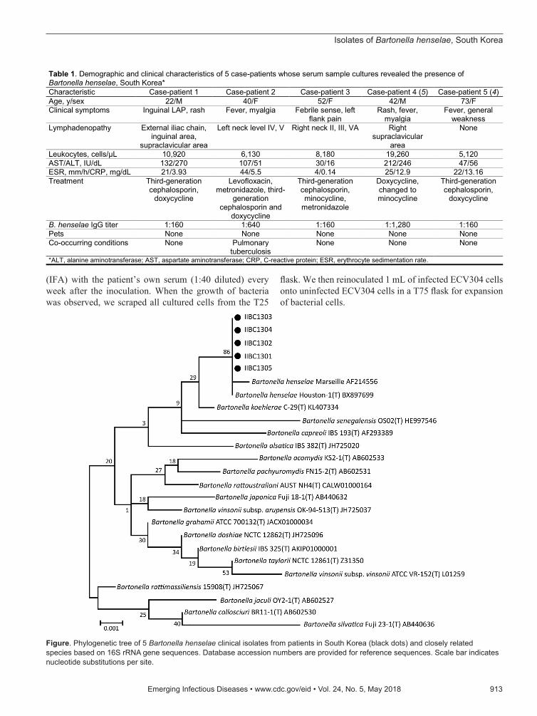
Figure. Phylogenetic tree of 5 Bartonella henselae clinical isolates from patients in South Korea (black dots) and closely related species based on 16S rRNA gene sequences. Database accession numbers are provided for reference sequences. Scale bar indicates nucleotide substitutions per site.

DISPATCHES
To identify the bacterial isolates, we amplified and se-quenced the 16S rRNA gene (7). The pathogens cultured from the specimens showed the highest sequence similari-ties with B. henselae Houston-1 strain (GenBank acces-sion nos. KY773227, KY773228, KY773229, KY773290, and KY885188). The similarity was >99% (Figure) (8,9). IFA results using a commercial Bartonella IFA IgG kit (FOCUS Diagnostics, DiaSorin Molecular, Cypress, CA, USA) also showed positive results for all patients’ serum samples; titers ranged from 1:40 to 1:1,280 (Table 1). We also performed multilocus sequence typing to determine the genotypes of B. henselae isolates (10) and found that all isolates belonged to sequence type 1 (Table 2).
ConclusionsWe cultured B. henselae isolates from clinical samples and compared characteristics of 5 patients: 3 new cases and 2 previously reported cases from which B. henselae was iso-lated (Table 2). Because of the diverse manifestations of B. henselae infection, the symptoms were similar to those of other bacterial infections. B. henselae infections in 3 pa-tients were initially misdiagnosed as other diseases: sexu-ally transmitted disease (case-patient 1), enteric fever-like syndrome (case-patient 2), and acute pyelonephritis (case-patient 3). The diagnosis of B. henselae infection was made even more difficult because none of these 5 patients report-ed a history of raising cats. However, the absence of contact with animals should not preclude infection; even though B. henselae infection is ususally related to cat scratches or bites, it may also occur without animal contacts (5). It is also noteworthy that the patient described in case 2 was co-infected with pulmonary tuberculosis. Co-infection with B. henselae and Mycoplasma spp. has also been reported in previous studies (11,12). Co-infection with other bacteria suggests that infection with Bartonella species may weak-en the host’s immune system, leaving the host vulnerable to secondary infections. In addition, these co-infections may cause difficulty in diagnosing Bartonella infection.
Multilocus sequence typing indicated that all isolates from this study belonged to B. henselae sequence type 1. This result is consistent with previous studies, which showed relatively less diversity among human strains than among the feline reservoir (10,13).
In summary, the clinical features of B. henselae infec-tion are diverse and nonspecific, which could initially lead to misdiagnosis as other diseases. Physicians and patients
should consider that Bartonella infection presents various clinical symptoms and might be a common cause of fever of unknown origin, irrespective of exposure to cats. Once Bartonella infection is suspected, cell culture should be considered to confirm the diagnosis.
This work was supported by a research grant from Inha University Hospital, Incheon, Korea.
About the AuthorDr. Kwon is a medical doctor at Inha University Hospital in Incheon, South Korea. Her research interests include infectious diseases, especially focusing on intracellular bacteria and epidemiology. Dr. Park is a research fellow at Inha University in Incheon. Her primary research interests include antimicrobial agents and vaccines for treatment of infectious diseases.
References 1. Maggi RG, Mozayeni BR, Pultorak EL, Hegarty BC, Bradley JM,
Correa M, et al. Bartonella spp. bacteremia and rheumatic symptoms in patients from Lyme disease–endemic region. Emerg Infect Dis. 2012;18:783–91. http://dx.doi.org/10.3201/eid1805.111366
2. Nelson CA, Saha S, Mead PS. Cat-scratch disease in the United States, 2005–2013. Emerg Infect Dis. 2016;22:1741–6. http://dx.doi.org/10.3201/eid2210.160115
3. Chung JY, Han TH, Kim BN, Yoo YS, Lim SJ. Detection of Bartonella henselae DNA by polymerase chain reaction in a patient with cat scratch disease: a case report. J Korean Med Sci. 2005;20:888–91. http://dx.doi.org/10.3346/jkms.2005.20.5.888
4. Im JH, Baek JH, Lee HJ, Lee JS, Chung MH, Kim M, et al. First case of Bartonella henselae bacteremia in Korea. Infect Chemother. 2013;45:446–50. http://dx.doi.org/10.3947/ic.2013.45.4.446
5. Durey A, Kwon HY, Im JH, Lee SM, Baek J, Han SB, et al. Bartonella henselae infection presenting with a picture of adult-onset Still’s disease. Int J Infect Dis. 2016;46:61–3. http://dx.doi.org/10.1016/j.ijid.2016.03.014
6. La Scola B, Raoult D. Culture of Bartonella quintana and Bartonella henselae from human samples: a 5-year experience (1993 to 1998). J Clin Microbiol. 1999;37:1899–905.
7. Lane DJ. 16S/23S rRNA sequencing. In: Stackebrandt E, Goodfellow M, editors. Nucleic acid techniques in bacterial systematics. Chichester (UK): Wiley; 1991. p. 115–75.
8. Tamura K, Nei M. Estimation of the number of nucleotide substitutions in the control region of mitochondrial DNA in humans and chimpanzees. Mol Biol Evol. 1993;10:512–26. http://dx.doi.org/10.1093/oxfordjournals.molbev.a040023
9. Tamura K, Stecher G, Peterson D, Filipski A, Kumar S. MEGA6: Molecular Evolutionary Genetics Analysis version 6.0. Mol Biol Evol. 2013;30:2725–9. http://dx.doi.org/10.1093/molbev/mst197
10. Iredell J, Blanckenberg D, Arvand M, Grauling S, Feil EJ, Birtles RJ. Characterization of the natural population of
914 Emerging Infectious Diseases • www.cdc.gov/eid • Vol. 24, No. 5, May 2018
Table 2. Characteristics of clinical Bartonella henselae isolates from 5 case-patients, South Korea*
Isolate Specimen type Allele at the 8 loci
Sequence type 16S batR ftsZ gltA groEL nlpD ribC rpoB IIBC1301 Blood 1 1 1 1 1 1 1 1 1 IIBC1302 Blood 1 1 1 1 1 1 1 1 1 IIBC1303 Blood 1 1 1 1 1 1 1 1 1 IIBC1304 Bone marrow 1 1 1 1 1 1 1 1 1 IIBC1305 Blood 1 1 1 1 1 1 1 1 1

Isolates of Bartonella henselae, South Korea
Bartonella henselae by multilocus sequence typing. J Clin Microbiol. 2003;41:5071–9. http://dx.doi.org/10.1128/JCM.41.11.5071-5079.2003
11. Sykes JE, Lindsay LL, Maggi RG, Breitschwerdt EB. Human coinfection with Bartonella henselae and two hemotropic mycoplasma variants resembling Mycoplasma ovis. J Clin Microbiol. 2010;48:3782–5. http://dx.doi.org/10.1128/JCM.01029-10
12. Pires dos Santos AP, Pires dos Santos RP, Biondo AW, Dora JM, Goldani LZ, Tostes de Oliveira ST, et al. Hemoplasma infection in HIV-positive patient, Brazil. Emerg Infect Dis. 2008;14:1922–4.
http://dx.doi.org/10.3201/eid1412.08096413. Dillon B, Valenzuela J, Don R, Blanckenberg D, Wigney DI,
Malik R, et al. Limited diversity among human isolates of Bartonella henselae. J Clin Microbiol. 2002;40:4691–9. http://dx.doi.org/10.1128/JCM.40.12.4691-4699.2002
Address for correspondence: Jin-Soo Lee, Inha University School of Medicine–Internal Medicine, 27 Inhang-ro, Joong-gu, Incheon, South Korea; email: [email protected]
Emerging Infectious Diseases • www.cdc.gov/eid • Vol. 24, No. 5, May 2018 915
Originally publishedin June 2008
https://wwwnc.cdc.gov/eid/article/14/6/08-0980_article
etymologia revisited
Bartonella henselae [bär′′ tə-nel′ə henz′ ə-lā]
Bartonella is a genus of gram-negative bacteria named after Peruvian scientist Alberto Leonardo Barton. He identified
a unique bacterium in 1905 during an outbreak among workers building a railway between Lima and La Oroya, a mining town in the Andes. The illness, usually fatal, was characterized by fever and severe anemia. Many of the sick were brought to Guadalupe Hospital in Lima, where Dr. Barton isolated the etiologic agent (which had been transmitted by sandflies) in patients’ blood cells. It was later called Bartonella bacilliformis.
The species B. henselae was named after Diane Hensel, a technologist in the clinical microbiology laboratory, University Hospitals, Oklahoma City, who in 1985 observed a Campylobacter-like organism in blood cultures of HIV-infected patients. The organism was first named Rochalimaea henselae and then B. henselae, when sequencing showed identity with that genus.
Sources: Dorland’s illustrated medical dictionary, 31st edition. Philadelphia: Saunders; 2007; http://www.whonamedit.com; Barton AL. Descripción de elementos endo-globulares hallados en las enfermos de fiebre verrucosa. La Crónica médica de Lima. 1909;26:7–10; http://sisbib.unmsm.edu.pe/BVrevistas/folia/Vol8_N4_dic97/bartonella.htm


