Embed Size (px)
Citation preview

C H A P T E R
1112
40-1 In this chapter we continue with a primary goal of physics—discover-ing and understanding the properties of atoms. About 100 years ago, researchersstruggled to find experiments that would prove the existence of atoms. Now wetake their existence for granted and even have photographs (scanning tunnelingmicroscope images) of atoms. We can drag them around on surfaces, such as tomake the quantum corral shown in the photograph of Fig. 39-12. We can evenhold an individual atom indefinitely in a trap (Fig. 40-1) so as to study its proper-ties when it is completely isolated from other atoms.
40-2 Some Properties of AtomsYou may think the details of atomic physics are remote from your daily life.However, consider how the following properties of atoms—so basic that werarely think about them—affect the way we live in our world.
Atoms are stable. Essentially all the atoms that form our tangible world haveexisted without change for billions of years. What would the world be like ifatoms continually changed into other forms, perhaps every few weeks orevery few years?
Atoms combine with each other. They stick together to form stable moleculesand stack up to form rigid solids. An atom is mostly empty space, but you canstand on a floor—made up of atoms—without falling through it.
These basic properties of atoms can be explained by quantum physics, as can thethree less apparent properties that follow.
A L L A B O U T AT O M S40W H AT I S P H YS I C S ?
Fig. 40-1 The blue dot is a photograph of thelight emitted from a single barium ion held for along time in a trap at the University of Washington.Special techniques caused the ion to emit light overand over again as it underwent transitions betweenthe same pair of energy levels.The dot representsthe cumulative emission of many photons.(Courtesy Warren Nagourney)
halliday_c40_1112-1141hr.qxd 14-01-2010 16:26 Page 1112

111340-2 SOM E PROPE RTI E S OF ATOM SPART 5
Atoms Are Put Together SystematicallyFigure 40-2 shows an example of a repetitive property of the elements as afunction of their position in the periodic table (Appendix G). The figure is a plotof the ionization energy of the elements; the energy required to remove the mostloosely bound electron from a neutral atom is plotted as a function of theposition in the periodic table of the element to which the atom belongs. Theremarkable similarities in the chemical and physical properties of the elements ineach vertical column of the periodic table are evidence enough that the atoms areconstructed according to systematic rules.
The elements are arranged in the periodic table in six complete horizontalperiods (and a seventh incomplete period): except for the first, each period startsat the left with a highly reactive alkali metal (lithium, sodium, potassium, and soon) and ends at the right with a chemically inert noble gas (neon, argon, krypton,and so on). Quantum physics accounts for the chemical properties of these elements.The numbers of elements in the six periods are
2, 8, 8, 18, 18, and 32.
Quantum physics predicts these numbers.
Atoms Emit and Absorb LightWe have already seen that atoms can exist only in discrete quantum states, eachstate having a certain energy. An atom can make a transition from one state toanother by emitting light (to jump to a lower energy level Elow) or by absorbinglight (to jump to a higher energy level Ehigh).As we first discussed in Section 39-3,the light is emitted or absorbed as a photon with energy
hf 5 Ehigh 2 Elow. (40-1)
Thus, the problem of finding the frequencies of light emitted or absorbed by anatom reduces to the problem of finding the energies of the quantum states of thatatom. Quantum physics allows us—in principle at least—to calculate these energies.
Atoms Have Angular Momentum and MagnetismFigure 40-3 shows a negatively charged particle moving in a circular orbit arounda fixed center. As we discussed in Section 32-7, the orbiting particle has both an
Fig. 40-2 A plot of the ionization energiesof the elements as a function of atomicnumber, showing the periodic repetition ofproperties through the six complete horizon-tal periods of the periodic table.The numberof elements in each of these periods is indi-cated.
He
H
Li
Ne
Ar
Na K
Kr
Xe
Rb Cs
Rn
2 8 8 18 18 32
100
5
20 30 40 50 60 70 80
10
15
20
25
Atomic number ZIo
niz
atio
n e
ner
gy (
eV)
Fig. 40-3 A classical model showing aparticle of mass m and charge2e movingwith speed v in a circle of radius r.Themoving particle has an angular momentum
given by where is its linear mo-mentum .The particle’s motion isequivalent to a current loop that has an as-sociated magnetic moment that is di-rected opposite .L
:�:
mv:p:r: � p:,L
:
µm, –e
r
z
v
L
halliday_c40_1112-1141hr.qxd 14-01-2010 16:26 Page 1113

1114 CHAPTE R 40 ALL ABOUT ATOM S
Fig. 40-4 The Einstein–de Haas experi-mental setup. (a) Initially, the magneticfield in the iron cylinder is zero and themagnetic dipole moment vectors of itsatoms are randomly oriented. (b) When amagnetic field is set up along the cylin-der’s axis, the magnetic dipole moment vec-tors line up parallel to and the cylinder be-gins to rotate.
B:
B:
�:
B = 0
Solenoid
Thin fiber
(a) (b)
Iron cylinder
µ µ
B
Lnet
Lrot
Aligning the magnetic moment vectors rotates the cylinder.
angular momentum and (because its path is equivalent to a tiny current loop) amagnetic dipole moment . As Fig. 40-3 shows, vectors and are both perpen-dicular to the plane of the orbit but, because the charge is negative, they point inopposite directions.
The model of Fig. 40-3 is strictly classical and does not accurately represent anelectron in an atom. In quantum physics, the rigid orbit model has been replacedby the probability density model, best visualized as a dot plot. In quantum physics,however, it is still true that in general, each quantum state of an electron in anatom involves an angular momentum and a magnetic dipole moment thathave opposite directions (those vector quantities are said to be coupled).
The Einstein–de Haas ExperimentIn 1915, well before the discovery of quantum physics,Albert Einstein and Dutchphysicist W. J. de Haas carried out a clever experiment designed to show that theangular momentum and magnetic moment of individual atoms are coupled.
Einstein and de Haas suspended an iron cylinder from a thin fiber, as shownin Fig. 40-4. A solenoid was placed around the cylinder but not touching it.Initially, the magnetic dipole moments of the atoms of the cylinder point inrandom directions, and so their external magnetic effects cancel (Fig. 40-4a).However, when a current is switched on in the solenoid (Fig. 40-4b) so that amagnetic field is set up parallel to the long axis of the cylinder, the magneticdipole moments of the atoms of the cylinder reorient themselves, lining up withthat field. If the angular momentum of each atom is coupled to its magneticmoment , then this alignment of the atomic magnetic moments must cause analignment of the atomic angular momenta opposite the magnetic field.
No external torques initially act on the cylinder; thus, its angular momentummust remain at its initial zero value. However, when is turned on and the atomicangular momenta line up antiparallel to , they tend to give a net angular momen-tum to the cylinder as a whole (directed downward in Fig. 40-4b). To maintainzero angular momentum, the cylinder begins to rotate around its central axis to pro-duce an angular momentum in the opposite direction (upward in Fig. 40-4b).
The twisting of the fiber quickly produces a torque that momentarily stops thecylinder’s rotation and then rotates the cylinder in the opposite direction as the twist-ing is undone. Thereafter, the fiber will twist and untwist as the cylinder oscillatesabout its initial orientation in angular simple harmonic motion.
Observation of the cylinder’s rotation verified that the angular momentum andthe magnetic dipole moment of an atom are coupled in opposite directions.Moreover, it dramatically demonstrated that the angular momenta associated withquantum states of atoms can result in visible rotation of an object of everyday size.
L:
rot
L:
net
B:
B:
�:L:
B:
�:
�:L:
�:L:
�:L:
halliday_c40_1112-1141hr.qxd 14-01-2010 16:26 Page 1114

40-3 Electron SpinAs we discussed in Section 32-7, whether an electron is trapped in an atom or isfree, it has an intrinsic spin angular momentum , often called simply spin.(Recall that intrinsic means that is a basic characteristic of an electron, likeits mass and electric charge.) As we shall discuss in the next section, the magni-tude of is quantized and depends on a spin quantum number s, which is always
for electrons (and for protons and neutrons). In addition, the component of measured along any axis is quantized and depends on a spin magnetic quantumnumber ms, which can have only the value
The existence of electron spin was postulated on an empirical basis by twoDutch graduate students, George Uhlenbeck and Samuel Goudsmit, from theirstudies of atomic spectra. The quantum physics basis for electron spin wasprovided a few years later, by British physicist P. A. M. Dirac, who developed (in1929) a relativistic quantum theory of the electron.
It is tempting to account for electron spin by thinking of the electron as a tinysphere spinning about an axis. However, that classical model, like the classicalmodel of orbits, does not hold up. In quantum physics, spin angular momentum isbest thought of as a measurable intrinsic property of the electron.
In Section 39-9, we briefly discussed the quantum numbers generated byapplying Schrödinger’s equation to the electron in a hydrogen atom (Table39-2). We can now extend the list of quantum numbers by including s and ms, asshown in Table 40-1. This set of five quantum numbers completely specifies thequantum state of an electron in a hydrogen atom or any other atom. All stateswith the same value of n form a shell. By counting the allowed values of and and then doubling the number to account for the two allowed values of ms, youcan verify that a shell defined by quantum number n has 2n2 states.All states withthe same value of n and form a subshell and have the same energy. You canverify that a subshell defined by quantum number has states.
40-4 Angular Momenta and Magnetic Dipole MomentsEvery quantum state of an electron in an atom has an associated orbital angularmomentum and a corresponding orbital magnetic dipole moment. Every elec-tron, whether trapped in an atom or free, has a spin angular momentum and acorresponding spin magnetic dipole moment. Let’s discuss these quantities.
Orbital Angular Momentum and MagnetismThe magnitude L of the orbital angular momentum of an electron in an atom isquantized; that is, it can have only certain values.These values are
(40-2)L � 2/(/ � 1)�,
L:
2(2/ � 1)/
�
m/
�
�12 or �1
2.
S:1
2
S:
S:
S:
Electron States for an Atom
Quantum Number Symbol Allowed Values Related to
Principal n 1, 2, 3, . . . Distance from the nucleusOrbital 0, 1, 2, . . . , (n � 1) Orbital angular momentumOrbital magnetic Orbital angular momentum
(z component)Spin s Spin angular momentumSpin magnetic ms Spin angular momentum
(z component)�1
2
12
0, �1, �2, . . . , ��m�
�
Table 40-1
111540-4 ANG U LAR MOM E NTA AN D MAG N ETIC DI POLE MOM E NTSPART 5
halliday_c40_1112-1141hr.qxd 14-01-2010 16:26 Page 1115

1116 CHAPTE R 40 ALL ABOUT ATOM S
in which is the orbital quantum number and is h/2p. According to Table 40-1,must be either zero or a positive integer no greater than n 2 1. For a state with
n 5 3, for example, only , and are permitted.As we discussed in Section 32-7, a magnetic dipole is associated with the
orbital angular momentum of an electron in an atom. This magnetic dipole hasan orbital magnetic dipole moment , which is related to the angular momen-tum by Eq. 32-28:
(40-3)
The minus sign in this relation means that is directed opposite . Becausethe magnitude of is quantized (Eq. 40-2), the magnitude of must also bequantized and given by
(40-4)
Neither nor can be measured in any way. However, we can measuretheir components along a given axis. Suppose that the atom is located in a mag-netic field , with a z axis extending in the direction of the field lines at the atom’slocation.Then we can measure the z components of and along that axis.
The components morb,z are quantized and given by
(40-5)
Here is the orbital magnetic quantum number of Table 40-1 and mB is theBohr magneton:
(Bohr magneton), (40-6)
where m is the electron mass.The components Lz of the angular momentum are also quantized, and they
are given by
(40-7)
Figure 40-5 shows the five quantized components Lz of the orbital angularmomentum for an electron with , as well as the associated orientations of theangular momentum . However, do not take the figure literally because we cannotdetect in any way.Thus, drawing it in a figure like Fig. 40-5 is merely a visual aide.We can extend that visual aide by saying that makes a certain angle u with the zaxis, such that
(40-8)
We can call u the semi-classical angle between vector and the z axis because u is aclassical measure of something that quantum theory tells us cannot be measured.
Spin Angular Momentum and Spin Magnetic Dipole MomentThe magnitude S of the spin angular momentum of any electron, whether freeor trapped, has the single value given by
(40-9)
where is the spin quantum number of the electron.s (� 12)
� 2(12)(1
2 � 1)� � 0.866�,
S � 2s(s � 1)�
S:
L:
cos � �Lz
L.
L:
L:
L:
� � 2
Lz � m��.
�B �eh
4m�
e�
2m� 9.274 � 10�24 J/T
m�
�orb,z � �m��B.
L:
�:orb
B:
L:
�:orb
�orb �e
2m 2/(/ � 1)�.
�:orbL:
L:
�:orb
�:orb � �e
2m L
:.
�:orb
L:
� � 0/ � 2, / � 1�
��
Fig. 40-5 The allowed values of Lz foran electron in a quantum state with .For every orbital angular momentum vec-tor in the figure, there is a vector pointingin the opposite direction, representing themagnitude and direction of the orbitalmagnetic dipole moment .�:orb
L:
� � 2
z
0
� = 2 L = √6 h
Lz = +2h
–2h
–h
+h L
The vector andits componentare quantized.
halliday_c40_1112-1141hr.qxd 14-01-2010 16:26 Page 1116

111740-4 ANG U LAR MOM E NTA AN D MAG N ETIC DI POLE MOM E NTSPART 5
As we discussed in Section 32-7, an electron has an intrinsic magneticdipole that is associated with its spin angular momentum , whether the elec-tron is confined to an atom or free.This magnetic dipole has a spin magnetic dipolemoment , which is related to the spin angular momentum by Eq. 32-22:
(40-10)
The minus sign in this relation means that is directed opposite . Because themagnitude of is quantized (Eq. 40-9), the magnitude of must also be quan-tized and given by
(40-11)
Neither nor can be measured in any way. However, we can measuretheir components along any given axis—call it the z axis. The components Sz ofthe spin angular momentum are quantized and given by
(40-12)
where ms is the spin magnetic quantum number of Table 40-1. That quantumnumber can have only two values: (the electron is said to be spin up)and (the electron is said to be spin down).
The components ms,z of the spin magnetic dipole moment are also quantized,and they are given by
ms,z 5 22msmB. (40-13)
Figure 40-6 shows the two quantized components Sz of the spin angular momen-tum for an electron and the associated orientations of vector . It also shows thequantized components ms,z of the spin magnetic dipole moment and the associ-ated orientations of .
Orbital and Spin Angular Momenta CombinedFor an atom containing more than one electron, we define a total angularmomentum , which is the vector sum of the angular momenta of the individualelectrons—both their orbital and their spin angular momenta. Each element inthe periodic table is defined by the number of protons in the nucleus of an atomof the element.This number of protons is defined as being the atomic number (orcharge number) Z of the element. Because an electrically neutral atom containsequal numbers of protons and electrons, Z is also the number of electrons in theneutral atom, and we use this fact to indicate a value for a neutral atom:
(40-14)
Similarly, the total magnetic dipole moment of a multielectron atom is thevector sum of the magnetic dipole moments (both orbital and spin) of its individ-ual electrons. However, because of the factor 2 in Eq. 40-13, the resultantmagnetic dipole moment for the atom does not have the direction of vector ;instead, it makes a certain angle with that vector. The effective magnetic dipolemoment for the atom is the component of the vector sum of the individualmagnetic dipole moments in the direction of (Fig. 40-7). In typical atoms theorbital angular momenta and the spin angular momenta of most of the electronssum vectorially to zero. Then and of those atoms are due to a relativelysmall number of electrons, often only a single valence electron.
�:effJ:
�J:
�:eff
�J:
J:
� (L:
1 � L:
2 � L:
3 � � L:
Z) � (S:
1 � S:
2 � S:
3 � � S:
Z).
J:
J:
�:s
S:
ms � � 12
ms � � 12
Sz � ms�,
�:sS:
�s �em
2s(s � 1)�.
�:sS:
S:
�:s
�:s � �em
S:
.
S:
�:s
S:
Fig. 40-6 The allowed values of Sz
and mz for an electron.
s,z = – B
z
s = 1_2
S = h√3__2
Sz = + h1_2
µ µ
s,z = + B
s
µ µ
Sz = – h1_2
S
µ
CHECKPOINT 1
An electron is in a quantum state for which the magnitude of the electron’s orbital angular momentum is . How many projections of the electron’s orbital mag-netic dipole moment on a z axis are allowed?
2√3�L:
Fig. 40-7 A classical modelshowing the total angular momen-tum vector and the effectivemagnetic moment vector .�:eff
J:
z
µ µ eff,zeff
Jz J
halliday_c40_1112-1141hr.qxd 14-01-2010 16:26 Page 1117

1118 CHAPTE R 40 ALL ABOUT ATOM S
40-5 The Stern–Gerlach ExperimentIn 1922, Otto Stern and Walther Gerlach at the University of Hamburg inGermany showed experimentally that the magnetic moment of silver atoms isquantized. In the Stern–Gerlach experiment, as it is now known, silver is vaporizedin an oven, and some of the atoms in that vapor escape through a narrow slit in theoven wall and pass into an evacuated tube. Some of those escaping atoms then passthrough a second narrow slit, to form a narrow beam of atoms (Fig. 40-8). (Theatoms are said to be collimated—made into a beam—and the second slit is called acollimator.) The beam passes between the poles of an electromagnet and then landson a glass detector plate where it forms a silver deposit.
When the electromagnet is off, the silver deposit is a narrow spot. However, whenthe electromagnet is turned on, the silver deposit should be spread vertically.The rea-son is that silver atoms are magnetic dipoles, and so vertical magnetic forces act onthem as they pass through the vertical magnetic field of the electromagnet; theseforces deflect them slightly up or down. Thus, by analyzing the silver deposit on theplate, we can determine what deflections the atoms underwent in the magnetic field.When Stern and Gerlach analyzed the pattern of silver on their detector plate, theyfound a surprise. However, before we discuss that surprise and its quantum implica-tions, let us discuss the magnetic deflecting force acting on the silver atoms.
The Magnetic Deflecting Force on a Silver AtomWe have not previously discussed the type of magnetic force that deflects thesilver atoms in a Stern–Gerlach experiment. It is not the magnetic deflectingforce that acts on a moving charged particle, as given by Eq. 28-2 The reason is simple:A silver atom is electrically neutral (its net charge q is zero),and thus this type of magnetic force is also zero.
The type of magnetic force we seek is due to an interaction between themagnetic field of the electromagnet and the magnetic dipole of the individual silveratom.We can derive an expression for the force in this interaction by starting with theenergy U of the dipole in the magnetic field.Equation 28-38 tells us that
(40-15)
where is the magnetic dipole moment of a silver atom. In Fig. 40-8, the positivedirection of the z axis and the direction of are vertically upward. Thus, we canwrite Eq. 40-15 in terms of the component mz of the atom’s magnetic dipolemoment along the direction of :
U 5 2mzB. (40-16)
Then, using Eq. 8-22 (F 5 2dU/dx) for the z axis shown in Fig. 40-8, we obtain
(40-17)
This is what we sought—an equation for the magnetic force that deflects a silveratom as the atom passes through a magnetic field.
The term dB/dz in Eq. 40-17 is the gradient of the magnetic field along the zaxis. If the magnetic field does not change along the z axis (as in a uniformmagnetic field or no magnetic field), then dB/dz 5 0 and a silver atom is notdeflected as it moves between the magnet’s poles. In the Stern–Gerlach experi-ment, the poles are designed to maximize the gradient dB/dz, so as to verticallydeflect the silver atoms passing between the poles as much as possible, so thattheir deflections show up in the deposit on the glass plate.
According to classical physics, the components mz of silver atoms passingthrough the magnetic field in Fig. 40-8 should range in value from �m (the dipole
Fz � �dUdz
� �z dBdz
.
B:
B:
�:
U � ��: � B
:,
B:
(F:
� qv: � B:
).
Fig. 40-8 Apparatus used byStern and Gerlach.
Collimator
ElectromagnetGlass
detector plate
z
S
N
Beam
halliday_c40_1112-1141hr.qxd 14-01-2010 16:26 Page 1118

Fig. 40-9 Results of a modern repetitionof the Stern–Gerlach experiment.With theelectromagnet turned off, there is only asingle beam; with the electromagnet turnedon, the original beam splits into two sub-beams.The two subbeams correspond toparallel and antiparallel alignment of themagnetic moments of cesium atoms withthe external magnetic field.
Bea
m in
ten
sity
Beam detector position
Magnet off
Magnet on
The double peakproved thatis quantized.
µz
111940-5 TH E STE R N–G E R LACH EXPE R I M E NTPART 5
moment is directed straight down the z axis) to �m ( is directed straight upthe z axis). Thus, from Eq. 40-17, there should be a range of forces on the atoms,and therefore a range of deflections of the atoms, from a greatest downwarddeflection to a greatest upward deflection. This means that we should expect theatoms to land along a vertical line on the glass plate, but they don’t.
The Experimental SurpriseWhat Stern and Gerlach found was that the atoms formed two distinct spots onthe glass plate, one spot above the point where they would have landed with nodeflection and the other spot just as far below that point. The spots were initiallytoo faint to be seen, but they became visible when Stern happened to breathe onthe glass plate after smoking a cheap cigar. Sulfur in his breath (from the cigar)combined with the silver to produce a noticeably black silver sulfide.
This two-spot result can be seen in the plots of Fig. 40-9, which shows the out-come of a more recent version of the Stern–Gerlach experiment. In that version,a beam of cesium atoms (magnetic dipoles like the silver atoms in the originalStern–Gerlach experiment) was sent through a magnetic field with a large verti-cal gradient dB/dz. The field could be turned on and off, and a detector could bemoved up and down through the beam.
When the field was turned off, the beam was, of course, undeflected and thedetector recorded the central-peak pattern shown in Fig. 40-9.When the field wasturned on, the original beam was split vertically by the magnetic field into twosmaller beams, one beam higher than the previously undeflected beam and theother beam lower.As the detector moved vertically up through these two smallerbeams, it recorded the two-peak pattern shown in Fig. 40-9.
The Meaning of the ResultsIn the original Stern–Gerlach experiment, two spots of silver were formed on theglass plate, not a vertical line of silver.This means that the component mz along (andalong z) could not have any value between 2m and 1m as classical physics predicts.Instead, mz is restricted to only two values, one for each spot on the glass. Thus, theoriginal Stern–Gerlach experiment showed that mz is quantized, implying (correctly)that is also. Moreover, because the angular momentum of an atom is associatedwith , that angular momentum and its component Lz are also quantized.
With modern quantum theory, we can add to the explanation of the two-spotresult in the Stern–Gerlach experiment.We now know that a silver atom consistsof many electrons, each with a spin magnetic moment and an orbital magneticmoment. We also know that all those moments vectorially cancel out except for asingle electron, and the orbital dipole moment of that electron is zero. Thus, thecombined dipole moment of a silver atom is the spin magnetic dipole momentof that single electron. According to Eq. 40-13, this means that mz can have onlytwo components along the z axis in Fig. 40-8. One component is for quantumnumber (the single electron is spin up), and the other component is forquantum number (the single electron is spin down). Substituting intoEq. 40-13 gives us
. (40-18)
Then substituting these expressions for mz in Eq. 40-17, we find that the force compo-nent Fz deflecting the silver atoms as they pass through the magnetic field can haveonly the two values
(40-19)
which result in the two spots of silver on the glass.
Fz � ��B� dBdz � and Fz � ��B� dB
dz �,
�s,z � �2(�12)�B � ��B and �s,z � �2(�1
2)�B � ��B
ms � � 12
ms � � 12
�:
�:L:
�:
B:
�:�:
halliday_c40_1112-1141hr.qxd 14-01-2010 16:26 Page 1119
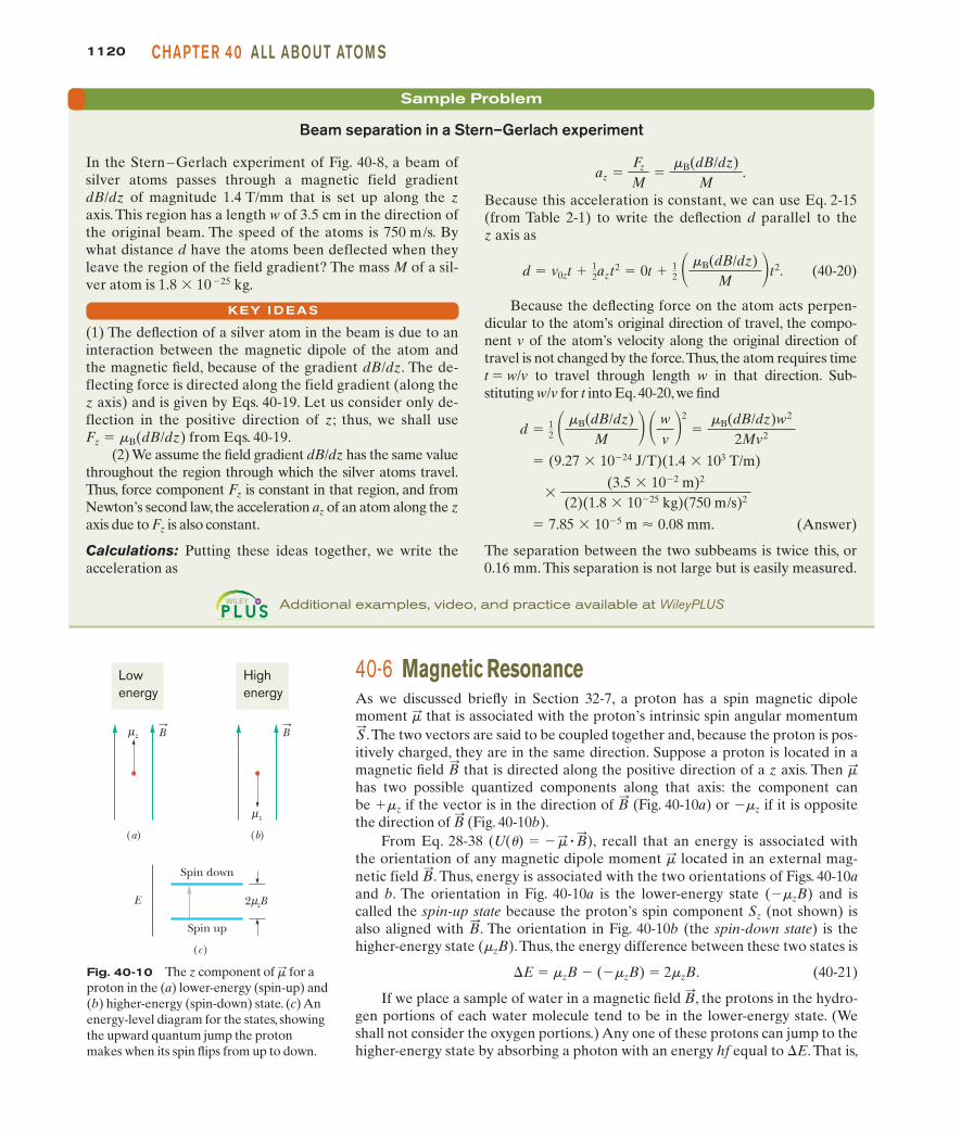
Fig. 40-10 The z component of for aproton in the (a) lower-energy (spin-up) and(b) higher-energy (spin-down) state. (c) Anenergy-level diagram for the states, showingthe upward quantum jump the protonmakes when its spin flips from up to down.
�:
µ
(a)
2 zB µ
(b)
z B
µ
(c)
z
B
E
Spin down
Spin up
Lowenergy
Highenergy
1120 CHAPTE R 40 ALL ABOUT ATOM S
40-6 Magnetic ResonanceAs we discussed briefly in Section 32-7, a proton has a spin magnetic dipolemoment that is associated with the proton’s intrinsic spin angular momentum
.The two vectors are said to be coupled together and, because the proton is pos-itively charged, they are in the same direction. Suppose a proton is located in amagnetic field that is directed along the positive direction of a z axis. Then has two possible quantized components along that axis: the component canbe �mz if the vector is in the direction of (Fig. 40-10a) or �mz if it is oppositethe direction of (Fig. 40-10b).
From Eq. 28-38 recall that an energy is associated withthe orientation of any magnetic dipole moment located in an external mag-�:
(U(�) � ��: � B:
),B:
B:
�:B:
S:
�:
Sample Problem
Because this acceleration is constant, we can use Eq. 2-15(from Table 2-1) to write the deflection d parallel to thez axis as
(40-20)
Because the deflecting force on the atom acts perpen-dicular to the atom’s original direction of travel, the compo-nent v of the atom’s velocity along the original direction oftravel is not changed by the force.Thus, the atom requires timet 5 w/v to travel through length w in that direction. Sub-stituting w/v for t into Eq. 40-20, we find
(Answer)
The separation between the two subbeams is twice this, or0.16 mm. This separation is not large but is easily measured.
� 7.85 � 10�5 m � 0.08 mm.
�(3.5 � 10�2 m)2
(2)(1.8 � 10�25 kg)(750 m/s)2
� (9.27 � 10�24 J/T)(1.4 � 103 T/m)
d � 12 � �B(dB/dz)
M � � wv �
2
��B(dB/dz)w2
2Mv2
d � v0zt � 12azt2 � 0t � 1
2 � �B(dB/dz)M �t2.
az �Fz
M�
�B(dB/dz)M
.
Additional examples, video, and practice available at WileyPLUS
Beam separation in a Stern–Gerlach experiment
In the Stern – Gerlach experiment of Fig. 40-8, a beam ofsilver atoms passes through a magnetic field gradientdB/dz of magnitude 1.4 T/mm that is set up along the zaxis. This region has a length w of 3.5 cm in the direction ofthe original beam. The speed of the atoms is 750 m/s. Bywhat distance d have the atoms been deflected when theyleave the region of the field gradient? The mass M of a sil-ver atom is 1.8 3 10 225 kg.
(1) The deflection of a silver atom in the beam is due to aninteraction between the magnetic dipole of the atom andthe magnetic field, because of the gradient dB/dz. The de-flecting force is directed along the field gradient (along thez axis) and is given by Eqs. 40-19. Let us consider only de-flection in the positive direction of z; thus, we shall use Fz 5 mB(dB/dz) from Eqs. 40-19.
(2) We assume the field gradient dB/dz has the same valuethroughout the region through which the silver atoms travel.Thus, force component Fz is constant in that region, and fromNewton’s second law, the acceleration az of an atom along the zaxis due to Fz is also constant.
Calculations: Putting these ideas together, we write the acceleration as
KEY I DEAS
netic field . Thus, energy is associated with the two orientations of Figs. 40-10aand b. The orientation in Fig. 40-10a is the lower-energy state (2mzB) and iscalled the spin-up state because the proton’s spin component Sz (not shown) isalso aligned with . The orientation in Fig. 40-10b (the spin-down state) is thehigher-energy state (mzB).Thus, the energy difference between these two states is
�E 5 mzB 2 (2mzB) 5 2mzB. (40-21)
If we place a sample of water in a magnetic field , the protons in the hydro-gen portions of each water molecule tend to be in the lower-energy state. (Weshall not consider the oxygen portions.) Any one of these protons can jump to thehigher-energy state by absorbing a photon with an energy hf equal to �E. That is,
B:
B:
B:
halliday_c40_1112-1141hr.qxd 14-01-2010 16:26 Page 1120

the proton can jump by absorbing a photon of energy
hf 5 2mzB. (40-22)
Such absorption is called magnetic resonance or, as originally, nuclear magneticresonance (NMR), and the consequent reversal of Sz is called spin-flipping.
In practice, the photons required for magnetic resonance have an associatedfrequency in the radio-frequency (RF) range and are provided by a small coilwrapped around the sample undergoing resonance. An electromagnetic oscilla-tor called an RF source drives a sinusoidal current in the coil at frequency f. Theelectromagnetic (EM) field set up within the coil and sample also oscillates atfrequency f. If f meets the requirement of Eq. 40-22, the oscillating EM field cantransfer a quantum of energy to a proton in the sample via a photon absorption,spin-flipping the proton.
The magnetic field magnitude B that appears in Eq. 40-22 is actually the mag-nitude of the net magnetic field at the site where a given proton undergoes spin-flipping. That net field is the vector sum of the external field set up by the mag-netic resonance equipment (primarily a large magnet) and the internal field setup by the magnetic dipole moments of the atoms and nuclei near the given proton.For practical reasons we do not discuss here, magnetic resonance is usually detectedby sweeping the magnitude Bext through a range of values while the frequency f ofthe RF source is kept at a predetermined value and the energy of the RF source ismonitored. A graph of the energy loss of the RF source versus Bext shows a reso-nance peak when Bext sweeps through the value at which spin-flipping occurs. Such agraph is called a nuclear magnetic resonance spectrum, or NMR spectrum.
Figure 40-11 shows the NMR spectrum of ethanol,which is a molecule consistingof three groups of atoms: CH3, CH2, and OH. Protons in each group can undergomagnetic resonance, but each group has its own unique magnetic-resonance value ofBext because the groups lie in different internal fields due to their arrangementwithin the CH3CH2OH molecule. Thus, the resonance peaks in the spectrum of Fig.40-11 form a unique NMR signature by which ethanol can be indentified.
40-7 The Pauli Exclusion PrincipleIn Chapter 39 we considered a variety of electron traps, from fictional one-dimensional traps to the real three-dimensional trap of a hydrogen atom. In allthose examples, we trapped only one electron. However, when we discuss traps con-taining two or more electrons (as we shall in the next two sections), we must con-sider a principle that governs any particle whose spin quantum number s is not zeroor an integer. This principle applies not only to electrons but also to protons andneutrons, all of which have The principle is known as the Pauli exclusion prin-ciple after Wolfgang Pauli, who formulated it in 1925. For electrons, it states that
s � 12.
B:
int
B:
int
B:
ext
B:
Fig. 40-11 A nuclear magnetic reso-nance spectrum for ethanol, CH3CH2OH.The spectral lines represent the absorptionof energy associated with spin-flips of pro-tons.The three groups of lines correspond,as indicated, to protons in the OH group,the CH2 group, and the CH3 group of theethanol molecule. Note that the two pro-tons in the CH2 group occupy four differentlocal environments.The entire horizontalaxis covers less than 1024 T.
En
ergy
abs
orbe
d
Bext
OH groupCH2 group
CH3 group
112140-8 M U LTI PLE E LECTRON S I N R ECTANG U LAR TRAPSPART 5
No two electrons confined to the same trap can have the same set of values for theirquantum numbers.
As we shall discuss in Section 40-9, this principle means that no two electronsin an atom can have the same four values for the quantum numbers n, , , andms. All electrons have the same quantum number Thus, any two electronsin an atom must differ in at least one of these other quantum numbers. Were thisnot true, atoms would collapse, and thus you and the world could not exist.
40-8 Multiple Electrons in Rectangular TrapsTo prepare for our discussion of multiple electrons in atoms, let us discuss twoelectrons confined to the rectangular traps of Chapter 39. We shall again use the
s � 12.
m/
/
halliday_c40_1112-1141hr.qxd 14-01-2010 16:26 Page 1121

1122 CHAPTE R 40 ALL ABOUT ATOM S
quantum numbers we found for those traps when only one electron was confined.However, here we shall also include the spin angular momenta of the two elec-trons.To do this, we assume that the traps are located in a uniform magnetic field.Then according to Eq. 40-12 , an electron can be either spin up with
or spin down with (We assume that the field is very weak so thatwe can neglect the energies of the electrons due to it.)
As we confine the two electrons to one of the traps, we must keep the Pauliexclusion principle in mind; that is, the electrons cannot have the same set of values for their quantum numbers.
1. One-dimensional trap. In the one-dimensional trap of Fig. 39-2, fitting an elec-tron wave to the trap’s width L requires the single quantum number n. There-fore, any electron confined to the trap must have a certain value of n, and itsquantum number ms can be either or . The two electrons could havedifferent values of n, or they could have the same value of n if one of them isspin up and the other is spin down.
2. Rectangular corral. In the rectangular corral of Fig. 39-13, fitting an electronwave to the corral’s widths Lx and Ly requires the two quantum numbers nx
and ny. Thus, any electron confined to the trap must have certain values forthose two quantum numbers, and its quantum number ms can be either or
; so now there are three quantum numbers.According to the Pauli exclusionprinciple, two electrons confined to the trap must have different values for atleast one of those three quantum numbers.
3. Rectangular box. In the rectangular box of Fig. 39-14, fitting an electron waveto the box’s widths Lx, Ly, and Lz requires the three quantum numbers nx, ny,and nz. Thus, any electron confined to the trap must have certain values forthese three quantum numbers, and its quantum number ms can be either or
; so now there are four quantum numbers. According to the Pauli exclusionprinciple, two electrons confined to the trap must have different values for atleast one of those four quantum numbers.
Suppose we add more than two electrons, one by one, to a rectangular trapin the preceding list. The first electrons naturally go into the lowest possibleenergy level—they are said to occupy that level. However, eventually the Pauliexclusion principle disallows any more electrons from occupying that lowestenergy level, and the next electron must occupy the next higher level. When anenergy level cannot be occupied by more electrons because of the Pauli exclusionprinciple, we say that level is full or fully occupied. In contrast, a level that is notoccupied by any electrons is empty or unoccupied. For intermediate situations,the level is partially occupied. The electron configuration of a system of trappedelectrons is a listing or drawing either of the energy levels the electrons occupy orof the set of the quantum numbers of the electrons.
Finding the Total EnergyTo find the energy of a system of two or more electrons confined to a trap, we as-sume that the electrons do not electrically interact with one another; that is, weshall neglect the electric potential energies of pairs of electrons. Then we can cal-culate the total energy for the system by calculating the energy of each electron(as in Chapter 39) and then summing those energies.
A good way to organize the energy values of a given system of electrons iswith an energy-level diagram for the system, just as we did for a single electron inthe traps of Chapter 39. The lowest level, with energy Egr, corresponds to theground state of the system. The next higher level, with energy Efe, corresponds tothe first excited state of the system. The next level, with energy Ese, correspondsto the second excited state of the system, and so on.
�12
� 12
�12
� 12
�12� 1
2
ms � � 12.ms � 1
2
(Sz � ms�)
halliday_c40_1112-1141hr.qxd 14-01-2010 16:26 Page 1122

Sample Problem
ter and draw a down arrow (to represent spin down) on theE1,1 level in Fig. 40-12a. The second electron also goes intothe E1,1 level but must have so that one of itsquantum numbers differs from those of the first electron.We represent this second electron with an up arrow (forspin up) on the E1,1 level in Fig. 40-12b.
Electrons, one by one: The level for energy E1,1 is fullyoccupied, and thus the third electron cannot have that en-ergy. Therefore, the third electron goes into the next higherlevel, which is for the equal energies E2,1 and E1,2 (the level isdegenerate). This third electron can have quantum numbersnx and ny of either 1 and 2 or 2 and 1, respectively. It can alsohave a quantum number ms of either or . Let us arbi-trarily assign it the quantum numbers nx 5 2, ny 5 1, and
.We then represent it with a down arrow on the levelfor E1,2 and E2,1 in Fig. 40-12c.
You can show that the next three electrons can also gointo the level for energies E2,1 and E1,2, provided that no setof three quantum numbers is completely duplicated. Thatlevel then contains four electrons (Fig. 40-12d), with quan-tum numbers (nx, ny, ms) of
(2, 1,� 12), (2, 1, � 1
2), (1, 2,� 12), (1, 2, � 1
2),
ms � �12
�12�1
2
ms � �12
Energy levels of multiple electrons in a 2D infinite potential well
Seven electrons are confined to a square corral (two-dimensional infinite potential well) with widths Lx 5 Ly 5L (Fig. 39-13). Assume that the electrons do not electricallyinteract with one another.(a) What is the electron configuration for the ground stateof the system of seven electrons?
One-electron diagram: We can determine the electron con-figuration of the system by placing the seven electrons in thecorral one by one, to build up the system. Because we assumethe electrons do not electrically interact with one another, wecan use the energy-level diagram for a single trapped electronin order to keep track of how we place the seven electrons inthe corral. That one-electron energy-level diagram is given inFig. 39-15 and partially reproduced here as Fig. 40-12a. Recallthat the levels are labeled as Enx,ny for their associated energy.For example, the lowest level is for energy E1,1, where quan-tum number nx is 1 and quantum number ny is 1.
Pauli principle: The trapped electrons must obey thePauli exclusion principle; that is, no two electrons can havethe same set of values for their quantum numbers nx, ny,and ms. The first electron goes into energy level E1,1 andcan have or We arbitrarily choose the lat-ms � � 1
2.ms � 12
112340-8 M U LTI PLE E LECTRON S I N R ECTANG U LAR TRAPSPART 5
10
8
5
(a)
2 E1,1
E2,1, E1,2
E3,1, E1,3
E2,2
E
10
8
5
(b)
2 E1,1
E
10
8
5
(c)
2 E1,1
E2,1, E1,2
E1,1
E2,1, E1,2
E2,2
E
10
8
5
(d)
2
E
These are the four lowestenergy levels of the corral.The first electron is in the lowest level.
A second electron can be there only if it has the opposite spin. The level is then full.
Two quantum states have that energy. Two electrons (with opposite spins) can be in each state. Then thatlevel is also full.
The lowest energy fora third electron is onthe next level up.
Fig. 40-12 (a) Energy-level diagramfor one electron in a square corral.(Energy E is in multiples of h2/8mL2.) Aspin-down electron occupies the lowestlevel. (b) Two electrons (one spin down,the other spin up) occupy the lowestlevel of the one-electron energy-leveldiagram. (c) A third electron occupiesthe next energy level. (d) Four electronscan be put into the second level.(Figure continues on page 1125.)
A
halliday_c40_1112-1141hr.qxd 14-01-2010 16:26 Page 1123

1124 CHAPTE R 40 ALL ABOUT ATOM S
2. If that jump is to occur, the energy change �E of theelectron (and thus of the system) must be �E 5 Ehigh 2Elow (Eq. 39-5), where Elow is the energy of the levelwhere the jump begins and Ehigh is the energy of thelevel where the jump ends.
3. The Pauli exclusion principle must still apply; an electroncannot jump to a level that is fully occupied.
First-excited-state energy: Let us consider the threejumps shown in Fig. 40-12f; all are allowed by the Pauli ex-clusion principle because they are jumps to either empty orpartially occupied states. In one of those possible jumps, anelectron jumps from the E1,1 level to the partially occupiedE2,2 level.The change in the energy is
(We shall assume that the spin orientation of the electronmaking the jump can change as needed.)
�E � E2,2 � E1,1 � 8 h2
8mL2 � 2 h2
8mL2 � 6 h2
8mL2 .
and the level is fully occupied. Thus, the seventh electrongoes into the next higher level, which is the E2,2 level. Let usassume this electron is spin down, with .
Figure 40-12e shows all seven electrons on a one-elec-tron energy-level diagram. We now have seven electrons inthe corral, and they are in the configuration with the lowestenergy that satisfies the Pauli exclusion principle. Thus, theground-state configuration of the system is that shown inFig. 40-12e and listed in Table 40-2.
(b) What is the total energy of the seven-electron system inits ground state, as a multiple of h2/8mL2?
The total energy Egr is the sum of the energies of the indi-vidual electrons in the system’s ground-state configuration.
Ground-state energy: The energy of each electron can beread from Table 39-1, which is partially reproduced in Table40-2, or from Fig. 40-12e. Because there are two electrons inthe first (lowest) level, four in the second level, and one inthe third level, we have
(Answer)
(c) How much energy must be transferred to the system forit to jump to its first excited state, and what is the energy ofthat state?
1. If the system is to be excited, one of the seven electronsmust make a quantum jump up the one-electron energy-level diagram of Fig. 40-12e.
� 32 h2
8mL2 .
Egr � 2�2 h2
8mL2 � � 4�5 h2
8mL2 � � 1�8 h2
8mL2 �
ms � �12
KEY I DEA
Table 40-2
Ground-State Configuration and Energies
nx ny ms Energya
2 2 8
2 1 5
2 1 5
1 2 5
1 2 5
1 1 2
1 1 2Total 32
aIn multiples of h2/8mL2.
�12
� 12
�12
� 12
�12
� 12
�12
KEY I DEAS
40-9 Building the Periodic TableThe four quantum numbers , and ms identify the quantumstates of individual electrons in a multielectron atom.The wave func-tions for these states, however, are not the same as the wave func-tions for the corresponding states of the hydrogen atom because, inmultielectron atoms, the potential energy associated with a givenelectron is determined not only by the charge and position of theatom’s nucleus but also by the charges and positions of all the otherelectrons in the atom. Solutions of Schrödinger’s equation for multi-electron atoms can be carried out numerically—in principle atleast—using a computer.
As we discussed in Sections 39-9 and 40-3, all states with thesame values of the quantum numbers n and form a subshell. For agiven value of , there are possible values of the quantum2� � 1�
�
n, �, m�
halliday_c40_1112-1141hr.qxd 14-01-2010 16:26 Page 1124

112540-9 B U I LDI NG TH E PE R IODIC TAB LEPART 5
In another of the possible jumps in Fig. 40-12f, an elec-tron jumps from the degenerate level of E2,1 and E1,2 to thepartially occupied E2,2 level.The change in the energy is
In the third possible jump in Fig. 40-12f, the electron inthe E2,2 level jumps to the unoccupied, degenerate level ofE1,3 and E3,1.The change in energy is
Of these three possible jumps, the one requiring the leastenergy change �E is the last one. We could consider evenmore possible jumps, but none would require less energy.
�E � E1,3 � E2,2 � 10 h2
8mL2 � 8 h2
8mL2 � 2 h2
8mL2 .
�E � E2,2 � E2,1 � 8 h2
8mL2 � 5 h2
8mL2 � 3 h2
8mL2 .
Thus, for the system to jump from its ground state to its firstexcited state, the electron in the E2,2 level must jump to theunoccupied, degenerate level of E1,3 and E3,1, and the re-quired energy is
(Answer)
The energy Efe of the first excited state of the system is then
(Answer)
We can represent this energy and the energy Egr for theground state of the system on an energy-level diagram forthe system, as shown in Fig. 40-12g.
� 32 h2
8mL2 � 2 h2
8mL2 � 34 h2
8mL2 .
Efe � Egr � �E
�E � 2 h2
8mL2 .
number and, for each , there are two possible values for the quantum num-ber ms.Thus, there are states in a subshell. It turns out that all states in agiven subshell have the same energy, its value being determined primarily by thevalue of n and to a lesser extent by the value of .
For the purpose of labeling subshells, the values of are represented by letters:
For example, the n 5 3, subshell would be labeled the 3d subshell.When we assign electrons to states in a multielectron atom, we must be
guided by the Pauli exclusion principle of Section 40-7; that is, no two electrons inan atom can have the same set of the quantum numbers , and ms. If thisimportant principle did not hold, all the electrons in any atom could jump to theatom’s lowest energy level, which would eliminate the chemistry of atoms andmolecules, and thus also eliminate biochemistry and us. Let us examine the atoms
n, �, m�
� � 2
s p d f g h . . . . � � 0 1 2 3 4 5 . . .
��
2(2� � 1)m�m�
Additional examples, video, and practice available at WileyPLUS
E1,1
E2,1, E1,2
E2,2
10
8
5
(e)
2
E
Electrons can jumpup only to levelsthat are not full.Here are threeallowed jumps.Which uses theleast energy?If that jump ismade, the systemis then in its firstexcited state.
The lowest energy forthe seventh electron ison the next level up. The system of 7electrons is in its lowest energy (system ground state).
E1,1
E2,1, E1,2
E3,1, E1,3
E2,2
10
8
5
( f )
2 Egr
Efe
EseE
34
(g)
32
E Here are the three lowest energylevels of thesystem.
A
Fig. 40-12 (Continued from page 1123) (e) The system’s ground-state configuration. (f) Threetransitions to consider for the first excited state. ( g) The system’s lowest three total energies.
halliday_c40_1112-1141hr.qxd 14-01-2010 16:26 Page 1125

1126 CHAPTE R 40 ALL ABOUT ATOM S
of a few elements to see how the Pauli exclusion principle operates in the build-ing up of the periodic table.
NeonThe neon atom has 10 electrons. Only two of them fit into the lowest-energysubshell, the 1s subshell. These two electrons both have n 5 1, , and ,but one has and the other has . The 1s subshell contains2[2(0) 1 1] 5 2 states. Because this subshell then contains all the electrons permitted by the Pauli principle, it is said to be closed.
Two of the remaining eight electrons fill the next lowest energy subshell, the2s subshell. The last six electrons just fill the 2p subshell which, with , holds2[2(1) 1 1] 5 6 states.
In a closed subshell, all allowed z projections of the orbital angular momen-tum vector are present and, as you can verify from Fig. 40-5, these projectionscancel for the subshell as a whole; for every positive projection there is a corre-sponding negative projection of the same magnitude. Similarly, the z projectionsof the spin angular momenta also cancel. Thus, a closed subshell has no angularmomentum and no magnetic moment of any kind. Furthermore, its probabilitydensity is spherically symmetric. Then neon with its three closed subshells (1s, 2s,and 2p) has no “loosely dangling electrons” to encourage chemical interactionwith other atoms. Neon, like the other noble gases that form the right-hand col-umn of the periodic table, is almost chemically inert.
SodiumNext after neon in the periodic table comes sodium, with 11 electrons. Ten ofthem form a closed neon-like core, which, as we have seen, has zero angularmomentum. The remaining electron is largely outside this inert core, in the 3ssubshell—the next lowest energy subshell. Because this valence electron ofsodium is in a state with (that is, an s state using the lettering systemabove), the sodium atom’s angular momentum and magnetic dipole momentmust be due entirely to the spin of this single electron.
Sodium readily combines with other atoms that have a “vacancy” into whichsodium’s loosely bound valence electron can fit. Sodium, like the other alkalimetals that form the left-hand column of the periodic table, is chemically active.
ChlorineThe chlorine atom, which has 17 electrons, has a closed 10-electron, neon-likecore, with 7 electrons left over. Two of them fill the 3s subshell, leaving five tobe assigned to the 3p subshell, which is the subshell next lowest in energy. Thissubshell, which has , can hold electrons, and so there is a vacancy, or a “hole,” in this subshell.
Chlorine is receptive to interacting with other atoms that have a valenceelectron that might fill this hole. Sodium chloride (NaCl), for example, is a verystable compound. Chlorine, like the other halogens that form column VIIA of theperiodic table, is chemically active.
IronThe arrangement of the 26 electrons of the iron atom can be represented asfollows:
1s2 2s2 2p6 3s2 3p6 3d6 4s2.
2[2(1) � 1] � 6� � 1
� � 0
L:
/ � 1
ms � �12ms � � 1
2
m/
� 0/ � 0
halliday_c40_1112-1141hr.qxd 14-01-2010 16:26 Page 1126

The subshells are listed in numerical order and, following convention, a super-script gives the number of electrons in each subshell. From Table 40-1 we can seethat an s subshell can hold 2 electrons, a p subshell can hold 6,and a d subshell can hold 10. Thus, iron’s first 18 electrons form thefive filled subshells that are marked off by the bracket, leaving 8 electrons to beaccounted for. Six of the eight go into the 3d subshell, and the remaining two gointo the 4s subshell.
The reason the last two electrons do not also go into the 3d subshell (whichcan hold 10 electrons) is that the 3d6 4s2 configuration results in a lower-energystate for the atom as a whole than would the 3d8 configuration.An iron atom with8 electrons (rather than 6) in the 3d subshell would quickly make a transition tothe 3d6 4s2 configuration, emitting electromagnetic radiation in the process. Thelesson here is that except for the simplest elements, the states may not be filled inwhat we might think of as their “logical” sequence.
40-10 X Rays and the Ordering of the ElementsWhen a solid target, such as solid copper or tungsten, is bombarded with elec-trons whose kinetic energies are in the kiloelectron-volt range, electromagneticradiation called x rays is emitted. Our concern here is what these rays—whosemedical, dental, and industrial usefulness is so well known and widespread—canteach us about the atoms that absorb or emit them. Figure 40-13 shows thewavelength spectrum of the x rays produced when a beam of 35 keV electronsfalls on a molybdenum target. We see a broad, continuous spectrum of radiationon which are superimposed two peaks of sharply defined wavelengths. The continuous spectrum and the peaks arise in different ways, which we next discussseparately.
The Continuous X-Ray SpectrumHere we examine the continuous x-ray spectrum of Fig. 40-13, ignoring for thetime being the two prominent peaks that rise from it. Consider an electron ofinitial kinetic energy K0 that collides (interacts) with one of the target atoms, as inFig. 40-14. The electron may lose an amount of energy �K, which will appear asthe energy of an x-ray photon that is radiated away from the site of the collision.(Very little energy is transferred to the recoiling atom because of the relativelylarge mass of the atom; here we neglect that transfer.)
The scattered electron in Fig. 40-14, whose energy is now less than K0, mayhave a second collision with a target atom, generating a second photon, with adifferent photon energy. This electron-scattering process can continue until theelectron is approximately stationary. All the photons generated by these colli-sions form part of the continuous x-ray spectrum.
A prominent feature of that spectrum in Fig. 40-13 is the sharply defined cutoff wavelength lmin, below which the continuous spectrum does not exist. Thisminimum wavelength corresponds to a collision in which an incident electronloses all its initial kinetic energy K0 in a single head-on collision with a targetatom. Essentially all this energy appears as the energy of a single photon, whoseassociated wavelength—the minimum possible x-ray wavelength—is found from
or (cutoff wavelength). (40-23)�min �hcK0
K0 � hf �hc
�min,
(/ � 2)(/ � 1)(/ � 0)
Rel
ativ
e in
ten
sity
30 40 50 60 70 80 90Wavelength (pm)
λ min
K β
K α
Continuousspectrum
Fig. 40-13 The distribution by wave-length of the x rays produced when 35 keVelectrons strike a molybdenum target.Thesharp peaks and the continuous spectrumfrom which they rise are produced by dif-ferent mechanisms.
112740-10 X RAYS AN D TH E OR DE R I NG OF TH E E LE M E NTSPART 5
Targetatom
K 0Incidentelectron
K0 – K
hf (= K) X-ray
photon
∆
∆
Fig. 40-14 An electron of kineticenergy K0 passing near an atom in thetarget may generate an x-ray photon,the electron losing part of its energy inthe process. The continuous x-rayspectrum arises in this way.
halliday_c40_1112-1141hr.qxd 14-01-2010 16:26 Page 1127

1128 CHAPTE R 40 ALL ABOUT ATOM S
The cutoff wavelength is totally independent of the target material. If we were toswitch from a molybdenum target to a copper target, for example, all features ofthe x-ray spectrum of Fig. 40-13 would change except the cutoff wavelength.
Fig. 40-15 A simplified energy-level diagram for a molybdenumatom, showing the transitions (ofholes rather than electrons) that giverise to some of the characteristicx rays of that element. Each horizon-tal line represents the energy of theatom with a hole (a missing electron)in the shell indicated.
20
15
10
5
0
L βL αK β
K α
L (n = 2)
M (n = 3)N (n = 4)
K (n = 1)E
ner
gy (
keV
)
CHECKPOINT 2
Does the cutoff wavelength lmin of the continuous x-ray spectrum increase, decrease, or remain the same if you (a) increase the kinetic energy of the electrons that strike the x-raytarget, (b) allow the electrons to strike a thin foil rather than a thick block of the target material, (c) change the target to an element of higher atomic number?
The Characteristic X-Ray SpectrumWe now turn our attention to the two peaks of Fig. 40-13, labeled Ka and Kb.These (and other peaks that appear at wavelengths beyond the range displayedin Fig. 40-13) form the characteristic x-ray spectrum of the target material.
The peaks arise in a two-part process. (1) An energetic electron strikes anatom in the target and, while it is being scattered, the incident electron knocksout one of the atom’s deep-lying (low n value) electrons. If the deep-lying elec-tron is in the shell defined by n 5 1 (called, for historical reasons, the K shell),there remains a vacancy, or hole, in this shell. (2) An electron in one of the shellswith a higher energy jumps to the K shell, filling the hole in this shell. During thisjump, the atom emits a characteristic x-ray photon. If the electron that fills theK-shell vacancy jumps from the shell with n 5 2 (called the L shell), the emittedradiation is the Ka line of Fig. 40-13; if it jumps from the shell with n 5 3 (calledthe M shell), it produces the Kb line, and so on. The hole left in either the L or Mshell will be filled by an electron from still farther out in the atom.
In studying x rays, it is more convenient to keep track of where a hole iscreated deep in the atom’s “electron cloud” than to record the changes in thequantum state of the electrons that jump to fill that hole. Figure 40-15 doesexactly that; it is an energy-level diagram for molybdenum, the element to whichFig. 40-13 refers. The baseline (E 5 0) represents the neutral atom in its groundstate. The level marked K (at E 5 20 keV) represents the energy of the molyb-denum atom with a hole in its K shell, the level marked L (at E 5 2.7 keV)represents the atom with a hole in its L shell, and so on.
The transitions marked Ka and Kb in Fig.40-15 are the ones that produce the twox-ray peaks in Fig. 40-13. The Ka spectral line, for example, originates when an elec-tron from the L shell fills a hole in the K shell.To state this transition in terms of whatthe arrows in Fig.40-15 show,a hole originally in the K shell moves to the L shell.
Ordering the ElementsIn 1913, British physicist H. G. J. Moseley generated characteristic x rays for asmany elements as he could find—he found 38—by using them as targets forelectron bombardment in an evacuated tube of his own design. By means of atrolley manipulated by strings, Moseley was able to move the individual targetsinto the path of an electron beam. He measured the wavelengths of the emittedx rays by the crystal diffraction method described in Section 36-10.
Moseley then sought (and found) regularities in these spectra as he moved fromelement to element in the periodic table. In particular, he noted that if, for a givenspectral line such as Ka, he plotted for each element the square root of the frequencyf against the position of the element in the periodic table, a straight line resulted.Figure 40-16 shows a portion of his extensive data. Moseley’s conclusion was this:
We have here a proof that there is in the atom a fundamental quantity, which increases by regular steps as we pass from one element to the next. This quantitycan only be the charge on the central nucleus.
halliday_c40_1112-1141hr.qxd 14-01-2010 16:26 Page 1128

112940-10 X RAYS AN D TH E OR DE R I NG OF TH E E LE M E NTSPART 5
As a result of Moseley’s work, the characteristic x-ray spectrum became the uni-versally accepted signature of an element, permitting the solution of a number ofperiodic table puzzles. Prior to that time (1913), the positions of elements in thetable were assigned in order of atomic mass, although it was necessary to invertthis order for several pairs of elements because of compelling chemical evidence;Moseley showed that it is the nuclear charge (that is, atomic number Z) that isthe real basis for ordering the elements.
In 1913 the periodic table had several empty squares, and a surprising num-ber of claims for new elements had been advanced. The x-ray spectrum provideda conclusive test of such claims. The lanthanide elements, often called the rare earthelements, had been sorted out only imperfectly because their similar chemical prop-erties made sorting difficult.Once Moseley’s work was reported, these elements wereproperly organized.
It is not hard to see why the characteristic x-ray spectrum shows such im-pressive regularities from element to element whereas the optical spectrum inthe visible and near-visible region does not: The key to the identity of an elementis the charge on its nucleus. Gold, for example, is what it is because its atoms havea nuclear charge of 179e (that is, Z 5 79). An atom with one more elementarycharge on its nucleus is mercury; with one fewer, it is platinum. The K electrons,which play such a large role in the production of the x-ray spectrum, lie very closeto the nucleus and are thus sensitive probes of its charge. The optical spectrum,on the other hand, involves transitions of the outermost electrons, which areheavily screened from the nucleus by the remaining electrons of the atom andthus are not sensitive probes of nuclear charge.
Accounting for the Moseley PlotMoseley’s experimental data, of which the Moseley plot of Fig. 40-16 is but a part,can be used directly to assign the elements to their proper places in the periodictable. This can be done even if no theoretical basis for Moseley’s results can beestablished. However, there is such a basis.
According to Eqs. 39-32 and 39-33, the energy of the hydrogen atom is
(40-24)
Consider now one of the two innermost electrons in the K shell of a multi-electron atom. Because of the presence of the other K-shell electron, our electron“sees” an effective nuclear charge of approximately (Z 2 1)e, where e is the
En � �me4
8 02 h2
1n2 � �
13.60 eVn2 , for n � 1, 2, 3, . . . .
Fig. 40-16 AMoseley plot of the Ka
line of the characteris-tic x-ray spectra of 21elements.The fre-quency is calculatedfrom the measuredwavelength.
0
0.5
1.0
1.5
2.0
2.5
10 20 30 40 50Element number in periodic table
(10
9 Hz1/
2 )√
AlSi Cl
K Ca
TiV
CrMn
FeCo
NiCu
Zn
ZrY Nb
Mo Ru
Pd Ag
f
halliday_c40_1112-1141hr.qxd 14-01-2010 16:26 Page 1129

1130 CHAPTE R 40 ALL ABOUT ATOM S
elementary charge and Z is the atomic number of the element. The factor e 4 inEq. 40-24 is the product of e 2 —the square of hydrogen’s nuclear charge—and(2e)2 —the square of an electron’s charge. For a multielectron atom, we canapproximate the effective energy of the atom by replacing the factor e 4 inEq. 40-24 with (Z 2 1)2e 2 3 (2e)2, or e 4(Z 2 1)2.That gives us
(40-25)
We saw that the Ka x-ray photon (of energy hf ) arises when an electron makes atransition from the L shell (with n 5 2 and energy E2) to the K shell (with n 5 1and energy E1).Thus, using Eq. 40-25, we may write the energy change as
Then the frequency f of the Ka line is
(40-26)
Taking the square root of both sides yields
(40-27)
in which C is a constant (5 4.96 3 107 Hz1/2). Equation 40-27 is the equation ofa straight line. It shows that if we plot the square root of the frequency of the Ka
x-ray spectral line against the atomic number Z, we should obtain a straight line.As Fig. 40-16 shows, that is exactly what Moseley found.
2 f � CZ � C,
� (2.46 � 10 15 Hz)(Z � 1)2.
f ��Eh
�(10.2 eV)(Z � 1)2
(4.14 � 10 �15 eV s)
� (10.2 eV)(Z � 1)2.
��(13.60 eV)(Z � 1)2
22 ��(13.60 eV)(Z � 1)2
12
�E � E2 � E1
En � �(13.60 eV)(Z � 1)2
n2 .
Sample Problem
Dividing the second equation by the first neatly eliminatesC, yielding
Substituting the given data yields
Solving for the unknown, we find that
ZX � 30.0. (Answer)
Thus, the number of protons in the impurity nucleus is 30,and a glance at the periodic table identifies the impurity aszinc. Note that with a larger value of Z than cobalt, zinc hasa smaller value of the Ka line.This means that the energy as-sociated with that jump must be greater in zinc than cobalt.
A
178.9 pm143.5 pm
�ZX � 127 � 1
.
A
�Co
�X�
ZX � 1ZCo � 1
.
Additional examples, video, and practice available at WileyPLUS
Characteristic spectrum in x-ray production
A cobalt target is bombarded with electrons, and the wave-lengths of its characteristic x-ray spectrum are measured.There is also a second, fainter characteristic spectrum, whichis due to an impurity in the cobalt. The wavelengths of theKa lines are 178.9 pm (cobalt) and 143.5 pm (impurity), andthe proton number for cobalt is ZCo � 27. Determine the impurity using only these data.
The wavelengths of the Ka lines for both the cobalt (Co)and the impurity (X) fall on a Ka Moseley plot, and Eq.40-27 is the equation for that plot.
Calculations: Substituting c/l for f in Eq. 40-27, we obtain
A
c�Co
� CZCo � C and A
c�X
� CZX � C.
KEY I DEA
halliday_c40_1112-1141hr.qxd 14-01-2010 16:26 Page 1130

113140-11 LAS E RS AN D LAS E R LIG HTPART 5
40-11 Lasers and Laser LightIn the early 1960s, quantum physics made one of its many contributions to tech-nology: the laser. Laser light, like the light from an ordinary lightbulb, is emittedwhen atoms make a transition from one quantum state to a lower one. However,in a lightbulb the emissions are random, both in time and direction, and in a laserthey are coordinated so that the emissions are at the same time and in the samedirection. As a result, laser light has the following characteristics:
1. Laser light is highly monochromatic. Light from an ordinary incandescentlightbulb is spread over a continuous range of wavelengths and is certainly notmonochromatic.The radiation from a fluorescent neon sign is monochromatic,true, to about 1 part in 106, but the sharpness of definition of laser light can bemany times greater, as much as 1 part in 1015.
2. Laser light is highly coherent. Individual long waves (wave trains) for laserlight can be several hundred kilometers long. When two separated beamsthat have traveled such distances over separate paths are recombined, they“remember” their common origin and are able to form a pattern of interfer-ence fringes. The corresponding coherence length for wave trains emitted by alightbulb is typically less than a meter.
3. Laser light is highly directional. A laser beam spreads very little; it departsfrom strict parallelism only because of diffraction at the exit aperture of thelaser. For example, a laser pulse used to measure the distance to the Moongenerates a spot on the Moon’s surface with a diameter of only a few kilo-meters. Light from an ordinary bulb can be made into an approximatelyparallel beam by a lens, but the beam divergence is much greater than forlaser light. Each point on a lightbulb’s filament forms its own separatebeam, and the angular divergence of the overall composite beam is setby the size of the filament.
4. Laser light can be sharply focused. If two light beams transport thesame amount of energy, the beam that can be focused to the smaller spotwill have the greater intensity (power per unit area) at that spot. Forlaser light, the focused spot can be so small that an intensity of1017 W/cm2 is readily obtained. An oxyacetylene flame, by contrast, hasan intensity of only about 103 W/cm2.
Lasers Have Many UsesThe smallest lasers, used for voice and data transmission over opticalfibers, have as their active medium a semiconducting crystal about thesize of a pinhead. Small as they are, such lasers can generate about 200mW of power. The largest lasers, used for nuclear fusion research and forastronomical and military applications, fill a large building. The largestsuch laser can generate brief pulses of laser light with a power level, dur-ing the pulse, of about 1014 W. This is a few hundred times greater thanthe total electrical power generating capacity of the United States. Toavoid a brief national power blackout during a pulse, the energy requiredfor each pulse is stored up at a steady rate during the relatively longinterpulse interval.
Among the many uses of lasers are reading bar codes, manufacturingand reading compact discs and DVDs, performing surgery of many kinds(both as a surgical aid as in Fig. 40-17 and as a cutting and cauterizingtool), surveying, cutting cloth in the garment industry (several hundredlayers at a time), welding auto bodies, and generating holograms.
Fig. 40-17 A patient's head is scanned andmapped by (red) laser light in preparation for brainsurgery. During the surgery, the laser-derived imageof the head will be superimposed on the model ofthe brain shown on the monitor, to guide the surgicalteam into the region shown in green on the model.(Sam Ogden/Photo Researchers)
halliday_c40_1112-1141hr.qxd 14-01-2010 16:26 Page 1131
1132 CHAPTE R 40 ALL ABOUT ATOM S
40-12 How Lasers WorkBecause the word “laser” is an acronym for “light amplification by the stimulatedemission of radiation,” you should not be surprised that stimulated emission isthe key to laser operation. Einstein introduced this concept in 1917.Although theworld had to wait until 1960 to see an operating laser, the groundwork for itsdevelopment was put in place decades earlier.
Consider an isolated atom that can exist either in its state of lowest energy(its ground state), whose energy is E0, or in a state of higher energy (an excitedstate), whose energy is Ex. Here are three processes by which the atom can movefrom one of these states to the other:
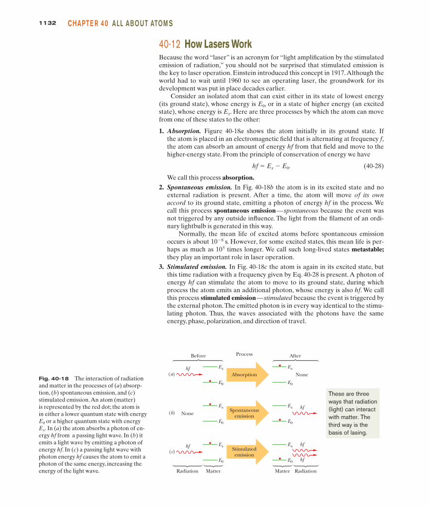
1. Absorption. Figure 40-18a shows the atom initially in its ground state. Ifthe atom is placed in an electromagnetic field that is alternating at frequency f,the atom can absorb an amount of energy hf from that field and move to thehigher-energy state. From the principle of conservation of energy we have
hf 5 Ex 2 E0. (40-28)
We call this process absorption.
2. Spontaneous emission. In Fig. 40-18b the atom is in its excited state and noexternal radiation is present. After a time, the atom will move of its ownaccord to its ground state, emitting a photon of energy hf in the process. Wecall this process spontaneous emission— spontaneous because the event wasnot triggered by any outside influence. The light from the filament of an ordi-nary lightbulb is generated in this way.
Normally, the mean life of excited atoms before spontaneous emissionoccurs is about 1028 s. However, for some excited states, this mean life is per-haps as much as 105 times longer. We call such long-lived states metastable;they play an important role in laser operation.
3. Stimulated emission. In Fig. 40-18c the atom is again in its excited state, butthis time radiation with a frequency given by Eq. 40-28 is present. A photon ofenergy hf can stimulate the atom to move to its ground state, during whichprocess the atom emits an additional photon, whose energy is also hf. We callthis process stimulated emission— stimulated because the event is triggered bythe external photon.The emitted photon is in every way identical to the stimu-lating photon. Thus, the waves associated with the photons have the sameenergy, phase, polarization, and direction of travel.
Fig. 40-18 The interaction of radiationand matter in the processes of (a) absorp-tion, (b) spontaneous emission, and (c)stimulated emission.An atom (matter) is represented by the red dot; the atom is in either a lower quantum state with energyE0 or a higher quantum state with energyEx. In (a) the atom absorbs a photon of en-ergy hf from a passing light wave. In (b) itemits a light wave by emitting a photon ofenergy hf. In (c) a passing light wave withphoton energy hf causes the atom to emit aphoton of the same energy, increasing theenergy of the light wave.
Ex
E0
hf(a)
Ex
E0
None(b)
Ex
E0
hf(c)
Before
Radiation Matter
Ex
E0
hf
Ex
E0
None
Ex
E0
hf
After
RadiationMatter
Process
Absorption
Spontaneousemission
Stimulatedemission
hf
These are threeways that radiation(light) can interactwith matter. Thethird way is thebasis of lasing.
halliday_c40_1112-1141hr.qxd 14-01-2010 16:26 Page 1132

113340-12 HOW LAS E RS WOR KPART 5
Figure 40-18c describes stimulated emission for a single atom. Suppose nowthat a sample contains a large number of atoms in thermal equilibrium at temper-ature T. Before any radiation is directed at the sample, a number N0 of theseatoms are in their ground state with energy E0 and a number Nx are in a state ofhigher energy Ex. Ludwig Boltzmann showed that Nx is given in terms of N0 by
(40-29)
in which k is Boltzmann’s constant.This equation seems reasonable.The quantitykT is the mean kinetic energy of an atom at temperature T. The higher thetemperature, the more atoms—on average—will have been “bumped up” bythermal agitation (that is, by atom–atom collisions) to the higher energy state Ex.Also, because Ex . E0, Eq. 40-29 requires that Nx , N0; that is, there will alwaysbe fewer atoms in the excited state than in the ground state. This is what weexpect if the level populations N0 and Nx are determined only by the action ofthermal agitation. Figure 40-19a illustrates this situation.
If we now flood the atoms of Fig. 40-19a with photons of energy Ex 2 E0,photons will disappear via absorption by ground-state atoms and photons will begenerated largely via stimulated emission of excited-state atoms. Einsteinshowed that the probabilities per atom for these two processes are identical.Thus, because there are more atoms in the ground state, the net effect will be theabsorption of photons.
To produce laser light, we must have more photons emitted than absorbed;that is, we must have a situation in which stimulated emission dominates.Thus, weneed more atoms in the excited state than in the ground state, as in Fig. 40-19b.However, because such a population inversion is not consistent with thermalequilibrium, we must think up clever ways to set up and maintain one.
The Helium–Neon Gas LaserFigure 40-20 shows a common type of laser developed in 1961 by Ali Javan andhis coworkers. The glass discharge tube is filled with a 20 : 80 mixture of heliumand neon gases, neon being the medium in which laser action occurs.
Figure 40-21 shows simplified energy-level diagrams for the two types of atoms.An electric current passed through the helium–neon gas mixture serves—through
Nx � N 0e�(Ex�E0)/kT,
Fig. 40-19 (a) The equilibrium distribution of atoms between the groundstate E0 and excited state Ex accounted forby thermal agitation. (b) An inverted population, obtained by special methods.Such a population inversion is essential forlaser action.
Ex
E0
(a) (b)
Ex
E0
Fig. 40-20 The elements of a helium–neon gas laser.An applied poten-tial Vdc sends electrons through a dischargetube containing a mixture of helium gasand neon gas. Electrons collide with heliumatoms, which then collide with neon atoms,which emit light along the length of thetube.The light passes through transparentwindows W and reflects back and forththrough the tube from mirrors M1 and M2
to cause more neon atom emissions. Someof the light leaks through mirror M2 toform the laser beam.
Discharge tube
M2(leaky)
M1
W W
Vdc+ –
Laserbeam
Fig. 40-21 Five essential en-ergy levels for helium and neonatoms in a helium–neon gaslaser. Laser action occurs be-tween levels E2 and E1 of neonwhen more atoms are at the E2
level than at the E1 level.
20
15
10
5
0
En
ergy
(eV
)
Metastablestate
He–Necollisions
E3 E2
E1
Excitationvia collisions
Rapiddecay
Heliumstates
Neonstates
E0Commonground state
Laser light(632.8 nm)
Then the helium atomsexcite the neon atomsto level E2 by collisions.Those neon atoms staylong enough to be forcedinto stimulated emission.
The current (electrons)excite the helium atomsby collisions (but not themore massive neonatoms).
halliday_c40_1112-1141hr.qxd 14-01-2010 16:26 Page 1133

1134 CHAPTE R 40 ALL ABOUT ATOM S
collisions between helium atoms and electrons of the current—to raise many heliumatoms to state E3, which is metastable with a mean life of at least 1 ms. (The neonatoms are too massive to be excited by collisions with the (low-mass) electrons.)
The energy of helium state E3 (20.61 eV) is very close to the energy of neonstate E2 (20.66 eV). Thus, when a metastable (E3) helium atom and a ground-state (E0) neon atom collide, the excitation energy of the helium atom is oftentransferred to the neon atom, which then moves to state E2. In this manner, neonlevel E2 (with a mean life of 170 ns) can become more heavily populated thanneon level E1 (which, with a mean life of only 10 ns, is almost empty).
This population inversion is relatively easy to set up because (1) initially thereare essentially no neon atoms in state E1, (2) the long mean life of helium level E3
means that there is always a good chance that collisions will excite neon atoms totheir E2 level, and (3) once those neon atoms undergo stimulated emission and fall totheir E1 level, they almost immediately fall down to their ground state (viaintermediate levels not shown) and are then ready to be re-excited by collisions.
Suppose now that a single photon is spontaneously emitted as a neon atomtransfers from state E2 to state E1. Such a photon can trigger a stimulated emis-sion event, which, in turn, can trigger other stimulated emission events. Throughsuch a chain reaction, a coherent beam of laser light, moving parallel to the tubeaxis, can build up rapidly.This light, of wavelength 632.8 nm (red), moves throughthe discharge tube many times by successive reflections from mirrors M1 and M2
shown in Fig. 40-20, accumulating additional stimulated emission photons witheach passage. M1 is totally reflecting, but M2 is slightly “leaky” so that a small fraction of the laser light escapes to form a useful external beam.
CHECKPOINT 3
The wavelength of light from laser A (ahelium–neon gas laser) is 632.8 nm; thatfrom laser B (a carbon dioxide gas laser)is 10.6 mm; that from laser C (a galliumarsenide semiconductor laser) is 840 nm.Rank these lasers according to the energyinterval between the two quantum statesresponsible for laser action, greatest first.
Sample Problem
To solve Eq. 40-30, we also need the mean energy of thermalagitation kT for an atom at room temperature (assumed tobe 300 K), which is
kT � (8.62 � 10�5 eV/K)(300 K) � 0.0259 eV,
in which k is Boltzmann’s constant.Substituting the last two results into Eq. 40-30 gives us
the population ratio at room temperature:
(Answer)
This is an extremely small number. It is not unreasonable,however. Atoms with a mean thermal agitation energy ofonly 0.0259 eV will not often impart an energy of 2.26 eV toanother atom in a collision.
(b) For the conditions of (a), at what temperature wouldthe ratio Nx /N0 be 1/2?
Calculation: Now we want the temperature T such thatthermal agitation has bumped enough neon atoms up to thehigher-energy state to give Nx /N0 � 1/2. Substituting that ra-tio into Eq. 40-30, taking the natural logarithm of both sides,and solving for T yield
(Answer) � 38 000 K.
T �Ex � E0
k(ln 2)�
2.26 eV(8.62 � 10 �5 eV/K)(ln 2)
� 1.3 � 10�38.
Nx /N0 � e�(2.26 eV)/(0.0259 eV)
Population inversion in a laser
In the helium–neon laser of Fig. 40-20, laser action occursbetween two excited states of the neon atom. However, inmany lasers, laser action (lasing) occurs between the groundstate and an excited state, as suggested in Fig. 40-19b.
(a) Consider such a laser that emits at wavelength l � 550nm. If a population inversion is not generated, what is theratio of the population of atoms in state Ex to the populationin the ground state E0, with the atoms at room temperature?
(1) The naturally occurring population ratio Nx/N0 of the twostates is due to thermal agitation of the gas atoms (Eq. 40-29):
(40-30)
To find Nx/N0 with Eq. 40-30, we need to find the energy sep-aration Ex � E0 between the two states. (2) We can obtainEx � E0 from the given wavelength of 550 nm for the lasing between those two states.
Calculation: The lasing wavelength gives us
� 2.26 eV.
�(6.63 � 10�34 J s)(3.00 � 10 8 m/s)(550 � 10�9 m)(1.60 � 10�19 J/eV)
Ex � E0 � hf �hc�
Nx/N0 � e�(Ex�E0)/kT.
KEY I DEAS
halliday_c40_1112-1141hr.qxd 14-01-2010 16:26 Page 1134

1135R EVI EW & S U M MARYPART 5
Additional examples, video, and practice available at WileyPLUS
This is much hotter than the surface of the Sun. Thus, it isclear that if we are to invert the populations of these twolevels, some specific mechanism for bringing this about is
needed—that is, we must “pump” the atoms. No tempera-ture, however high, will naturally generate a population inversion by thermal agitation.
Some Properties of Atoms The energies of atoms arequantized; that is, the atoms have only certain specific values of energy associated with different quantum states. Atoms can maketransitions between different quantum states by emitting or absorb-ing a photon; the frequency f associated with that light is given by
hf � Ehigh � Elow, (40-1)
where Ehigh is the higher energy and Elow is the lower energy of thepair of quantum states involved in the transition. Atoms also havequantized angular momenta and magnetic dipole moments.
Angular Momenta and Magnetic Dipole Moments Anelectron trapped in an atom has an orbital angular momentumwith a magnitude given by
(40-2)
where is the orbital quantum number (which can have the valuesgiven by Table 40-1) and where the constant “h-bar” is .The projection Lz of on an arbitrary z axis is quantized and mea-surable and can have the values
(40-7)
where is the orbital magnetic quantum number (which can havethe values given by Table 40-1).
A magnetic dipole is associated with the angular momentum of an electron in an atom. This magnetic dipole has an orbital mag-netic dipole moment that is directed opposite :
(40-3)
where the minus sign indicates opposite directions. The projectionmorb,z of the orbital magnetic dipole moment on the z axis is quan-tized and measurable and can have the values
(40-5)
where mB is the Bohr magneton:
(40-6)
An electron, whether trapped or free, has an intrinsic spinangular momentum (or just spin) with a magnitude given by
(40-9)
where s is the spin quantum number of the electron, which is al-ways . The projection Sz of on an arbitrary z axis is quantizedS
:12
S � 1s(s � 1)�,
S:
�B �eh
4m� 9.274 � 10 �24 J/T.
�orb,z � �m/�B,
�:orb � �e
2m L
: ,
L:
�:orb
L:
m/
Lz � m/�,
L:
� � h/2/
L � 2/(/ � 1)�,
L:
and measurable and can have the values
(40-12)
where ms is the spin magnetic quantum number of the electron,which can be or .
An electron has an intrinsic magnetic dipole that is associatedwith its spin angular momentum , whether the electron is con-fined to an atom or free. This magnetic dipole has a spin magneticdipole moment that is directed opposite :
(40-10)
The projection ms,z of the spin magnetic dipole moment on anarbitrary z axis is quantized and measurable and can have the values
ms,z � �2msmB. (40-13)
Spin and Magnetic Resonance A proton has an intrinsicspin angular momentum and an associated spin magnetic dipolemoment that is always in the same direction as . If a proton islocated in magnetic field , the projection mz of on a z axis (de-fined to be along the direction of ) can have only two quantizedorientations: parallel to or antiparallel to . The energy differ-ence between these orientations is 2mzB. The energy required of aphoton to spin-flip the proton between the two orientations is
hf � 2mzB. (40-22)
In general, is the vector sum of an external field set up by themagnetic-resonance equipment and an internal field set up bythe atoms and nuclei surrounding the proton. Detection of suchspin-flips can lead to nuclear magnetic resonance spectra by whichspecific substances can be identified.
Pauli Exclusion Principle Electrons in atoms and othertraps obey the Pauli exclusion principle, which requires that no twoelectrons in a trap can have the same set of quantum numbers.
Building the Periodic Table The elements are listed in theperiodic table in order of increasing atomic number Z; the nuclearcharge is Ze, and Z is both the number of protons in the nucleusand the number of electrons in the neutral atom.
States with the same value of n form a shell, and those withthe same values of both n and form a subshell. In closed shellsand subshells, which are those that contain the maximum numberof electrons, the angular momenta and the magnetic moments ofthe individual electrons sum to zero.
/
B:
int
B:
extB:
B:
B:
B:
�:B:
S:
�:S:
�:s
�:s � �em
S:
.
S:
�:s
S:
�12� 1
2
Sz � ms�,
halliday_c40_1112-1141hr.qxd 14-01-2010 16:26 Page 1135

1136 CHAPTE R 40 ALL ABOUT ATOM S
X Rays and the Numbering of the Elements A continu-ous spectrum of x rays is emitted when high-energy electrons losesome of their energy in a collision with atomic nuclei. The cutoffwavelength lmin is the wavelength emitted when such electronslose all their initial energy in a single such encounter and is
(40-23)
in which K0 is the initial kinetic energy of the electrons that strikethe target.
The characteristic x-ray spectrum arises when high-energy elec-trons eject electrons from deep within the atom; when a resulting“hole” is filled by an electron from farther out in the atom, a photon ofthe characteristic x-ray spectrum is generated.
In 1913, British physicist H. G. J. Moseley measured the fre-quencies of the characteristic x rays from a number of elements.He noted that when the square root of the frequency is plottedagainst the position of the element in the periodic table, a straight
�min �hcK0
,
line results, as in the Moseley plot of Fig. 40-16. This allowedMoseley to conclude that the property that determines the posi-tion of an element in the periodic table is not its atomic mass (aswas thought) but its atomic number Z — that is, the number ofprotons in its nucleus.
Lasers and Laser Light Laser light arises by stimulatedemission. That is, radiation of a frequency given by
hf � Ex � E0 (40-28)
can cause an atom to undergo a transition from an upper energylevel (of energy Ex) to a lower energy level, with a photon of frequency f being emitted. The stimulating photon and the emittedphoton are identical in every respect (such as wavelength, phase,and direction of travel) and combine to form laser light.
For the emission process to predominate, there must normallybe a population inversion; that is, there must be more atoms in theupper energy level than in the lower one.
1 How many (a) subshells and (b) electron states are in the n � 2shell? How many (c) subshells and (d) electron states are in the n � 5 shell?
2 An electron in an atom of gold is in a state with n � 4.Which ofthese values of are possible for it: �3, 0, 2, 3, 4, 5?
3 Label these statements as true or false: (a) One (and only one)of these subshells cannot exist: 2p, 4f, 3d, 1p. (b) The number of val-ues of that are allowed depends only on and not on n. (c)There are four subshells with n � 4. (d) The smallest value of n fora given value of is . (e) All states with also have
. (f) There are n subshells for each value of n.
4 An atom of uranium has closed 6p and 7s subshells.Which sub-shell has the greater number of electrons?
5 An atom of silver has closed 3d and 4d subshells. Which sub-shell has the greater number of electrons, or do they have the samenumber?
6 From which atom of each of the following pairs is it easier toremove an electron: (a) krypton or bromine, (b) rubidium orcerium, (c) helium or hydrogen?
7 An electron in a mercury atom is in the 3d subshell. Which ofthe following values are possible for it: �3, �1, 0, 1, 2?
8 Figure 40-22 shows three points at which a spin-up electron canbe placed in a nonuniform magnetic field (there is a gradient along
m/
m� � 0� � 0/ � 1�
�m/
�
the z axis). (a) Rank the three points according to the energy U ofthe electron’s intrinsic magnetic dipole moment , most positivefirst. (b) What is the direction of the force on the electron due to themagnetic field if the spin-up electron is at point 2?
9 The Ka x-ray line for any element arises because of a transitionbetween the K shell (n � 1) and the L shell (n � 2). Figure 40-13shows this line (for a molybdenum target) occurring at a singlewavelength. With higher resolution, however, the line splits intoseveral wavelength components because the L shell does not havea unique energy. (a) How many components does the Ka linehave? (b) Similarly, how many components does the Kb line have?
10 Consider the elements krypton and rubidium. (a) Which ismore suitable for use in a Stern–Gerlach experiment of the kinddescribed in connection with Fig. 40-8? (b) Which, if either, wouldnot work at all?
11 On which quantum numbers does the energy of an electrondepend in (a) a hydrogen atom and (b) a vanadium atom?
12 Which (if any) of the following are essential for laser action tooccur between two energy levels of an atom? (a) There are moreatoms in the upper level than in the lower. (b) The upper level ismetastable. (c) The lower level is metastable. (d) The lower level isthe ground state of the atom. (e) The lasing medium is a gas.
13 Figure 40-21 shows partial energy-level diagrams for the he-lium and neon atoms that are involved in the operation of a he-lium–neon laser. It is said that a helium atom in state E3 can col-lide with a neon atom in its ground state and raise the neon atomto state E2.The energy of helium state E3 (20.61 eV) is close to, butnot exactly equal to, the energy of neon state E2 (20.66 eV). Howcan the energy transfer take place if these energies are not exactlyequal?
14 The x-ray spectrum of Fig. 40-13 is for 35.0 keV electronsstriking a molybdenum (Z � 42) target. If you substitute a silver(Z � 47) target for the molybdenum target, will (a) lmin, (b) thewavelength for the Ka line, and (c) the wavelength for the Kb lineincrease, decrease, or remain unchanged?
�:s
Fig. 40-22 Question 8.
z
B
3
2
1
halliday_c40_1112-1141hr.qxd 14-01-2010 16:26 Page 1136

sec. 40-5 The Stern–Gerlach Experiment•13 What is the acceleration of a silver atom as it passesthrough the deflecting magnet in the Stern–Gerlach experiment ofFig. 40-8 if the magnetic field gradient is 1.4 T/mm?
•14 Suppose that a hydrogen atom in its ground state moves 80cm through and perpendicular to a vertical magnetic field that hasa magnetic field gradient dB/dz � 1.6 � 102 T/m. (a) What is themagnitude of force exerted by the field gradient on the atom dueto the magnetic moment of the atom’s electron, which we take tobe 1 Bohr magneton? (b) What is the vertical displacement of theatom in the 80 cm of travel if its speed is 1.2 � 105 m/s?
•15 Calculate the (a) smaller and (b) larger value of the semiclas-sical angle between the electron spin angular momentum vectorand the magnetic field in a Stern–Gerlach experiment. Bear inmind that the orbital angular momentum of the valence electron inthe silver atom is zero.
•16 Assume that in the Stern–Gerlach experiment as described forneutral silver atoms, the magnetic field has a magnitude of 0.50 T.(a) What is the energy difference between the magnetic moment ori-entations of the silver atoms in the two subbeams? (b) What is thefrequency of the radiation that would induce a transition betweenthese two states? (c) What is the wavelength of this radiation, and (d)to what part of the electromagnetic spectrum does it belong?
sec. 40-6 Magnetic Resonance•17 In an NMR experiment, the RF source oscillates at 34 MHzand magnetic resonance of the hydrogen atoms in the sample being in-vestigated occurs when the external field has magnitude 0.78 T.Assume that and are in the same direction and take the protonmagnetic moment component mz to be 1.41 � 10�26 J/T. What is themagnitude of ?
•18 A hydrogen atom in its ground state actually has two possi-ble, closely spaced energy levels because the electron is in the mag-netic field of the proton (the nucleus). Accordingly, an energy isassociated with the orientation of the electron’s magnetic moment
relative to , and the electron is said to be either spin up (higherenergy) or spin down (lower energy) in that field. If the electron isexcited to the higher-energy level, it can de-excite by spin-flippingand emitting a photon. The wavelength associated with thatphoton is 21 cm. (Such a process occurs extensively in the MilkyWay galaxy, and reception of the 21 cm radiation by radio tele-scopes reveals where hydrogen gas lies between stars.) What is theeffective magnitude of as experienced by the electron in theground-state hydrogen atom?
•19 What is the wavelength associated with a photon that will in-duce a transition of an electron spin from parallel to antiparallel ori-entation in a magnetic field of magnitude 0.200 T? Assume that .
sec. 40-8 Multiple Electrons in Rectangular Traps•20 A rectangular corral of widths Lx � L and Ly � 2L containsseven electrons. What multiple of h2/8mL2 gives the energy of the
� � 0
B:
B:
�:
B:
B:
int
B:
extB:
int
B:
ext
B:
SSM
Tutoring problem available (at instructor’s discretion) in WileyPLUS and WebAssign
SSM Worked-out solution available in Student Solutions Manual
• – ••• Number of dots indicates level of problem difficulty
Additional information available in The Flying Circus of Physics and at flyingcircusofphysics.com
WWW Worked-out solution is at
ILW Interactive solution is at http://www.wiley.com/college/halliday
sec. 40-4 Angular Momenta and Magnetic Dipole Moments•1 An electron in a hydrogen atom is in a state with . Whatis the minimum possible value of the semiclassical angle between
and Lz?
•2 How many electron states are there in a shell defined by thequantum number n � 5?
•3 (a) What is the magnitude of the orbital angular momentum ina state with ? (b) What is the magnitude of its largestprojection on an imposed z axis?
•4 How many electron states are there in the following shells: (a)n � 4, (b) n � 1, (c) n � 3, (d) n � 2?
•5 (a) How many values are associated with n � 3? (b) Howmany values are associated with ?
•6 How many electron states are in these subshells: (a) n � 4,; (b) n � 3, ; (c) n � 4, ; (d) n � 2, ?
•7 An electron in a multielectron atom has . For thiselectron, what are (a) the value of , (b) the smallest possible valueof n, and (c) the number of possible values of ms?
•8 In the subshell , (a) what is the greatest (most positive) value, (b) how many states are available with the greatest value,and (c) what is the total number of states available in the subshell?
••9 An electron is in a state with . (a) Whatmultiple of gives the magnitude of ? (b) What multiple of mB
gives the magnitude of ? (c) What is the largest possible valueof , (d) what multiple of gives the corresponding value of Lz,and (e) what multiple of mB gives the corresponding value of morb,z?(f) What is the value of the semiclassical angle u between the direc-tions of Lz and ? What is the value of angle u for (g) the secondlargest possible value of and (h) the smallest (that is, most nega-tive) possible value of ?
••10 An electron is in a state with n � 3.What are (a) the numberof possible values of , (b) the number of possible values of , (c)the number of possible values of ms, (d) the number of states in then � 3 shell, and (e) the number of subshells in the n � 3 shell?
••11 If orbital angular momentum is measured along,say, a z axis to obtain a value for Lz, show that
is the most that can be said about the other two components of theorbital angular momentum.
•••12 A magnetic field is applied to a freely floating uniformiron sphere with radius The sphere initially had nonet magnetic moment, but the field aligns 12% of the magnetic mo-ments of the atoms (that is, 12% of the magnetic moments of theloosely bound electrons in the sphere, with one such electron peratom). The magnetic moment of those aligned electrons is thesphere’s intrinsic magnetic moment . What is the sphere’s result-ing angular speed ?�
�:s
R � 2.00 mm.
(Lx2 � Ly
2)1/2 � [�(� � 1) � m�2]1/2�
L:
SSM
m��
m�
m�
L:
�m�
�:L:
�� � 3WWWSSM
m�
m�� � 3
�m� � �4
� � 0� � 1� � 1� � 3
� � 1m�
�
� � 3
L:
� � 5
1137PROB LE M SPART 5
halliday_c40_1112-1141hr.qxd 14-01-2010 16:26 Page 1137

1138 CHAPTE R 40 ALL ABOUT ATOM S
ground state of this system? Assume that the electrons do not inter-act with one another, and do not neglect spin.
•21 Seven electrons are trapped in a one-dimensional infinite po-tential well of width L. What multiple of h2/8mL2 gives the energy ofthe ground state of this system? Assume that the electrons do not in-teract with one another,and do not neglect spin.
•22 Figure 40-23 is an energy-level di-agram for a fictitious infinite potential wellof length L that contains one electron. Thenumber of degenerate states of the levelsare indicated: “non” means nondegenerate(which includes the ground state),“double”means 2 states, and “triple” means 3 states.We put a total of 11 electrons in the well. Ifthe electrostatic forces between the elec-trons can be neglected, what multiple of
gives the energy of the first excitedstate of the 11-electron system?
••23 A cubical box of widths Lx � Ly � Lz � L con-tains eight electrons. What multiple of h2/8mL2 gives the energy ofthe ground state of this system? Assume that the electrons do notinteract with one another, and do not neglect spin.
••24 For Problem 20, what multiple of h2/8mL2 gives the energyof (a) the first excited state, (b) the second excited state, and (c) thethird excited state of the system of seven electrons? (d) Constructan energy-level diagram for the lowest four energy levels.
••25 For the situation of Problem 21, what multiple of h2/8mL2
gives the energy of (a) the first excited state, (b) the second excitedstate, and (c) the third excited state of the system of seven elec-trons? (d) Construct an energy-level diagram for the lowest fourenergy levels of the system.
•••26 For the situation of Problem 23, what multiple of h2/8mL2
gives the energy of (a) the first excited state, (b) the second excitedstate, and (c) the third excited state of the system of eight elec-trons? (d) Construct an energy-level diagram for the lowest fourenergy levels of the system.
sec. 40-9 Building the Periodic Table•27 Two of the three electrons in a lithium atomhave quantum numbers (n, , ms) of and .What quantum numbers are possible for the third electron if theatom is (a) in the ground state and (b) in the first excited state?
•28 Show that the number of states with the same quantum num-ber n is 2n2.
•29 A recently named element is darmstadtium (Ds), whichhas 110 electrons. Assume that you can put the 110 electrons intothe atomic shells one by one and can neglect any electron–electroninteraction. With the atom in ground state, what is the spectro-scopic notation for the quantum number for the last electron?
•30 For a helium atom in its ground state, what are quantumnumbers (n, ,, and ms) for the (a) spin-up electron and(b) spin-down electron?
•31 Consider the elements selenium (Z � 34), bromine (Z � 35),and krypton (Z � 36). In their part of the periodic table, the sub-shells of the electronic states are filled in the sequence
1s 2s 2p 3s 3p 3d 4s 4p . . . .
�, m�
�
(1, 0, 0,�12)(1, 0, 0,�1
2)�, m�
WWWSSM
SSM
h2/8mL2
What are (a) the highest occupied subshell for selenium and (b) thenumber of electrons in it, (c) the highest occupied subshell forbromine and (d) the number of electrons in it, and (e) the highestoccupied subshell for krypton and (f) the number of electrons in it?
•32 Suppose two electrons in an atom have quantum numbers n � 2 and . (a) How many states are possible for those twoelectrons? (Keep in mind that the electrons are indistinguishable.)(b) If the Pauli exclusion principle did not apply to the electrons,how many states would be possible?
sec. 40-10 X Rays and the Ordering of the Elements•33 Through what minimum potential difference must an elec-tron in an x-ray tube be accelerated so that it can produce x rayswith a wavelength of 0.100 nm?
••34 The wavelength of the Ka line from iron is 193 pm. What isthe energy difference between the two states of the iron atom thatgive rise to this transition?
••35 In Fig. 40-13, the x rays shown are producedwhen 35.0 keV electrons strike a molybdenum (Z � 42) target. Ifthe accelerating potential is maintained at this value but a silver (Z � 47) target is used instead, what values of (a) lmin, (b) the wavelength of the Ka line, and (c) the wavelength of the Kb line result? The K, L, and M atomic x-ray levels for silver (compare Fig. 40-15) are 25.51, 3.56, and 0.53 keV.
••36 When electrons bombard a molybdenum target, they pro-duce both continuous and characteristic x rays as shown in Fig. 40-13.In that figure the kinetic energy of the incident electrons is 35.0 keV. Ifthe accelerating potential is increased to 50.0 keV, (a) what is the meanvalue of lmin, and (b) do the wavelengths of the Ka and Kb lines in-crease,decrease,or remain the same?
••37 Show that a moving electron cannot spontaneously changeinto an x-ray photon in free space. A third body (atom or nucleus)must be present. Why is it needed? (Hint: Examine the conserva-tion of energy and momentum.)
••38 Here are the Ka wavelengths of a few elements:
Element l (pm) Element l (pm)
Ti 275 Co 179V 250 Ni 166Cr 229 Cu 154Mn 210 Zn 143Fe 193 Ga 134
Make a Moseley plot (like that in Fig. 40-16) from these data and ver-ify that its slope agrees with the value given for C in Section 40-10.
••39 Calculate the ratio of the wavelength of the Ka line forniobium (Nb) to that for gallium (Ga). Take needed data from theperiodic table of Appendix G.
••40 (a) From Eq. 40-26, what is the ratio of the photon energiesdue to Ka transitions in two atoms whose atomic numbers are Zand Z�? (b) What is this ratio for uranium and aluminum? (c) Foruranium and lithium?
••41 The binding energies of K-shell and L-shell electrons in cop-per are 8.979 and 0.951 keV, respectively. If a Ka x ray from copperis incident on a sodium chloride crystal and gives a first-order
SSM
WWWSSM
� � 1
Fig. 40-23Problem 22.
Non
E (h2/8mL2)
12Triple11
Double7Triple6
Ground4
halliday_c40_1112-1141hr.qxd 14-01-2010 16:26 Page 1138

1139PROB LE M SPART 5
Bragg reflection at an angle of 74.1° measured relative to parallelplanes of sodium atoms, what is the spacing between these parallelplanes?
••42 From Fig. 40-13, calculate approximately the energy differ-ence EL � EM for molybdenum. Compare it with the value thatmay be obtained from Fig. 40-15.
••43 A tungsten (Z � 74) target is bombarded by electrons in an x-raytube. The K, L, and M energy levels for tungsten (compare Fig. 40-15)have the energies 69.5, 11.3, and 2.30 keV, respectively. (a) What is theminimum value of the accelerating potential that will permit the produc-tion of the characteristic Ka and Kb lines of tungsten? (b) For this sameaccelerating potential,what is lmin? What are the (c) Ka and (d) Kb wave-lengths?
••44 A 20 keV electron is brought to rest by colliding twice withtarget nuclei as in Fig. 40-14. (Assume the nuclei remain station-ary.) The wavelength associated with the photon emitted in thesecond collision is 130 pm greater than that associated with thephoton emitted in the first collision. (a) What is the kinetic energyof the electron after the first collision? What are (b) the wave-length l1 and (c) the energy E1 associated with the first photon?What are (d) l2 and (e) E2 associated with the second photon?
••45 X rays are produced in an x-ray tube by electrons acceler-ated through an electric potential difference of 50.0 kV. Let K0 bethe kinetic energy of an electron at the end of the acceleration.Theelectron collides with a target nucleus (assume the nucleus remainsstationary) and then has kinetic energy K1 � 0.500K0. (a) Whatwavelength is associated with the photon that is emitted? The elec-tron collides with another target nucleus (assume it, too, remainsstationary) and then has kinetic energy K2 � 0.500K1. (b) Whatwavelength is associated with the photon that is emitted?
•••46 Determine the constant C in Eq. 40-27 to five significant fig-ures by finding C in terms of the fundamental constants in Eq. 40-24 and then using data from Appendix B to evaluate those con-stants. Using this value of C in Eq. 40-27, determine the theoreticalenergy Etheory of the Ka photon for the low-mass elements listed inthe following table. The table includes the value (eV) of the mea-sured energy Eexp of the Ka photon for each listed element. Thepercentage deviation between Etheory and Eexp can be calculated as
What is the percentage deviation for (a) Li, (b) Be, (c) B, (d) C, (e)N, (f) O, (g) F, (h) Ne, (i) Na, and (j) Mg?
Li 54.3 O 524.9Be 108.5 F 676.8B 183.3 Ne 848.6C 277 Na 1041N 392.4 Mg 1254
(There is actually more than one Ka ray because of the splitting ofthe L energy level, but that effect is negligible for the elementslisted here.)
sec. 40-12 How Lasers Work•47 The active volume of a laser constructed of the semi-conductor GaAlAs is only 200 mm3 (smaller than a grain of sand),
percentage deviation �Etheory � Eexp
Eexp 100.
and yet the laser can continuously deliver 5.0 mW of power at awavelength of 0.80 mm.At what rate does it generate photons?
•48 A high-powered laser beam (l � 600 nm) with a beam diam-eter of 12 cm is aimed at the Moon, 3.8 � 105 km distant.The beamspreads only because of diffraction. The angular location of theedge of the central diffraction disk (see Eq. 36-12) is given by
where d is the diameter of the beam aperture. What is the diameterof the central diffraction disk on the Moon’s surface?
•49 Assume that lasers are available whose wavelengths can beprecisely “tuned” to anywhere in the visible range—that is, in therange 450 nm � l � 650 nm. If every television channel occupies abandwidth of 10 MHz, how many channels can be accommodatedwithin this wavelength range?
•50 A hypothetical atom has only two atomic energy levels, sepa-rated by 3.2 eV. Suppose that at a certain altitude in the atmos-phere of a star there are 6.1 � 1013/cm3 of these atoms in thehigher-energy state and 2.5 � 1015/cm3 in the lower-energy state.What is the temperature of the star’s atmosphere at that altitude?
•51 A hypothetical atom has energy levels uniformly sepa-rated by 1.2 eV.At a temperature of 2000 K, what is the ratio of thenumber of atoms in the 13th excited state to the number in the11th excited state?
•52 A laser emits at 424 nm in a single pulse that lasts 0.500 ms. The power of the pulse is 2.80 MW. If we assume that theatoms contributing to the pulse underwent stimulated emission onlyonce during the 0.500 ms,how many atoms contributed?
•53 A helium–neon laser emits laser light at a wavelength of632.8 nm and a power of 2.3 mW.At what rate are photons emittedby this device?
•54 A certain gas laser can emit light at wavelength 550 nm,which involves population inversion between ground state and anexcited state. At room temperature, how many moles of neon areneeded to put 10 atoms in that excited state by thermal agitation?
•55 A pulsed laser emits light at a wavelength of 694.4 nm. Thepulse duration is 12 ps, and the energy per pulse is 0.150 J. (a) Whatis the length of the pulse? (b) How many photons are emitted ineach pulse?
•56 A population inversion for two energy levels is often de-scribed by assigning a negative Kelvin temperature to the system.What negative temperature would describe a system in which thepopulation of the upper energy level exceeds that of the lowerlevel by 10% and the energy difference between the two levels is2.26 eV?
••57 A hypothetical atom has two energy levels, with a transitionwavelength between them of 580 nm. In a particular sample at 300K, 4.0 � 1020 such atoms are in the state of lower energy. (a) Howmany atoms are in the upper state, assuming conditions of thermalequilibrium? (b) Suppose, instead, that 3.0 � 1020 of these atomsare “pumped” into the upper state by an external process, with 1.0 � 1020 atoms remaining in the lower state. What is the maxi-mum energy that could be released by the atoms in a single laserpulse if each atom jumps once between those two states (either viaabsorption or via stimulated emission)?
SSM
sin � �1.22�
d,
halliday_c40_1112-1141hr.qxd 14-01-2010 16:26 Page 1139

65 Excited sodium atoms emit two closely spaced spectrum linescalled the sodium doublet (Fig. 40-27) with wavelengths 588.995nm and 589.592 nm. (a) What is the difference in energy betweenthe two upper energy levels (n � 3, )? (b) This energy differ-ence occurs because the electron’s spin magnetic moment can beoriented either parallel or antiparallel to the internal magneticfield associated with the electron’s orbital motion. Use your resultin (a) to find the magnitude of this internal magnetic field.
� � 1
••60 The beam from an argon laser (of wavelength 515 nm) has a di-ameter d of 3.00 mm and a continuous energy output rate of 5.00 W.The beam is focused onto a diffuse surface by a lens whose focal lengthf is 3.50 cm. A diffraction pattern such as that of Fig. 36-10 is formed,the radius of the central disk being given by
(see Eq. 36-12 and Fig. 36-14).The central disk can be shown to con-tain 84% of the incident power. (a) What is the radius of the centraldisk? (b) What is the average intensity (power per unit area) in theincident beam? (c) What is the average intensity in the central disk?
••61 The active medium in a particular laser that generates laserlight at a wavelength of 694 nm is 6.00 cm long and 1.00 cm in di-ameter. (a) Treat the medium as an optical resonance cavity analo-gous to a closed organ pipe. How many standing-wave nodes arethere along the laser axis? (b) By what amount �f would the beamfrequency have to shift to increase this number by one? (c) Showthat �f is just the inverse of the travel time of laser light for oneround trip back and forth along the laser axis. (d) What is the cor-responding fractional frequency shift �f /f? The appropriate indexof refraction of the lasing medium (a ruby crystal) is 1.75.
••62 Ruby lases at a wavelength of 694 nm. A certain rubycrystal has Cr ions (which are the atoms that lase).Thelasing transition is between the first excited state and the groundstate, and the output is a light pulse lasting 2.00 ms.As the pulse be-gins, 60.0% of the Cr ions are in the first excited state and the restare in the ground state. What is the average power emitted duringthe pulse? (Hint: Don’t just ignore the ground-state ions.)
4.00 � 1019
R �1.22 f�
d
1140 CHAPTE R 40 ALL ABOUT ATOM S
••58 The mirrors in the laser of Fig. 40-20, which are separated by8.0 cm, form an optical cavity in which standing waves of laser lightcan be set up. Each standing wave has an integral number n of halfwavelengths in the 8.0 cm length, where n is large and the wavesdiffer slightly in wavelength. Near l � 533 nm, how far apart inwavelength are the standing waves?
••59 Figure 40-24 shows the energy levels of two types ofatoms. Atoms A are in one tube, and atoms B are in another tube.The energies (relative to a ground-state energy of zero) are indicated; the average lifetime of atoms in each level is also indi-cated. All the atoms are initially pumped to levels higher than thelevels shown in the figure. The atoms then drop down through thelevels, and many become “stuck” on certain levels, leading to popu-lation inversion and lasing. The light emitted by A illuminates Band can cause stimulated emission of B. What is the energy perphoton of that stimulated emission of B?
Additional Problems63 Figure 40-25 is an energy-leveldiagram for a fictitious infinite potentialwell of length L that contains one elec-tron. The number of degenerate states ofthe levels are indicated:“non” means non-degenerate (which includes the groundstate) and “triple”means 3 states. If we puta total of 22 electrons in the well, whatmultiple of gives the energy ofthe ground state of the 22-electron sys-tem? Assume that the electrostatic forcesbetween the electrons are negligible.
64 Martian CO2 laser. Where sunlight shines on the atmosphereof Mars, carbon dioxide molecules at an altitude of about 75 kmundergo natural laser action. The energy levels involved in the ac-tion are shown in Fig. 40-26; population inversion occurs betweenenergy levels E2 and E1. (a) What wavelength of sunlight excitesthe molecules in the lasing action? (b) At what wavelength doeslasing occur? (c) In what region of the electromagnetic spectrumdo the excitation and lasing wavelengths lie?
h2/8mL2
Fig. 40-25Problem 63.
Non
E (h2/8mL2)
12
Triple14
Triple11
Triple9
Triple6
Ground3
Fig. 40-26 Problem 64.
E2 = 0.289 eV
E1 = 0.165 eV
E0 = 0
Fig. 40-27 Problem 65.
n = 3, � = 1
n = 3, � = 0
588.
995
nm
589.
592
nm
Fig. 40-24 Problem 59.
6.9 eV, 3 ms
4.2 eV, 3 ms
3.9 eV, 3 s
0
A
µ
6.1 eV, 3 sµ
5.4 eV, 3 ms
3.7 eV, 3 s
0
B
µ
6.2 eV, 3 s
10.8 eV, 3 ms
9.5 eV, 3 ms
11.5 eV, 3 ms
µ
7.8 eV, 3 sµ
66 Comet stimulated emission. When a comet approaches theSun, the increased warmth evaporates water from the ice on thesurface of the comet nucleus, producing a thin atmosphere of watervapor around the nucleus. Sunlight can then dissociate H2O mole-cules in the vapor to H atoms and OH molecules. The sunlight canalso excite the OH molecules to higher energy levels.
halliday_c40_1112-1141hr.qxd 14-01-2010 16:26 Page 1140

1141PROB LE M SPART 5
When the comet is still relatively far from the Sun, the sun-light causes equal excitation to the E2 and E1 levels (Fig. 40-28a).Hence, there is no population inversion between the two levels.However, as the comet approaches the Sun, the excitation to the E1
level decreases and population inversion occurs. The reason has to dowith one of the many wavelengths—said to be Fraunhofer lines—thatare missing in sunlight because,as the light travels outward through theSun’s atmosphere, those particular wavelengths are absorbed by the at-mosphere.
As a comet approaches the Sun, the Doppler effect due to thecomet’s speed relative to the Sun shifts the Fraunhofer lines inwavelength, apparently overlapping one of them with the wave-length required for excitation to the E1 level in OH molecules.Population inversion then occurs in those molecules, and they radi-ate stimulated emission (Fig. 40-28b). For example, as cometKouhoutek approached the Sun in December 1973 and January1974, it radiated stimulated emission at about 1666 MHz duringmid-January. (a) What was the energy difference E2 � E1 for thatemission? (b) In what region of the electromagnetic spectrum wasthe emission?
changed, what maximum value would work? Use the equation forthe central diffraction maximum as given by Eq. 36-12 (sin u �1.22l/d).
70 A molybdenum (Z � 42) target is bombarded with 35.0 keVelectrons and the x-ray spectrum of Fig. 40-13 results. The Kb andKa wavelengths are 63.0 and 71.0 pm, respectively. What photonenergy corresponds to the (a) Kb and (b) Ka radiation? The two ra-diations are to be filtered through one of the substances in the fol-lowing table such that the substance absorbs the Kb line morestrongly than the Ka line. A substance will absorb radiation x1
more strongly than it absorbs radiation x2 if a photon of x1 hasenough energy to eject a K electron from an atom of the substancebut a photon of x2 does not. The table gives the ionization energyof the K electron in molybdenum and four other substances.Whichsubstance in the table will serve (c) best and (d) second best as thefilter?
Zr Nb Mo Tc Ru
Z 40 40 42 43 44EK (keV) 18.00 18.99 20.00 21.04 22.12
71 An electron in a multielectron atom is known to have thequantum number . What are its possible , and ms quan-tum numbers?
72 Show that if the 63 electrons in an atom of europium were as-signed to shells according to the “logical” sequence of quantumnumbers, this element would be chemically similar to sodium.
73 Lasers can be used to generate pulses of light whose du-rations are as short as 10 fs. (a) How many wavelengths of light (l � 500 nm) are contained in such a pulse? (b) In
what is the missing quantity X (in years)?
74 Show that .
75 Suppose that the electron had no spin and that the Pauli ex-clusion principle still held. Which, if any, of the present noble gaseswould remain in that category?
76 (A correspondence principle problem.) Estimate (a) thequantum number for the orbital motion of Earth around the Sunand (b) the number of allowed orientations of the plane of Earth’sorbit. (c) Find umin, the half-angle of the smallest cone that can beswept out by a perpendicular to Earth’s orbit as Earth revolvesaround the Sun.
77 Knowing that the minimum x-ray wavelength produced by40.0 keV electrons striking a target is 31.1 pm, determine thePlanck constant h.
�
� � 1.06 � 10 �34 J s � 6.59 � 10 �16 eV s
10 fs1 s
�1 sX
,
SSM
n, m�� � 3
E2
E1
E0
E2
E1
E0
(a) (b)
Fig. 40-28 Problem 66.
67 Show that the cutoff wavelength (in picometers) in the continu-ous x-ray spectrum from any target is given by lmin � 1240/V, whereV is the potential difference (in kilovolts) through which the elec-trons are accelerated before they strike the target.
68 By measuring the go-and-return time for a laser pulse totravel from an Earth-bound observatory to a reflector on theMoon, it is possible to measure the separation between these bod-ies. (a) What is the predicted value of this time? (b) The separationcan be measured to a precision of about 15 cm. To what uncer-tainty in travel time does this correspond? (c) If the laser beamforms a spot on the Moon 3 km in diameter, what is the angular di-vergence of the beam?
69 Can an incoming intercontinental ballistic missile bedestroyed by an intense laser beam? A beam of intensity 108 W/m2
would probably burn into and destroy a nonspinning missile in 1 s.(a) If the laser had 5.0 MW power, 3.0 mm wavelength, and a 4.0 mbeam diameter (a very powerful laser indeed), would it destroy amissile at a distance of 3000 km? (b) If the wavelength could be
SSM
halliday_c40_1112-1141hr.qxd 14-01-2010 16:26 Page 1141