Embed Size (px)
Citation preview

1
Chapter 1: General Introduction

2
1.1 Elasmobranch taxonomy
1.1.1 Evolution and phylogeny
Extant species of fish with jaws (Gnathostomes) are divided into two classes:
Osteichthyes, containing the bony fish in three subclasses (Acanthodii, Actinopterygii,
and Sarcopterygii); and Chondrichthyes, which is comprised of the cartilaginous fish.
Class Chondrichthyes is divided into two subclasses: Holocephali, containing the
ratfish; and Elasmobranchii, containing the sharks, skates, and rays. The extant species
of elasmobranchs are believed to have originated in the Early Triassic period (Cunny
and Benton 1999; Winchell et al. 2004), with the first appearance of sharks in the fossil
record being some 440 million years ago (Martin 2001). The vast majority of species
have been marine, and at present, only around 43 species of over 800 within the sub-
class elasmobranchii are know to exist in freshwater (FW) environments upstream of
tidal river mouths (Compagno and Cook 1995).
There has been much debate over the phylogeny of elasmobranchs because of their
importance as a basal position in the vertebrate tree. Winchell and co-workers (2004)
highlighted four major problems associated with anatomical cladistics for
elasmobranchs: firstly, the poor preservation of cartilaginous endoskeletons; secondly,
the divergent features in the musculoskeletal system of the closest extant outgroup, the
chimeras; thirdly, the widespread possibility of convergent evolution due to similar
ecological niches; and fourthly, the conserved nature of shark morphology and the lack
of recognisable synapomorphies.
These reasons have lead to many different phylogenies being suggested for the
elasmobranchs based on both morphological and molecular data (Compagno 1973;

3
Compagno 1977; Maisey 1980; Shirai 1996; Douady et al. 2003; Winchell et al. 2004).
Recent studies have suggested an early divergence of Batoids (skates and rays) and
other shark species, as well as a grouping of the Squaloid, Squatinoid, Hexanchoid, and
Pristiophoroid sharks as “Orbitostylic” sharks due to the presence of an orbital process
which projects from the upper-jaw cartilage inside the eye socket (Maisey 1980;
Douady et al. 2003; Winchell et al. 2004).
Ongoing research into this area has resulted in many different phylogenies being
published at relatively short intervals, with no definitive answer being reached. There
are some common patterns among the more compelling studies, such as the
“Orbitostylic” grouping noted above. The consensus of these studies gives strong
evidence for the following elasmobranch phylogeny (Figure 1.1.1). This represents an
amalgamation of the most convincing phylogenies available and is used as a descriptive
tool to give an evolutionary background to any comparisons which are made.
There are about 350 extant shark species, with around 55% of these comprising the
order Carcharhiniformes (Compagno 1988). These have been divided into 8 families:
Scyliorhinidae (catsharks), Proscylliidae (finback catsharks), Pseudotriakidae (false
catsharks), Leptochariidae (barbeled houndsharks), Triakidae (houndsharks),
Hemigaleidae (weasel and snaggletoothed sharks), Carcharhinidae (requiem sharks),
and Sphyrnidae (hammerhead sharks) (Compagno 1988).

4
Figure 1.1.1 – Amalgamative phylogeny of the Gnathostomes and the subclass
Elasmobranchii (Compagno 1977; Maisey 1980; Douady et al. 2003; Winchell et al.
2004).
Osteichthyes
Holocephalans
Batoids
Squaloids
Squatinoids
Heterodontoids
Carcharhinoids
Lamnoids
Orectoloboids
Orbitostylic

5
Despite the large concentration of species within the order Carcharhiniformes it is
morphologically and biologically far less diverse than the other shark orders, with few
highly specialised sharks or unusual trophic adaptations. Notable exceptions to this are
the bowplane cephalofoils of the hammerheads, and the ability of the swell sharks to
gulp air or water to expand their bodies, similar to bony fish such as the puffers
(Tetraodontidae) (Compagno 1988). Little work has been conducted on the phylogeny
of Carcharhiniform sharks, but it has been suggested that scyliorhinids represent the
basal lineage, that triakids branched off second, and that carcharhinids and sphyrnids are
the most derived (White 1937; Compagno 1973; Winchell et al. 2004).
The relatively conserved nature of the Carcharhiniformes lends itself to comparative
studies for both morphology and physiology; the species chosen for use in this study
were taken from this order of sharks: Scyliorhinus canicula from the basal scyliorhinid
lineage, and Carcharhinus leucas from more derived carcharhinids. The two species
provided a unique possibility for the comparison of osmoregulation and the factors
affecting euryhalinity during this study. The ability to work on animals from
fundamentally different environments enabled particular osmoregulatory processes,
such as rectal gland oxygen consumption, to be measured at different salinities and
directly compared between a fully euryhaline (C. leucas) and a partially euryhaline (S.
canicula) species.

6
1.1.2 Species information
1.1.2.1 The lesser-spotted dogfish
The lesser-spotted dogfish, S. canicula, is a marine elasmobranch which can tolerate
moderate changes in salinity, around 60% - 120% seawater (SW). It is found
throughout the temperate waters of Europe, reaching a maximum size of around 100cm.
S. canicula typically gorge feeds on a diet which consists largely of small fish, molluscs
and crustaceans.
The majority of physiological research on elasmobranchs has been carried out on S.
canicula and the spiny dogfish, Squalus acanthias. This is due to their manageable
average sizes, ability to be maintained in aquaria and relative abundance in the waters of
Europe and North America respectively, rather than any particular scientific
significance. S. canicula in particular is a very robust species; recent studies have shown
98% survival rates in discarded animals from beam trawl fisheries following periods of
high stress (Revill et al. 2005). This bias in the fundamental research has resulted in a
good depth of understanding concerning the mechanisms involved in the
osmoregulation of these species, compared to that of other elasmobranchs. A good
illustration of this is the species specific model for stimulation and secretion in the
rectal gland of S. acanthias suggested by Silva and co-workers (1996) (Section 1.11.1).

7
1.1.2.2 The bull shark
The bull shark, C. leucas, is a fully euryhaline elasmobranch inhabiting SW, estuarine,
and FW environments. C. leucas is found along many coastlines around the world in
tropical and subtropical seas, as well as inland FW systems (Taylor 1997). This is the
only species of shark which is known to stay for extended periods in FW. An example
of this is the population found in Brisbane, Queensland, Australia: female C. leucas
give birth to live young in the estuarine reaches of the Brisbane River. The juveniles
then migrate upstream into FW for an undetermined length of time, and may then move
downstream and finally into SW at Moreton Bay.
The use of different habitats by adults and juveniles is thought to be an adaptation that
helps improve the survival of young sharks through a decreased risk of predation from
the adults. C. leucas is a large species which grows to a length of 3 - 4m. It has an
omnivorous diet which includes fishes (including other sharks), dolphins, turtles, birds,
molluscs, echinoderms and even terrestrial mammals (Taylor 1997).
Research on C. leucas has focused on distribution patterns, population studies, and basic
haematic parameters (Thorson et al. 1973; Sosa-Nishizaki et al. 1998; Wintner et al.
2002; Pillans and Franklin 2004). This is largely due to the problems associated with
capture, transport, and maintenance of the species in captivity (Sections 2.2.2 and 2.4).
Nevertheless, the species is of great scientific importance in terms of osmoregulation
due to its fully euryhaline nature.

8
1.2 Elasmobranch osmoregulation
The majority of extant elasmobranch species inhabit a marine environment and maintain
body fluid osmolality slightly hyperosmotic to SW. This is achieved through a
combination of organic and inorganic osmolytes, as well as regulating fluid volume.
Sodium (Na+) and chloride (Cl-) are two of the major osmolytes and in SW
elasmobranchs plasma concentrations are lower than the surrounding environment,
typically around 250 mmol l-1 (typical values for the water are around 500 mmol l-1).
Plasma osmolality is rendered to a hyperosmotic level via the retention of nitrogenous
compounds in the extracellular fluids, the major constituent being urea with a
concentration of around 350 mmol l-1 (Table 1.2.1) (Ballantyne et al. 1987). A
ureosmotic strategy is unusual but has also been studied in other species; notably
holocephalans, coelacanths, lungfish, the killifish (Rivulus marmoratus), and the crab-
eating frog (Rana cancrivora) (Griffith 1991; Frick and Wright 2001; Wright et al.
2004).
Urea is formed by the ornithine urea cycle (OUC) (Section 1.7) and retention of such a
high concentration would ordinarily have toxic effects via protein denaturation (Yancey
and Somero 1978; Yancey and Somero 1980; Yancey et al. 1982). In elasmobranchs
some proteins function optimally in elevated urea levels (Yancey and Somero 1978),
whilst others require the toxicity of urea to be offset by the action of methylamines such
as trimethylamine oxide (TMAO) (Yancey and Somero 1979; Yancey and Somero
1980). TMAO is the major methylamine and the second largest constituent of
nitrogenous osmolytes in elasmobranchs.

9
Fluid Osmolality
(mOsm Kg-1)
Na+
(mmol l-1)
Cl-
(mmol l-1)
Urea
(mmol l-1)
Plasma 1018 286 246 351
Urine 780 337 203 14.5
Rectal Gland 1018 540 533 ~0
Seawater 930 440 495 ~0
Table 1.2.1 - Osmotic activity and principle osmolytes in the fluids of S. acanthias
(Burger and Hess 1960).
Osmolality
(mOsm Kg-1)
Na+
(mmol l-1)
Cl-
(mmol l-1)
Urea
(mmol l-1)
SW 1067 289 296 370
FW 642 208 203 192
Potamotrygon 320 178 146 1.2
Table 1.2.2 - Osmotic activity and principle osmolytes in the blood plasma of C. leucas
from SW and FW environments (Pillans and Franklin 2004) and a FW Potamotrygon
stingray (Wood et al. 2002a).

10
These differences in osmolyte concentrations for marine elasmobranchs result in
gradients for the following movements across the semi-permeable surfaces:
• A large efflux of urea
• Influxes of ions, notably Na+ and Cl-
• A small influx of water
The relative concentrations of these osmolytes are regulated by the gills, the gut, the
rectal gland, and the kidney. The function of these principle osmoregulatory organs is
described later (Sections 1.3, 1.4, 1.5, and 1.6). Through the action of these organs
elasmobranchs are able to selectively alter the relative concentrations of principle
osmolytes in the body fluids in relation to SW. In this way the internal concentrations of
individual osmolytes can be maintained at different levels to those in the external
environment.
Euryhaline elasmobranchs such as C. leucas and the Atlantic stingray, Dasyatis sabina,
adopt a similar osmoregulatory strategy in SW (Smith 1931b; Smith 1931a; Pillans and
Franklin 2004). Through the action of the organs noted above, elasmobranchs in FW
maintain reduced levels of urea along with a less severe reduction in Na+ and Cl- (Table
1.2.2) (Thorson et al. 1973; Piermarini and Evans 1998; Pillans and Franklin 2004).
These concentrations of principle osmolytes in FW lead to the following fluxes:
• A large influx of water
• Effluxes of ions, notably Na+ and Cl-
• A large efflux of urea
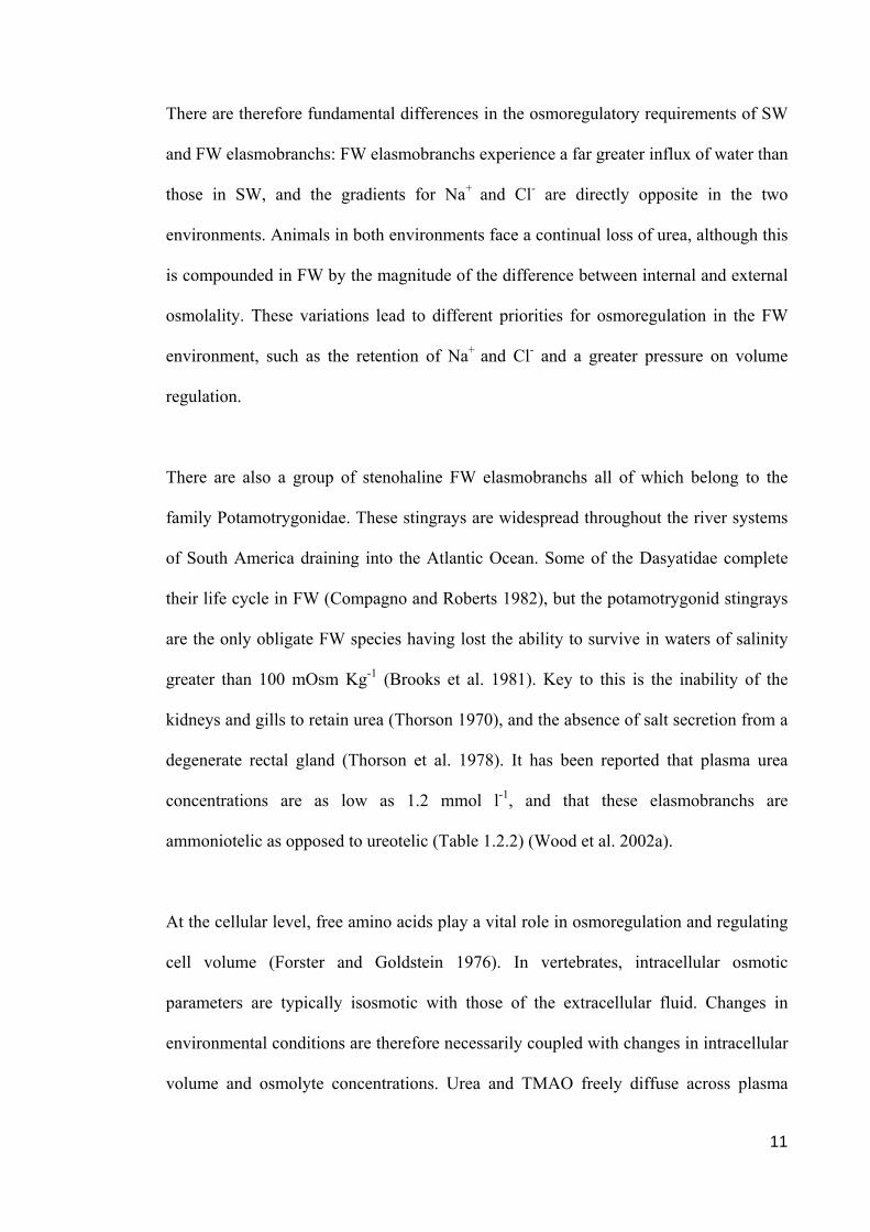
11
There are therefore fundamental differences in the osmoregulatory requirements of SW
and FW elasmobranchs: FW elasmobranchs experience a far greater influx of water than
those in SW, and the gradients for Na+ and Cl- are directly opposite in the two
environments. Animals in both environments face a continual loss of urea, although this
is compounded in FW by the magnitude of the difference between internal and external
osmolality. These variations lead to different priorities for osmoregulation in the FW
environment, such as the retention of Na+ and Cl- and a greater pressure on volume
regulation.
There are also a group of stenohaline FW elasmobranchs all of which belong to the
family Potamotrygonidae. These stingrays are widespread throughout the river systems
of South America draining into the Atlantic Ocean. Some of the Dasyatidae complete
their life cycle in FW (Compagno and Roberts 1982), but the potamotrygonid stingrays
are the only obligate FW species having lost the ability to survive in waters of salinity
greater than 100 mOsm Kg-1 (Brooks et al. 1981). Key to this is the inability of the
kidneys and gills to retain urea (Thorson 1970), and the absence of salt secretion from a
degenerate rectal gland (Thorson et al. 1978). It has been reported that plasma urea
concentrations are as low as 1.2 mmol l-1, and that these elasmobranchs are
ammoniotelic as opposed to ureotelic (Table 1.2.2) (Wood et al. 2002a).
At the cellular level, free amino acids play a vital role in osmoregulation and regulating
cell volume (Forster and Goldstein 1976). In vertebrates, intracellular osmotic
parameters are typically isosmotic with those of the extracellular fluid. Changes in
environmental conditions are therefore necessarily coupled with changes in intracellular
volume and osmolyte concentrations. Urea and TMAO freely diffuse across plasma

12
membranes (Fenstermacher et al. 1972), therefore the intra- and extracellular
concentrations are equivalent. This is not so with free amino acids which constitute 1%
of extracellular fluid osmolality and 19% of that of intracellular fluid (Perlman and
Goldstein 1988). Acclimation of Batoids to decreases in salinity has been proven to
affect free amino acid concentrations. In the little skate, Raja erinacea, significant
decreases in free amino acid concentrations were measured in wing muscle and
erythrocytes upon acclimation to 50% SW, although concentrations in the heart were
unaffected (Boyd et al. 1977). Similar effects were also observed in the brain of the D.
sabina acclimated to 50% SW (Boyd et al. 1977). Clearly free amino acids play an
important role in regulating cell volume, particularly during salinity transfer in
euryhaline elasmobranchs.
It is therefore evident that osmoregulation is of fundamental importance to euryhalinity
in elasmobranchs, at both the cellular and whole animal levels. Through the action of
the gills, gut, rectal gland, and kidneys elasmobranch fish have the ability to
independently regulate the concentrations of Na+, Cl- and urea in both SW and FW
environments, as part of their hyperosmoregulatory strategy. The mechanisms by which
this osmoregulatory strategy is controlled are poorly understood, particularly during
migration between FW and SW. However, the principle organs involved have been
reasonably well studied and their modes of action are well described. The principle
osmoregulatory organs named above will be described in detail, along with their modes
of action and importance in SW and FW. In addition there are a number of other organs
that are believed to play an important role in osmoregulation, such as the liver as the
main site of urea production, and the pituitary gland, the interrenal gland, and the heart
as endocrine organs effecting osmoregulatory control. These too will be discussed.

13
1.3 The gills
The gills of elasmobranchs have been the subject of many anatomical studies (Wright
1973; Olson and Kent 1980; DeVries and DeJaeger 1984; Metcalfe and Butler 1986).
There are usually five pairs of gills, although six and seven are not uncommon. Each
gill arch is made up of lateral rods of cartilage (the gill filaments) supporting a sheet of
muscular and connective tissue (the interbranchial septum). The dorsal and the ventral
surfaces of each gill filament have a row of secondary lamellae; these are the principal
sites of gas exchange.
Branchial vasculature is highly complex and varies greatly from species to species.
Evans and co-workers (2005) recently published a thorough review of the fish gill, in
which detailed descriptions of the vasculature are made. A general model for blood flow
through the elasmobranch gills can be drawn. The entire cardiac output enters the
afferent branchial arteries (ABAs) via the ventral aorta. Blood flowing through an ABA
feeds two hemibranchs of a gill arch where it is oxygenated at the lamellae of the
filaments (Evans et al. 2005). The vasculature which supplies the secondary epithelium
can be mediated by sphincters located on the efferent primary artery, and on both
afferent and efferent secondary arteries (Laurent and Dunel 1980). Oxygenated blood
flows into an efferent branchial artery (EBA) which in turn flows into the dorsal aorta
for systemic distribution (Evans et al. 2005). There are two distinct but interconnected
circulations within the gill filaments: the arterio-arterial pathway which is involved in
respiratory gas exchange; and the arteriovenous pathway, a nonrespiratory pathway
possibly involved in supplying nutrients to the epithelium and structural tissues (Figure
1.3.1).

14
Figure 1.3.1 – Generalised blood flow through an elasmobranch gill arch and filament.
Arterio-arterial pathway: blood travels ( ) from the afferent branchial artery
(ABA) to an afferent filamental artery (AFA), which runs the length of the filament.
This blood is distributed to the lamellae (L) via afferent lamellar arterioles (ALA’s).
Lamellar blood flows through efferent lamellar arterioles (ELA’s) into an efferent
filamental artery (EFA). Oxygenated blood then flows to the efferent branchial artery
(EBA) and on to the dorsal aorta for systemic distribution. Arteriovenous pathway:
blood in the EFA can be distributed to interlamellar vessels (ILV’s) via postlamellar
arteriovenous anastomoses (>) or nutrient arteries (NA). The ILV’s are drained by
branchial veins (BV). The direction of water flow ( ) over the gills is also shown
(Evans et al. 2005).

15
The elevated concentrations of urea and TMAO in the blood plasma of elasmobranchs
results in a substantial concentration gradient for the diffusive efflux of these osmolytes.
The internal concentration of Na+ and Cl- result in a gradient for the diffusive influx of
these ions across epithelial membranes from the marine environment. Even though the
permeability of elasmobranch gill epithelia to urea is the lowest recorded (Boylan 1967)
the gills are still the major site of diffusive urea efflux, as well as Na+ and Cl- influx in
SW. It has been suggested that rates of urea loss are reduced through a combination of
structural and active transport mechanisms. The basolateral membranes of S. acanthias
gill epithelia have the highest cholesterol to phospholipid ratios recorded for a natural
membrane (Fines et al. 2001). This could be a means of reducing the diffusion of urea
into the cell as cholesterol is know to reduce urea permeability (Mourtisen and
Jorgensen 1994). There is also evidence for Na+ dependent active urea transport by
basolateral membrane vesicles (Fines et al. 2001). These findings lead Evans and co–
workers (2005) to suggest that the gill epithelium acts as an intermediary compartment
where the urea concentration gradient with the environment is lowered below that of
blood plasma, thereby reducing diffusive urea loss (Figure 1.3.2).

16
Figure 1.3.2 – Proposed model of urea retention in the elasmobranch gill epithelia. The
basolateral membrane has a decreased permeability for urea, in part due to the high
cholesterol content (represented by the thick line for the membrane). This greatly
reduces the amount of urea in the blood which diffuses into the cell. The concentration
of urea which actually enters the cell is then further reduced by an unidentified Na+-
dependent urea transporter in the basolateral membrane. The Na+ gradient required for
the urea transporter is thought to be maintained by the action of Na+, K+-ATPase. The
relatively low intracellular concentration of urea, as compared to that of the blood
plasma, reduces the gradient for the diffusive loss of urea to the external environment
(Evans et al. 2005).

17
Conversely to the situation described for urea there is active accumulation of Na+ and
Cl- at the gills, despite the osmotic consequences of the salt load. This accumulation is
related to the acid-base regulatory system (Bentley et al. 1976) which is involved in the
excretion of acidic (e.g. hydrogen, H+) and basic (e.g. bicarbonate, HCO3-) ions (Figure
1.3.3) (Evans 1982; Evans 1984). Studies on teleosts and elasmobranchs have shown
consistently that acid secretion is linked to Na+ absorption, and that base secretion is
linked to Cl- absorption (Evans 1982; Cooper and Morris 2004b; Evans et al. 2005).
Faster and more complete compensation for hypercapnia in SW acclimated D. sabina
(Choe and Evans 2003), and a persistence in alkalosis in Heterodontus portusjacksoni
acclimated to reduced salinity (Cooper and Morris 2004b), further support the role of
Na+ in branchial acid excretion. It has been suggested that there are two acid secretion
mechanisms: an apical V-ATPase which is electrically linked to Na+ absorption, and an
electroneutral exchange of Na+ and H+ via the Na+/H+ exchange proteins; and two base
secretion mechanisms via two apical Cl-/HCO3- exchangers: AE1, and pendrin (Evans
et al. 2005).
Accumulation of Na+ and Cl- at the gills may also act as a means of decreasing the
influx of these ions from the external environment. Just as a lower intracellular urea
concentration in the gill epithelia decreases the gradient for the diffusional efflux of
urea, elevated intracellular Na+ and Cl- concentrations would decrease the gradient for
the diffusional influx of these ions from the marine environment. However, given the
specific evolution of the rectal gland towards secreting excess Na+ and Cl- (Section 1.5)
the necessity for decreasing the influxes of these ions is not as great as that for the
retention of urea.

18
Figure 1.3.3 – A working model of NaCl-linked acid base extrusion in the chloride
cells of D. sabina. One type of chloride cell (A MRC) expresses Na+, K+-ATPase
(NKA) on its basolateral membrane and is hypothesized to draw in Na+ across the apical
surface in exchange for cytoplasmic H+. The other type of chloride cell (B MRC)
expresses V-H+-ATPase (V) on its basolateral membrane and draws Cl- into the cell via
pendrin (PDN) in exchange for HCO3-. The pathway for basolateral Cl- movement is
unknown (Evans et al. 2005).

19
There is also substantial efflux of Na+ and Cl- across the gills by the chloride cells or
mitochondria-rich cells (MRC’s) (Figures 1.3.3 and 4). The rate of Na+ and Cl- efflux by
the chloride cells is still less than the rate of influx. Branchial activity of Na+, K+-
ATPase, the active protein in Na+ and Cl- transport (Section 1.5), is ten to fifteen times
below that of marine teleosts, and hence there is net accumulation of Na+ and Cl- at the
gills and no net efflux (Jampol and Epstein 1970; Shuttleworth 1988). Chloride cells are
pear-shaped secretory cells in the epithelia of the gills. As well as being rich in
mitochondria, there is an extensive network of smooth endoplasmic reticulum, and
copious basolateral infoldings of the plasma membrane so as to increase surface area
(Wright 1973).
Comparative studies of Raja clavata and S. canicula revealed two types of chloride
cells. In one cell type the apical membrane is buried deep in a cul-de-sac and connects
to the external milieu by a narrow opening; conversely, the other cell type has a
protruding apical membrane (Laurent and Dunel 1980). Both of these cell types lack the
tubular system which is found in teleost chloride cells. In elasmobranchs these are
functionally replaced by copious infoldings of the basolateral membrane.

20
Figure 1.3.4 – Light micrograph of S. acanthias gill lamellae showing the darkly
stained chloride cells (*). Scale bar of 50 µm (Wilson et al. 2002).
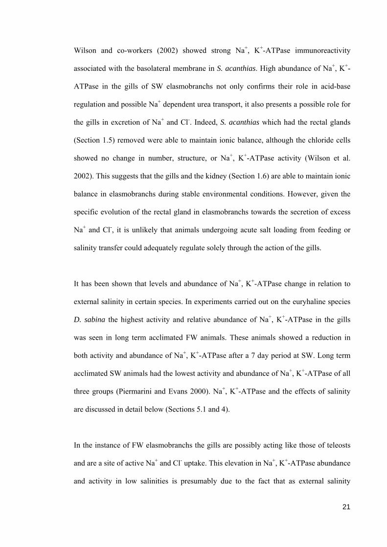
21
Wilson and co-workers (2002) showed strong Na+, K+-ATPase immunoreactivity
associated with the basolateral membrane in S. acanthias. High abundance of Na+, K+-
ATPase in the gills of SW elasmobranchs not only confirms their role in acid-base
regulation and possible Na+ dependent urea transport, it also presents a possible role for
the gills in excretion of Na+ and Cl-. Indeed, S. acanthias which had the rectal glands
(Section 1.5) removed were able to maintain ionic balance, although the chloride cells
showed no change in number, structure, or Na+, K+-ATPase activity (Wilson et al.
2002). This suggests that the gills and the kidney (Section 1.6) are able to maintain ionic
balance in elasmobranchs during stable environmental conditions. However, given the
specific evolution of the rectal gland in elasmobranchs towards the secretion of excess
Na+ and Cl-, it is unlikely that animals undergoing acute salt loading from feeding or
salinity transfer could adequately regulate solely through the action of the gills.
It has been shown that levels and abundance of Na+, K+-ATPase change in relation to
external salinity in certain species. In experiments carried out on the euryhaline species
D. sabina the highest activity and relative abundance of Na+, K+-ATPase in the gills
was seen in long term acclimated FW animals. These animals showed a reduction in
both activity and abundance of Na+, K+-ATPase after a 7 day period at SW. Long term
acclimated SW animals had the lowest activity and abundance of Na+, K+-ATPase of all
three groups (Piermarini and Evans 2000). Na+, K+-ATPase and the effects of salinity
are discussed in detail below (Sections 5.1 and 4).
In the instance of FW elasmobranchs the gills are possibly acting like those of teleosts
and are a site of active Na+ and Cl- uptake. This elevation in Na+, K+-ATPase abundance
and activity in low salinities is presumably due to the fact that as external salinity

22
increases the requirement for active Na+ and Cl- uptake across the gills will decrease as
the ion flux gradient is reversed. These results also demonstrate the capacity for
modification of gill physiology and morphology to changing environmental conditions
in a euryhaline elasmobranch. The discrepancy between S. acanthias (SW) and D.
sabina (euryhaline) suggests that plasticity in chloride cell structure and/or abundance,
and associated branchial Na+, K+-ATPase may therefore be a key factor in
elasmobranch euryhalinity.
23
1.4 The gut
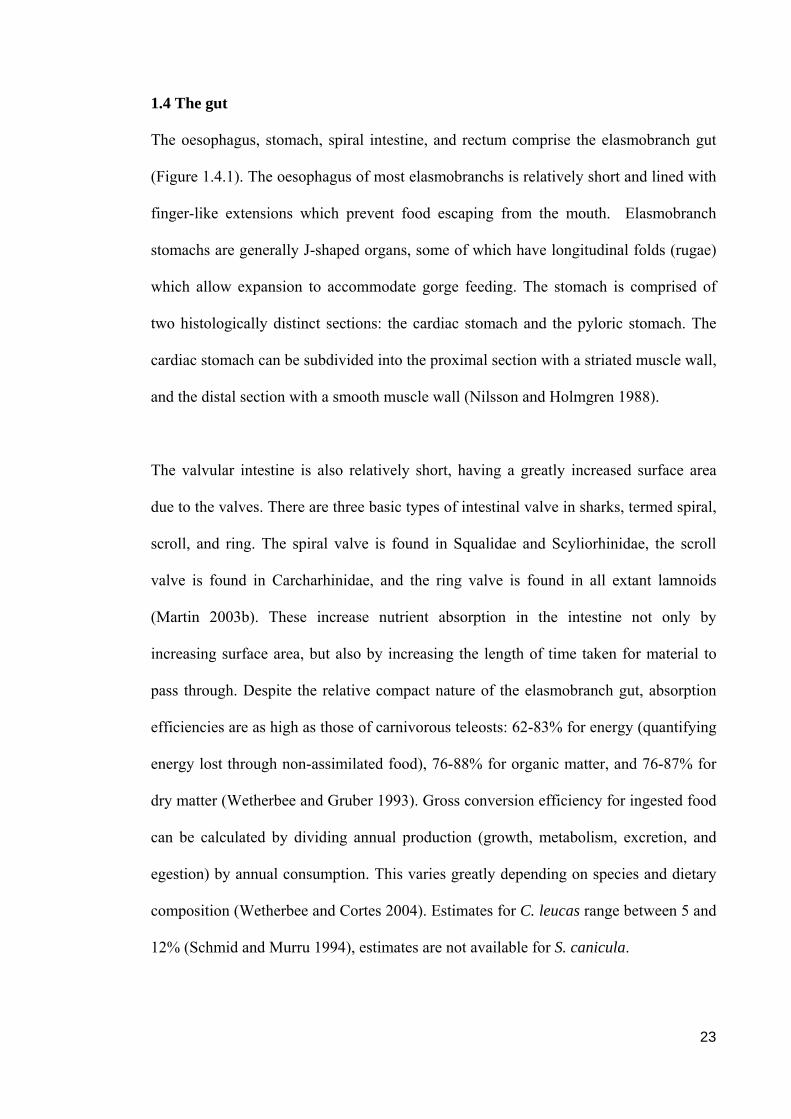
The oesophagus, stomach, spiral intestine, and rectum comprise the elasmobranch gut
(Figure 1.4.1). The oesophagus of most elasmobranchs is relatively short and lined with
finger-like extensions which prevent food escaping from the mouth. Elasmobranch
stomachs are generally J-shaped organs, some of which have longitudinal folds (rugae)
which allow expansion to accommodate gorge feeding. The stomach is comprised of
two histologically distinct sections: the cardiac stomach and the pyloric stomach. The
cardiac stomach can be subdivided into the proximal section with a striated muscle wall,
and the distal section with a smooth muscle wall (Nilsson and Holmgren 1988).
The valvular intestine is also relatively short, having a greatly increased surface area
due to the valves. There are three basic types of intestinal valve in sharks, termed spiral,
scroll, and ring. The spiral valve is found in Squalidae and Scyliorhinidae, the scroll
valve is found in Carcharhinidae, and the ring valve is found in all extant lamnoids
(Martin 2003b). These increase nutrient absorption in the intestine not only by
increasing surface area, but also by increasing the length of time taken for material to
pass through. Despite the relative compact nature of the elasmobranch gut, absorption
efficiencies are as high as those of carnivorous teleosts: 62-83% for energy (quantifying
energy lost through non-assimilated food), 76-88% for organic matter, and 76-87% for
dry matter (Wetherbee and Gruber 1993). Gross conversion efficiency for ingested food
can be calculated by dividing annual production (growth, metabolism, excretion, and
egestion) by annual consumption. This varies greatly depending on species and dietary
composition (Wetherbee and Cortes 2004). Estimates for C. leucas range between 5 and
12% (Schmid and Murru 1994), estimates are not available for S. canicula.

24
Figure 1.4.1 – Diagram of the gut of S. acanthias (Nilsson and Holmgren 1988). The Cardiac stomach is comprised of the striated muscle
cardiac stomach (st) and the smooth muscle cardiac stomach (sm).
Coeliac artery Pyloric stomach
Cardiac stomach
Spiral intestine
Rectum
Mesenteric artery
Rectal gland artery
Rectal glandOesophagus
st sm

25
There are two major factors which influence the role of the gut in osmoregulation: diet
and drinking rate. Due to the nature of aquatic environments the surrounding media is
necessarily imbibed during a feeding event. Elasmobranchs were not thought to actively
drink their environmental media, because a hyperosmotic strategy means the osmotic
gradient is for water to enter the animal, hence there is no requirement to imbibe water.
However Hazon and co-workers (1989) demonstrated that S. canicula does indeed drink
and pharmacological manipulation of the endogenous renin angiotensin system (RAS)
resulted in an increase in drinking rate. A detailed description of the RAS is provided
below (Section 1.10). Basal drinking rates are considerably lower in SW elasmobranchs
than in teleosts, even when compared to Anguilla anguilla which has one of the lowest
recorded teleost drinking rates (Table 1.4.1). This is due to the fact that marine teleosts
are hyposmotic and face a continual loss of water to the environment across semi-
permeable surfaces. The basal rate of drinking in S. canicula increases with
environmental salinity and the ingested Na+ does enter the blood stream (Hazon et al.
1989). Drinking rate also increases during acute transfer to increased salinity in both S.
canicula and Triakis scyllia (Anderson et al. 2002b). Drinking rate may therefore be a
key factor in elasmobranch euryhalinity, by elevating plasma osmolality during transfer
to increased salinity.

26
Species Drinking rate
(ml Kg-1 h-1) Reference
S. canicula 0.3 (Hazon et al. 1997b)
T. scyllia 0.4 (Anderson et al. 2001)
Anguilla anguilla* 1.0 (Perrott et al. 1992)
Pleuronectes platessa* 2.5 (Carroll et al. 1995)
Ammodytes lanceolatus* 3.0 (Perrott et al. 1992)
Limanda limanda* 3.6 (Perrott et al. 1992)
Myxocephalus scorpius* 7.8 (Perrott et al. 1992)
Table 1.4.1 – Drinking rates in SW elasmobranch and teleost (*) fish.
Captivity period Osmolality
(mOsm Kg-1)
Wild 681
1-7 days 638
+ 12 days 558
Table 1.4.2 – Mean blood plasma osmolality in wild, short term, and long term captive
FW C. leucas (n = 9, 13, and 14 respectively). Captive animals were not fed whereas
wild animals had unrestricted access to natural prey species.

27
When examining the role of the gut in elasmobranch osmoregulation a large
consideration must go to dietary composition. By definition the effects of this will vary
greatly between species, and also between populations. Not only will the diet itself vary,
but the requirements from that diet will vary depending on the environment in which the
elasmobranch inhabits, whether the species is an active or ambush predator, and
whether or not the species is ram ventilating. Many marine elasmobranchs, including S.
canicula, are typically gorge feeders. One of the consequences of this is that the animal
is subjected to large and infrequent salt loading during feeding events. This situation is
exaggerated if the diet is also particularly rich in salts, such as one comprised largely of
invertebrates as in S. canicula.
Dietary intake may also be a key source of salts for FW elasmobranchs. Potamotrygonid
rays experienced negative salt balance with their native ion-poor waters during periods
of starvation (Wood et al. 2002a). FW elasmobranchs may therefore require dietary salts
to maintain osmotic stasis.
Metabolic urea is also important for osmoregulation, and this is directly related to food
availability. Infrequently fed Poroderma africanum could not adequately osmoregulate
during acclimation to changes in salinity. Reduced metabolic urea production resulted
in decreases in plasma osmolality and hyposmotic regulation (Haywood 1973). Similar
effects of starvation were seen in C. leucas during captivity trials in this study (Table
1.4.2). This highly active species displayed a visible loss in body condition prior to a
sharp decrease in plasma osmolality during periods of starvation.

28
Armour and co-workers (1993a) showed that S. canicula fed on a low protein diet
showed an impaired osmoregulatory ability when acclimating to hypersaline water.
Animals adopted a strategy utilising increased plasma Na+ and Cl- concentrations to
compensate for the lack of metabolic urea. This further supports the idea of the gut
being an important source for elevating osmolyte levels during salinity transfer.
There is therefore large scope for the gut to be involved in the overall osmoregulatory
mechanisms in elasmobranchs given that imbibed Na+ does enter the blood, the
possibility of large salt loads entering the interstitial fluid during feeding, and the
importance of dietary derived salts and urea. Clearly more research is required into this
area to discover the specific role of the gut for both osmolyte and water exchange, and
possible humoral effects on other osmoregulatory organs.

29
1.5 The rectal gland
The rectal gland is the only organ in elasmobranchs which is capable of producing a
NaCl solution more concentrated than blood plasma levels, and has evolved specifically
for this purpose. The cost of NaCl secretion by the rectal gland has been estimated at
0.5% of the standard metabolic rate (Morgan et al. 1997). The gland itself is a blind-
ending, usually bullet-shaped tube in the dorsal mesentery, which is suspended above
the valvular intestine. It is attached to the intestine postvalvularly. Rectal glands vary in
size and shape depending on the species of elasmobranch, and its life history. Glands
may be smaller in euryhaline, and particularly in freshwater, than in marine animals
(Oguri 1976). This is presumably due to the lower influxes and variations of Na+ and Cl-
in a more dilute environment. The structure and vasculature of the rectal gland are
highly complex and a detailed description is provided later (Section 4.1).
The mechanisms involved in ion transport in the tissues of the rectal gland have been
well documented (Shuttleworth 1988; Silva et al. 1997; Olson 1999). Localised on the
basolateral membrane of the epithelial cells of the secretory tubules is the protein Na+,
K+-ATPase (Dubinsky and Monti 1986). This actively pumps Na+ into the extracellular
space as well as transporting K+ into the secretory cell. Also located on the basolateral
membrane is the Na+K+-2Cl- cotransporter. The action of this protein is passive as it is
driven by the inward Na+ gradient set up by the action of Na+, K+-ATPase. Along this
concentration gradient Na+ enters the cell facilitating the coupled translocation of K+
and Cl- into the intracellular space. Na+, K+-ATPase then actively pumps Na+ back out
of the cell (Haas and Forbush 1998). The internal accumulation of excess K+ is
prevented by passive flow through the basolateral potassium specific channel, thereby
maintaining equilibrium (Riordan et al. 1994).

30
These processes result in a high concentration of Cl- in the secretory cells and a high
Na+ concentration in the intercellular space. Located on the apical membrane of the
secretory cells are chloride-selective channels. Through these channels Cl- ions move
passively into the lumen of the secretory tubule so as to restore the intercellular
electrochemical equilibrium. The Na+K+-2Cl- cotransporter is stimulated by a fall in
intracellular Cl- concentration pursuant to increased Cl- efflux across the apical
membrane. Na+ then passively moves paracellularly through the Na+-selective tight
junctions into the lumen to balance the electrical potential created by the movement of
Cl- ions (Fig 1.5.1) (Olson 1999).
In contrast to the situation described in the gills (Section 1.3), activity and abundance of
Na+, K+-ATPase in the rectal gland is lowest in long term acclimated FW animals.
Levels in acclimated and wild caught SW animals are relatively constant (Piermarini
and Evans 2000; Pillans et al. 2005). This is due to a relative influx of Na+ and Cl-
across semi-permeable membranes in SW and an efflux in FW. Hence there is a reduced
requirement for rectal gland secretion of Na+ and Cl- in more dilute environments.

31
Figure 1.5.1 - Mechanism of Na+ and Cl- ion secretion by secretory tubule cells and
their control. ADP = adenosine diphosphate; ANP = atrial natriuretic peptide; ATP =
adenosine triphosphate; cAMP = cyclic adenosine monophosphate; cGMP = cyclic
guanosine monophosphate; Cl- = chloride ion; K+ = potassium ion; Na+ = sodium ion;
Pi = phosphatidylinositol; VIP = vasoactive intestinal peptide (Olson 1999). Also shown
are some of the hormones affecting the mechanism: the stimulatory actions of
scyliorhinin II, VIP, and two natriuretic peptides (Section 1.11.1), and the inhibitory
action of somatostatin.

32
The hormonal control of rectal gland secretion is detailed below (Sections 1.11.1 and
6.1), but secretion rates can also be affected by neurotransmitters present in the nerves
of the rectal gland. Vasoactive intestinal peptide (VIP) is found in the rectal gland
nerves of S. acanthias (Holmgren and Nilsson 1983; Chipkin et al. 1988) and stimulates
Cl- secretion by activating adenylate cyclase (Stoff et al. 1979). The species specific
model of rectal gland activation involving VIP is detailed below (Section 1.11.1). The
rectal gland of S. acanthias also contains inhibitory neuropeptides including
somatostatin, bombesin, cholecystokinin and neuropeptide Y (Holmgren and Nilsson
1983; Bjenning and Holmgren 1988; Silva et al. 1993). Somatostatin has a direct
inhibitory effect on rectal gland cells both proximally and distally to the release of
cAMP (Stoff et al. 1979; Silva et al. 1985) (Figure 1.5.1), while bombesin inhibits
indirectly through the release of somatostatin (Silva et al. 1990). The method of
inhibition by cholecystokinin has yet to be defined. The inhibitory action of
neuropeptide Y does not affect adenylate cyclase activity, having a direct effect on Cl-
secretion at a site distal to the generation of cAMP. Neuropeptide Y also inhibits VIP-
stimulated transport related oxygen consumption by Na+, K+-ATPase (Silva et al. 1993).
There is therefore much scope for mediating the activity of the elasmobranch rectal
gland with many factors having stimulatory (Sections 1.11.1 and 6.1) and inhibitory
effects.
Given the highly specialised nature of the rectal gland as a means of NaCl secretion, and
the depth of factors which influence its function, it must be of key osmoregulatory
importance during acclimation to salinity changes. Expectations would be for high
levels of activity and secretion during acclimation to reduced salinity in order to rapidly
decrease plasma osmolality and minimise the osmotic influx of water. This is of

33
paramount importance as excess water is excreted via the kidneys (Section 1.6) and
increases in urine volume may increase the loss of urea. The importance of urea
retention is discussed in detail elsewhere (Sections 1.2, 2.1, 2.4, 7.1.2 and 3). Low
levels of activity and secretion would be expected during acclimation to increased
salinity as a means of increasing plasma osmolality.

34
1.6 The Kidney:
Elasmobranch kidneys consist of a pair of elongate structures found on either side of the
dorsal aorta. In sharks they have a thread-like appearance at the anterior end, midway
along the dorsal surface of the abdominal cavity. They gradually widen posteriorly and
fuse below the cloaca (Shuttleworth 1988). Elasmobranchs posses a renal portal system
in which portal veins are formed from the bifurcation of the caudal vein. Upon entering
the kidney these divide to form a matrix of smaller vessels. Blood from the portal
system mixes freely with that from the glomerular vasa efferentia before exiting the
kidney through the renal vein (Hentschel 1988). There is also evidence of a glomerular
bypass vessel which permits blood to flow from the afferent to the efferent vessel,
thereby avoiding filtration (Brown and Green 1992).
The functional unit of the kidney, the nephron, is a complex tubular system. It has been
extensively reviewed by Lacy and Reale (1995) and Hentschel and co-workers (1993).
There is strong evidence of a counter current exchange system involving specialised
epithelial transport (Hentschel and Zierold 1993). There are two regions of renal tissue
in S. canicula: firstly, a dorsal 'bundle' region which is contained in a urea impermeable
sheath (Figure 1.6.1) and the tubules are closely packed into discrete bundles. It is
hypothesised that the counter current exchange system operates in this region (Stolte et
al. 1977). The second region of renal tissue is the ventral 'sinus' which lies outside of the
sheath but has two further loops with the potential for counter current exchange. In this
region the tubules are loosely arranged and segregated by blood sinuses (Lacy and
Reale 1995). The division between the two zones is also marked by large renal
corpuscles.

35
Each individual nephron forms two loops in the bundle zone and two long convolutions
in the sinus region (Hentschel 1988). There is much diversity and specialisation of
epithelial tissue throughout the length of the nephron (Hazon et al. 1997b).
Due to the osmolyte concentrations described above, marine elasmobranchs face a slight
continual influx of water across their semi-permeable surfaces. This excess water is
excreted by the kidneys by an increase in renal clearance, primarily through increased
glomerular filtration rate (GFR) and urine flow rate (Goldstein and Forster 1971;
Forster et al. 1972). Upon exposure to reduced salinity and the associated increase in
water influx, H. portusjacksoni displays a doubling of GFR (Cooper and Morris 2004b).

36
Figure 1.6.1 - Schematic diagram of a single nephron from S. canicula. Filtrate from
the renal corpuscle/glomerulus (RC) flows through the neck segment (NS) and into
loop 1 in the bundle region. Filtrate then passes through proximal segments I and II (PI
and PII) of loop 2 in the sinus region. Then the filtrate passes through the intermediate
segment (IS) into the early distal segment (EDT) and loop 3 in the bundle region.
Filtrate then flows into the late distal segment (LDT) and loop 4 in the sinus region
before entering the collecting tubule (CT). Filtrate then passes into the collecting duct
(CD) (Hazon et al. 1997b).
37
Elasmobranch kidneys cannot produce hyperosmotic urine, typically urine is
hyposmotic relative to blood plasma (Henderson et al. 1988). This fact, coupled with
the use of urea as a plasma osmolyte means that the major roles of the elasmobranch
kidney are urea retention and volume regulation. S. canicula acclimating to reduced
salinity show a marked diuresis along with a reduction of plasma osmolality (Wells et
al. 2002). However, FW acclimated euryhaline elasmobranchs appear to be able to
selectively reduce the urinary concentration of Na+ and Cl- (Shuttleworth 1988; Janech
et al. 1998). The kidney in FW is therefore also capable of regulating the concentration
of Na+ and Cl- in the blood plasma. Lacy and Reale (1991b; 1991a) discovered that
tubular cells in the early distal tubule (EDT) have similar characteristics to cells which
are known to actively transport Na+.
The major role of the elasmobranch kidney is urea retention. There is active urea
transport and reabsorption in the elasmobranch nephron, micropuncture studies have
implicated the second proximal segment (PII) (Figure 1.6.1) as a possible site of Na+-
linked urea reabsorption (Stolte et al. 1977). Levels of skate kidney urea transporter
(SkUT) significantly decreased in response to a decrease in salinity in the marine
elasmobranch Raja erinacea (Morgan et al. 2003). This suggests there is scope for
physiological modification within the kidney to changes in environmental salinity.
The kidney is also the site of the elasmobranch RAS, a key osmoregulatory enzyme
cascade. A detailed description of the RAS is offered below (Section 1.10).

38
1.7 The liver
The Elasmobranch liver performs a number of functions for hydrodynamics and
metabolism. The liver is the main store for energy reserves in the form of fatty acids
although these do perform another function. To elaborate, elasmobranchs lack the swim
bladder of teleost species and are heavier than the surrounding environment. Dynamic
lift is generated from the pectoral fins whilst the animal is in motion. This imposes
hydrodynamic constraints on shark size as a doubling of body length equates to a square
of fin surface area but a cube of body mass. This reduction in relative lift is offset by an
increase in proportional liver size in larger animals which increases the relative amount
of body fatty acids which are less dense than SW. An example of this can be found in
the basking shark, Cetorhinus maximus: the liver from an 8.8 m, 5.9 tonne specimen
accounted for nearly 25% of total body mass yielding 2270 l of oil (Martin 2003a).
With the exception of the FW Potamotrygonid stingrays, elasmobranch fish are
ureotelic with urea production largely occurring in the liver via the OUC (Figure 1.7.1).
This has been extensively reviewed by Goldstein (1967), Anderson (1995; 2001), and
Walsh and Mommsen (2001). A synopsis of the OUC in S. acanthias has been
produced: a mitochondrial glutamine synthase converts a CO2- group of glutamic acid
into an amide group of glutamine; a glutamine-dependent carbamoyl phosphate
synthase (CPS III) and an ornithine carbamoyl transferase make citrulline, then
arginine; and finally a mitochondrial arginase splits arginine into urea and ornithine
(Perlman and Goldstein 1988; Acher 1996).

39
Figure 1.7.1 – The OUC. Numbered circles represent the following enzymes: 1)
Carbamoyl phosphate synthase, 2) Ornithine carbamoyl transferase, 3)
Argininosuccinate synthase, 4) Argininosuccinate lyase, 5) Arginase (Saunders 2002).
Elasmobranchs posses an OUC in the liver which utilises CPS III as enzyme number 1.
This is located in the mitochondria and preferentially uses glutamine (to NH3) as a
nitrogen donor (Tam et al. 2003).

40
Potamotrygonid rays do possess an OUC, but have been demonstrated as being
ammoniotelic (Wood et al. 2002a). Marine elasmobranchs acclimating to reduced
salinity show a reduction in plasma urea levels which can be due to increased renal
clearance of urea, as seen in Negaprion brevirostris (Goldstein et al. 1968); or a
combination of increased clearance and decreased biosynthesis, as seen in R. erinacea
(Goldstein and Forster 1971), S. canicula (Hazon and Henderson 1984), and the FW
stingray Himantura signifer (Tam et al. 2003).
Recent studies have demonstrated a functional OUC in the stomach of H. signifer, with
70% capacity of that of the liver (based on CPS III activity) (Tam et al. 2003). This is
also found in SW Taeniura lymma, although the capacity of this was only around 1% of
that of the liver. Furthermore, ammonia excretion via this route decreases in response to
elevated salinity. It has been suggested that this localised urea production provides a
means of preventing loss of ingested nitrogen as ammonia and amino acids (Tam et al.
2003). This could be of vital importance in FW elasmobranchs when acclimating to
increases in salinity given that they may have a reduced capacity for renal urea retention
(Section 1.6).
The liver also secretes angiotensinogen, the first protein in the RAS protein cascade.
The process and osmoregulatory effects of the RAS are detailed below (Section 1.10).
The organs described above are therefore the major sites of osmoregulatory processes in
elasmobranch fish, and it has been shown that they have modified functions and
priorities in SW and FW. It is through the control of these organs that fully euryhaline
species are able to move between these two environments and maintain their

41
hyperosmotic state. Therefore in order to fully assess the roles these glands have in
elasmobranch osmoregulation it is necessary to detail the endocrine systems which
influence them.

42
1.8 The pituitary gland
The elasmobranch pituitary gland consists of the pars distalis (anterior), pars intermedia,
and neurohypophysis with a large pituitary cleft, similar to other vertebrates. The
elasmobranch gland differs from that of tetrapods through the presence of a partially
separated ventral lobe as opposed to the pars tuberalis seen in the pituitary gland of
other vertebrates. This unique structure contains both a gonadotropin and a thyrotropin
(Fig 1.8.1) (Young 1981).
The hypothalamus is directly linked to the pituitary gland through a portal system which
passes through the median eminence (Fig. 1.8.1). Some neurons within the
hypothalamus secrete hormones, carried via this route, which strictly control secretion
of hormones from the anterior pituitary.
The neurointermedia is permeated by numerous nerve fibres from the neurohypophysial
tract. In elasmobranchs, this contains melanophore-stimulating hormone (MSH)
whereas the teleost equivalent, melanin-concentrating hormone (MCH) is mostly found
in the pars lateralis (Kawauchi 1992). The neurointermedia also contains some
arginine vasotocin (AVT) the action of which is described below (Section 1.8.2), as
well as various neutral octapeptides whose functions remain unknown (Young 1981).

43
Figure 1.8.1 - Generalised elasmobranch pituitary gland. Solid arrows = veins; Broken arrows = arteries; Thin arrows = portal veins
(Young 1981).
44
Very little experimental work has been carried out on the elasmobranch pituitary itself,
but the actions of many hormones which it secretes have well documented affects on
osmoregulatory systems. For ease of description these have been separated into those
emanating from the anterior pituitary and the posterior pituitary. Elasmobranch specific
studies have been utilised where possible.

45
1.8.1 The anterior pituitary
De Vlaming and co-workers (1975) have investigated the effects of hypophysectomy on
D. sabina, resulting in a decrease in plasma osmolality, mostly through a decrease in
urea concentration. Removal of the rostral lobe of the pars distalis resulted in an
increase in plasma osmolality through greater concentrations of Na+ and urea, although
this could be reversed through an injection of mammalian prolactin. Injection of
adrenocorticotrophin (ACTH) alone into these animals had no effect, but when coupled
with the injection of prolactin, ACTH did negate the effects of prolactin. ACTH is
released from the anterior pituitary in response to corticotropin releasing hormone
(CRH) from the hypothalamus. ACTH stimulates the release of corticosteroids; for
example, 1α-hydroxycorticosterone from the interrenal gland (Klesch and Sage 1975;
Hazon and Henderson 1985) (Section 1.9).
Also released from the anterior pituitary is thyroid stimulating hormone (TSH) which
acts as a stimulus for the secretion of thyroid hormones. One such hormone is thyroxine
which has been shown to reduce renal Na+, K+-ATPase activity, and the intracellular
concentrations of cAMP and cGMP in Ginglymostoma cirratum (Honn and Chavin
1976). Removal of the thyroid gland caused increases in plasma urea concentrations and
osmolality in D. sabina (De Vlaming et al. 1975). Replacement therapy with thyroxine
returned plasma urea concentrations to normal levels in these animals (De Vlaming et
al. 1975). This is illustrative of the importance of the pituitary as a means of stimulating
other organs which have important osmoregulatory roles.
Other hormones are also released from the anterior pituitary such, as growth hormone
(GH) and prolactin. Prolactin cells are located in the pars distalis and transfer from SW

46
to FW has been shown to activate prolactin release from these cells in teleosts
(Olivereau and Ball 1970). The role of these hormones in elasmobranch osmoregulation
is largely unknown.

47
1.8.2 The posterior pituitary
The posterior pituitary is the site of neurohypophysial hormone secretion. These can be
divided into the vasotocin-vasopressin lineage and oxytocin-like hormones, both of
which have been well reviewed for elasmobranchs (Acher 1996; Acher et al. 1999).
From the former AVT is one of the key osmoregulatory hormones secreted by the
posterior section of the pituitary gland. All elasmobranchs studied thus far posses AVT,
a homologue of mammalian arginine vasopressin (AVP) (Acher 1996; Acher et al.
1999). AVT is the major neurohypophysial peptide in lower vertebrates. In teleosts,
dose-dependent decreases in urine flow rates, GFR, and tubular transport maxima for
glucose have been seen in trout, Oncorhynchus mykiss, in response to AVT (Amer and
Brown 1995). AVT was thought to have similar antidiuretic effects in elasmobranchs
and this has recently been demonstrated in S. canicula (Wells et al. 2002). In Triakis
scyllium vasotocin levels in the hypothalamus and blood plasma significantly increased
in response to elevated salinity (Hyodo et al. 2004). Clearly neurohypophysial
hormones such as AVT are important endocrine signals for osmoregulation, particularly
during salinity transfer.
Hormones from the oxytocin lineage display much structural variation within the
Chondrichthyes (Table 1.8.2.1). Examination of the effects of these hormones could
therefore become species specific. From the oxytocin lineage both asvatocin and
phasvatocin have been identified in the posterior pituitary of S. canicula (Chauvet et al.
1994). Despite high concentrations of oxytocin-like hormones in the neurohypophysis
no clear function of these peptides has been discovered (Acher et al. 1999), perhaps
because of the high degree of structural variation.

48
Structure
Classification/hormone 1 2 3 4 5 6 7 8 9
Holocephali
Oxytocin Cys Tyr Ile Gln Asn Cys Pro Leu Gly (NH2)
Elasmobranchii
Sharks
Aspargtocin Cys Tyr Ile Asn Asn Cys Pro Leu Gly (NH2)
Valitocin Cys Tyr Ile Gln Asn Cys Pro Val Gly (NH2)
Asvatocin Cys Tyr Ile Asn Asn Cys Pro Val Gly (NH2)
Phasvatocin Cys Tyr Phe Asn Asn Cys Pro Val Gly (NH2)
Rays
Glumitocin Cys Tyr Ile Ser Asn Cys Pro Gln Gly (NH2)
Table 1.8.2.1 – Structure of the oxytocin-like hormones of cartilaginous fish (Acher et
al. 1999).
49
1.9 The interrenal gland
Separate from the kidney is the interrenal gland (Fig 1.9.1), the site of 1α-
hydroxycorticosterone secretion. 1α-hydroxycorticosterone is synthesised in the tissue
from corticosterone (Kime 1987). This steroid was first isolated in the blood plasma of
Raja radiata (Idler and Truscott 1966). Plasma concentration of 1α-
hydroxycorticosterone increases at low salinities, corresponding to the point at which
Na+ becomes regulated at a lower level and urea concentrations continue to decrease
(Armour et al. 1993a). It is likely therefore that 1α-hydroxycorticosterone acts to
minimise Na+ (and Cl-) excretion from the rectal gland, kidney, and gills (Armour et al.
1993a).
Homologous renal extract and heterologous angiotensin II (Ang II) cause in vivo
increases in the plasma concentration of 1α-hydroxycorticosterone (Hazon and
Henderson 1985), as well as increasing secretion from isolated perfused interrenal
glands (O'Toole et al. 1990; Armour et al. 1993b). This suggests the RAS (Section 1.10)
may have a regulatory effect on the interrenal gland. However, the pituitary gland has
been proposed as a major site of regulation for 1α-hydroxycorticosterone secretion from
the interrenal gland (Section 1.8.1) (Hazon and Henderson 1985). Secretion of 1α-
hydroxycorticosterone is stimulated by ACTH, through synergistic action of
intracellular Ca2+ and cAMP.

50
Figure 1.9.1 - Ventral view of the aorta and paired circulatory branches, kidneys, and interrenal gland of Scyllium catulus. White = kidney;
Stippled = interrenal tissue; Black = chromaffin bodies (Chester-Jones 1957).

51
Ang II also stimulates secretion via the action of both intracellular and extracellular
Ca2+ (Armour et al. 1993b). Alterations in Na+ concentration to perifused sections of
glands have inconsistent effects on secretion rates. An increase in urea concentration in
the perifusate increased 1α-hydroxycorticosterone secretion, however a decrease of urea
had no affect (O'Toole et al. 1990). Therefore the factors affecting the interrenal gland
have documented effects on osmoregulation but, although clearly influential, the exact
osmoregulatory role of 1α-hydroxycorticosterone remains to be established.
The chromaffin tissue of elasmobranchs is also discrete from the renal and interrenal
tissue, again contrasting with teleosts. Chromaffin tissue is a site of catecholamine
secretion. The physiological effects of catecholamines are detailed below (Section
1.12).

52
1.10 The RAS
The RAS is a peptide cascade beginning with angiotensinogen which is released from
the liver. This is acted upon by renin which occurs in the kidneys to initiate the cascade
(Fig 1.10.1) (Hazon et al. 1999). The juxtaglomerular apparatus, the site of renin in
other vertebrate species, is located at the vascular pole of the renal corpuscle in
granulated peripolar cells and has been found in a number of elasmobranchs (Lacy et al.
1987; Lacy and Reale 1989; Lacy and Reale 1990). The macula densa, an important part
of the juxtaglomerular apparatus has also been identified (Lacy and Reale 1990). The
presence of the juxtaglomerular apparatus suggest that the RAS may be involved in the
control of GFR, as is the case in teleosts (Brown et al. 1980). In addition to a systemic
RAS, this is strong evidence for the presence of an intrarenal RAS in the elasmobranch
kidney.
There is also a possible presence of Ang II receptors in the elasmobranch kidney
(Tierney et al. 1997). Only two studies have examined the physiological actions of Ang
II in the elasmobranch kidney. Wells and co-workers (2003) demonstrated that
inhibition of angiotensin-converting enzyme resulted in a glomerular diuresis, an
increase in urea and Cl- clearance, and an increase in transport maxima for glucose.
Later work showed that Ang II caused a glomerular antidiuresis and decreases in
perfusion flow rate, transport maxima for glucose, and the proportion of filtering
glomeruli in S. canicula. In addition to this renal urea, Na+, and Cl- clearance were all
significantly reduced by Ang II (Wells et al. In Press).

53
Figure 1.10.1 - The vertebrate RAS showing the action of renin and angiotensin
converting enzyme (ACE), and inhibition by captopril (X) (Hazon et al. 1999). Also
shown in detail are the structures for elasmobranch angiotensin I and II, mammalian
Ang II has Val at position 3 (Takei et al. 1993).
Liver
Angiotensinogen
Angiotensin I
Renin
Angiotensin II
ACE
Angiotensin III
Degradation products
Asn – Arg – Pro – Tyr – Ile – His – Pro – Phe – Gln – Leu Angiotensin I
Asn – Arg – Pro – Tyr – Ile – His – Pro – Phe Angiotensin II
ACE
X
X

54
The structure of elasmobranch Ang I was first deduced in T. scyllia (Takei et al. 1993).
Elasmobranch Ang II has an asparagine residue at position 1, like teleosts, an isoleucine
residue at position 5, like mammals, and a unique proline residue at position 3
(Kobayashi and Takei 1996).
Circulating levels of Ang II in S. canicula have been calculated at 100 - 150 pg/ml
(Tierney et al. 1998). Receptors for Ang II have been suggested in the heart (high and
low affinity receptors) (Cerra et al. 2001), interrenal gland, gills and intestine (Tierney
et al. 1997), as well as in the rectal gland (Masini et al. 1993; Tierney et al. 1997), with
most binding occurring in the subcapsular region (Hazon et al. 1997a).
Ang II has been shown to have a variety of effects in elasmobranchs. Homologous Ang
II causes a dose-dependent increase in drinking rate in both S. canicula and T. scyllia
(Anderson et al. 2001). Furthermore, inhibition of ACE significantly reduces the
dipsogenic effect of the smooth muscle relaxant papaverine in the same two species
(Anderson et al. 2001).
Heterologous Ang II shows vasopressor activity in S. acanthias (Opdyke and Holcombe
1976), S. canicula (Hazon et al. 1989), and T. scyllia (Hazon et al. 1995). Homologous
Ang II shows a response almost 23 times greater than heterologous peptides in T. scyllia
(Takei et al. 1993). This pressor response seems to be mediated by catecholamines
(Section 1.12) (Opdyke and Holcombe 1976; Opdyke et al. 1981). Possible receptors for
Ang II have been identified in the tissues of the rectal gland (Tierney et al. 1997),
although Ang II had no effect on the vascular perfusion of the secretory parenchyma nor
on Cl- secretion rates (Anderson et al. 2002a).

55
The heart of elasmobranchs (Section 1.11) may also be affected by angiotensin.
Angiotensin II binding sites have been found in tissues of the hearts of S. canicula
(Cerra et al. 2001). The action of Ang II is potentially complex with distinct receptor
subtypes, high and low affinity, each having different distributions (Cerra et al. 2001).
These findings suggest that the RAS is an important endocrine control system in
elasmobranch osmoregulation which affects many of the organs outlined above,
although its role in the control of the rectal gland remains unclear.
56
1.11 The Heart
The elasmobranch cardiovascular system consists of a single circulation of a closed
circuit. The heart consists of four contractile chambers which are arranged in the order
of sinus venosus, atrium, ventricle, and conus arteriosus running posterior to anterior
(Fig 1.11.1). The walls of all four chambers contain myocardial tissue, lacking the
smooth muscle found in the heart of teleosts (Tota 1999; Ramos 2004). Venous blood
flows to the atrium via the sinus venosus. Connecting these two chambers is the
sinoatrial orifice where a sinoatrial valve prevents the back flow of blood during atrial
systole. The atrium lies ventral to the sinus venosus and dorsal to the ventricle. The
atrioventricular orifice lies on the ventral side of the boundary between the two
chambers. The opening is circular and surrounded by two ellipsoid flaps, the
atrioventricular valves. The ventricle contains the largest amount of myocardial tissue of
all the chambers. The ventricle is composed of two layers of tissue: the compacta and
the spongiosa (Fig 1.11.1). The compacta is a dense layer of tissue and lies exterior to
the spongiosa which is more diffuse in structure. In S. canicula which have been
acclimated to 120% SW there is a decrease in the tissue of the spongiosa and an
increase in the tissue of the compacta as compared to fish acclimated to 100% and 70%
SW. This is coupled with an increase in the amount of collagen in the tissue (Anderson
and Good, unpublished). This change in tissue ratios could be a modification to the
more viscous blood of the volume depleted animals acclimated to increased salinity.
The conus arteriosus is the most anterior chamber and is confluent with the ventral aorta
(Tota 1999).

57
Figure 1.11.1 - Schematic illustration showing the four chambers of the elasmobranch heart. The S-shaped arrangement of the chambers
means atrial contraction may assist ventricle filling. SA = sinoatrial valve; AV = atrioventricular valve (Tota 1999).

58
Changes in environmental salinity can also affect the heart in other ways. Heart
mitochondria from a variety of species of pelagic sharks have been shown to exhibit
decreased respiratory control ratios (a marker for functional integrity of the isolated
mitochondria) at osmolalities above and below 1000 mOsm (Lewiston et al. 1979).
Increases in extracellular concentrations of organic osmolytes can also have a profound
effect on the tissues of the heart. With increasing concentrations of mannitol, sucrose,
and betaine S. acanthias showed an increase in respiratory control ratio and the rate of
oxygen uptake. However at high concentrations a decrease below basal levels was
demonstrated. This pattern was not seen with increases in extracellular urea
concentration, but was also observed with increases in concentration of TMAO. Unlike
the other osmolytes tested, urea and TMAO readily permeate the mitochondria of the
heart and therefore the cellular osmotic gradient is not present (Lea and Hillman 1990).
TMAO may therefore affect respiratory control ratios by other means. These findings
illustrate the potential consequences when animals fail to completely osmoregulate or
acclimate slowly to external salinity. It is also important to note for the planning of
experimental procedures.
The heart has been shown to affect the kidneys of elasmobranchs. The action of the
heart over the kidneys appears to be in two methods: neural reflex regulation and
endocrine regulation. These two pathways combine to show an increase in renal
clearance with increased blood volume (Peterson and Benjamin 1992).

59
1.11.1 Natriuretic peptides
The natriuretic peptide (NP) system in vertebrates is antagonistic to the RAS and
consists of three types of hormones (Figure 1.11.2). Atrial natriuretic peptides (ANP)
are secreted by mammalian atria in response to an increase in central venous volume or
pressure (Olson 1999). Brain or B-type natriuretic peptide is also found in most
vertebrates, although is replaced by ventricular NP in teleosts (Takei 1999). However,
in elasmobranch fish C-type natriuretic peptide (CNP) is the only circulating natriuretic
peptide (Schofield et al. 1991; Suzuki et al. 1991b; Suzuki et al. 1992; Suzuki et al.
1994). Circulating levels of CNP in T. scyllia far exceed those recorded for any other
species (1.97 pmol ml-1) (Suzuki et al. 1994). CNP is a brain peptide in most vertebrate
species and is therefore present at very low levels in the blood plasma of other species.
This circulating CNP is believed to originate from the heart. A high molecular weight
form of CNP has been isolated from both the atrium and ventricle of the heart of S.
canicula (Suzuki et al. 1991b). The highest concentration of CNP was found in the
atrium, then the ventricle, brain, and pituitary gland respectively (Suzuki et al. 1994). In
the heart the majority of CNP is the pro-hormone CNP-115, whereas in the brain the
majority is the mature peptide CNP-22 (Figure 1.11.2). Contrasting to teleost and
mammals, most CNP in elasmobranch blood is the pro-hormone, not a processed form
(Suzuki et al. 1994). This suggests that the heart is the main storage organ for pro-CNP
which is the major circulating hormone. It is only at specific sites within tissues that the
pro-hormone is processed into other forms.

60
Figure 1.11.2 – (a) A schematic drawing of the generalised processing of natriuretic peptides in vertebrates. The numbers of amino acids in the
mature peptides are noted. (b) Sequential processing of natriuretic peptides in vertebrates. The different patterns in the boxes of preprohormones
denote the signal peptide (striped), N-terminal prohormone (hatched), and mature peptide (solid). The number of amino acid residues are noted
(Takei 1999).

61
As the only natriuretic peptide in elasmobranchs CNP has wide scope for effecting
osmoregulatory processes. Recently CNP has been shown to have effects on the
elasmobranch kidney. Wells and co-workers (In Press) illustrated that CNP caused a
glomerular diuresis, an increase in transport maxima for glucose, but no change in the
proportion of filtering glomeruli. CNP did cause significant increases in renal clearance
of urea, Na+, and Cl-. These effects are antagonistic to those reported for Ang II (Section
1.10).
It has been proposed that CNP may also influence the vasculature of the rectal gland
(Hazon et al. 1997b). Recently it was illustrated experimentally that CNP significantly
increased blood flow to the secretory parenchyma (Anderson et al. 2002a). CNP has
also been shown to dilate the smooth muscle of the capsular region in the rectal gland of
S. acanthias (Evans and Piermarini 2001). This could affect the degree of perfusion
seen in the inner regions of the gland by reducing pressure in the capillaries, and
therefore potentially affect secretion rate.
CNP also has direct effects on the secretory tubules of the rectal gland (Solomon et al.
1992b) (Figure 1.5.1). Endogenous CNP has been shown to stimulate rectal gland
secretion 7 to 8 times above basal levels in isolated perfused glands of S. acanthias
(Solomon et al. 1992a) via guanylyl cyclase-linked receptors (Gunning et al. 1997).
Similar stimulation with CNP was also seen in S. canicula (Anderson et al. 1995b).
CNP released from the heart can therefore have a direct influence on the secretory
activity of the rectal gland.

62
A slightly different model of stimulation has been suggested for the rectal gland of S.
acanthias. In response to a volume stimulus the tissues of the heart release CNP. This
circulates to the rectal gland where it stimulates the nerves to release VIP (Section 1.5).
CNP and VIP then both induce effects on the secretory tissues of the rectal gland and
activate NaCl secretion (Silva et al. 1996) (Figure 1.5.1). Experimental evidence for the
presence of a receptor mechanism in the atrial and cardiac region which triggers the
sequence to activate glandular secretion has been gathered (Erlij and Rubio 1986).
It is important to note here that VIP does not stimulate the rectal glands of all
elasmobranch species; this is a species specific model of activation although the
outlying principles on the direct effects of CNP may be extrapolated to elasmobranchs
in general. Unlike S. acanthias, the rectal glands of S. canicula and R. clavata have been
shown to be unaffected by VIP (Anderson et al. 1995a).
The rectal glands of both S. canicula and R. clavata are stimulated by an intestinal
factor which was first termed rectin, but has been identified as scyliorhinin II (Anderson
et al. 1995a) (Figure 1.5.1). Scyliorhinin II has been isolated from the intestine of S.
canicula and Torpedo marmorata (Conlon and Thim 1988; Anderson et al. 1995a).
The different intestinal peptides which stimulate the rectal glands of S. canicula and S.
acanthias are perhaps a reflection of the difference in feeding behaviour and therefore
the resulting salt loads: S. canicula is typically a gorge feeder, whereas S. acanthias
does not gorge feed.

63
Regardless of which intestinal factor they react to, elasmobranch rectal glands are
stimulated to secrete NaCl by CNP, both directly and indirectly. The heart must
therefore have a role to play in the overall osmoregulatory strategy of elasmobranchs,
principally as a major source of one of the main osmoregulatory hormones, CNP, but
also exerting neural control over other organs. CNP is possibly the most important
endocrine factor with regards to the control of rectal gland secretion (Section 6.1).

64
1.12 Catecholamines
Catecholamines are secreted by the chromaffin tissue in the elasmobranch interrenal
gland. They have been shown to increase blood flow to the gills of S. canicula (Davies
and Rankin 1973), and decrease blood flow to the gut of S. acanthias (Holmgren et al.
1992). The vasculature of the rectal gland has also been shown to be constricted by
catecholamines (Shuttleworth 1983).
The principle catecholamines in elasmobranchs are adrenaline and noradrenaline. Both
are known to have major effects on blood pressure: increasing dorsal aortic blood
pressure and decreasing coeliac arterial blood flow in S. acanthias (Holmgren et al.
1992). Branchial vasculature can be manipulated by the action of adrenaline and
noradrenaline. Both of these hormones appear to act via β–adrenoceptor-mediated
vasodilation (Davies and Rankin 1973; Capra and Satchell 1977). This vasodilatory
response masks a smaller α–adrenoceptor-mediated vasoconstriction (Davies and
Rankin 1973; Capra and Satchell 1977). Adrenaline has been shown to have effects on
the kidney, reducing the proportion of filtering glomeruli but causing an overall diuresis
in S. canicula (Brown and Green 1987).
Two of the major osmoregulatory peptides have been shown to affect catecholamine
release. Circulating levels of noradrenaline increase 15-fold in S. acanthias in response
to CNP (McKendry et al. 1999). Ang II has been shown to increase plasma
concentrations of adrenaline and noradrenaline in S. acanthias (Bernier et al. 1999).
Given that two of the major osmoregulatory hormones affect catecholamine release
from chromaffin tissue, and that catecholamines affect blood flow and function of
different organs, it is likely that they play a role in mediating osmoregulatory responses.

65
1.13 Objectives
Elasmobranchs require the combined actions of the gills, gut, rectal gland and kidney in
order to maintain osmotic stasis and alter osmolality during changes in salinity. There
appears to be scope for interspecific differences in the processes behind this. The aim of
this study was to examine the differences between a partially and a fully euryhaline
species in terms of their osmotic profiles at different salinities, and the processes by
which these are achieved and maintained. Specific interest was given to the rectal gland
due to its role in Na+ and Cl- balance, and the difference in activity between SW and
FW. The specific aims are:
1) To produce replicable chronic salinity transfer protocols for S. canicula to 80,
100, and 120% SW; and for C. leucas to FW and SW in captivity. Also to
produce experimental protocols for the acute transfer of both species to 100%
SW from all chronic acclimation conditions.
2) To produce plasma osmotic profiles for S. canicula chronically acclimated to 80,
100, and 120% SW conditions; and for C. leucas in FW and SW. Also, to gain
equivalent data for both species during acute salinity transfer to 100% SW.
3) To assess blood volume in S. canicula during chronic and acute salinity
transfers.
4) To measure in vivo rectal gland secretion rates of S. canicula during chronic and
acute salinity transfer.

66
5) To examine the histology of the rectal gland from both species chronically
acclimated to the different salinities.
6) To measure maximal Na+, K+-ATPase activity in the gills, gut, rectal gland, and
kidney of both species chronically acclimated to the different salinities.
7) To measure oxygen consumption of the rectal gland in both species chronically
acclimated to the different salinities, measure what proportion of that was
attributable to Na+, K+-ATPase, and to asses the effects of CNP on both
parameters.

67
Ambiguous citations
(Smith 1931b; Smith 1931a; Silva et al. 1977; Stoff et al. 1977a; Stoff et al. 1977b;
Silva et al. 1979; Silva et al. 1983; Silva et al. 1985; Silva et al. 1987; Silva et al. 1990;
Lacy and Reale 1991b; Lacy and Reale 1991a; Suzuki et al. 1991a; Suzuki et al. 1991b;
Solomon et al. 1992a; Solomon et al. 1992b; Armour et al. 1993a; Armour et al. 1993b;
Hentschel et al. 1993; Hentschel and Zierold 1993; Silva et al. 1993; Anderson 1995;
Anderson et al. 1995a; Anderson et al. 1995b; Cornelius 1995a; Cornelius 1995b;
Solomon et al. 1995a; Solomon et al. 1995b; Silva et al. 1996; Hazon et al. 1997a;
Hazon et al. 1997b; Silva et al. 1997; Silva et al. 1999; Anderson 2001; Anderson et al.
2001; Anderson et al. 2002a; Anderson et al. 2002b; Silva and Epstein 2002; Wells
2002; Wells et al. 2002; Wood et al. 2002a; Wood et al. 2002b; Cooper and Morris
2004b; Cooper and Morris 2004a)

68
References Acher, R. (1996). "Molecular evolution of fish neurohypophysial hormones:
neutral and selective evolutionary mechanisms." Gen. Comp. Endocrinol. 102: 157-172.
Acher, R., Chauvet, J., et al. (1999). "Unique evolution of neurohypophysial hormones in cartilaginous fishes: possible implications for urea-based osmoregulation." J. Exp. Zool. 284: 475-484.
Amer, S. and Brown, J. A. (1995). "Glomerular actions of arginine vasotocin in the in situ perfused trout kidney." Am. J. Physiol. 269: R775-R780.
Anderson, P. M. (1995). Urea cycle in fish: molecular and mitochondrial studies. Ionoregulation: Cellular and molecular approaches to fish ionic regulation. C. M. Wood and T. J. Shuttleworth. New York, Academic Press. 14: 57-83.
Anderson, P. M. (2001). Urea and glutamine synthesis: environmental influences on nitrogen excretion. Nitrogen excretion. P. A. Wright and P. M. Anderson. San Diego, Academic Press. 20: 239-278.
Anderson, W. G., Conlon, J. M., et al. (1995a). "Characterization of the endogenous intestinal peptide that stimulates the rectal gland of Scyliorhinus canicula." Am. J. Physiol. 268: R1359-R1364.
Anderson, W. G., Good, J. P., et al. (2002a). "Changes in chloride secretion rate and vascular perfusion in the rectal gland of the European lesser spotted dogfish in response to environmental and hormonal stimuli." J. Fish. Biol. 60: 1580-1590.
Anderson, W. G., Takei, Y., et al. (2001). "The dipsogenic effect of the renin-angiotensin system in elasmobranch fish." Gen. Comp. Endocrinol. 124: 300-307.
Anderson, W. G., Takei, Y., et al. (2002b). "Osmotic and volaemic effects on drinking rate in elasmobranch fish." J. Exp. Biol. 205: 1115-1122.
Anderson, W. G., Tierney, M. L., et al. (1995b). "Natriuretic hormones in elasmobranch fish; possible interactions with other endocrine systems." Physiol. Zool. 68: 184.
Armour, K. J., O'Toole, L. B., et al. (1993a). "The effect of dietary protein restriction on the secretory dynamics of 1α-hydroxycorticosterone and urea in the dogfish, Scyliorhinus canicula: a possible role for 1α-hydroxycorticosterone in sodium retention." J. Endocrinol. 138: 275-282.
Armour, K. J., O'Toole, L. B., et al. (1993b). "Mechanisms of ACTH-and angiotensin II-stimulated Iα-hydroxycorticosterone secretion in the dogfish, Scyliorhinus canicula." J. Mol. Endocrinol. 10: 235-244.
Ballantyne, J. S., Moyes, C. D., et al. (1987). "Compatible and counteracting solutes and the evolution of ion and osmoregulation in fishes." Can. J. Zool. 65: 1883-1888.
Bentley, P. J., Maetz, J., et al. (1976). "A study of the unidirectional fluxes of Na and Cl across the gills of the dogfish Scyliorhinus canicula (Chondrichthyes)." J Exp Biol 64: 629-637.
Bernier, N. J., Gilmour, K. M., et al. (1999). "Cardiovascular control via angiotensin II and circulating catecholamines in the spiny dogfish, Squalus acanthias." J. Comp. Physiol. B 169(4-5): 237-248.

69
Bjenning, C. and Holmgren, S. (1988). "Neuropeptides in the fish gut. An immunohistochemical study of evolutionary patterns." Histochemistry 88: 155-163.
Boyd, T. A., Cha, C. J., et al. (1977). "Free amino acids in tissues of the skate Raja erinacea and the stingray Dasyatis sabina: effects of environmental dilution." J. Exp. Zool. 199: 435-442.
Boylan, J. W. (1967). Gill permeability in Squalus acanthias. Sharks, Skates and Rays. P. W. Gilbert, R. F. Mathewson and D. P. Rall. Baltimore, John Hopkins University Press: 197-206.
Brooks, D. R., Thorson, T. B., et al. (1981). Fresh-water stingrays (Potamotrygonidae) and their helminth parasites: testing hypotheses of evolution and co-evolution. Advances in cladistics, Proceedings of the First Meeting of the Willi Hennig Society., The New York Botanical Garden, Bronx, New York.
Brown, J. A. and Green, C. (1987). "Single nephron function of the lesser spotted dogfish, Scyliorhinus canicula, and the effects of adrenaline." J. Exp. Biol. 129: 265-278.
Brown, J. A. and Green, C. (1992). "Glomerular bypass shunts and distribution of glomeruli in the kidney of the lesser spotted dogfish, Scyliorhinus canicula." Cell. Tissue. Res. 269(2): 299-304.
Brown, J. A., Oliver, J. A., et al. (1980). "Angiotensin and single nephron glomerular function in the trout Salmo gairdneri." Am. J. Physiol. 239: R509-R514.
Burger, J. W. and Hess, W. N. (1960). "Function of the rectal gland in the spiny dogfish." Science 131: 670-671.
Capra, M. F. and Satchell, G. H. (1977). "The adrenergic responses of isolated saline perfused prebranchial arteries and gills of the elasmobranch Squalus acanthias." Gen. Pharmacol. 8: 67-71.
Carroll, S., Hazon, N., et al. (1995). "Drinking rates and Na+ effluxes in response to temperature change in two species of marine flatfish: dab, Limanda limanda and plaice, Pleuronectes platessa." J. Comp. Physiol. 164B: 579-584.
Cerra, M. C., Tierney, M. L., et al. (2001). "Angiotensin II binding sites in the heart of Scyliorhinus canicula: An autoradiographic study." Gen. Comp. Endocrinol. 121: 126-134.
Chauvet, J., Rouille, Y., et al. (1994). "Special evolution of neurohypophysial hormones in cartilagenous fishes: asvatocin and phasvatocin, two oxytocin-like peptides isolated from the spotted dogfish (Scyliorhinus canicula)." Proc. Natl. Acad. Sci. USA 91: 11266-11270.
Chester-Jones, I. (1957). The adrenal cortex. Cambridge, Cambridge University Press.
Chipkin, S. R., Stoff, J. S., et al. (1988). "Immunohistochemical evidence for the neural mediation of VIP activity in the dogfish rectal gland." Peptides 9: 119-124.
Choe, K. P. and Evans, D. H. (2003). "Compensation for hypercapnia by a euryhaline elasmobranch: Effect of salinity and roles of gills and kidneys in freshwater." J. Exp. Zool. A. 297A: 52-63.
Compagno, L. J. V. (1973). "Interrelationships of living elasmobranchs." Zool. J. Linn. Soc. 53(1): 15-61.

70
Compagno, L. J. V. (1977). "Phyletic relationships of living sharks and rays." Am. Zool. 17: 303-322.
Compagno, L. J. V. (1988). Sharks of the Order Carcharhiniformes. Princeton, Princeton University Press.
Compagno, L. J. V. and Cook, S. F. (1995). "The exploitation and conservation of freshwater elasmobranchs: status of taxa and prospects for the future." J. Aquari. Aquat. Sci. 7: 62-90.
Compagno, L. J. V. and Roberts, T. R. (1982). "Freshwater stingrays (Dasyatidae) of Southeast Asia and New Guinea, with a description of a new species Himantura and reports of unidentified species." Environ. Biol. Fish. 7: 321-339.
Conlon, J. M. and Thim, L. (1988). "Isolation of the tachykinin, des[ser1pro2] scyliorhinin-II from the intestine of the ray, Torpedo marmorata." Gen. Comp. Endocrinol. 71(3): 383-388.
Cooper, A. R. and Morris, S. (2004a). "Haemoglobin function and respiratory status of the Port Jackson shark, Heterodontus portusjacksoni, in response to lowered salinity." J. Comp. Physiol. B 174: 223-236.
Cooper, A. R. and Morris, S. (2004b). "Osmotic, sodium, carbon dioxide and acid-base state of the Port Jackson shark, Heterodontus portusjacksoni, in response to lowered salinity." J. Comp. Physiol. B. 174: 211-222.
Cornelius, F. (1995a). "Hydrophobic ion interaction on Na+ activation and dephosphorylation of reconstituted Na+, K+-ATPase." Biochim. Biophys. Acta. 1235: 183-196.
Cornelius, F. (1995b). "Phosphorylation/dephosphorylation of reconstituted shark Na+, K+-ATPase: one phosphorylation site per alpha beta promoter." Biochim. Biophys. Acta. 1235: 197-204.
Cunny, C. and Benton, M. J. (1999). "Early radiation of the neoselachian sharks in Western Europe." Geobios 32: 193-204.
Davies, D. T. and Rankin, J. C. (1973). "Adrenergic receptors and vascular responses to catecholamines of perfused dogfish gills." Comp. Gen. Pharmacol. 60: 830-840.
De Vlaming, V. L., Sage, M., et al. (1975). "Pituitary, adrenal and thyroid influences on osmoregulation in the euryhaline elasmobranch, Dasyatis sabina." Comp. Biochem. Physiol. 52A: 505-513.
DeVries, R. and DeJaeger, S. (1984). "The gill of the spiny dogfish, Squalus acanthias: respiratory and non-respiratory function." Am. J. Anat. 169: 1-29.
Douady, C. J., Dosay, M., et al. (2003). "Molecular phylogenetic evidence refuting the hypothesis of Batoidea (rays and skates) as derived sharks." Mol. Phylogenet. Evol. 26: 215-221.
Dubinsky, W. P. and Monti, L. B. (1986). "Resolution of apical from basolateral membrane of shark rectal gland." Am. J. Physiol. 251(20): C721-C726.
Erlij, D. and Rubio, R. (1986). "Control of rectal gland secretion in the dogfish (Squalus acanthias) - Steps in the sequence of activation." J. Exp. Biol. 122: 99-112.
Evans, D. H. (1982). "Mechanisms of acid extrusion by two marine fishes: the teleost Opsanus beta and the elasmobranch Squalus acanthias." J. Exp. Biol. 97: 289-299.

71
Evans, D. H. (1984). The roles of gill permeability and transport mechanisms in euryhalinity. Fish Physiology. W. S. Hoar and D. J. Randall. New York, Academic Press. 10: 239-283.
Evans, D. H. and Piermarini, P. M. (2001). "Contractile properties of the elasmobranch rectal gland." J. Exp. Biol. 204: 59-67.
Evans, D. H., Piermarini, P. M., et al. (2005). "The multifunctional fish gill: dominant site of gas exchange, osmoregulation, acid-base regulation, and excretion of nitrogenous waste." Physiol. Rev. 85: 97-177.
Fenstermacher, J., Sheldon, F., et al. (1972). "The blood to tissue distribution of various polar materials in the dogfish, Squalus acanthias." Comp. Biochem. Physiol. 42A: 195-204.
Fines, G. A., Ballantyne, J. S., et al. (2001). "Active urea transport and an unusual basolateral membrane composition in the gills of a marine elasmobranch." Am. J. Physiol. 280: R16-R24.
Forster, R. P. and Goldstein, L. (1976). "Intracellular osmoregulatory role of amino acids and urea in marine elasmobranchs." Am. J. Physiol. 230: 925-931.
Forster, R. P., Goldstein, L., et al. (1972). "Intrarenal control of urea reabsorption by renal tubules of the marine elasmobranch, Squalus acanthias." Comp. Biochem. Physiol. 42A: 3-12.
Frick, N. T. and Wright, P. A. (2001). "Nitrogen metabolism and excretion in the mangrove killifish Rivulus marmoratus I. The influence of environmental salinity and external ammonia." J. Exp. Biol. 205: 79-89.
Goldstein, L. (1967). Urea biosynthesis in elasmobranchs. Sharks, Skates and Rays. P. W. Gilbert, R. F. Mathewson and D. P. Rall. Baltimore, John Hopkins University Press: 207-214.
Goldstein, L. and Forster, R. P. (1971). "Osmoregulation and urea metabolism in the little skate Raja erinacea." Am. J. Physiol. 220(3): 742-746.
Goldstein, L., Oppelt, W. W., et al. (1968). "Osmotic regulation and urea metabolism in the lemon shark Negaprion brevirostris." Am. J. Physiol. 215: 1493-1497.
Griffith, R. W. (1991). "Guppies, Toadfish, Lungfish, Coelacanths and frogs - A scenario for the evolution of urea retention in fishes." Environ. Biol. Fish. 32: 199-218.
Gunning, M., Solomon, R. J., et al. (1997). "Role of guanylyl cyclase receptors for CNP in salt secretion by shark rectal gland." Am. J. Physiol. 273: R1400-R1406.
Haas, M. and Forbush, B. (1998). "The Na-K-Cl cotransporters." Bioenerg. Biomem. 30: 161-172.
Haywood, G. P. (1973). "Hypo-osmotic regulation coupled with reduced metabolic urea in the dogfish Poroderma africanum: an analysis of serum osmolarity, chloride, and urea." Mar. Biol. 23: 121-127.
Hazon, N., Balment, R. J., et al. (1989). "The renin-angiotensin system and vascular and dipsogenic regulation in elasmobranchs." Gen. Comp. Endocrinol. 74: 230-236.
Hazon, N., Cerra, M. C., et al. (1997a). Elasmobranch renin angiotensin system and the angiotensin receptor. Proceedings of the XIIIth International Congress of Comparative Endocrinology., Yokohama, Japan, Monduzzi Editore.

72
Hazon, N. and Henderson, I. W. (1984). "Secretory dynamics of 1a-hydroxycorticosterone in the elasmobranch fish, Scyliorhinus canicula." J. Endocrinol. 103: 205-211.
Hazon, N. and Henderson, I. W. (1985). "Factors affecting the secretory dynamics of 1α-hydroxycorticosterone in the dogfish, Scyliorhinus canicula." Gen. Comp. Endocrinol. 59: 50-55.
Hazon, N., Tierney, M., et al. (1995). "Endogenous angiotensins, angiotensin-II competitive binding inhibitors and converting enzyme inhibitor in elasmobranch fish." Neth. J. Zool. 45(1-2): 117-120.
Hazon, N., Tierney, M. L., et al. (1997b). Ion and Water balance in Elasmobranch fishes. Ionic Regulation in Animals. N. Hazon, B. Eddy and G. Flik. Berlin, Heidelberg, Springer-Verlag: 70-86.
Hazon, N., Tierney, M. L., et al. (1999). "Renin-angiotensin system in elasmobranch fish: A review." J. Exp. Zool. 284(5): 526-534.
Henderson, I. W., O'Toole, L. B., et al. (1988). Kidney Function. Physiology of Elasmobranch Fishes. T. J. Shuttleworth. Berlin, Springer Verlag: 201-214.
Hentschel, H. (1988). "Renal blood vascular system in the elasmobranch, Raja erinacea Mitchill, in relation to kidney zones." Am. J. Anat. 183: 130-147.
Hentschel, H., Mahler, S., et al. (1993). "Renal tubule of dogfish, Scyliorhinus canicula - a comprehensive study of structure with emphasis on intramembrane particles and immunoreactivity for H+-K+-adenosine triphosphatase." Anat. Rec. 235(4): 511-532.
Hentschel, H. and Zierold, K. (1993). "Morphology and element distribution of magnesium-secreting epithelium: the proximal tubule segment PII of dogfish, Scyliorhinus canicula (L.)." Eur. J. Cell Biol. 63: 32-42.
Holmgren, S., Axelsson, M., et al. (1992). "The effect of catecholamines, substance-p and vasoactive intestinal polypeptide on blood-flow to the gut in the dogfish Squalus acanthias." J. Exp. Biol. 168: 161-175.
Holmgren, S. and Nilsson, S. (1983). "Bombesin-, gastrin/cck-, 5-hydroxytryptamine-, neurotensin-, somatostatin-, and VIP-like immunoreactivity and catecholamine fluorescence in the gut of the elasmobranch, Squalus acanthias." Cell Tiss. Res. 234(3): 595-618.
Honn, K. V. and Chavin, W. (1976). "In vitro trophic action of ACTH and insulin upon adrenocortical enzymes of the Squaliform elasmobranch Ginglymostoma cirratum (Bonnaterre)." Gen. Comp. Endocrinol. 29: 360-368.
Hyodo, S., Tsukada, T., et al. (2004). "Neurohypophysial hormones of dogfish, Triakis scyllium: structures and salinity-dependent secretion." Gen. Comp. Endocrinol. 138: 97-104.
Idler, D. R. and Truscott, B. (1966). "Iα-Hydroxycorticosterone from cartilaginous fish: a new adrenal steroid in blood." J. Fish. Res. Bd. Canada 23(4): 615-619.
Jampol, L. M. and Epstein, F. M. (1970). "Sodium-potassium-activated adenosinetriphosphate and osmotic regulation by fishes." Am. J. Physiol. 218(2): 607-611.
Janech, M. G., Fitzgibbon, W. R., et al. (1998). "Effect of low salinity on the renal function of Dasyatis sabina, a marine euryhaline elasmobranch." J. Invest. Med. 46(1): 63A-63A.
73
Kawauchi, H. (1992). Advances of fish pituitary hormone research. Trends in Comp. Physiology and Biochemistry Proceedings 8th Int. symposium., Tokyo, Japan.
Kime, D. E. (1987). The Steroids. Fundamentals of comparative vertebrate Endocrinology. I. Chester-Jones, P. M. Ingleton and J. G. Phillips. New York, Plenum Press. 1: 3-56.
Klesch, W. and Sage, M. (1975). "The stimulation of corticosteroidogenesis in the interrenal of the elasmobranch Dasyatis sabina by mammalian ACTH." Comp. Biochem. Physiol. A 52: 145-146.
Kobayashi, H. and Takei, Y. (1996). The Renin-Angiotensin System comparative aspects. Berlin, Heidelberg, Springer-Verlag.
Lacy, E. R., Castellucci, M., et al. (1987). "The elasmobranch renal corpuscle: Fine structure of Bowman's capsule and the glomerular capillary wall." Anat. Rec. 218: 294-305.
Lacy, E. R. and Reale, E. (1989). "Granulated peripolar epithelial cells in the renal corpuscle of marine elasmobranch fish." Cell Tiss. Res. 257: 61-67.
Lacy, E. R. and Reale, E. (1990). "The presence of a juxtaglomerular apparatus in elasmobranch fish." Anat. Embryol. 182(3): 249-262.
Lacy, E. R. and Reale, E. (1991a). "Fine structure of the elasmobranch renal tubule: intermediate, distal and collecting duct segments of the little skate." Am. J. Anat. 192: 478-497.
Lacy, E. R. and Reale, E. (1991b). "Fine structure of the elasmobranch renal tubule: Neck and proximal segments of the little skate." Am. J. Anat. 190: 118-132.
Lacy, E. R. and Reale, E. (1995). Functional morphology of the elasmobranch nephron and retention of urea. Cellular and Molecular Approaches to Fish Ionic Regulation. C. M. Wood and T. J. Shuttleworth. New York, Academic Press: 107-146.
Laurent, P. and Dunel, S. (1980). "Morphology of gill epithelia in fish." Am. J. Physiol. 238: R147-R159.
Lea, M. and Hillman, S. (1990). "Effects of Osmolality and solutes on performance of shark heart mitochondria." J. Exp. Zool. 255: 9-15.
Lewiston, N., Newman, A., et al. (1979). "Shark heart mitochondria: Effects of external osmolality on respiration." Science 206: 75-76.
Maisey, J. G. (1980). "An evaluation of Jaw suspension in sharks." Am. Mus. Novit. 2706: 1-17.
Martin, A. (2001). "The phylogenetic placement of Chondrichthyes: inferences from analysis of multiple genes and implications for comparative studies." Genetica 111: 349-357.
Martin, R. A. (2003a). Does liver size limit shark body size? 2005. Martin, R. A. (2003b). No guts, no glory. Masini, M. A., Uva, B., et al. (1993). "Renin-like activity, angiotensin I-converting
enzyme-like activity, and osmoregulatory peptides in the dogfish rectal gland." Gen. Comp. Endocrinol. 93: 246-254.
McKendry, J. E., Bernier, N. J., et al. (1999). "Natriuretic peptides and the control of catecholamine release in two freshwater teleost and a marine elasmobranch fish." Fish Physiol. Biochem. 20: 61-77.
Metcalfe, J. D. and Butler, P. J. (1986). "The functional anatomy of the gills of the dogfish (Scyliorhinus canicula)." J. Zool. Lond. (A) 208: 519-530.

74
Morgan, J. D., Wilson, J. M., et al. (1997). "Oxygen consumption and Na+, K+-ATPase activity of the rectal gland and gill tissue in the spiny dogfish, Squalus acanthias." Can. J. Zool. 75: 820-825.
Morgan, R. L., Ballantyne, J. S., et al. (2003). "Regulation of a renal urea transporter with reduced salinity in a marine elasmobranch, Raja erinacea." J. Exp. Biol. 206: 3285-3292.
Mourtisen, O. G. and Jorgensen, K. (1994). "Dynamic order and disorder in lipid bilayers." Chem. Phys. Lipids 73: 3-25.
Nilsson, S. and Holmgren, S. (1988). The autonomic nervous system. Physiology of Elasmobranch Fishes. T. J. Shuttleworth. London, Springer-Verlag: 143-169.
Oguri, M. (1976). Rectal glands of marine and freshwater sharks: Comparative histology. Investigations of the Ichthyofauna of Nicaraguan Lakes. T. B. Thorson. Lincoln, School of Life Sciences: 613-614.
Olivereau, M. and Ball, J. N. (1970). "Pituitary influences on osmoregulation in teleosts." Mem. Soc. Endocrinol. 18: 57-85.
Olson, K. R. (1999). Rectal gland and volume homeostasis. Sharks, Skates and Rays. W. C. Hamlett. Baltimore, John Hopkins University Press: 329-352.
Olson, K. R. and Kent, B. (1980). "The microvasculature of the elasmobranch gill." Cell Tiss. Res. 209: 49-63.
Opdyke, D. F., Carroll, R. G., et al. (1981). "Angiotensin-II releases catecholamines in dogfish." Comp. Biochem. Physiol. 70(1): 131-134.
Opdyke, D. F. and Holcombe, R. (1976). "Response to angiotensin I and II and to AI-converting enzyme inhibitor in a shark." Am. J. Physiol. 231: 1750-1753.
O'Toole, L. B., Armour, K. J., et al. (1990). "Secretory patterns of 1-alpha-hydroxycorticosterone in the isolated perifused interrenal gland of the dogfish, Scyliorhinus canicula." J. Mol. Endocrinol. 5(1): 55-60.
Perlman, D. F. and Goldstein, L. (1988). Nitrogen Metabolism. Physiology of Elasmobranch Fishes. T. J. Shuttleworth. London, Springer-Verlag: 253-275.
Perrott, M. N., Grierson, C. E., et al. (1992). "Drinking behaviour in sea water and fresh water teleosts, the role of the renin-angiotensin system." Fish Physiol. Biochem. 10(2): 161-168.
Peterson, T. V. and Benjamin, B. A. (1992). "The heart and control of renal excretion: neural and endocrine mechanisms." FASEB 6: 2923-2932.
Piermarini, P. M. and Evans, D. H. (1998). "Osmoregulation of the Atlantic stingray (Dasyatis sabina) from the freshwater lake Jesup of the St. Johns River, Florida." Physiol. Zool. 71(5): 553-560.
Piermarini, P. M. and Evans, D. H. (2000). "Effects of environmental salinity on Na+/K+-ATPase in the gills and rectal gland of a euryhaline elasmobranch (Dasyatis sabina)." J. Exp. Biol. 203: 2957-2966.
Pillans, R. P. and Franklin, C. E. (2004). "Plasma osmolyte concentrations of bull sharks Carcharhinus leucas, captured along a salinity gradient." Comp Biochem Physiol A 138: 363 - 371.
Pillans, R. P., Good, J. P., et al. (2005). "Freshwater to seawater acclimation of juvenile bull sharks (Carcharhinus leucas): plasma osmolytes and Na+/K+-ATPase activity in gill, rectal gland, kidney and intestine." J Comp Physiol B 175: 37 - 44.

75
Ramos, C. (2004). "The structure and ultrastructure of the sinus venosus in the mature dogfish (Scyliorhinus canicula): the endocardium, the epicardium and the subepicardial space." Tissue Cell 36: 399-407.
Revill, A. S., Dulvy, N. K., et al. (2005). "The survival of discarded lesser-spotted dogfish (Scyliorhinus canicula) in the Western English Channel beam trawl fishery." Fish. Res. 71: 121-124.
Riordan, J. R., Forbush, B., et al. (1994). "The molecular basis of chloride transport in shark rectal gland." J. Exp. Biol. 196: 405-418.
Saunders, B. (2002). Merck Source Resource Library., Merck Source. 2005. Schmid, T. H. and Murru, F. L. (1994). "Bioenergetics of the bull shark,
Carcharhinus leucas, maintained in captivity." Zool. Biol. 13: 177-185. Schofield, J. P., Stephen, D., et al. (1991). "Identification of C-type natriuretic
peptide in heart of spiny dogfish shark (Squalus acanthias)." Am. J. Physiol. 261(4 Pt2): F734-F739.
Shirai, S. (1996). Phylogenetic relationships of neoselachians (Chondrichthyes: Euselachii). Interrelationships of Fishes. M. L. J. Stiassny, L. R. Parenti and G. D. Johnson. San Diego, Academic Press: 9-34.
Shuttleworth, T. J. (1983). "Haemodynamic effects of secretory agents on the isolated elasmobranch rectal gland." J. Exp. Biol. 103: 193-204.
Shuttleworth, T. J. (1988). Salt and water balance - extrarenal mechanisms. Physiology of Elasmobranch fishes. T. J. Shuttleworth. Berlin, Heidelberg, Springer-Verlag: 171-199.
Silva, P. and Epstein, F. H. (2002). "Role of the cytoskeleton in secretion of chloride by shark rectal gland." J. Comp. Physiol. B 172: 719-723.
Silva, P., Epstein, F. H., et al. (1993). "Neuropeptide-Y inhibits chloride secretion in the shark rectal gland." Am. J. Physiol. 265(2 Pt2): R439-R446.
Silva, P., Epstein, J. A., et al. (1983). "Ouabain binding in rectal gland of Squalus acanthias." J. Memb. Biol. 75: 105-114.
Silva, P., Lear, S., et al. (1990). "Somatostatin mediates bombesin inhibition of chloride secretion by rectal gland." Am. J. Physiol. 258: R1459-R1463.
Silva, P., Solomon, R. J., et al. (1996). "The rectal gland of Squalus acanthias: A model for the transport of chlorine." Kidney Int. 49: 1552-1556.
Silva, P., Solomon, R. J., et al. (1997). "Transport mechanisms that mediate the secretion of chloride by the rectal gland of Squalus acanthias." J. Exp. Zool. 279: 504-508.
Silva, P., Solomon, R. J., et al. (1999). "Mode of activation of salt secretion by C-type natriuretic peptide in the shark rectal gland." Am. J. Physiol. 277: R1725-R1732.
Silva, P., Stoff, J., et al. (1979). "Indirect evidence for enhancement of Na-K-ATPase activity with stimulation of rectal gland secretion." Am. J. Physiol. 237: F468-F472.
Silva, P., Stoff, J., et al. (1977). "Mechanism of active chloride secretion by shark rectal gland: role of Na-K-ATPase in chloride transport." Am. J. Physiol. 233: F298-F306.
Silva, P., Stoff, J. S., et al. (1985). "Mode of action of somatostatin to inhibit secretion by shark rectal gland." Am. J. Physiol. 249: R329-R334.
Silva, P., Stoff, J. S., et al. (1987). "Atrial natriuretic peptide stimulates salt secretion by shark rectal gland by releasing VIP." Am. J. Physiol. 252: F99-F103.

76
Smith, H. W. (1931a). "The absorption and excretion of water and salts by the elasmobranch fishes. I. Fresh water Elasmobranchs." Am. J. Physiol. 98(2): 279-295.
Smith, H. W. (1931b). "The absorption and excretion of water and salts by the elasmobranch fishes. II Marine Elasmobranchs." Am. J. Physiol. 98(2): 296-310.
Solomon, R., Castelo, L., et al. (1995a). "Preliminary data on intracellular signalling mechanisms in the rectal gland of Squalus acanthias: a pharmalogical approach." Bull. Mt. Desert Isl. Biol. Lab. 34: 47-48.
Solomon, R., Nathanson, M., et al. (1995b). "An increase in intracellular calcium is associated with inhibition and not stimulation of the rectal gland of Squalus acanthias." Bull. Mt. Desert Isl. Biol. Lab. 34: 42-43.
Solomon, R., Protter, A., et al. (1992a). "C-type natriuretic peptides stimulate chloride secretion in the rectal gland of Squalus acanthias." Am. J. Physiol. 262: R707-R711.
Solomon, R., Solomon, H., et al. (1992b). "Chloride secretion in the rectal gland of Squalus acanthias the role of C-type natriuretic peptide (CNP)." Bull. Mt. Des. Mar. Lab. 31: 62-64.
Sosa-Nishizaki, O., Taniuchi, T., et al. (1998). "The bull shark, Carcharhinus leucas (Valenciennes, 1841), from the Usumacinta river, Tabasco, Mexico, with notes on its serum composition and osmolarity." Ciencias Mar. 24(2): 183-192.
Stoff, J. S., Hallac, R., et al. (1977a). "The role of vasoactive intestinal peptide (VIP) in the regulation of active chloride secretion in the rectal gland of Squalus acanthias." Bull. Mt. Desert Isl. Biol. Lab. 17: 66.
Stoff, J. S., Rosa, R., et al. (1979). "Hormonal regulation of active chloride transport in the dogfish rectal gland." Am. J. Physiol. 237: F138-F144.
Stoff, J. S., Silva, P., et al. (1977b). "Cyclic AMP regulation of active chloride transport in the rectal gland of marine elasmobranchs." J. Exp. Zool. 199: 443-448.
Stolte, H., Galaske, R. G., et al. (1977). "Renal tubule ion transport and collecting duct function in the elasmobranch little skate, Raja erinacea." J. Exp. Zool. 199: 403-410.
Suzuki, R., Kaneko, T., et al. (1991a). "Effects of osmotic pressure on prolactin and growth hormone secretion from organ-cultured eel pituitary." J. Comp. Physiol. 161B: 147-153.
Suzuki, R., Takahashi, A., et al. (1991b). "Isolation of high-molecular-weight C-type natriuretic peptide from the heart of a cartilaginous fish (European dogfish, Scyliorhinus canicula)." Febs Letts. 282(2): 321-325.
Suzuki, R., Takahashi, A., et al. (1992). "Different molecular forms of C-type natriuretic peptide isolated from the brain and heart of an elasmobranch, Triakis scyllia." J. Endocrinol. 135: 317-323.
Suzuki, R., Togashi, K., et al. (1994). "Distribution and molecular forms of C-type natriuretic peptide in plasma and tissue of a dogfish, Triakis scyllia." Gen. Comp. Endocrinol. 96: 378-384.
Takei, Y. (1999). Structural and functional evolution of the natriuretic peptide system in vertebrates. International review of cytology. K. W. Jeon. San Diego, Academic Press. 194: 1-66.
Takei, Y., Hasegawa, Y., et al. (1993). "A novel angiotensin I isolated from an elasmobranch fish." J. Endocrinol. 139: 281-285.

77
Tam, W. L., Wong, W. P., et al. (2003). "The osmotic response of the Asian freshwater stingray (Himantura signifer) to increased salinity: a comparison with the marine (Taeniura lymma) and Amazonian freshwater (Potamotrygon motoro) stingrays." J. Exp. Biol. 206: 2931-2940.
Taylor, L. R. (1997). Sharks & rays. London, HarperCollins. Thorson, T. B. (1970). "Freshwater stingrays, Potamotrygon spp.: failure to
concentrate urea when exposed to a saline medium." Life Sci. 9: 893-900.
Thorson, T. B., Cowan, C. M., et al. (1973). "Body fluid solutes of juveniles and adults of the euryhaline bull shark Carcharhinus leucas from freshwater and saline environments." Physiol. Zool. 46: 29-42.
Thorson, T. B., Wooton, R. M., et al. (1978). "Rectal gland of freshwater stingrays, Potamotrygon spp. (Chondrichthyes: Potamotrygonidae)." Biol. Bull. 154: 508-516.
Tierney, M. L., Takei, Y., et al. (1997). "The presence of angiotensin II receptors in elasmobranchs." Gen. Comp. Endocrinol. 105: 9-17.
Tierney, M. L., Takei, Y., et al. (1998). "A radioimmunoassay for the determination of angiotensin II in elasmobranch fish." Gen. Comp. Endocrinol. 111(299-305).
Tota, B. (1999). Heart. Sharks, Skates and Rays. W. C. Hamlett. Baltimore, John Hopkins University Press: 238-272.
Walsh, P. J. and Mommsen, T. P. (2001). Evolutionary considerations of nitrogen metabolism and excretion. Nitrogen excretion. P. A. Wright and P. M. Anderson. San Diego, Academic Press. 20: 1-30.
Wells, A. (2002). The endocrine control of renal function in elasmobranch fish. School of Biological and Medical Sciences. St Andrews, University of St Andrews: 226.
Wells, A., Anderson, W. G., et al. (In Press). "Effects of angiotensin II and C-type natriuretic peptide on the in situ perfused trunk preparation of the dogfish." Gen. Comp. Endocrinol.
Wells, A., Anderson, W. G., et al. (2002). "Development of an in situ perfused kidney preparation for elasmobranch fish: action of arginine vasotocin." Am. J. Physiol. 282: R1636-R1642.
Wells, A., Anderson, W. G., et al. (2003). "Evidence for an intrarenal renin-angiotensin system in the European lesser-spotted dogfish." J. Fish. Biol. 63: 1337-1340.
Wetherbee, B. M. and Cortes, E. (2004). Food consumption and feeding habits. Biology of sharks and their relatives. J. C. Carrier, J. A. Musick and M. R. Heithaus. London, CRC Press: 223-244.
Wetherbee, B. M. and Gruber, S. H. (1993). "Absorption efficiency of the lemon shark Negaprion brevirostris at varying rates of energy intake." Copeia 2: 416-425.
White, E. G. (1937). "Interrelationships of the elasmobranchs with a key to the order Galea." Bull. Am. Mus. Nat. Hist. 74: 25-138.
Wilson, J. M., Morgan, J. D., et al. (2002). "Branchial mitochondria-rich cells in the dogfish Squalus acanthias." Comp. Biochem. Physiol. A 132: 365-374.
78
Winchell, C. J., Martin, A. P., et al. (2004). "Phylogeny of elasmobranchs based on LSU and SSU ribosomal RNA genes." Mol. Phylogenet. Evol. 31(1): 214-224.
Wintner, S. P., Dudley, S. F. J., et al. (2002). "Age and growth estimates for the Zambezi shark, Carcharhinus leucas, from the east coast of South Africa." Mar. Freshwater Res. 53: 557-566.
Wood, C. M., Matsuo, A. Y. O., et al. (2002a). "Mechanisms of ion transport in Potamotrygon, a stenohaline freshwater elasmobranch native to the ion-poor blackwaters of the Rio Negro." J. Exp. Biol. 205: 3039-3054.
Wood, C. M., Wilson, P., et al. (2002b). "Obligatory urea production and the cost of living in the Magadi tilapia revealed by acclimation to reduced salinity and alkalinity." Physiol. Biochem. Zool. 75: 111-122.
Wright, D. E. (1973). "The structure of the gills of the elasmobranch Scyliorhinus canicula." Z. Zellforch 144: 489-509.
Wright, P., Anderson, P., et al. (2004). "The crab-eating frog, Rana cancrivora, up-regulates hepatic carbamoyl phosphate synthetase I activity and tissue osmolyte levels in response to increased salinity." J. Exp. Zool. 301A: 559-568.
Yancey, P. H., Clark, M. E., et al. (1982). "Living with water stress: evolution of osmolyte systems." Science 217: 1214-1222.
Yancey, P. H. and Somero, G. N. (1978). "Urea-requiring lactate dehydrogenases of marine elasmobranch fish." J. Comp. Physiol. 125: 135-141.
Yancey, P. H. and Somero, G. N. (1979). "Counteraction of urea destabilization of protein structure by methylamine osmoregulatory compounds of elasmobranch fishes." Biochem. J. 183: 317-323.
Yancey, P. H. and Somero, G. N. (1980). "Methylamine osmoregulatory solutes of elasmobranch fishes counteract urea inhibition of enzymes." J. Exp. Zool. 212: 205-213.
Young, J. Z. (1981). The life of vertebrates. London, Oxford University Press.