Embed Size (px)
Citation preview
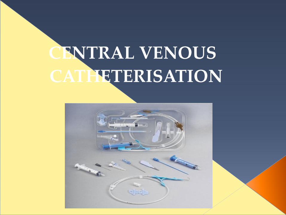
CENTRAL VENOUS
CATHETERISATION
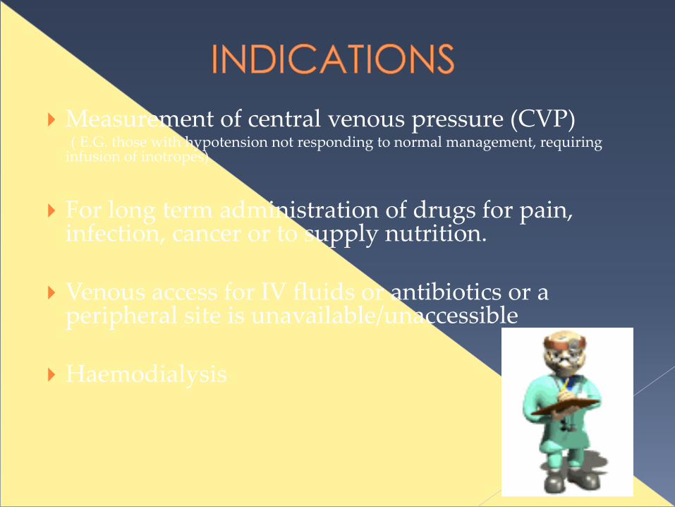
Measurement of central venous pressure (CVP) ( E.G. those with hypotension not responding to normal management, requiring
infusion of inotropes)
For long term administration of drugs for pain, infection, cancer or to supply nutrition.
Venous access for IV fluids or antibiotics or a peripheral site is unavailable/unaccessible
Haemodialysis

Patients undergoing thrombolytic or anticoagulative therapy
Bleeding disorders
Vasculitis
Distorted local anatomy
Overlying skin infections( dermatitis), burns
Uncooperative patient
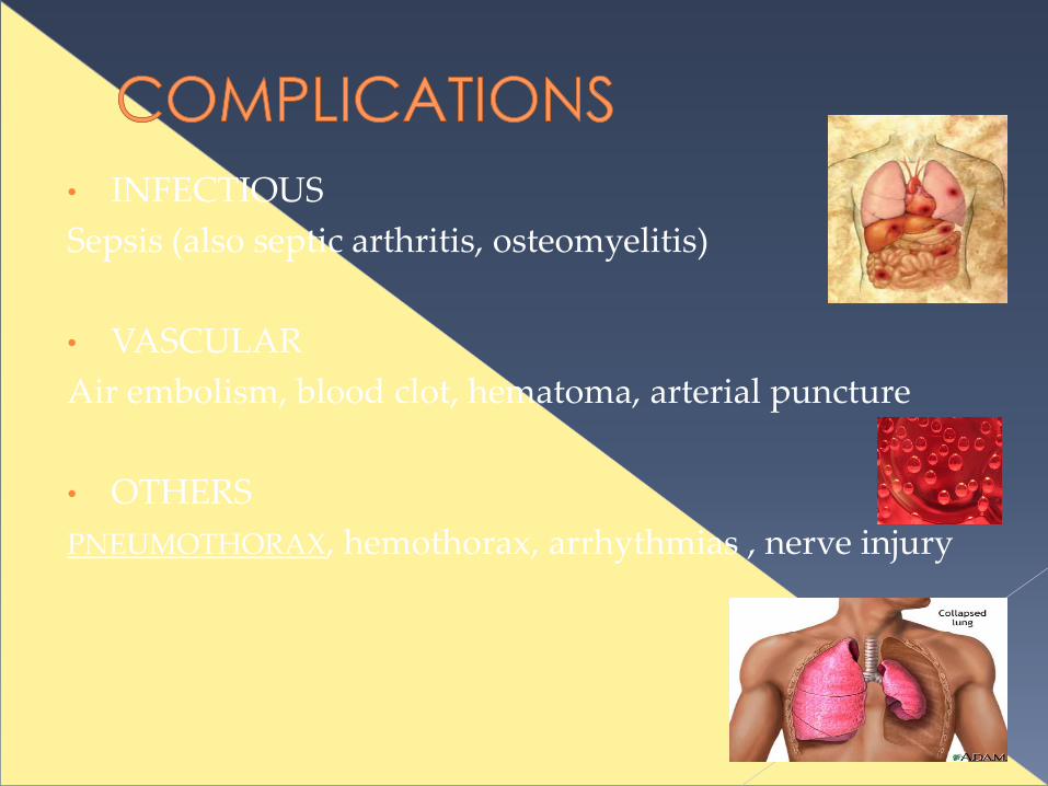
• INFECTIOUS
Sepsis (also septic arthritis, osteomyelitis)
• VASCULAR
Air embolism, blood clot, hematoma, arterial puncture
• OTHERS
PNEUMOTHORAX, hemothorax, arrhythmias , nerve injury

INTERNAL JUGULAR VEIN
SUBCLAVIAN VEIN
FEMORAL VEIN
Length of catheters
15cm catheters for subclavian and internal jugular lines, and 60cm catheters for femoral line

Patient on a tilting bed, trolley or operating table
Standard multiple lumen kit
Guide wire
Sterile gloves
Sterile gown
Drapes
Disinfectant (Povidone-iodine solution/ chlorhexidine)
Suturing needle
Scalpel
Local Anaesthetic (lidocaine)
Sterile saline flush
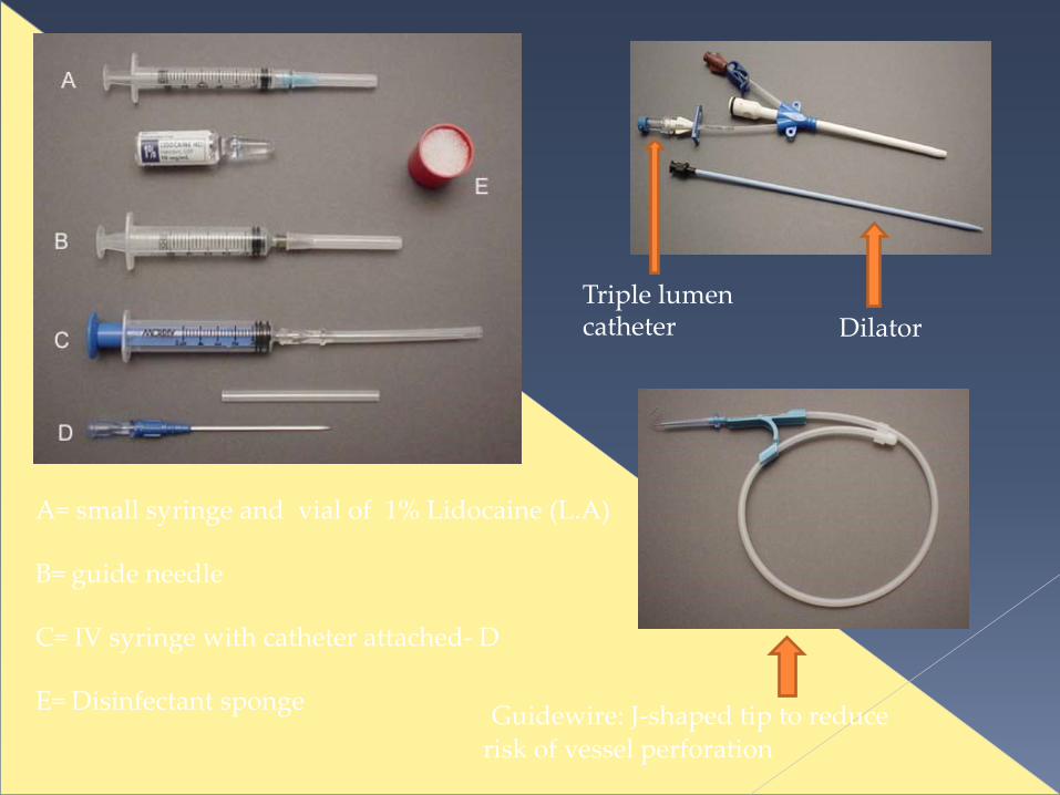
A= small syringe and vial of 1% Lidocaine (L.A)
B= guide needle
C= IV syringe with catheter attached- D
E= Disinfectant sponge Guidewire: J-shaped tip to reduce
risk of vessel perforation
DilatorTriple lumen catheter
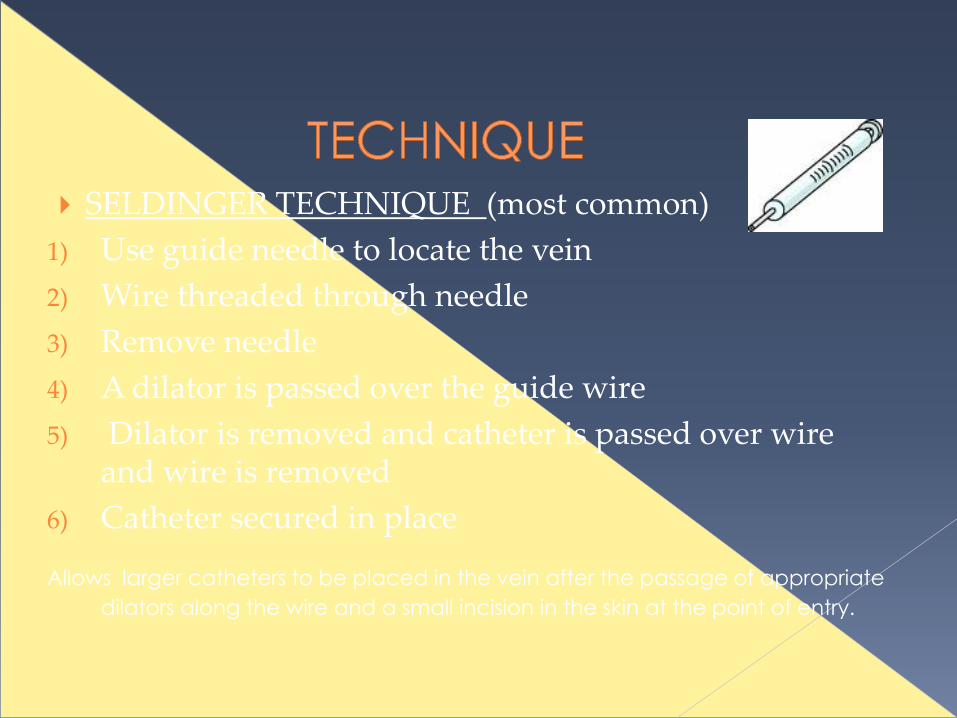
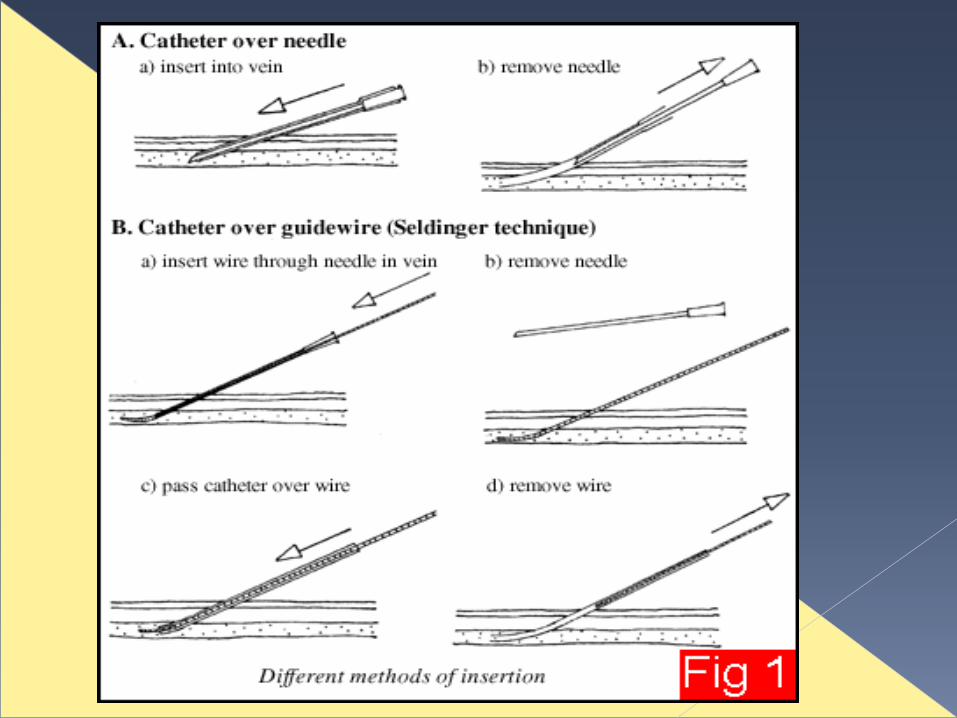
SELDINGER TECHNIQUE (most common)
1) Use guide needle to locate the vein
2) Wire threaded through needle
3) Remove needle
4) A dilator is passed over the guide wire
5) Dilator is removed and catheter is passed over wire and wire is removed
6) Catheter secured in place
Allows larger catheters to be placed in the vein after the passage of appropriate
dilators along the wire and a small incision in the skin at the point of entry.

Obtain informed consent and explain risks and benefits of procedure
Optimal patient positioning and cooperation, make sure patient is comfortable
Take your time
Sterile technique
Local anaesthetic should be used
Always have a hand on your wire
Aspirate while advancing as you withdraw the needle slowly
Withdraw needle to the level of the skin before redirecting the angle
Don’t poke yourself with the needle
The tip of the catheter can lie in either the superior or inferior vena cava (SVC or IVC) or into the right atrium (RA).
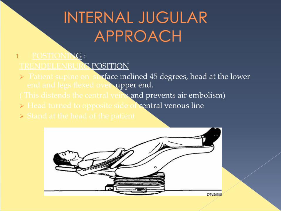
1. POSTIONING :
TRENDELENBURG POSITION
Patient supine on surface inclined 45 degrees, head at the lower end and legs flexed over upper end.
( This distends the central veins and prevents air embolism)
Head turned to opposite side of central venous line
Stand at the head of the patient

Ultrasound and landmarks can be used
IJV is between the clavicular and sternal heads of the sternocleidomastiod muscle
Point of needle insertion is midway between sternal head of SCM and mastoid process behind ear

Disinfect area , apply L.A and fenestrated drape
Place three fingers on carotid artery
Place needle about 45 degrees to the skin, lateral to the carotid artery
Direct needle in sagittal plane angled towards feet
Vein should be 1-1.5 cm deep, avoid deep probing in the neck
Seldinger technique used
http://www.youtube.com/watch?v=QHiuYc22pfE

1.Disinfection, L.A and
sterile drape
2. Insert needle into
IJV and aspirate
3. Hold tip of needle with
one hand
4. Place wire through
needle and remove needle5. Insert catheter over wire
then remove wire
6. Once catheter is in
place , secure and
apply dressing
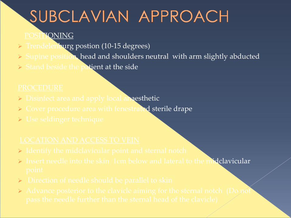
POSITIONING
Trendelenburg postion (10-15 degrees)
Supine position, head and shoulders neutral with arm slightly abducted
Stand beside the patient at the side
PROCEDURE
Disinfect area and apply local anaesthetic
Cover procedure area with fenestrated sterile drape
Use seldinger technique
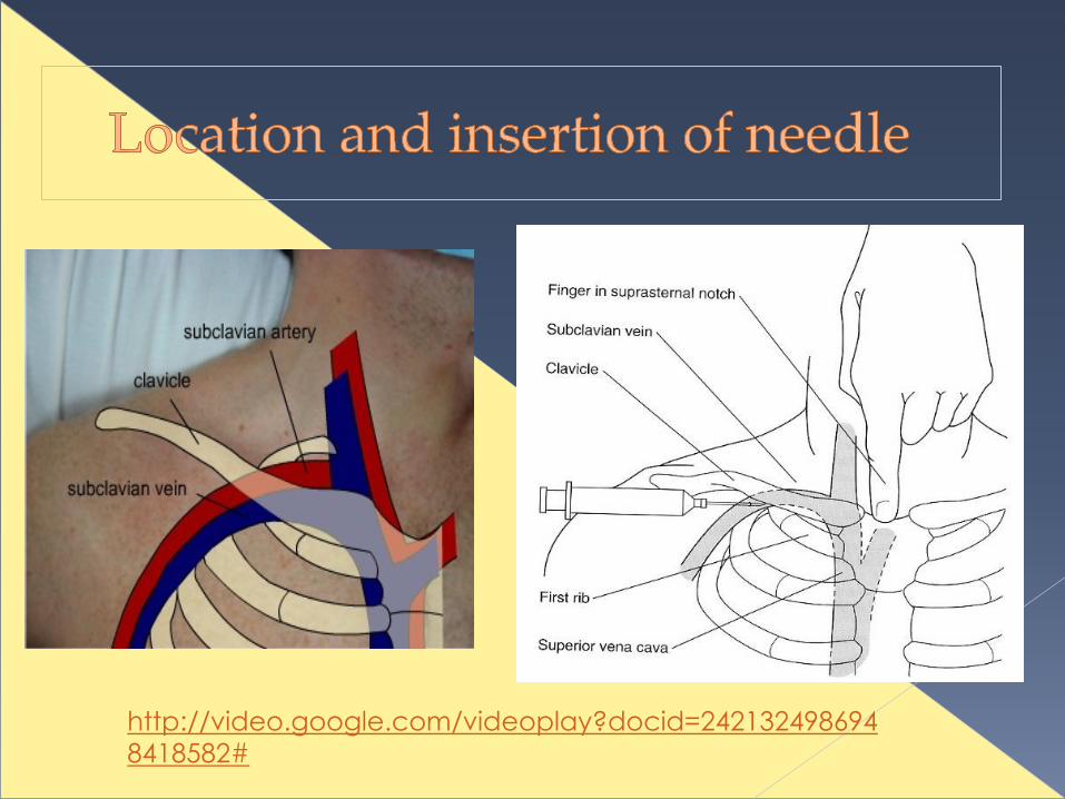
LOCATION AND ACCESS TO VEIN
Identify the midclavicular point and sternal notch
Insert needle into the skin 1cm below and lateral to the midclavicular point
Direction of needle should be parallel to skin
Advance posterior to the clavicle aiming for the sternal notch (Do not pass the needle further than the sternal head of the clavicle)
http://video.google.com/videoplay?docid=242132498694
8418582#

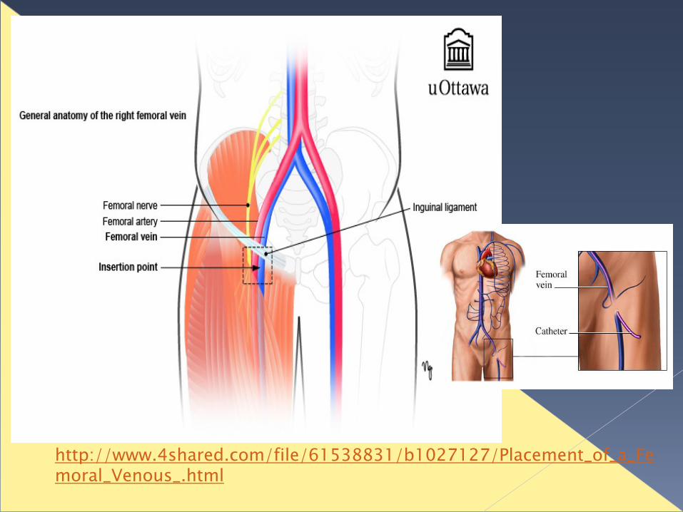
POSITIONING Supine patient Extend the patient’s leg and abduct slightly at the hip
PROCEDURE Disinfect area and apply local anaesthetic Cover procedure area with fenestrated sterile drape Use seldinger technique
LOCALISATION AND ACCESS TO VEIN Vein is medial to femoral artery Identify the pulsation of the femoral artery 1-2 cm below the inguinal
ligament. Position needle at 45 degree angle and about 1cm medial to pulsation Needle inserted at skin about 2 cm below inguinal ligament Aiming towards the umbilicus (In adults, the vein normally found 2-4cm from the skin. In small children
reduce elevation on the needle to 10-15° as the vein is more superficial)
http://www.4shared.com/file/61538831/b1027127/Placement_of_a_Femoral_Venous_.html
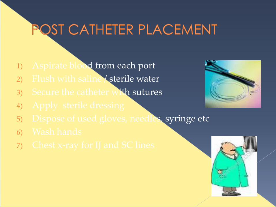
1) Aspirate blood from each port
2) Flush with saline / sterile water
3) Secure the catheter with sutures
4) Apply sterile dressing
5) Dispose of used gloves, needles, syringe etc
6) Wash hands
7) Chest x-ray for IJ and SC lines
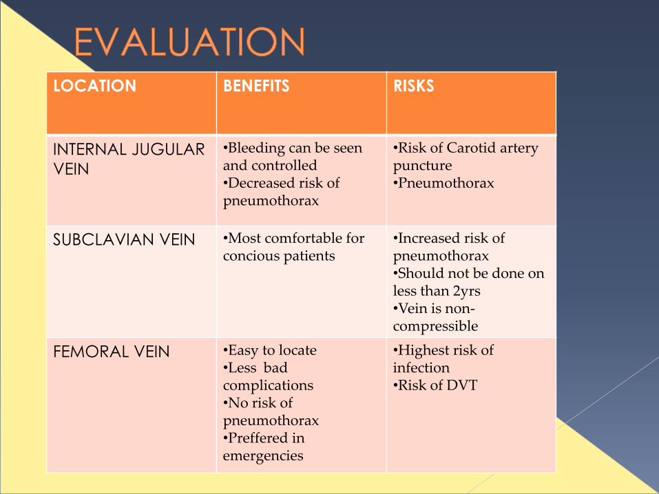
LOCATION BENEFITS RISKS
INTERNAL JUGULAR
VEIN
•Bleeding can be seen and controlled•Decreased risk of pneumothorax
•Risk of Carotid artery puncture•Pneumothorax
SUBCLAVIAN VEIN •Most comfortable for concious patients
•Increased risk of pneumothorax•Should not be done on less than 2yrs •Vein is non-compressible
FEMORAL VEIN •Easy to locate •Less bad complications•No risk of pneumothorax•Preffered in emergencies
•Highest risk of infection•Risk of DVT

THANX FOR LISTENING!