Embed Size (px)
DESCRIPTION
Cells and Tissues. Cells and Tissues. Carry out all chemical activities needed to sustain life Cells are the building blocks of all living things Tissues are groups of cells that are similar in structure and function. Good Morning Anatomy Students!. - PowerPoint PPT Presentation
Citation preview
ELAINE N. MARIEB
EIGHTH EDITION
3
Copyright © 2006 Pearson Education, Inc., publishing as Benjamin Cummings
PowerPoint® Lecture Slide Presentation by Jerry L. Cook, Sam Houston University
ESSENTIALSOF HUMANANATOMY
& PHYSIOLOGY
PART A
Cells and Tissues
Copyright © 2006 Pearson Education, Inc., publishing as Benjamin Cummings
Cells and Tissues Carry out all chemical activities needed to
sustain life
Cells are the building blocks of all living things
Tissues are groups of cells that are similar in structure and function
Copyright © 2006 Pearson Education, Inc., publishing as Benjamin Cummings
Good Morning Anatomy Students!
Warm Up: Record and answer the following question on p.9 of your notebook (10 min)
Name the four elements that compose living matter.
Distinguish between cell, organelles and inclusion.
EQ: How are cell organelles like a factory?
(Write this question and be prepared to answer at end of class).
Copyright © 2006 Pearson Education, Inc., publishing as Benjamin Cummings
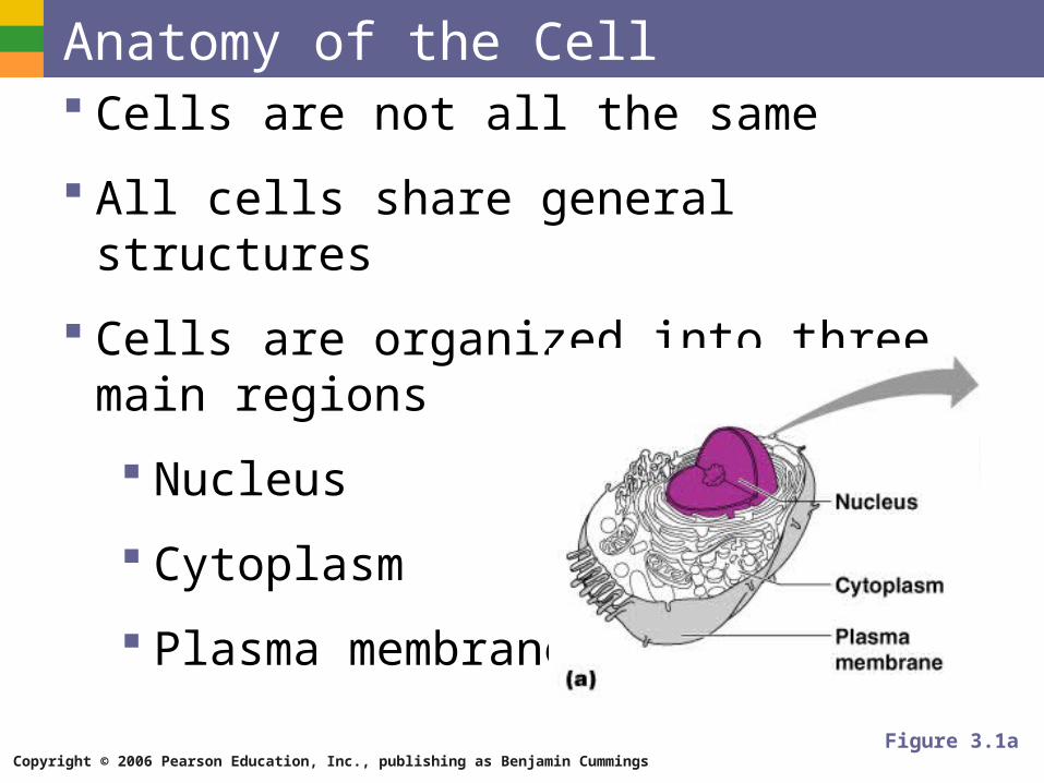
Anatomy of the Cell Cells are not all the same
All cells share general structures
Cells are organized into three main regions
Nucleus
Cytoplasm
Plasma membrane
Figure 3.1a
Copyright © 2006 Pearson Education, Inc., publishing as Benjamin Cummings
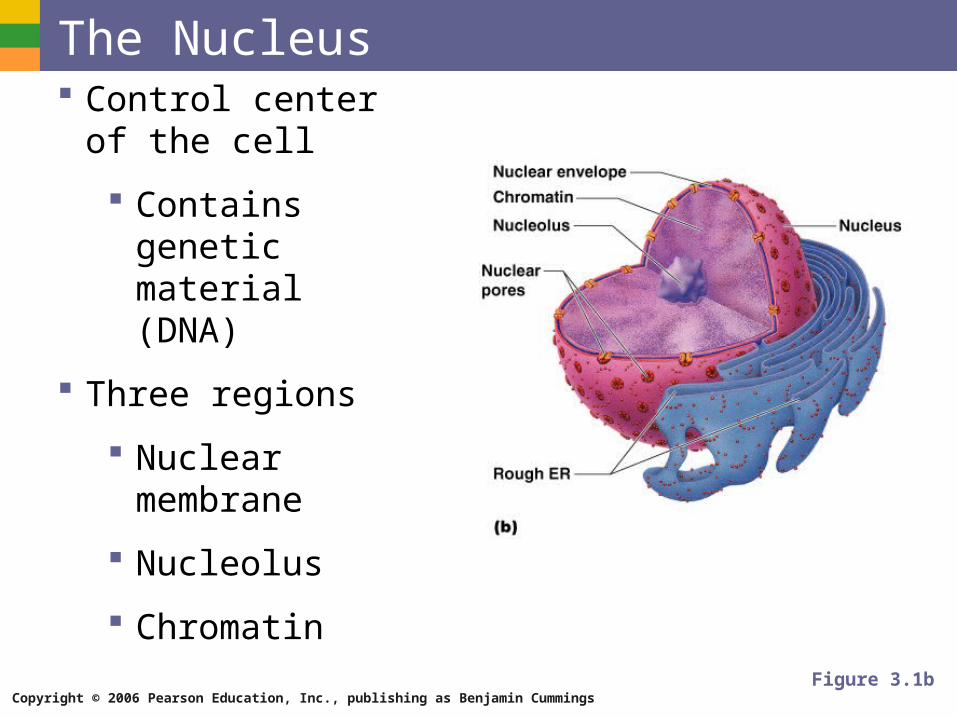
The Nucleus Control center of the
cell
Contains genetic material (DNA)
Three regions
Nuclear membrane
Nucleolus
Chromatin
Figure 3.1b
Copyright © 2006 Pearson Education, Inc., publishing as Benjamin Cummings
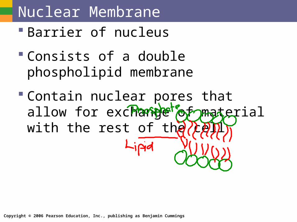
Nuclear Membrane Barrier of nucleus
Consists of a double phospholipid membrane
Contain nuclear pores that allow for exchange of material with the rest of the cell
Copyright © 2006 Pearson Education, Inc., publishing as Benjamin Cummings
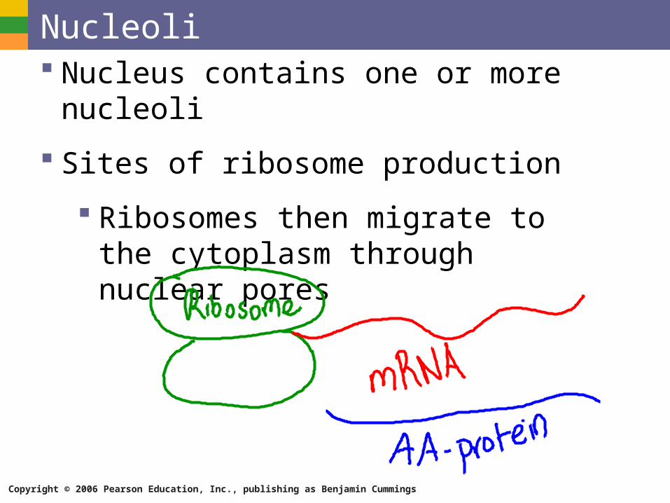
Nucleoli Nucleus contains one or more nucleoli
Sites of ribosome production
Ribosomes then migrate to the cytoplasm through nuclear pores
Copyright © 2006 Pearson Education, Inc., publishing as Benjamin Cummings
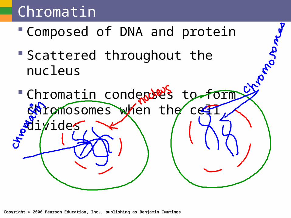
Chromatin Composed of DNA and protein
Scattered throughout the nucleus
Chromatin condenses to form chromosomes when the cell divides
Copyright © 2006 Pearson Education, Inc., publishing as Benjamin Cummings
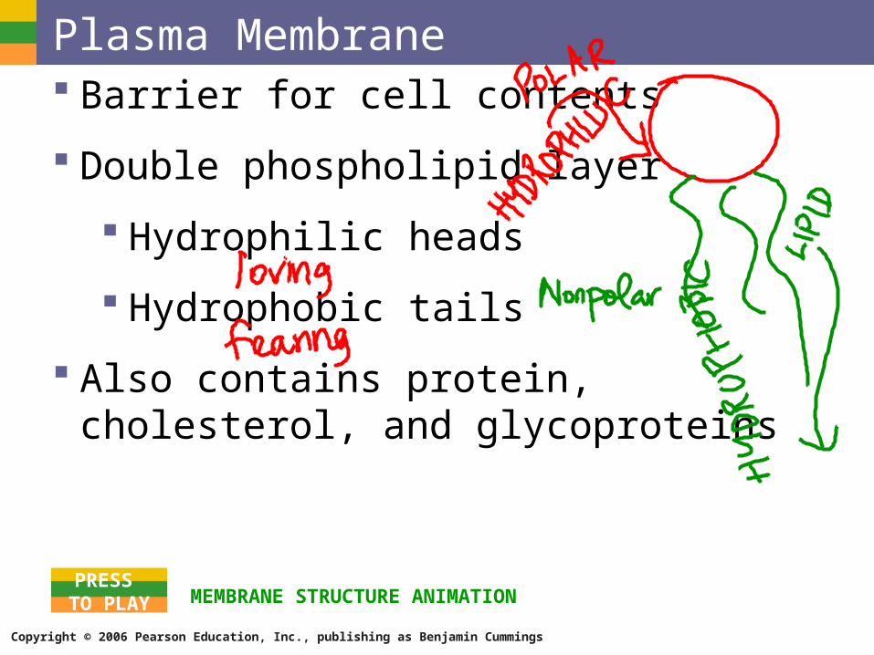
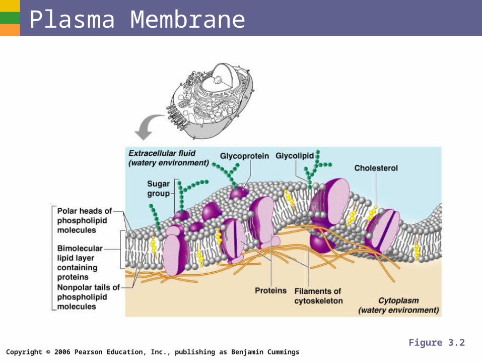
Plasma Membrane Barrier for cell contents
Double phospholipid layer
Hydrophilic heads
Hydrophobic tails
Also contains protein, cholesterol, and glycoproteins
MEMBRANE STRUCTURE ANIMATIONPRESS
TO PLAY
Copyright © 2006 Pearson Education, Inc., publishing as Benjamin Cummings
Plasma Membrane
Figure 3.2
Copyright © 2006 Pearson Education, Inc., publishing as Benjamin Cummings
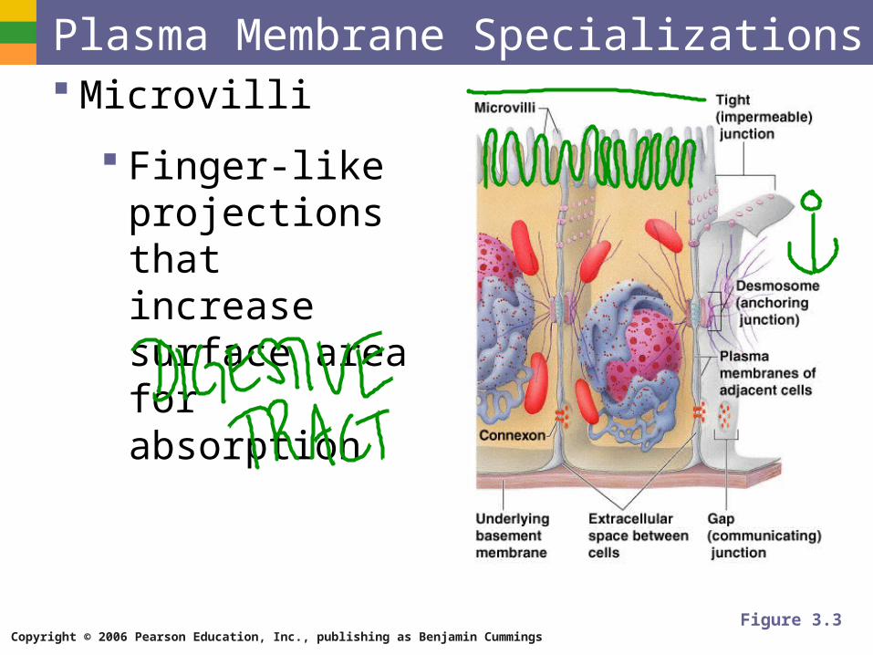
Plasma Membrane Specializations Microvilli
Finger-like projections that increase surface area for absorption
Figure 3.3
Copyright © 2006 Pearson Education, Inc., publishing as Benjamin Cummings
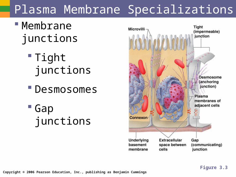
Plasma Membrane Specializations Membrane junctions
Tight junctions
Desmosomes
Gap junctions
Figure 3.3
Copyright © 2006 Pearson Education, Inc., publishing as Benjamin Cummings
Warm Up Define the following terms on p. 10 of your
Notebook (NB):
Selective permeability
Diffusion (Simple & Facilitated)
Active transport
Exocytosis
Endocytosis
Hypertonic/ Hypotonic/Isotonic
Copyright © 2006 Pearson Education, Inc., publishing as Benjamin Cummings
Cytoplasm Material outside the nucleus and inside the
plasma membrane
Cytosol
Fluid that suspends other elements
Organelles
Metabolic machinery of the cell
Inclusions
Non-functioning units
Copyright © 2006 Pearson Education, Inc., publishing as Benjamin Cummings
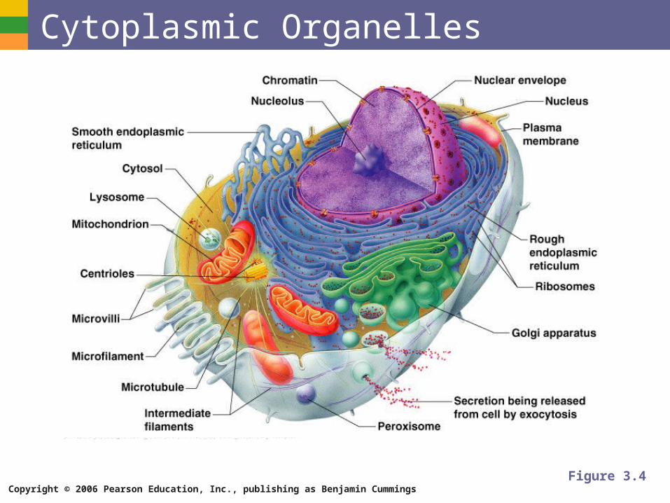
Cytoplasmic Organelles
Figure 3.4
Copyright © 2006 Pearson Education, Inc., publishing as Benjamin Cummings
Cytoplasmic Organelles Ribosomes
Made of protein and RNA
Sites of protein synthesis
Found at two locations
Free in the cytoplasm
Attached to rough endoplasmic reticulum
Copyright © 2006 Pearson Education, Inc., publishing as Benjamin Cummings
Cytoplasmic Organelles Endoplasmic reticulum (ER)
Fluid-filled tubules for carrying substances
Two types of ER
Rough Endoplasmic Reticulum
Studded with ribosomes
Site where building materials of cellular membrane are formed
Smooth Endoplasmic Reticulum
Functions in cholesterol synthesis and breakdown, fat metabolism, and detoxification of drugs
Copyright © 2006 Pearson Education, Inc., publishing as Benjamin Cummings
Cytoplasmic Organelles Golgi apparatus
Modifies and packages proteins
Produces different types of packages
Secretory vesicles
Cell membrane components
Lysosomes
Copyright © 2006 Pearson Education, Inc., publishing as Benjamin Cummings
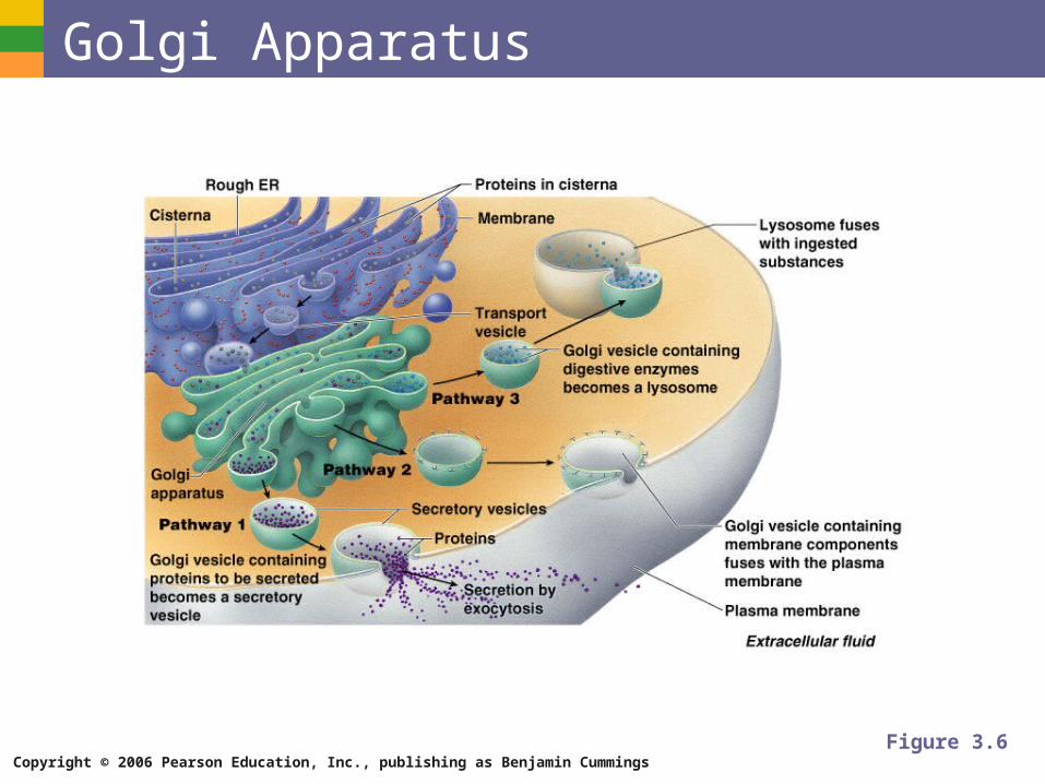
Golgi Apparatus
Figure 3.6
Copyright © 2006 Pearson Education, Inc., publishing as Benjamin Cummings
Cytoplasmic Organelles Lysosomes
Contain enzymes that digest nonusable materials within the cell
Peroxisomes
Membranous sacs of oxidase enzymes
Detoxify harmful substances
Break down free radicals (highly reactive chemicals)
Replicate by pinching in half
Copyright © 2006 Pearson Education, Inc., publishing as Benjamin Cummings
Cytoplasmic Organelles Mitochondria
“Powerhouses” of the cell
Change shape continuously
Carry out reactions where oxygen is used to break down food
Provides ATP for cellular energy
Copyright © 2006 Pearson Education, Inc., publishing as Benjamin Cummings
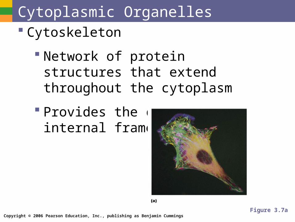
Cytoplasmic Organelles Cytoskeleton
Network of protein structures that extend throughout the cytoplasm
Provides the cell with an internal framework
Figure 3.7a
Copyright © 2006 Pearson Education, Inc., publishing as Benjamin Cummings
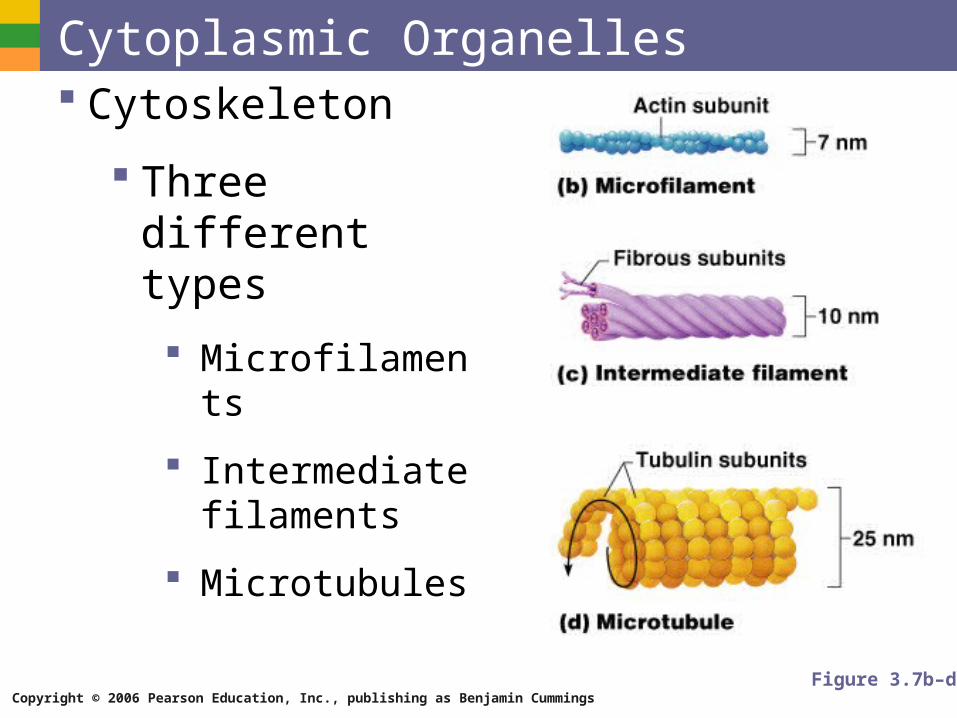
Cytoplasmic Organelles Cytoskeleton
Three different types
Microfilaments
Intermediate filaments
Microtubules
Figure 3.7b–d
Copyright © 2006 Pearson Education, Inc., publishing as Benjamin Cummings
Cytoplasmic Organelles Centrioles
Rod-shaped bodies made of microtubules
Direct formation of mitotic spindle during cell division
Copyright © 2006 Pearson Education, Inc., publishing as Benjamin Cummings
Cellular Projections Not found in all cells
Used for movement
Cilia moves materials across the cell surface
Flagellum propels the cell
Copyright © 2006 Pearson Education, Inc., publishing as Benjamin Cummings
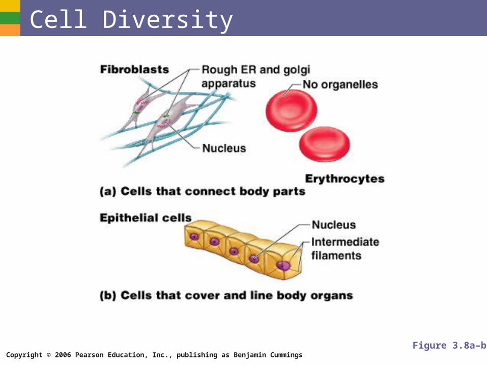
Cell Diversity
Figure 3.8a–b
Copyright © 2006 Pearson Education, Inc., publishing as Benjamin Cummings
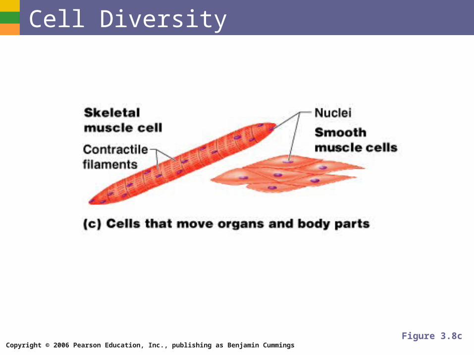
Cell Diversity
Figure 3.8c
Copyright © 2006 Pearson Education, Inc., publishing as Benjamin Cummings
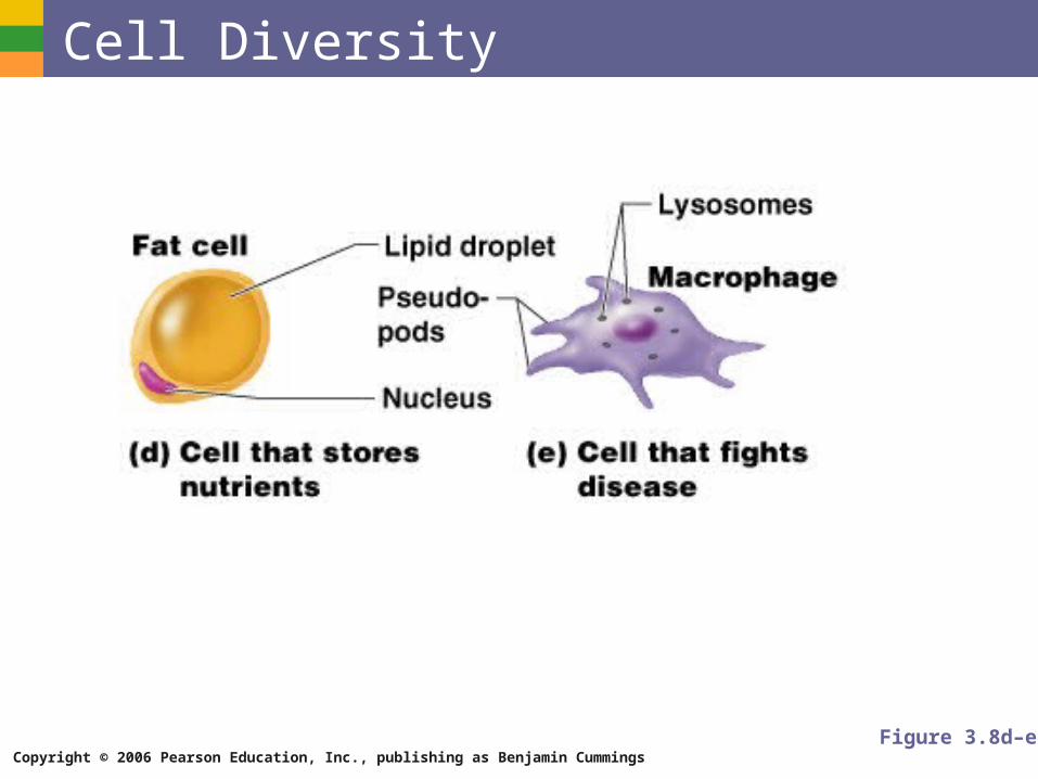
Cell Diversity
Figure 3.8d–e
Copyright © 2006 Pearson Education, Inc., publishing as Benjamin Cummings
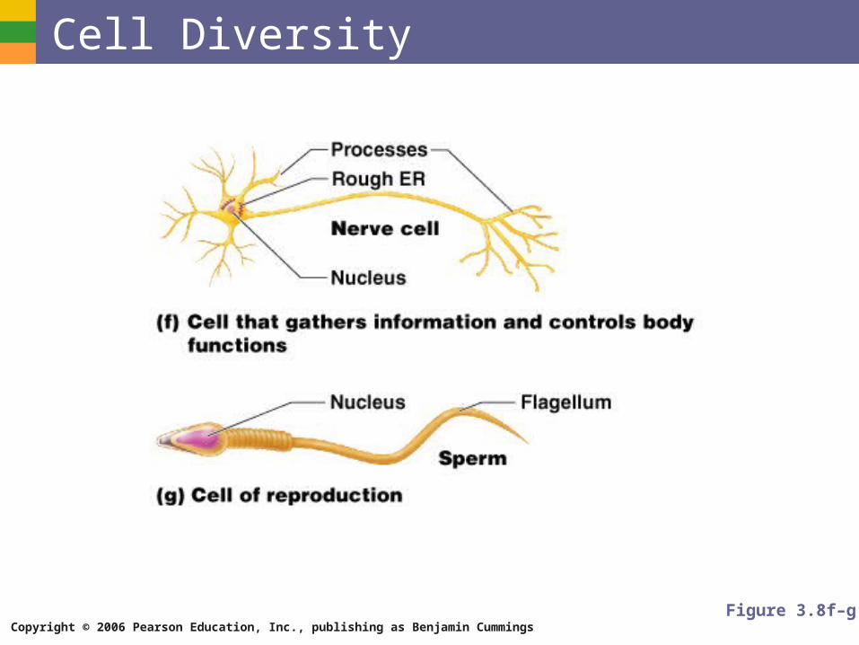
Cell Diversity
Figure 3.8f–g