Embed Size (px)
Citation preview
1159
administered before the initial reading was taken,but all cases were given iodine prior to the operation.It can be seen that shortly after subtotal thyroid-ectomy the B.M.R. has fallen to within normallimits, but that this was not associated with anychange in the LA.
Of those shown in Table III. Case 10 was a well-marked case of "
spontaneous "
myxcedema whichwas treated by means of thyroid (B.P. 1932) grs. 2 b.d.Sixteen days elapsed between the initial reading and
TABLE III.-Cases of Myxaedema Before and AfterThyroidFEMALES
the second here recorded. Case 11 was one of post-operative myx oedema and received thyroid (B.P. 1932)grs. 4 t.d.s. for six days with a view to producing arapid elevation of B.M.R. It can be seen that in thetwo cases of myxoedema studied, the LA. is notoutside the normal limits, while the B.M.R. reflectsthe clinical myxcedematous state. After thyroidmedication, clinical improvement took place, asso-
ciated with a rise in B.M.R. but without alterationin the I.A.The dissociation, shown in the cases here recorded,
between B.M.R. and LA. is also evident from a studyof the effects of severe exercise. When this is carriedout to the point of exhaustion it may elevate the
oxygen consumption by 200 per cent. or more andonly produce a slight change in the LA. The remark-able constancy of the LA. with marked alteration ofthe thyroid state suggested an investigation of it beforeand after death. No significant change was noted inthe LA. readings of cats in experiments of this kind.These results show that little or no change takes
place in the LA. values in spite of great alteration ofthe clinical state in thyrotoxic patients. They suggestthat any change which takes place in these valuesas a result of treatment is too slow to be of clinical use.
SUMMARY
(1) In eight consecutive cases of typical Graves’sdisease, the impedance angle (LA.) was found to beoutside the normal limits in the direction indicatedby Brazier.
(2) In these eight cases pre-operative iodine medica-tion caused no alteration of the impedance anglealthough this treatment produced in all of themclinical improvement, fall in pulse-rate, and fall inbasal metabolic rate.
(3) In four cases of Graves’s disease studied beforeand 9 to 14 days after subtotal thyroidectomy, noalteration was found in the I.A., although the opera-tion was followed by a fall of B.M.R. and pulse-rateand by clinical improvement.
(4) In two cases of well-marked myxcedema theLA. was found to be normal. Treatment with thyroidextract produced no alteration in the LA.
(5) Exercise to the point of exhaustion, producingan increase of 200 per cent. in O2 consumption,increased the LA. only by 10 Brazier units (i.e.,about 7 per cent).
(6) In cats there is no significant change in theLA. after death.
(7) It appears from these results that determinationof the impedance angle is of no value in assessingthe clinical progress of cases of Graves’s disease ormyxaedema.Thanks are due to the honorary staff of the
Middlesex Hospital for permission to examine theircases ; and to Dr. M. A. B. Brazier, Prof. E. C. Dodds,Mr. Olmwood of Gambrell Bros., and Prof. SamsonWright.
REFERENCES
1. Brazier, M. A. B. : Jour. Inst. Elect. Engineers, 1933, lxxiii.,203.
2. Brazier, M. A. B.: THE LANCET, 1933, ii., 742.3. Brazier, M. A. B., and Grant, F. M. : Ibid., 1934, i., 125.
Clinical and Laboratory NotesCAVERNOUS ANGIOMA OF THE
VERTEBRA
BY MARY F. LACEY, M.B. Liverp.RESIDENT MEDICAL OFFICER, SMITHDOWN ROAD HOSPITAL,
LIVERPOOL ; AND
E. SEYMOUR SMITH, M.B. Liverp.DEPUTY MEDICAL SUPERINTENDENT OF THE HOSPITAL
A SCHOOL teacher, aged 47, was admitted to theSmithdown-road Hospital with a history of completeloss of power in both legs for 26 years. Up to the ageof 21 she had been perfectly healthy, and the onset ofthe condition was probably gradual from this time.
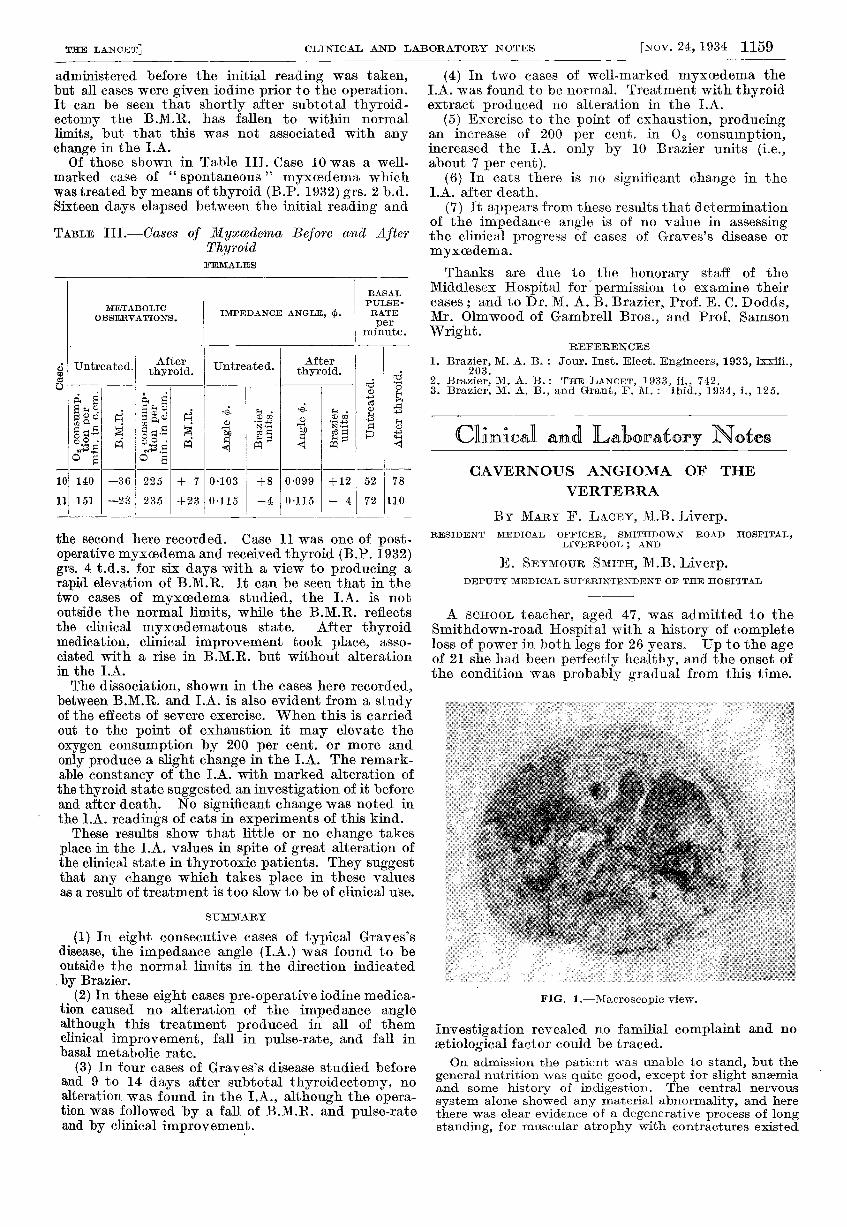
FIG. 1.-Macroscopic view.
Investigation revealed no familial complaint and noaetiological factor could be traced.On admission the patient was unable to stand, but the
general nutrition was quite good, except for slight anaemiaand some history of indigestion. The central nervous
system alone showed any material abnormality, and herethere was clear evidence of a degenerative process of longstanding, for muscular atrophy with contractures existed

1160
in the lower extremities. The cranial nerves were normal,the arm-jerks and abdominal reflexes were present andequal on both sides, the knee- and ankle-jerks were absent,and there was a bilateral extensor plantar response.Ankle-clonus was also bilateral. From the level of the ninthdorsal vertebra there was complete loss of pain and tactilesensation. There was complete incontinence of urine, butthe anal sphincter was not involved.The blood and cerebro-spinal fluid Wassermann reaction
were negative as was the reaction to Lange’s gold sol.
FIG. 2.-Ascending degeneration above the lesion. (x 7-5.)
There was no increase in cellular elements, and no alterationin the protein and sugar contents.An X ray examination of the spine was made and a
tentative diagnosis of haemangioma of the ninth thoracicvertebra arrived at. A macroscopic examination showedthe usual irregular absorption of the bony trabeculationwith a large swelling extending to either side of the
adjacent upper vertebra and to the lower limit of onevertebra below. The outline of the swelling was clearly
FIG. 4.-Extensive degeneration below the lesion; and it isobvious that there has been considerable pressure, for thedorsal columns are affected as well as the descending fibres.Even the anterior horn is affected and there are few cellsleft. There are still some relatively healthy ascending fibresin the dorsal region. ( x 7 -5.)
defined, and in its substance a fine network of bonytrabeculation enclosing large cystic spaces was seen.The patient died four months after admission to hospital,
and sections of the tumour taken at the autopsy revealedthe typical microscopic characteristics of a cavernous
angioma (Figs. 1-5).
Ewing 1 reports a similar case, and Nattrass andRamage 2 quote 11 cases as having been reportedbefore 1927, the first noted being that by Virchow in1862-63. In 1929 Bailey and Bucy 3 of Chicago putthe number of cases as 27 described up to that date.
The one now reported varies in many findings fromthose mentioned, but in the serological examinationsthere is almost no difference. It is unfortunate thatthe antecedent history of this patient is not available,but it seems to be established that from the time theillness was recognised there was no remission and thesymptoms of compression persisted and increased.Whether there was an early remission, as in the caseof Bailey and Bucy, or whether the onset was gradualas reported byNattrass and
Ramage, it is
impossible to
say ; but with a
history of 28
years of inabilityto walk one feelsthat the proba-bility lies with agradual onset.The duration ofthe condition isof interest, andthe contracturesand muscular
atrophy suggesta slow but
steady develop-ment.
Faecal incontinence was absent in this case, whichis not in accord with other reports, although Connelland Hay 4 instance a similar condition in which asensation of ful.lness of the rectum was experiencedprior to incontinent defalcation. In every patientcontrol of the bladder is reported to have been absent.The age of onset, in the early twenties, is interesting,
and is probably one of the earliest reported. Other
FIG. 3.-Nerve roots which are all thatis functional at the level of the lesion.(X7.5.)
.
FIG. 5.-Cavernous angioma showing large intercommunicatingspaces filled with blood. ( x 80.)
records show the ages of recent cases as being betweenthe age of 50 and 62 years.We wish to acknowledge our indebtedness to Dr.
John P. Steel, medical superintendent of the hospital,for permission to publish an account of this case.
REFERENCES
1. Ewing, J. : Neoplastic Diseases, Philadelphia, 1928.2. Nattrass, F. J., and Ramage, D. : Jour. Neurol. and Psycho-
path., 1932, xii., 231.3. Bailey, P., and Bucy, P. C. : Jour. A.M.A.,1929, xcii., 1748.4. Connell, W. T., and Hay, W. D. : Canad. Med. Assoc. Jour.,
1930, xxii., 75.